Embed Size (px)
Citation preview
Antepartum and Postpartum Hemorrhage
Dr. Megha Jain
www.anaesthesia.co.in email: [email protected]
University College of Medical Sciences & GTB Hospital, Delhi
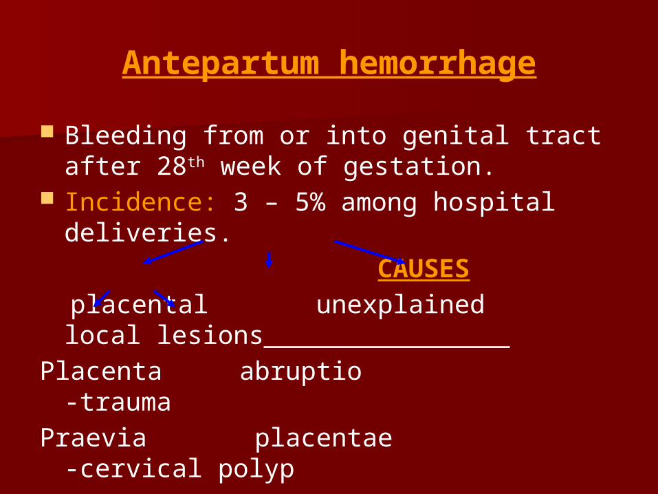
Antepartum hemorrhage
Bleeding from or into genital tract after 28th week of gestation.
Incidence: 3 – 5% among hospital deliveries.
CAUSES placental unexplained local lesions
Placenta abruptio -traumaPraevia placentae -cervical
polyp -carcinoma
cervix
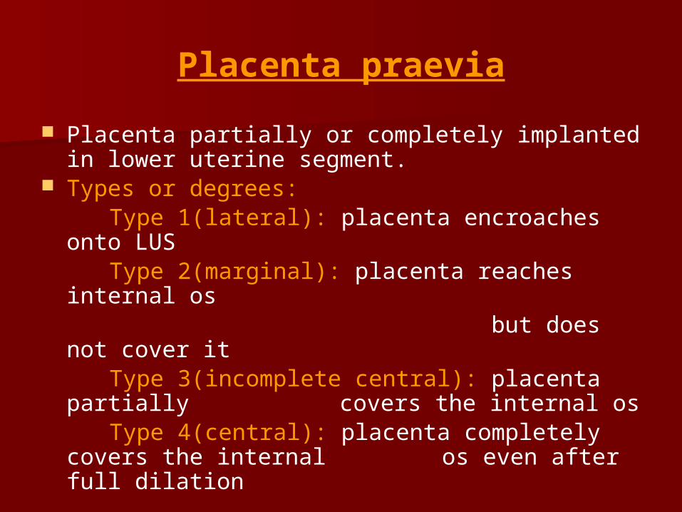
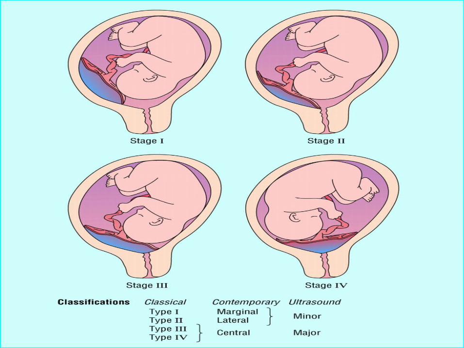
Placenta praevia
Placenta partially or completely implanted in lower uterine segment.
Types or degrees: Type 1(lateral): placenta encroaches onto LUS Type 2(marginal): placenta reaches internal os but does not cover it Type 3(incomplete central): placenta partially
covers the internal os Type 4(central): placenta completely covers the
internal os even after full dilation
Risk factor
Multiparity Increased maternal age History of previous CS or any other
scar in the uterus(myomectomy) Big size placenta
Clinical presentation
1st vaginal bleeding episode- after 36th week- 60% b/w 32 - 36 week- 30% before 32 week- 10% # painless and recurrent bleeding # GC and anemia – prop. to blood loss # Size of uterus prop. to POG # Presenting part is usually high up # FHS is usually present # Diagnosis by USG
The double set up examination
Vaginal examination in OT Preparation- # Two large bore i/v cannula # Blood for transfusion # Oral antacid # Oxygen # Skilled assistant If profuse bleeding CS GA(RSI) # Treat hypovolemia # Induction- Ketamine, intubate- Sch # Maintenance- O2, N2O # Awake extubation
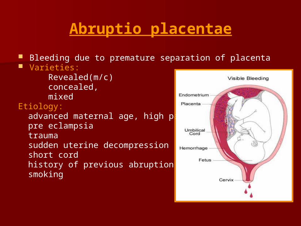
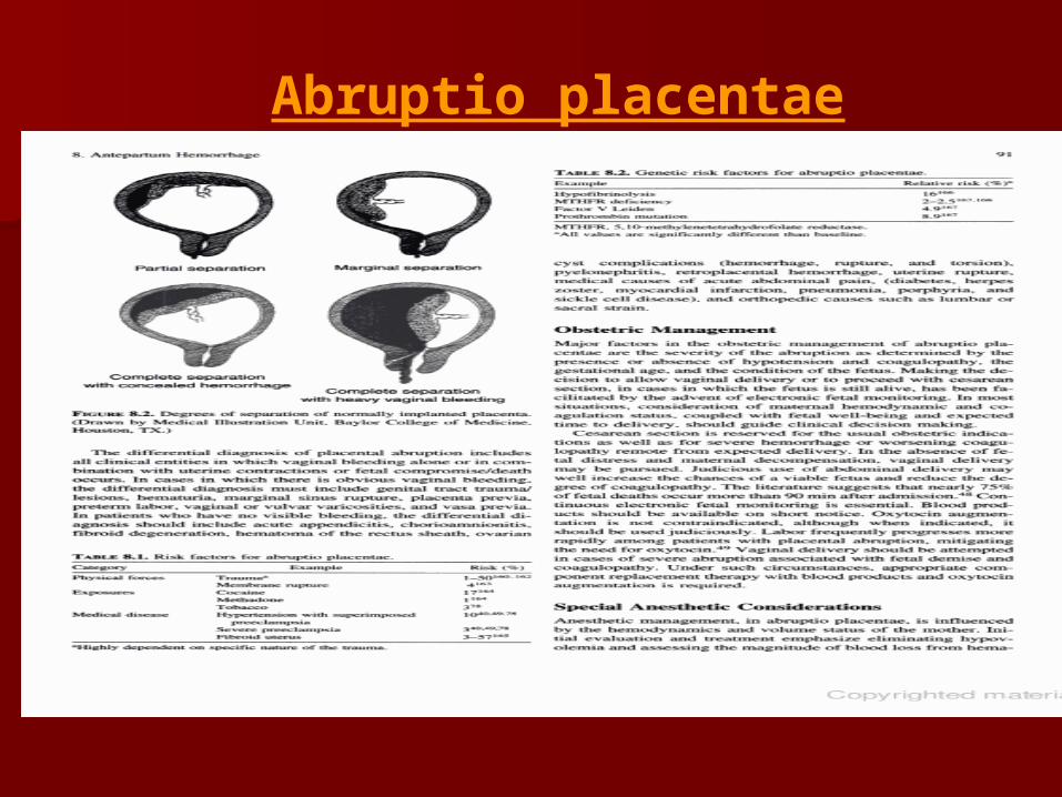
Abruptio placentae
Bleeding due to premature separation of placenta Varieties: Revealed(m/c) concealed, mixedEtiology: advanced maternal age, high parity pre eclampsia trauma sudden uterine decompression short cord history of previous abruption smoking
Abruptio placentae
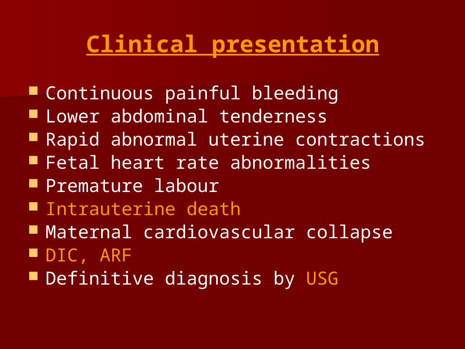
Clinical presentation
Continuous painful bleeding Lower abdominal tenderness Rapid abnormal uterine contractions Fetal heart rate abnormalities Premature labour Intrauterine death Maternal cardiovascular collapse DIC, ARF Definitive diagnosis by USG
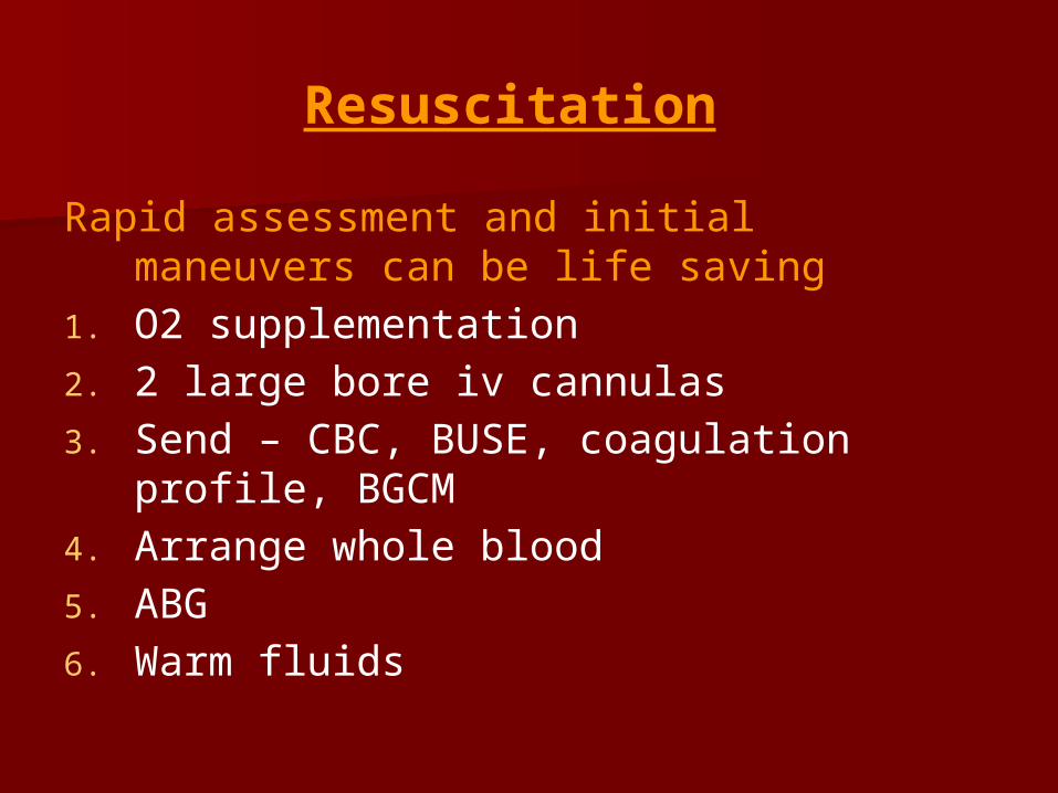
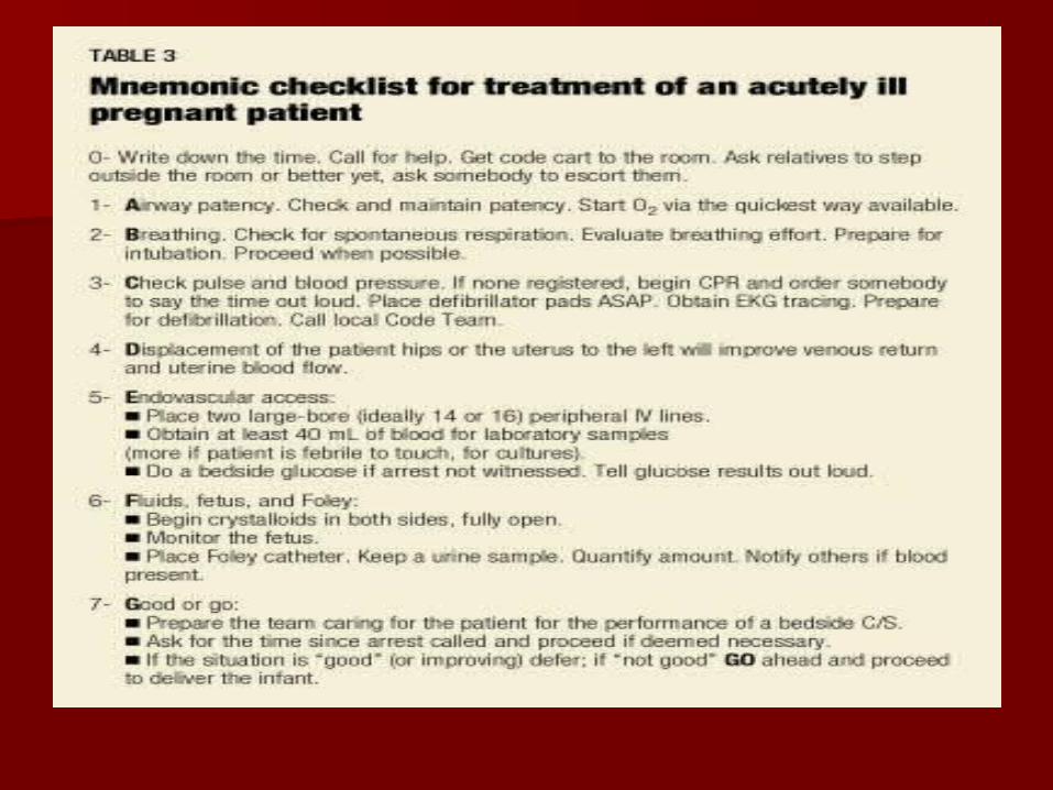
Resuscitation
Rapid assessment and initial maneuvers can be life saving
1. O2 supplementation
2. 2 large bore iv cannulas
3. Send – CBC, BUSE, coagulation profile, BGCM
4. Arrange whole blood
5. ABG
6. Warm fluids
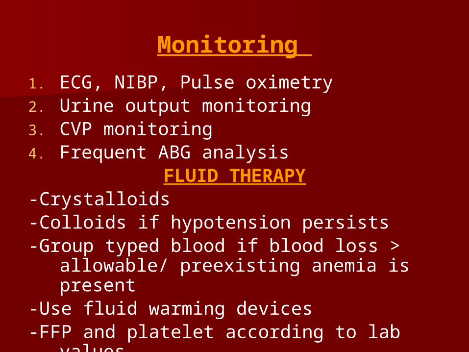
Monitoring
1. ECG, NIBP, Pulse oximetry2. Urine output monitoring3. CVP monitoring4. Frequent ABG analysis
FLUID THERAPY-Crystalloids-Colloids if hypotension persists-Group typed blood if blood loss > allowable/
preexisting anemia is present-Use fluid warming devices -FFP and platelet according to lab values.
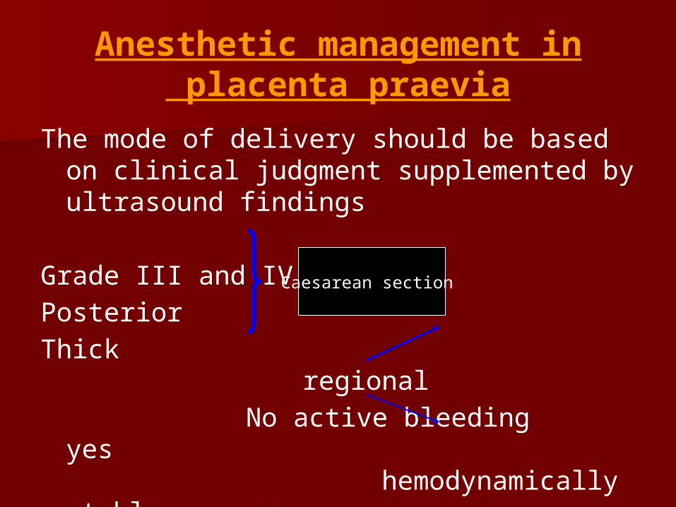
Anesthetic management in placenta praevia
The mode of delivery should be based on clinical judgment supplemented by ultrasound findings
Grade III and IV
Posterior
Thick regional
No active bleeding yes hemodynamically stable no
GA
Caesarean section
Regional anaesthesia –associated with more blood loss because
-placenta praevia patients are at increased risk of placenta accreta
-obstretician may cut into placenta during uterine incision
-LUS has lesser power of contraction and retraction

General anesthesia
RSI – preferred technique Induction agent- Ketamine safest for
hypovolemic patients(0.5 to 1mg/kg) Intubation with Sch(1.5mg/kg) Maintenance with- O2(50%)+ N2O(50%)+ low
conc of volatile agent if tolerated Extubate when fully awake and responding to
verbal commands
Anesthetic management of abruptio placentae
Definitive treatment is delivery of the fetus Route of delivery depends on – degree of abruption maternal hemodynamics status of the fetus If abruption is mild to moderate and the
mother is hemodynamically stable with fetus being mature – continuous lumbar epidural, caudal or SAB may be used for labour and vaginal delivery
Anesthetic management (contd….)
For severe abruption – Em LSCS ↓ GA(RSI) ↑ risk of persistent hemorrhage due to uterine
atony and coagulopathy Thus give oxytocin immediately after delivery Other drugs used are methergin and PG
analogues Transfuse blood, FFP and platelet
Postpartum hemorrhage
Definition: blood loss > 500 ml after vaginal delivery of fetus > 1000 ml after CS
Clinically it refers to any amount of bleeding from or into genital tract which adversely affects maternal condition
Incidence: 3-5% among all deliveries
Types
# Primary: in 24 hrs following delivery
Third stage hge- before placental expulsion
True PPH- after placental expulsion
# Secondary: beyond 24 hrs but within puerperium
Etiology
1. Atonic uterus: (80%) can be due to- # grand multipara # over distention of uterus (twins,
macrosomia) # malnutrition and anemia # APH # prolonged labour # uterine fibroid 2. Trauma3. Blood coagulation disorder
Assessment of obstetric hemorrhage
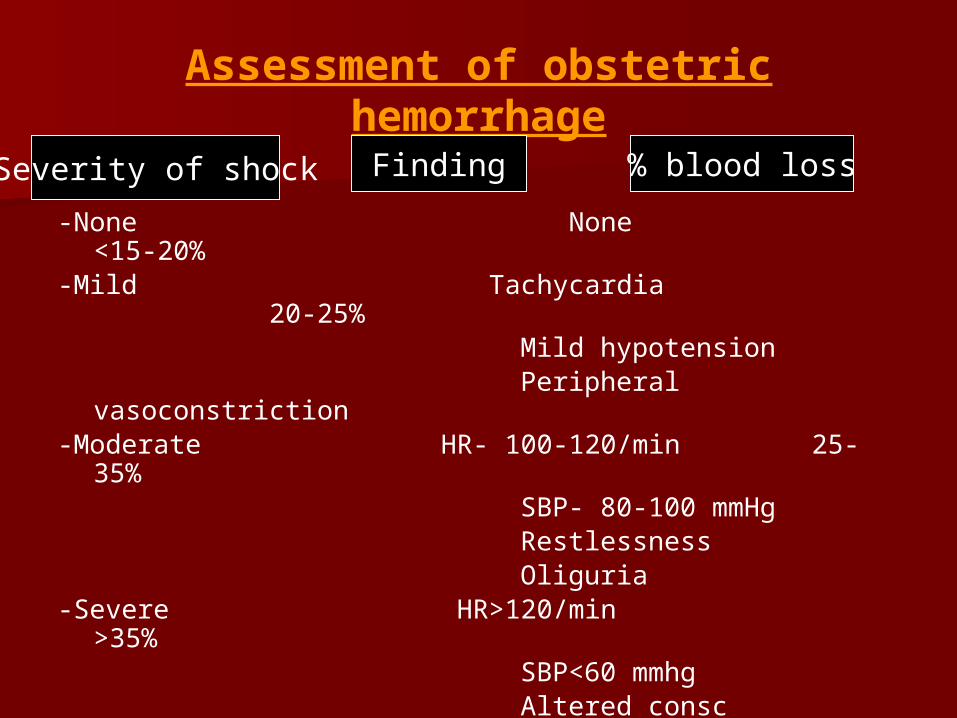
-None None <15-20%-Mild Tachycardia 20-25% Mild hypotension Peripheral vasoconstriction-Moderate HR- 100-120/min 25-35% SBP- 80-100 mmHg Restlessness Oliguria-Severe HR>120/min >35% SBP<60 mmhg Altered consc Anuria
Severity of shock Finding % blood loss
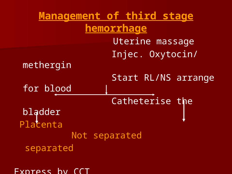
Management of third stage hemorrhage
Uterine massage Injec. Oxytocin/ methergin Start RL/NS arrange for blood Catheterise the bladder Placenta Not
separated separated
Express by CCT Manual removal under GA
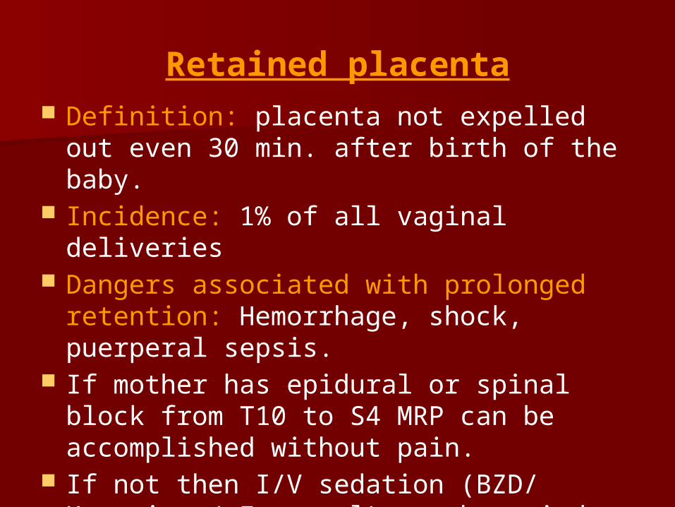
Retained placenta Definition: placenta not expelled out even
30 min. after birth of the baby. Incidence: 1% of all vaginal deliveries Dangers associated with prolonged
retention: Hemorrhage, shock, puerperal sepsis.
If mother has epidural or spinal block from T10 to S4 MRP can be accomplished without pain.
If not then I/V sedation (BZD/ Ketamine / Fentanyl) can be tried.
But if the patient is hemodynamically unstable GA should be administered.
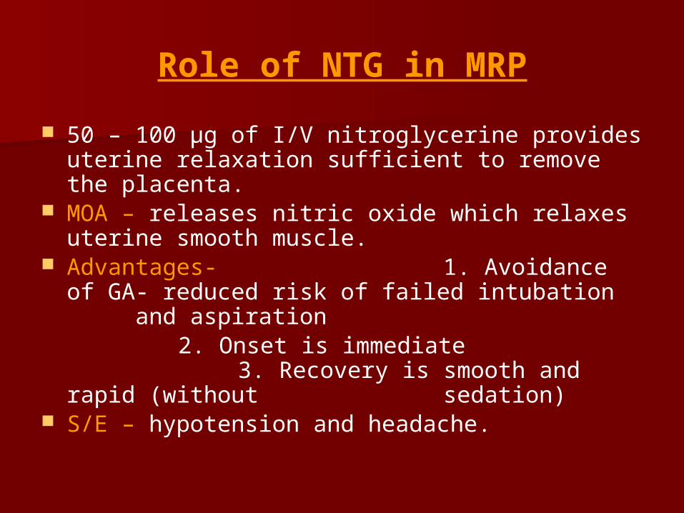
Role of NTG in MRP
50 – 100 µg of I/V nitroglycerine provides uterine relaxation sufficient to remove the placenta.
MOA – releases nitric oxide which relaxes uterine smooth muscle.
Advantages- 1. Avoidance of GA- reduced risk of failed
intubation and aspiration 2. Onset is immediate
3. Recovery is smooth and rapid (without sedation)
S/E – hypotension and headache.
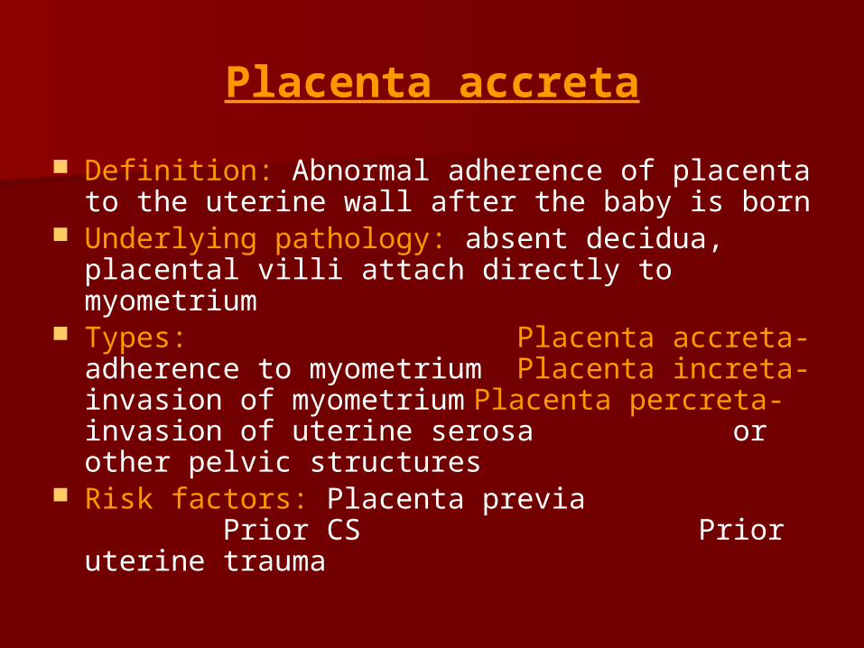
Placenta accreta
Definition: Abnormal adherence of placenta to the uterine wall after the baby is born
Underlying pathology: absent decidua, placental villi attach directly to myometrium
Types: Placenta accreta- adherence to
myometrium Placenta increta- invasion of myometrium Placenta percreta- invasion of uterine serosa or other pelvic structures
Risk factors: Placenta previa Prior CS Prior uterine trauma
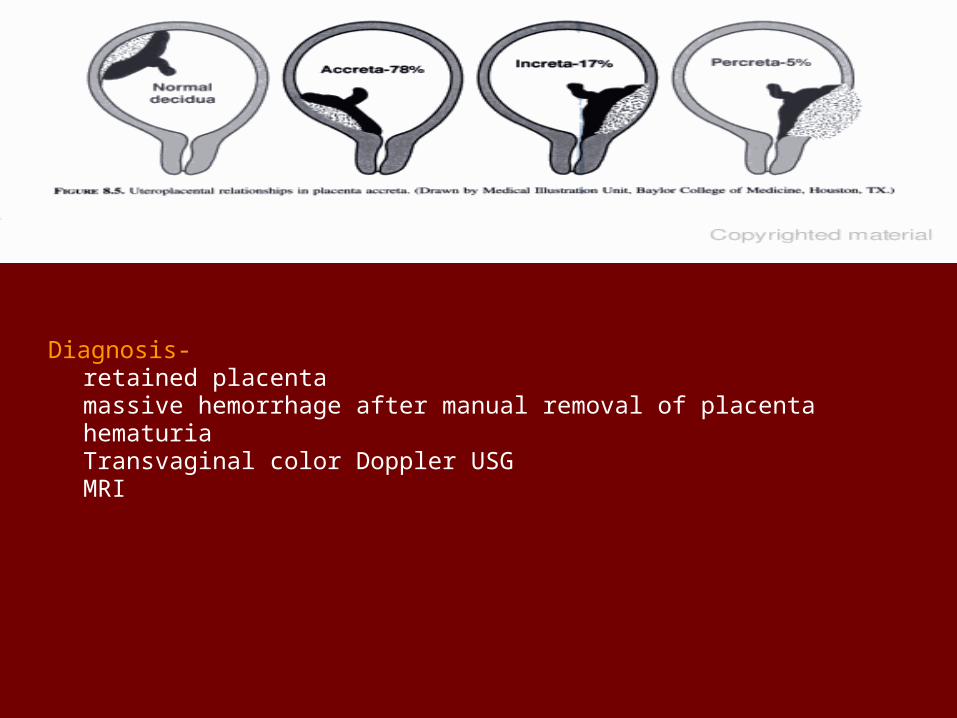
Diagnosis- retained placenta massive hemorrhage after manual removal of placenta hematuria Transvaginal color Doppler USG MRI
Anesthetic mgt. of placenta accreta
Most patients require hysterectomy ↓ GA Insert large bore I/V line Arrange blood Secure airway- Endotracheal intubation Routine monitoring-ECG , NIBP, Pulse oximetry, urine
output Consider CV line and arterial line Use fluid warming devices May require ICU care
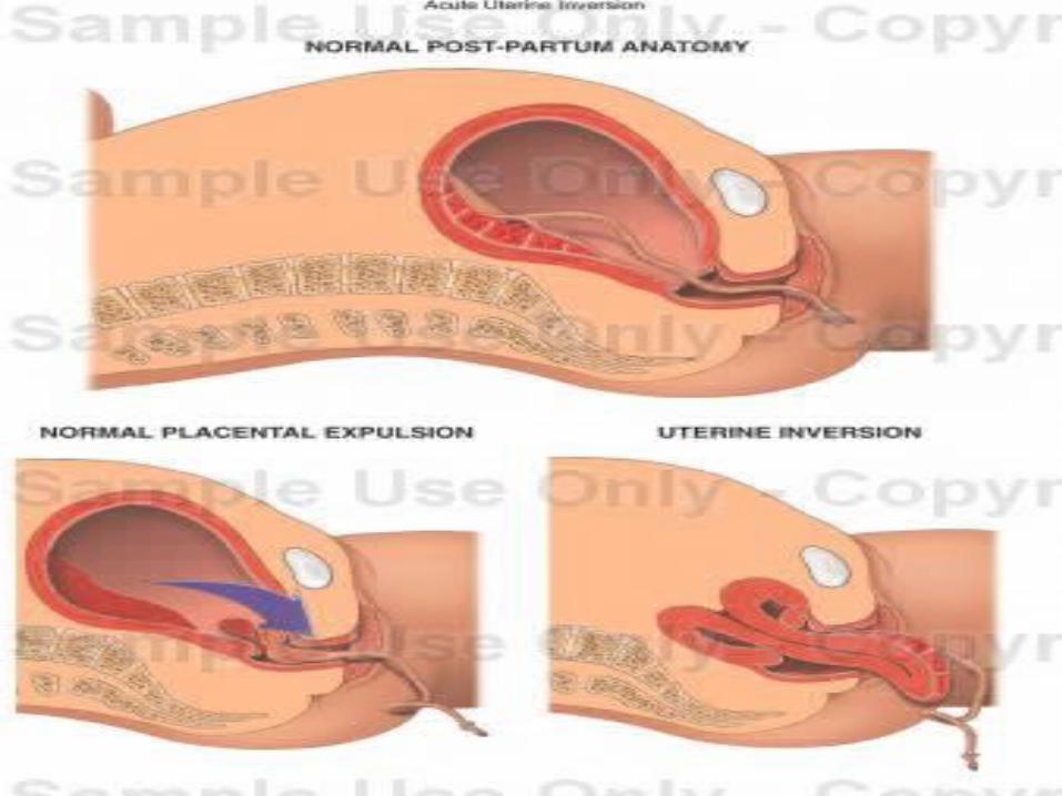
Uterine inversion
Rare C/C of 3rd stage where uterus turns inside out partially or completely
Etiology- may be spontaneous or - due to pulling of the cord - uterine atony - inappropriate fundal pressure - placenta accreta Dangers associated- hemorrhage, shock,
pulmonary embolism
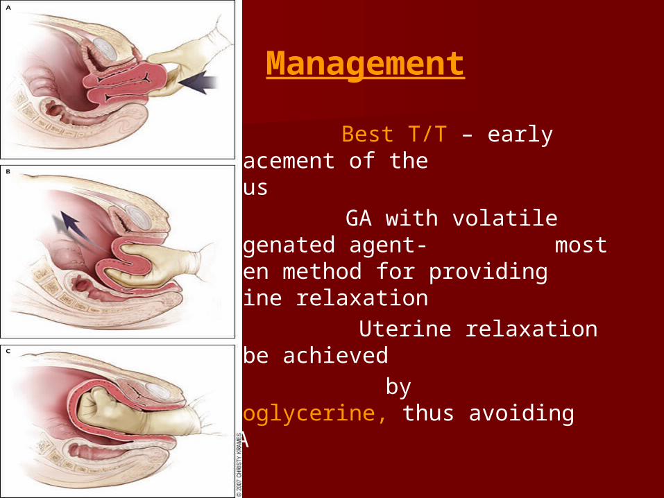
Management
Best T/T – early replacement of the uterus
GA with volatile halogenated agent- most proven method for providing uterine relaxation
Uterine relaxation may be achieved
by nitroglycerine, thus avoiding GA
Uterine atony
- M/C cause of PPH - Conditions associated with uterine atony
are- Multiple gestation Macrosomia Polyhydramnios High parity Prolonged/precipitous/augmented labor Tocolytic drugs High conc. Of volatile halogenated
agents
Management
Resuscitation and immediate management- Administer 100% O2 2 large bore I/V cannula and arrange blood Fluid resuscitation- crystalloid/colloid using
pressure bag Transfuse cross matched blood( O-Negative if
group specific not available) Use fluid warmer and warming blanket Monitor- ECG,NIBP,O2 sat., urine output, acid
base status, hemoglobin(using hemocue) and coagulation parameters
Consider arterial line and CVP line only after definitive treatment has commenced
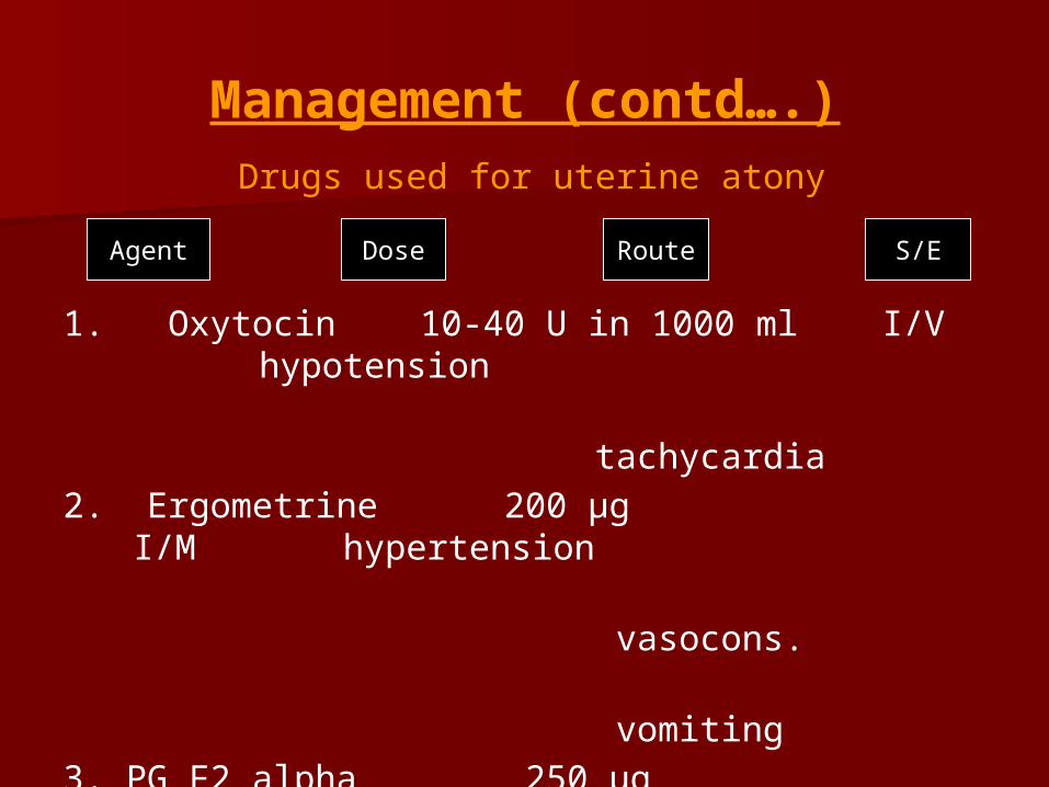
Management (contd….)Drugs used for uterine atony
1. Oxytocin 10-40 U in 1000 ml I/V hypotension
tachycardia
2. Ergometrine 200 µg I/M hypertension
vasocons.
vomiting3. PG F2 alpha 250 µg I/M
hypotension I/U
bronchocons.
Agent Dose Route S/E
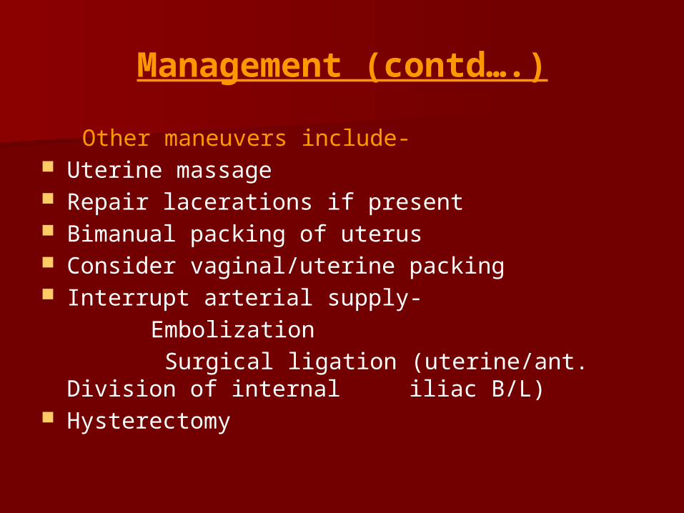
Management (contd….)
Other maneuvers include- Uterine massage Repair lacerations if present Bimanual packing of uterus Consider vaginal/uterine packing Interrupt arterial supply- Embolization Surgical ligation (uterine/ant. Division of
internal iliac B/L) Hysterectomy
Thank You
www.anaesthesia.co.in