Embed Size (px)
Citation preview

The Journ~ND SURGE
mit the)een:afts arethe lost
:s by
reconstruct
pe injurymferential,95-1011,~ injuries.
;t Surg
¯ 8:34-7,:raft as a;ureerv 20:3
ities or the h,1946~tz JE: The{ailure to
~lte
tvest
il plate.¯ J Invest
.’tal andDermatol
"antenna"eforrnity
procedure for the "hook-nail"
The hook-nail deformity is a relatively common problem after fingertip amputations. It is usuallyignored but can be quite disabling. This deformity can be corrected by a carefully planned
rration. Most of the curved nail plate is removed, :the pulp is reflected from the distal phalanxout to a normal contour, and then the full thickness of the nail bed is elevated off the distal
! danx and splinted by multiple small Kirschner pins in a straight position. The defect created is.’overed with a cross finger flap. (J HAND SURG 8:55-8, 1983.)
irdogan Atasoy, M.D., Alan Godfrey, M.D., and Michael Kalisman, M.D.,
L.~:Y,’,ville. Kv.
wide variety of flaps and grafting pro-been described for the repair of skin de-
amputations,1-8 but little has beent correction of the defects and deformities of
and nail bed that are very common aftere~aii bed injuries. Knowledge of the anat-
of the nail and surrounding struc-of good treatment of nail bed
reduce the incidence of secondary nail
that frequently follows distal finger
is the hook-nail deformity (Figs. I, A,usually occurs after loss of part of the
and nail bed. To some patientsand may be ignored; to others it
cosmetic significance or may be dis-to certain occupations. We have
procedure" for this methodale Kirschner wires protruding
finger like antennas.
been treated by this method with a4 years.
machine operator sustained a left
A
t of Surgery, University of Louisville School ofKy.
are former Fellows in Hand Surgery,Fellowship in Hand Surgery, University of
cine, Louisville, Kentucky.Sept. 22, 1981.
. Suite 1001. Doctors OfficeSt., Louisville, KY 40202.
04500.40/0 © 1983 American Society for Surgery of the Hand
Fig. 1o A, Hook-nail deformity and normal contour of finger-tip (dotted line). B, Removal of nail plate and skin marking.C, Reflection of pulp, elevation of full thickness of nail ma-trix and splinting of it with three small Kirschner wires likeantennae. D, Coverage of defect with a cross finger flap. E,Appea:rance after division of flap (2 weeks postoperative).
ring finger amputation by the gear of a machine in September1977. ’INe initial treatment consisted of primary closure of thefingertip in the emergency room of a hospital. The patientgradually developed a typical hook-nail deformity. On Jan. 6,1978, the antenna procedure was performed. Missing pulpwas reconstructed and straight nail growth was achieved. InDecember 1978. two-point discrimination was 4 mm; the pa-
THE JOURNAL OF HAND SURGERY 55
The56 Atasoy et al. HAND SUR~
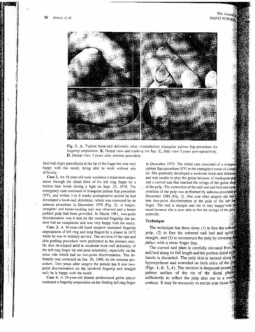
Fig. 2. A, Typical hook-nail deformity after unsatisfactory triangular palmar flap procedure forfingertip amputation. B, Dorsal view and marking the flap. C, Side view 3 years post-operatively.D, Dorsal view 3 years after antenna procedure.
tient had slight paresthesia at the tip of the finger but was veryhappy with the result, being able to work without anydifficulty.
Case 2. An 18-year-old male sustained a transverse ampu-tation through the distal third of his left ring finger by abroken beer bottle during a fight on Sept. 23, 1978. Theemergency care consisted of triangular palmar flap procedure(VY), and within 3 to 6 weeks postoperative period he haddeveloped a hook-nail deformity, which was corrected by anantenna procedure in December 1978 (Fig. 2). A longer,straighter and better-looking nail was obtained and a betterpadded pulp had been provided. In March 1981, two-pointdiscrimination was 4 mm on the corrected fingertip; the pa-tient had no complaints and was very happy with the result.
Case 3. A 36-year-old hand surgeon sustained fingertipamputations of left ring and long fingers by a planer in 1972while he was in military service. The revision of the tips andskin grafting procedure were performed as the primary care.He then developed mild to moderate hook-nail deformity ofthe left ring finger tip and poor sensibility, especially on theulnar side which had no two-point discrimination. The de-formity was corrected on Jan. 30, 1980, by the antenna pro-cedure. Two years after surgery the patient has 8 mm two-point discrimination on the involved fingertip and straightnail; he is happy with the result.
Case 4. A 34-year-old female professional guitar playersustained a fingertip amputation on her fretting left ring finger
in December 1979. The initial care consisted of a trianpalmar flap procedure (VY) in the emergency roomtal. She gradually developed a moderate hook-nailand was unable to play the guitar because of inade~and a curved nail that reached the strings of the guitarof the pulp. The correction of the nail and nail bedstruction of the pulp was performed by antennaDecember 1980 (Fig. 3). One year after surgery shemm two-polnt discrimination at the pulp of the leftfinger. The nail is straight and she is veryresult because she is now able to fret the strings of thecorrectly.
Technique
The technique has three aims: (1) to freepulp, (2) to free the tethered nail bed andstraight, and (3) to reconstruct the pulp ,defect with a cross finger flap.
The curved nail plate is carefully elevated
nail bed along its full length and the porti6nlunula is discarded. The pulp skin is incisedhyponychium and extended on both sides of the(Figs. 1, B, 3, A). The incision is deepenedpalmar surface of the tip of the distal
sufficiently to reflect the pulp skin out to acontour. It may be necessary to excise scar tissue J

$,No. 11983 "Antenna" procedttre for hool~-nail deformity 57
fa
~m of~ail
ed and
:ry she hadthe left
andcovering!
ttedn distal t6;eds of¯ .d aroundstaltoa
~r tissue
Fig. 3. A~ Hook-nail deformity on a female guitar player. Beginning of reconstruction, most of nailwas removed and incision marked. B, Pulp was reflected, and nail bed was elevated and splintedwith three small Kirschner wires as antennas. C, Dorsal view, marking of the flap. D, Cross finger
: flap covering the defect. E, Two weeks post-operatively. Division of the flap. F, Side view 6~, months post-operatively. G; Dorsal view 6 months after antenna precedure. "
surface and margin of the pulp in order to free
nail bed is carefully elevated fromThe elevation continues back to the
the nail bed is straight. The nail bed isposition with two or three 0.028 gauge
pins, which are inserted into the dorsum of~Figs. 1, C, 3, B).
created by the reflected pulpbed is covered with a cross finger
the dorsum of the adjacent finger (Figs.- 1, D,
3, C, 3, D). The involved fingers, wrist, and forearmare immobilized in a plaster splint.
The cross finger flap is divided in 2 weeks and thefingers are gently manipulated for mobility. The patientis encouraged in immediate active and passive exer-cises. The Kirschner pins are left in place for a total of3 weeks (Figs. 1, E, 3, E).
Discussion
Nail production is directed distally by the nail folds,and the nail remains adherent to the nail bed by virtue
The JournalHAND SUR~Atasoy et al.
of the continuing production of nail by the ventral nailmatrix.1° The contour of the nail bed is dependent on itsmechanical support. Normally, the distal phalanx main-tains a gentle convexity and the nail bed and nailgrowth follow this. Tension applied to the nail bed willdistort it and nail distortion will follow, but if the pro-longed support remain unaltered, the distortion will be
less. After loss of the tip of the distal phalanx, growthpattern of the nail bed and nail are influenced by thetension forces. This explains the production of a hook-nail deformity in a fingertip amputation that has healedafter tight primary closure, a split skin graft, or atension-loaded advancement flap.
The antenna procedure supports the nail bed, and thecross fifiger flap reconstructs the previously lost pulpand relieves the deforming tension.
The viability of the elevated nail bed is not disturbedas long as its continuity with the remaining proximalmatrix and paronychial skin fold is not interrupted.
REFERENCES
1. Flatt AE: The thenar39:80-5. 1957
flap. J Bone Joint Surg [Br]
2. Kutler W: A new method for fingertipJAMA 133:29, 1947
3. Atasoy E, Ioakimidis E, Kasdan ML, Kutz JE, KleHE: Reconstruction of the amputated fingertip withtriangular volar flap. J Bone Joint Surg [Am]1970
4. Atasoy E: The cross thumb to index finger pediclelHAND SURG 5:572, 1980
5. Onizuta T, Ichinose M: Lengthening of the amfin~er. J Trauma 14:572, 1974
6. Cr~nin TD: The cross finger flap, a new method of ~
pair. Plast Reconstr Surg 9:171, 19527. Gurdin M, Pangman JW: The repair of surface defec
the fingers by transdigital flaps. Plast Reconstr S~5:368, 1950
8. Moberg E: Aspects of sensation in reconstructiveof the upper extremity. J Bone Joint Surg [Am~ 46:8ti1964
9. Kleinert HE, Putcha SM, Ashbell TS, Kutz JE: Theformed fingernail, a frequent result of failure tonail bed injuries. J Trauma 7:177, 1967
10. Zook EG, Van Beck AL, Russell R, Beatty ME:omy and physiology of the perionychium: A revithe literature and anatomic studies. J HAND SURG 5:5:1980 ~
A circumferential fingernail ,Fingernailon the palmar aspect.o f the finger
A case of an unusual congenital deformity wiith circumferential nail growth over all sides of the
left small finger is presented. (J HAND SURG 8:58-60, 1983.)
Michael Kalisman, M.D.,* and Harold E. Kleinert, M.D., Louisville, Ky.
It is generally believed that congenital[ de-
formities occur in 1% to 2% of all live births? Theexact incidence of congenital hand malformations isunknown. The presence of a nail on the palmar aspectof the small finger is a very rare and unique deformity.
From th~ Department of Surgery, University of Louisville School ofMedicine, Louisville, Ky.
Received for publication Sept. 21, 1981.Reprint requests: Michael Kalisman, M.D., 117 East 72nd St., New
York, NY 10021.*Present address: St. Lukes-Roosevelt ~ospital Center, New
York, N. Y.
In our review of the literature we haveonly one previously reported case-~ in
." chromosome-6 abnormality was the suggested
this deformity. In this previously reposed casesal skin and fingernails appeared on the palmarof both small fingers.
Case report
The mother’s main concern with her 4-month-okisian male baby was the abnormal appearance of thefinger, which had a well-developed, totally
"nail.Both parents were in good health. There was no!
58 THE JOURNAL OF HAND SURGERY 0363-5023/83/010058+03500.30/0 © 1983 American Society for