Embed Size (px)
Citation preview

Trina Banerjee

Metabolic Abnormalities of Anorexia
Etiologies of Kidney Failure
Treatment

HypokalemiaHyponatremiaHypercalcemiaHypomagnesemiaHypophosphatemiaMetabolic Acidosis

Decreased Reabsorption of K:K is reabsorbed using the H/K ATPase, which requires an acceptor for the proton in the tubular fluidIn anorexia the phosphate stores are low and bicarb will have been reabsorbed proximally
Increased Secretion of K:Decreased effective circulating volume results in an increase in aldosterone

Impaired urinary concentration:Decreased collecting system responsiveness to ADH, with decreased expression of aquaporin-2, when K<3 meq/L
Increased renal ammonia production:As K leaves the tubular cell H enters, causing intracellular acidosisThe H is then secreted leading to increased ammonia formation

Low solute ingestion
Impaired Osmoregulation

Case control study
12 patients with anorexia, 10 on antidepressants. 2 control groups: 12 women without anorexia not on antidepressants and 11 women on antidepressants
Urine osmolarity measured at baseline and after 12 hour fast

Compared to women not on antidepressants, anorexics had:
Baseline: Identical urine urea and creatinine, identical serum ADH, higher baseline osmolarities
Following water deprivation: Minimal increase in ADH, minimal rise in urine osmolarity


Increased bone breakdown from acidosis, results in hypercalciuria
High calcium turns oiff the calcium sensing receptor in the thick ascending tubule shutting off ROM-K, leading to magnesium wasting

During starvation, tissue breakdown can cause total depletion of phosphateClinical Consequences:
Cardiac:Low ATP decreases contractility of the heart
Pulmonary:Low ATP causes diaphragmatic weaknessHematologic:Anemia because RBCs don’t get enough ATPImmune dysfunction because leukocytes don’t get enough ATP
RhabdoSeizures

Increased production of ketones: Starvation ketosis
Decreased phosphate buffer in the tubular fluid to excrete acid

Volume Depletion
Rhabdomyolysis
Hypokalemic Nephropathy

Polyuria, metabolic alkalosis, proteinuria
If short term can be reversed
If long term causes progressive renal dysfunction

Acute:Over at least 1 month chronic hypokalemiaproduces vacuolar lesions in the proximal and distal tubule, which can be reversed by K repletion
Chronic:Renal tubular cell hypertrophyMedullary collecting ducts and the thick ascending limb with tubular atrophy, interstitial macrophage infiltration, and fibrosis Hypertrophy of the juxtaglomerular apparatus
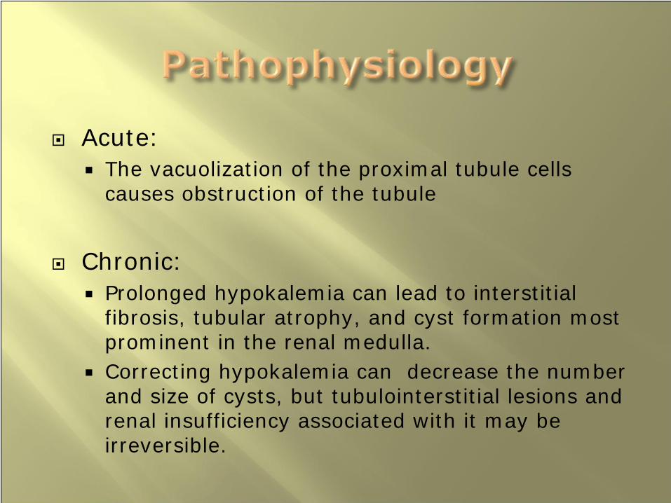
Acute:The vacuolization of the proximal tubule cells causes obstruction of the tubule
Chronic:Prolonged hypokalemia can lead to interstitial fibrosis, tubular atrophy, and cyst formation most prominent in the renal medulla.Correcting hypokalemia can decrease the number and size of cysts, but tubulointerstitial lesions and renal insufficiency associated with it may be irreversible.


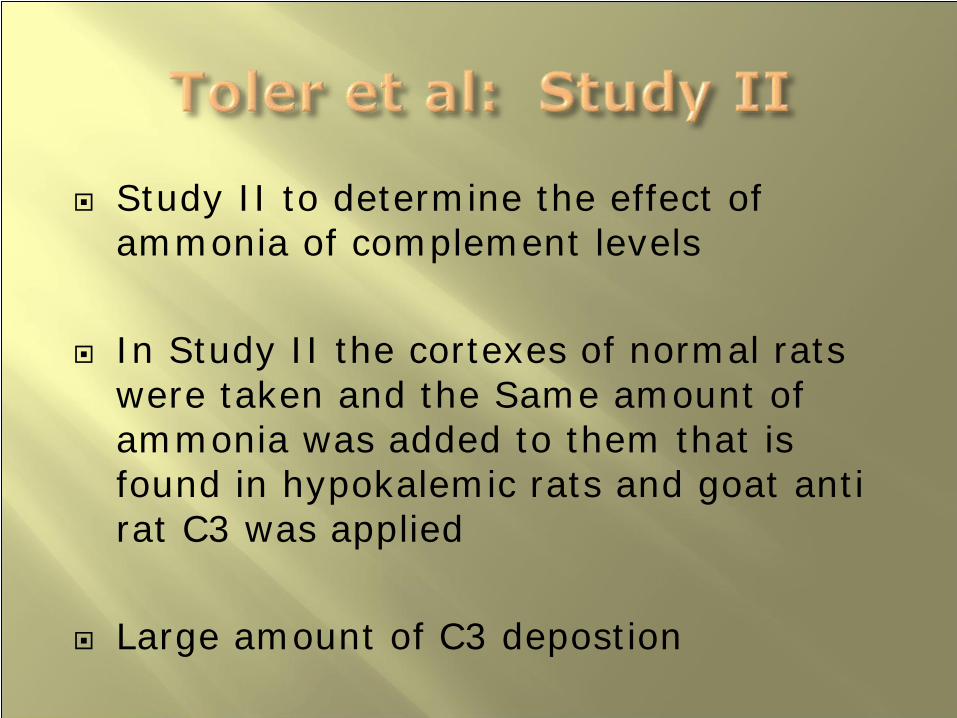
Study II to determine the effect of ammonia of complement levels
In Study II the cortexes of normal rats were taken and the Same amount of ammonia was added to them that is found in hypokalemic rats and goat anti rat C3 was applied
Large amount of C3 depostion

Vasoconstriction
Alternation in growth factors and cytokines: including VEG-F, IGF-1, IGFBP-1, Angiotensin II, MCP-1, and TGF-Beta
Decreased angiogenesis
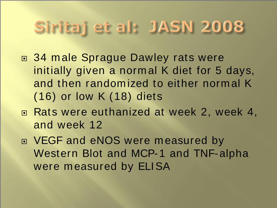
34 male Sprague Dawley rats were initially given a normal K diet for 5 days, and then randomized to either normal K (16) or low K (18) dietsRats were euthanized at week 2, week 4, and week 12VEGF and eNOS were measured by Western Blot and MCP-1 and TNF-alpha were measured by ELISA
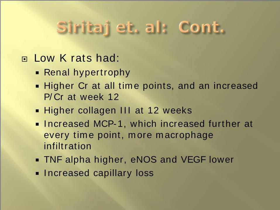
Low K rats had:Renal hypertrophyHigher Cr at all time points, and an increased P/Cr at week 12Higher collagen III at 12 weeksIncreased MCP-1, which increased further at every time point, more macrophage infiltrationTNF alpha higher, eNOS and VEGF lowerIncreased capillary loss

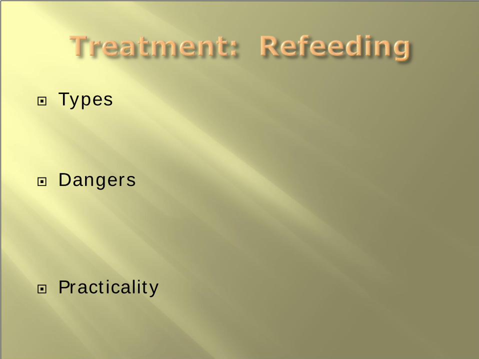
Types
Dangers
Practicality

Oral refeeding:Preferred
Indications for TPN:Failure of weight gain with standard oral feedingLife threatening weight lossWorsening psychological state
PEG

CardiovascularHypokalemiaHypophosphatemiaHypomagnesemiaGastrointestinal

Heart mass is reduced during anorexia
Increased circulation during refeeding
Heart failure may result

Increased glucose load causes insulin release, which caused K to shift into cells
Potassium will also be incorporated into tissues as the catabolic state switches to the anabolic state

Increased glucose load causes insulin release which causes phosphate to shift into cells
Inctreased glucose load causes insulin release which causes phosphate to shift into cells

Magnesium may be incorporated into newly formed tissue

LFTs:Early in refeeding LFTs may rise. Initially the AST and ALT, followed by the alkaline phosphatase, and then the bilirubin. There is no clinical significance
Motility:ConstipationDiarrhea:
Atrophy of the intestinal mucosa may have diarrhea early in refeeding

Calculation:Female: 100lbs for 5 feet, five lbs for every inch more than 5 feet tallMale: 106lbs for 5 feet, 6 lbs for every lb over
Patients may be calculated as mild, moderate, or severe depending on whether they are 10, 20, or 30% below IBWPt is acceptable when within 10% of IBW, or when menstruation

Basal metabolic rate is Based on the Harris-Benedict Formula:
6.55 + (9.6*body weight in kg) + (1.8*height in cm) – (4.7*age in years)
Total Energy Expenditure (TEE)Multipled by a factor of 1.2 to 2 to achieve the total energy expenditure

Intake levels usually start at 600-1000kcal/day and are increased by 300-400kcal every 3-4 days
Initially anorectics are metabolically inefficient, and the amount of calcoriesrequired for weight gain may vary between 1800 and 4500kcal

CV status should be check several times a day
Mag/Phos/K should be checked at least daily


![[PPT]Anorexia Nervosa - Mr Sitar's Website - homemrsitarswebsite.wikispaces.com/file/view/Anorexia Nervosa... · Web viewWhat is the definition to this illness? Anorexia nervosa is](https://img.pdfslide.us/doc/110x75/5af162f57f8b9ad0618f592d/pptanorexia-nervosa-mr-sitars-website-nervosaweb-viewwhat-is-the-definition.jpg)