Embed Size (px)
Citation preview

Revi
ews
and
Com
men
taRy
n R
evie
w f
oR R
esid
ents
28 radiology.rsna.org n Radiology: Volume 266: Number 1—January 2013
1 From the Department of Radiology, University of Alabama Medical Center, University of Alabama at Birmingham, 619 19th St South, JT N 331, Birmingham, AL 35249 (P.B., D.E.M.); and Department of Radiology, William Beaumont Hospital, Royal Oak, Mich (F.S., A.S.). Received December 21, 2011; revision requested February 9, 2012; revision received March 15; accepted April 3; final version accepted April 23. Address correspondence to P.B. (e-mail: [email protected]).
q RSNA, 2013
Peyman Borghei, MD Farnoosh Sokhandon, MD Ali Shirkhoda, MD Desiree E. Morgan, MD
Anomalies, Anatomic Variants, and Sources of Diagnostic Pitfalls in Pancreatic Imaging1
In this review, a brief discussion of the important events of pancreatic embryology is followed by presentation of con-genital anomalies and normal variants. For each variant, the appearance at different radiologic modalities including computed tomography, magnetic resonance (MR) imaging, endoscopic retrograde cholangiopancreatography, MR chol-angiopancreatography, and fluoroscopy will be demon-strated.
© RSNA, 2013
Note: This copy is for your personal non-commercial use only. To order presentation-ready copies for distribution to your colleagues or clients, contact us at www.rsna.org/rsnarights.

Review foR Residents: Anomalies, Anatomic Variants, and Diagnostic Pitfalls in Pancreatic Imaging Borghei et al
Radiology: Volume 266: Number 1—January 2013 n radiology.rsna.org 29
For the resident physician, differenti-ation of normal variants of the pan-creas from disease can be challeng-
ing. The goal of this review is to familiarize radiology residents and other practitioners with the basic knowledge of pancreatic imaging and to demonstrate normal con-ditions that, if not correctly diagnosed, might lead to unnecessary follow-up exam-inations or other diagnostic procedures.
Pancreatic Development
By the 4th week of embryologic develop-ment, the pancreatic duct develops from separate ventral and dorsal buds originat-ing from the endodermal lining of the du-odenum (1). The gallbladder, extrahe-patic bile ducts, central intrahepatic bile ducts, and ventral pancreas with its duc-tal network are derived from the ventral bud or outpouching. The dorsal bud is the precursor to dorsal pancreas and its ductal system (Fig 1a) (2). The ventral pancreas rotates clockwise posterior to the duodenum and comes into contact
with the dorsal pancreas in the 7th gesta-tional week to develop into the future pancreatic neck. The dorsal and ventral pancreatic buds grow into a pair of branching, arborizing ductal systems, each with its own central or main duct. The two anlagen fuse with each other and, together with the duodenum, fuse with the abdominal wall. After fusion, a new duct connects the distal portion of the dorsal pancreatic duct with the short-er duct of the ventral pancreas to form the main pancreatic duct, also known as the duct of Wirsung, which empties into the major papilla. The remnant of the dorsal duct forms the duct of Santorini, which drains into minor papilla (Fig 1b) (3). The process of pancreatic fusion is complicated, and a wide spectrum of an-atomic variants may occur during the course of pancreatic development.
Congenital Anomalies
Anatomic anomalies of the pancreas are classified as either a fusion anomaly (pan-creas divisum), migration anomaly (an-nular pancreas, ectopic pancreas), or duplication anomaly (number or form variation) (4). Pancreatic fusion or migra-tion anomalies may result in anatomic variants that predispose to specific pan-creatic or peripancreatic diseases.
Pancreas DivisumPancreas divisum results from a failure of ventral and dorsal bud fusion. The ventral (Wirsung) duct drains only the ventral pancreatic anlage, whereas the majority of the gland empties into the minor pa-pilla through the dorsal (Santorini) duct (3). It is the most common congenital anomaly, occurring in 4%–14% of the population, as evidenced by autopsy re-sults; 3%–8% are seen during endoscopic retrograde cholangiopancreatography (ERCP) and approximately 9% are de-picted at magnetic resonance (MR) chol-angiopancreatography (MRCP) (5–7). In most cases, there is no communication between the ventral and dorsal duct; however, in some individuals, there is a filamentous communication remaining and in others the ventral duct is totally absent (8). Although still debatable, pan-creas divisum is associated with acute
and recurrent pancreatitis. The reported frequency of complete pancreas divisum in patients with acute pancreatitis ranges from 25% to 38% (9–11).
In the past, ERCP was the modality of choice for diagnosing pancreas divisum; however, MRCP allows noninvasive multi-planar visualization of the biliary tree and pancreatic duct without injection of con-trast material and avoids risks of ERCP-induced pancreatitis or those associated with sedation required for the procedure. Advances in therapeutic endoscopic pro-cedures, such as minor papillotomy or insertion of stents into the minor papilla for treatment of patients with pancreatic divisum, make recognition of this variant important (12). The main anatomic fea-ture of pancreas divisum, continuity of the dorsal pancreatic duct with the duct of Santorini draining into the minor pa-pilla, is readily identified at both ERCP and MRCP. The ventral duct drains into the major papilla without communication with the dorsal pancreatic duct (13). On ERCP images, the pancreas divisum is optimally identified with the injection of contrast material into both the minor and major papillae (Fig 2). On MRCP images, the normal ventral system may be so small in caliber that the ducts are not vis-ible (Fig 3); however, it is the predomi-nant pancreatic duct drainage into the minor papilla at a level superior to the level of the bile duct emptying into the major papilla that indicates the presence of divisum. The administration of secre-tin during MRCP increases the visibility of the main pancreatic duct and its side branches and also increases the sensitiv-ity and specificity for detection of ana-tomic variants such as pancreas divisum (14–16). With the advent of multidetec-tor computer tomographic (CT) scanners and high-spatial-resolution thin-section imaging, pancreas divisum may be rou-tinely seen with use of CT as well (6).
Published online before print10.1148/radiol.12112469 Content code:
Radiology 2013; 266:28–36
Abbreviations:ERCP = endoscopic retrograde cholangiopancreatography MRCP = MR cholangiopancreatography
Conflicts of interest are listed at the end of this article.
Essentials
n Pancreatic fusion or migration anomalies may result in anatomic variants that predispose patients to specific pancreatic or peripan-creatic diseases such as recurrent acute pancreatitis, cystic dystro-phy of the duodenum, duodenal obstruction, cholangiocarcinoma, and gall bladder carcinoma.
n Pancreatic fusion or migration anomalies and normal variants such as fat deposition or acces-sory splenic tissue may give rise to imaging appearances that mimic more serious pancreatic conditions.
n Knowledge of pancreatic embry-ology and of normal anatomic variants is essential to identify these entities and to help differ-entiate them from pathologic conditions, thus preventing potential unnecessary imaging investigation or more invasive procedures such as biopsy or surgery.

Review foR Residents: Anomalies, Anatomic Variants, and Diagnostic Pitfalls in Pancreatic Imaging Borghei et al
30 radiology.rsna.org n Radiology: Volume 266: Number 1—January 2013
Figure 1: Schematic of pancreatic development. (a) Early development of the ventral (solid arrow) and dorsal buds (dashed arrow) prior to rotation. (b) Final configuration of the pancreas after periduodenal rotation of the ventral bud and approximation of the ventral and dorsal pancreas. The dorsal pancreatic duct merges with the ventral (Wirsung) duct and drains into major papilla. The rudi-mentary distal dorsal duct (Santorini) drains into minor papilla.
Figure 1
Figure 2: ERCP images of pancreas divisum in 26-year-old woman with abdominal pain. (a) The main pancreatic duct (arrows) drains through the minor papilla, consistent with pancreas divisum. (b) The short, terminally arborizing duct of Wirsung (arrow) is depicted by means of contrast material injection of the major papilla, with the common bile duct (arrowhead) also filling.
Figure 2
Figure 3: MRCP image of pancreas divisum in 46-year-old man. Angled coronal fat-suppressed half-Fourier acquisition single-shot turbo spin-echo (repetition time msec/echo time msec, 9450/740; 40-mm section thickness; 30-cm field of view) MR image shows the main pancreatic duct (dorsal San-torini duct, straight solid arrow) draining separately into the minor papilla (dashed arrow). The common bile duct (arrowheads) joins the smaller ventral pan-creatic duct (curved arrow) at a more inferior level and drains into the duodenum through the major papilla.
Figure 3
Figure 4: Schematic of annular pancreas. Ventral pancreas (arrowhead) encircles the second portion of the duodenum. The duct of the ventral pancreas (arrows) makes a turn around the duodenum and merges with the main dorsal duct in a normal fash-ion to form the Wirsung duct and drain to the major papilla.
Figure 4
Annular PancreasAnnular pancreas is a rare congenital anomaly in which incomplete rotation of the ventral anlage leads to a segment of the pancreas encircling the second part of duodenum (Fig 4). There are two types of annular pancreas: extramural and intramural. In the extramural type, the ventral pancreatic duct encircles the duodenum to join the main pancreatic duct. In the intramural type, the pancre-atic tissue is intermingled with muscle fibers in the duodenal wall, and small ducts drain directly into the duodenum. In patients with extramural annular pan-
creas, the presenting symptoms are those of high gastrointestinal obstruction. For patients with the intramural type, symp-toms are those of duodenal ulceration. Intervention for extramural obstructing annular pancreas is surgical and usually a bypass procedure; for intramural annular pancreas with duodenal ulceration, sub-total gastrectomy with or without vagot-omy is the procedure of choice (17). Pe-diatric patients with annular pancreas may have diagnostic findings on conven-tional abdominal radiographs, the classic double-bubble sign. The larger proximal bubble is caused by gastric distention and
the smaller distal bubble is caused by a dilated duodenal bulb (18,19). Barium examination reveals focal stenosis of the periampullary region, with an extrinsic eccentric defect of the second portion of the duodenum (Fig 5). Findings on CT scans include enlargement of the pancre-atic head, which encircles the second portion of the duodenum; this may be
Review foR Residents: Anomalies, Anatomic Variants, and Diagnostic Pitfalls in Pancreatic Imaging Borghei et al
Radiology: Volume 266: Number 1—January 2013 n radiology.rsna.org 31
identified either with or without oral con-trast material (Figs 6, 7). MR imaging demonstrates pancreatic tissue and occa-sionally the small annular duct encircling the descending duodenum. ERCP dem-onstrates normal main pancreatic duct
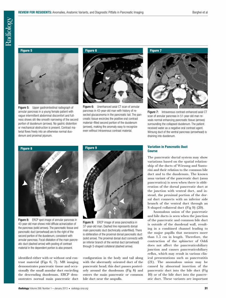
Figure 5: Upper gastrointestinal radiograph of annular pancreas in a young female patient with vague intermittent abdominal discomfort and full-ness shows slit-like smooth narrowing of the second portion of duodenum (arrows). No gastric distention or mechanical obstruction is present. Contrast ma-terial flows freely into an otherwise normal duo-denum and proximal jejunum.
Figure 5
Figure 6: Unenhanced axial CT scan of annular pancreas in 43-year-old man with history of re-sected glucaconoma in the pancreatic tail. The pan-creatic tissue encircles the positive oral contrast material–filled second portion of the duodenum (arrows), making the anomaly easy to recognize even without intravenous contrast material.
Figure 6
Figure 7: Intravenous contrast-enhanced axial CT scan of annular pancreas in 51-year-old man re-veals normal enhancing pancreatic tissue (arrows) surrounding the collapsed duodenum. The patient received water as a negative oral contrast agent. Wirsung duct of the ventral pancreas (arrowhead) is draining into duodenum.
Figure 7
Figure 8: ERCP spot image of annular pancreas in 43-year-old man shows mild diffuse acinarization of the pancreas (solid arrows). The pancreatic tissue and pancreatic duct (arrowhead) are to the right of the second portion of the duodenum, consistent with annular pancreas. Focal dilatation of the main pancre-atic duct (dashed arrow) with pooling of contrast material in the dependent portion is also present.
Figure 8
Figure 9: ERCP image of ansa pancreatica in 37-year-old man. Dashed line represents dorsal main pancreatic duct (technically underfilled). There is obliteration of the proximal dorsal pancreatic duct (solid arrow). The proximal dorsal duct connects with an inferior branch of the ventral duct (arrowhead) through S-shaped collateral (dashed arrow).
Figure 9 Variation in Pancreatic Duct Course
The pancreatic ductal system may show variations based on the spatial relation-ship of the ducts of Wirsung and Santo-rini and their relation to the common bile duct and to the duodenum. The known ansa variant of the pancreatic duct (ansa pancreatica) is seen when there is oblit-eration of the dorsal pancreatic duct at the junction with ventral duct, and in-stead, the proximal portion of the dor-sal duct connects with an inferior side branch of the ventral duct through an S-shaped collateral duct (Fig 9) (20).
Anomalous union of the pancreatic and bile ducts is seen when the junction of the pancreatic and common bile duct is outside of the duodenal wall, result-ing in a combined channel leading to the major papilla that measures more than 1.5 cm in length. Therefore, the contraction of the sphincter of Oddi does not affect the pancreaticobiliary junction and causes pancreaticobiliary reflux, which may result in various clin-ical presentations such as pancreatitis (21). The anomalous union may be caused by abnormal insertion of the pancreatic duct into the bile duct (Fig 10) or of the bile duct into the pancre-atic duct. These variants are important
configuration in the body and tail along with the aberrantly oriented duct of the pancreatic head; this duct passes posteri-orly around the duodenum (Fig 8) and enters the main pancreatic or common bile duct near the ampulla.

Review foR Residents: Anomalies, Anatomic Variants, and Diagnostic Pitfalls in Pancreatic Imaging Borghei et al
32 radiology.rsna.org n Radiology: Volume 266: Number 1—January 2013
Figure 10: ERCP spot image in 54-year-old woman with anomalous pancreaticobiliary duct union shows abnormal proximal insertion of the pancreatic duct to the normal-appearing common bile duct, resulting in a long combined pancreatic-common bile duct channel more than 1.5 cm in length (arrow).
Figure 10
Figure 11: Contrast-enhanced axial multidetector CT scans of congenital short pancreas (hypoplasia) in 28-year-old man show (a) the head and uncinate process of the pancreas (arrows) with (b) absence of the body and tail.
Figure 11
Figure 12: Spot radiograph from upper gastroin-testinal examination in 33-year-old woman reveals a focal, smooth, round filling defect (arrows) on the inferior wall of duodenal bulb (DB), with tiny umbili-cation (arrowhead) characteristic of ectopic pan-creas. At endoscopy, the lesion was raised and epithelialized, looking like a thickened papilla.
Figure 12
to recognize as there is an increased association with choledochal cysts (22) and gallbladder carcinoma (23).
Agenesis and Hypoplasia of the PancreasAgenesis of the pancreas is rare and generally incompatible with life. Hypo-plasia results from absence of the ven-tral or dorsal anlagen. Partial agenesis of the dorsal pancreas is more common than the agenesis of the ventral portion, but complete agenesis of the dorsal pan-creas is extremely rare (3). At imaging, dorsal pancreatic hypoplasia manifests as a short, rounded pancreatic head ad-jacent to the duodenum, with absence of the pancreatic neck, body, and tail (Fig 11). Pancreas hypoplasia may be associ-ated with an increased risk of pancreati-tis and polysplenia syndrome (24).
Ectopic PancreasEctopic pancreatic tissue is usually seen in the submucosa of the gastric antrum (30%), the proximal portion of the duo-denum (30%), the remaining duodenum (20%), or other regions of the small bowel (20%) (25). The most widely ac-cepted theory of the embryologic origin of this lesion is its development from resid-ual cells of the primitive ventral or dorsal
bud within the bowel lumen (26). The le-sions are composed of normal pancreatic tissue, often including islet cells, and usu-ally have a small pancreatic duct. Ectopic pancreatic tissue may be identified at bar-ium upper gastrointestinal examination, where features of a small collection of barium located within a central niche or umbilication on a small rounded mass are diagnostic (Fig 12). The umbilication rep-resents the orifice of the rudimentary duct through which the ectopic pancreas empties (27). If this feature is absent, the lesion cannot be reliably differentiated ra-diographically from other submucosal tu-mors such as Brunner gland adenoma, leiomyoma, or lymphoma. Ectopic pan-creatic tissue is functional and subject to the same inflammatory and neoplastic disorders that afflict the normal pancreas; however, the majority of cases are asymp-tomatic and found incidentally (28,29). Cystic dystrophy, a serious but uncom-mon complication, represents dilatation of the ectopic pancreatic ducts within the heterotopic pancreatic tissue and occurs most often in the second part of the duo-denum (30). It is thought to result from obstruction of the small ducts leading to repeated pancreatitis. Chronic alcohol consumption has been reported to trigger cystic dystrophy in ectopic pancreatic tis-sue (31). Imaging features include thick-ened duodenal wall containing multiple cysts, with moderate to strong enhance-ment. Adjacent inflammatory changes with or without enlarged lymph nodes can be seen in half of the patients (32).
Cystic dystrophy is one of several benign conditions that affect the duodenal wall in the region of the minor pancreatic papilla. Others include pancreatic hamartoma, paraduodenal wall cyst, myoadenomato-sis, and groove pancreatitis, which has been collectively termed paraduodenal pancreatitis (33,34). In general, these conditions produce a thickened duodenal wall that may contain dilated ducts and pseudocystic changes, Brunner gland hy-perplasia, or dense myxoid stromal prolif-eration with intervening rounded lobules of pancreatic tissue (35).
Review foR Residents: Anomalies, Anatomic Variants, and Diagnostic Pitfalls in Pancreatic Imaging Borghei et al
Radiology: Volume 266: Number 1—January 2013 n radiology.rsna.org 33
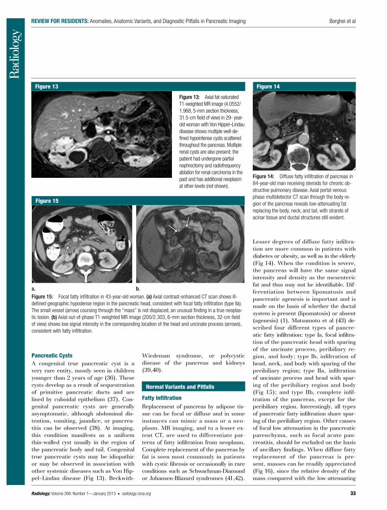
Figure 14: Diffuse fatty infiltration of pancreas in 84-year-old man receiving steroids for chronic ob-structive pulmonary disease. Axial portal venous phase multidetector CT scan through the body re-gion of the pancreas reveals low-attenuating fat replacing the body, neck, and tail, with strands of acinar tissue and ductal structures still evident.
Figure 14
Pancreatic CystsA congenital true pancreatic cyst is a very rare entity, mostly seen in children younger than 2 years of age (36). These cysts develop as a result of sequestration of primitive pancreatic ducts and are lined by cuboidal epithelium (37). Con-genital pancreatic cysts are generally asymptomatic, although abdominal dis-tention, vomiting, jaundice, or pancrea-titis can be observed (38). At imaging, this condition manifests as a uniform thin-walled cyst usually in the region of the pancreatic body and tail. Congenital true pancreatic cysts may be idiopathic or may be observed in association with other systemic diseases such as Von Hip-pel–Lindau dis ease (Fig 13), Beckwith-
Wiedeman syn drome, or polycystic disease of the pancreas and kidneys (39,40).
Normal Variants and Pitfalls
Fatty InfiltrationReplacement of pancreas by adipose tis-sue can be focal or diffuse and in some instances can mimic a mass or a neo-plasm. MR imaging, and to a lesser ex-tent CT, are used to differentiate pat-terns of fatty infiltration from neoplasm. Complete replacement of the pancreas by fat is seen most commonly in patients with cystic fibrosis or occasionally in rare conditions such as Schwachman-Diamond or Johansen-Blizzard syndromes (41,42).
Figure 13
Figure 13: Axial fat-saturated T1-weighted MR image (4.0552/ 1.968, 5-mm section thickness, 31.5-cm field of view) in 29- year-old woman with Von Hippel–Lindau disease shows multiple well-de-fined hypointense cysts scattered throughout the pancreas. Multiple renal cysts are also present; the patient had undergone partial nephrectomy and radiofrequency ablation for renal carcinoma in the past and has additional neoplasm at other levels (not shown).
Figure 15: Focal fatty infiltration in 43-year-old woman. (a) Axial contrast-enhanced CT scan shows ill-defined geographic hypodense region in the pancreatic head, consistent with focal fatty infiltration (type IIa). The small vessel (arrow) coursing through the “mass” is not displaced, an unusual finding in a true neoplas-tic lesion. (b) Axial out-of-phase T1-weighted MR image (200/2.303, 6-mm section thickness, 32-cm field of view) shows low signal intensity in the corresponding location of the head and uncinate process (arrows), consistent with fatty infiltration.
Figure 15
Lesser degrees of diffuse fatty infiltra-tion are more common in patients with diabetes or obesity, as well as in the elderly (Fig 14). When the condition is severe, the pancreas will have the same signal intensity and density as the mesenteric fat and thus may not be identifiable. Dif-ferentiation between lipomatosis and pancreatic agenesis is important and is made on the basis of whether the ductal system is present (lipomatosis) or absent (agenesis) (1). Matsumoto et al (43) de-scribed four different types of pancre-atic fatty infiltration: type Ia, focal infiltra-tion of the pancreatic head with sparing of the uncinate process, peribiliary re-gion, and body; type Ib, infiltration of head, neck, and body with sparing of the peribiliary region; type IIa, infiltration of uncinate process and head with spar-ing of the peribiliary region and body (Fig 15); and type IIb, complete infil-tration of the pancreas, except for the peribiliary region. Interestingly, all types of pancreatic fatty infiltration share spar-ing of the peribiliary region. Other causes of focal low attenuation in the pancreatic parenchyma, such as focal acute pan-creatitis, should be excluded on the basis of ancillary findings. When diffuse fatty replacement of the pancreas is pre-sent, masses can be readily appreciated (Fig 16), since the relative density of the mass compared with the low-attenuating

Review foR Residents: Anomalies, Anatomic Variants, and Diagnostic Pitfalls in Pancreatic Imaging Borghei et al
34 radiology.rsna.org n Radiology: Volume 266: Number 1—January 2013
fat alters perception and potentially af-fects mass characterization.
PseudomassNormal alteration of the pancreatic con-tour is referred to as pseudomass and can mimic pancreatic neoplasm. Lobu-lation of the pancreatic head and neck is defined as extension of the parenchyma beyond 1 cm relative to the anterior superior pancreaticoduodenal artery, whether anteriorly (type I), posteriorly (type II) (Fig 17), or horizontally (type III) (44). Enlargement of lymph nodes adjacent to the pancreas can mimic a pancreatic mass (Fig 18) and may re-sult in potential diagnostic challenges. Paraaortic and periportal lymphadenopa-thy should be differentiated from pan-creatic neoplasm to avoid unnecessary additional diagnostic procedures. Occa-sionally, accessory splenic tissue may be located in the body-tail region of the pancreas, mimicking an infiltrative process such as autoimmune pancreati-tis or neoplasm, especially a pancreatic endocrine tumor. This entity can gener-ally be recognized at multiphasic CT, since the tissue follows the enhancement pat-tern of the spleen (45). When CT is not definitive, MR imaging is very useful because the signal in the accessory splenic tissue is more readily defined (Fig 19), compared with the surround-ing pancreatic parenchyma (46). Heat-damaged red blood cells labeled with
Figure 16: Pancreatic adenocarcinoma in 64-year-old man with fatty replaced gland. Axial contrast-enhanced multidetector CT scan shows slightly heterogeneous hypovascular adenocarci-noma (arrow) in the region of the head. The fatty replacement provides inherent tissue contrast and thus increases conspicuity of this lesion.
Figure 16
Figure 18: Peripancreatic and periportal lymph nodes mimicking pancreatic mass in 52-year-old woman. Portal venous phase multidetector CT scan obtained to evaluate generalized abdominal pain shows cirrhotic liver contour and prominent node anterior to the common hepatic artery (arrow), mim-icking a partially exophytic pancreatic mass. Note the slight difference in attenuation of pancreatic parenchyma compared with the nodes, which helps in differentiation.
Figure 18
Figure 17: Prominent pancreatic head in 28-year-old woman. Note the lateral extension of the pancre-atic head (arrows) in relation to anterior superior pancreaticoduodenal artery (arrowhead), represent-ing type II (posterior) lobulated pancreas head var-iant. D 5 duodenum.
Figure 17
Figure 19: Intrapancreatic accessory spleen in 54-year-old man with an incidental pancreatic mass. (a) Contrast-enhanced axial multidetector CT scan shows a dense pancreatic body mass (arrow). (b) Contrast-enhanced T1-weighted fat-suppressed axial MR image (4.2011/2.069, 5-mm section thickness, 36-cm field of view) shows pancreatic body lesion that enhances slightly more than normal pancreatic parenchyma on (c) early postgadolinium-enhanced pancreatic phase MR image. (d) T2-weighted MR image (1800/70, 6-mm section thickness, 34-cm field of view) shows features also consistent with splenic tissue rather than a mass.
Figure 19

Review foR Residents: Anomalies, Anatomic Variants, and Diagnostic Pitfalls in Pancreatic Imaging Borghei et al
Radiology: Volume 266: Number 1—January 2013 n radiology.rsna.org 35
technetium 99m (99mTc) or 99mTc-la-beled sulfur colloid scans can also be used to diagnose accessory splenic tis-sue, showing increased uptake in the region of interest (47). Focal autoim-mune pancreatitis can also manifest as a pseudomass and focal parenchymal en-largement. One imaging feature that can help distinguish this entity from pancreatic adenocarcinoma is the pat-tern of enhancement. At the portal ve-nous phase, focal autoimmune pancrea-titis may appear hyperdense compared with the usual hypodense pancreatic ade-nocarcinoma (48).
In conclusion, congenital anomalies and normal variants of the pancreas can present a diagnostic challenge when en-countered. Knowledge of pancreatic em-bryology and of normal anatomic vari-ants is essential to identify these entities and help differentiate them from patho-logic conditions, thus preventing poten-tial unnecessary imaging investigation or more invasive procedures such as biopsy or surgery.
Disclosures of Conflicts of Interest: P.B. No rel-evant conflicts of interest to disclose. F.S. No rel-evant conflicts of interest to disclose. A.S. No relevant conflicts of interest to disclose. D.E.M. No relevant conflicts of interest to disclose.
References 1. Mortelé KJ, Rocha TC, Streeter JL, Taylor AJ.
Multimodality imaging of pancreatic and bil-iary congenital anomalies. RadioGraphics 2006;26(3):715–731.
2. Rizzo RJ, Szucs RA, Turner MA. Congenital abnormalities of the pancreas and biliary tree in adults. RadioGraphics 1995;15(1): 49–68; quiz 147–148.
3. Shirkhoda A, Borghei P. Anomalies and ana-tomic variants of the pancreas. In: Gore RM, Levine MS, eds. Textbook of gastrointestinal radiology. 3rd ed. Vol 2. Philadelphia, Pa: Elsevier, 2007; 1869–1884.
4. Siegel HJ. Radiologic interpretation: normal biliary system and variations; normal pan-creatic duct and variations. In: Siegel HJ, ed. Endoscopic retrograde cholangiopancre-atography: technique, diagnosis and therapy. New York, NY: Raven, 1992; 41–59.
5. Lehman GA, Sherman S. Diagnosis and ther-apy of pancreas divisum. Gastrointest Endosc Clin N Am 1998;8(1):55–77.
6. Soto JA, Lucey BC, Stuhlfaut JW. Pancreas divisum: depiction with multi-detector row CT. Radiology 2005;235(2):503–508.
7. Morgan DE, Logan K, Baron TH, Koehler RE, Smith JK. Pancreas divisum: implications for diagnostic and therapeutic pancreatog-raphy. AJR Am J Roentgenol 1999;173(1): 193–198.
8. Yu J, Turner MA, Fulcher AS, Halvorsen RA. Congenital anomalies and normal variants of the pancreaticobiliary tract and the pan-creas in adults. II. Pancreatic duct and pan-creas. AJR Am J Roentgenol 2006;187(6): 1544–1553.
9. Uomo G, Manes G, D’Anna L, Laccetti M, Di Gaeta S, Rabitti PG. Fusion and duplica-tion variants of pancreatic duct system: clin-ical and pancreatographic evaluation. Int J Pancreatol 1995;17(1):23–28.
10. Cotton PB. Congenital anomaly of pancreas divisum as cause of obstructive pain and pancreatitis. Gut 1980;21(2):105–114.
11. Bertin C, Pelletier AL, Vullierme MP, et al. Pancreas divisum is not a cause of pancrea-titis by itself but acts as a partner of genetic mutations. Am J Gastroenterol 2012;107(2): 311–317.
12. Lehman GA, Sherman S. Diagnosis and therapy of pancreas divisum. Gastrointest Endosc Clin N Am 1998;8(1):55–77.
13. Bret PM, Reinhold C, Taourel P, Guibaud L, Atri M, Barkun AN. Pancreas divisum: eval-uation with MR cholangiopancreatography. Radiology 1996;199(1):99–103.
14. Matos C, Metens T, Devière J, Delhaye M, Le Moine O, Cremer M. Pancreas divisum: evaluation with secretin-enhanced magnetic resonance cholangiopancreatography. Gas-trointest Endosc 2001;53(7):728–733.
15. Manfredi R, Costamagna G, Brizi MG, et al. Severe chronic pancreatitis versus sus-pected pancreatic disease: dynamic MR chol-angiopancreatography after secretin stimu-lation. Radiology 2000;214(3):849–855.
16. Manfredi R, Costamagna G, Brizi MG, et al. Pancreas divisum and “santorinicele”: diagno-sis with dynamic MR cholangiopancreatog-raphy with secretin stimulation. Radiology 2000;217(2):403–408.
17. Johnston DW. Annular pancreas: a new classi-fication and clinical observations. Can J Surg 1978;21(3):241–244.
18. Jimenez JC, Emil S, Podnos Y, Nguyen N. Annular pancreas in children: a recent de-cade’s experience. J Pediatr Surg 2004; 39(11):1654–1657.
19. Sencan A, Mir E, Günsar C, Akcora B. Symptomatic annular pancreas in newborns. Med Sci Monit 2002;8(6):CR434–CR437.
20. Ishii H, Arai K, Fukushima M, et al. Fusion variations of pancreatic ducts in patients with
anomalous arrangement of pancreatico-biliary ductal system. J Hepatobiliary Pan-creat Surg 1998;5(3):327–332.
21. Matsumoto Y, Fujii H, Itakura J, et al. Pan-creaticobiliary maljunction: pathophysiolog-ical and clinical aspects and the impact on biliary carcinogenesis. Langenbecks Arch Surg 2003;388(2):122–131.
22. Cha SW, Park MS, Kim KW, et al. Chole-dochal cyst and anomalous pancreaticobiliary ductal union in adults: radiological spec-trum and complications. J Comput Assist Tomogr 2008;32(1):17–22.
23. Kimura K, Ohto M, Saisho H, et al. Associ-ation of gallbladder carcinoma and anom-alous pancreaticobiliary ductal union. Gas-troenterology 1985;89(6):1258–1265.
24. Schnedl WJ, Piswanger-Soelkner C, Wallner SJ, et al. Agenesis of the dorsal pancreas and associated diseases. Dig Dis Sci 2009;54(3): 481–487.
25. Thoeni RF, Gedgaudas RK. Ectopic pan-creas: usual and unusual features. Gastro-intest Radiol 1980;5(1):37–42.
26. Sternberg SS, Mills SE, Carter D, eds. Sternberg’s diagnostic surgical pathology. 4th ed. Vol 1. Philadelphia, Pa: Lippincott Williams & Wilkins, 2004; 1486.
27. Harold KL, Sturdevant M, Matthews BD, Mishra G, Heniford BT. Ectopic pancre-atic tissue presenting as submucosal gastric mass. J Laparoendosc Adv Surg Tech A 2002;12(5):333–338.
28. Eisenberger CF, Gocht A, Knoefel WT, et al. Heterotopic pancreas: clinical presentation and pathology with review of the literature. Hepatogastroenterology 2004;51(57):854–858.
29. Cho JS, Shin KS, Kwon ST, et al. Hetero-topic pancreas in the stomach: CT findings. Radiology 2000;217(1):139–144.
30. Tison C, Regenet N, Meurette G, et al. Cystic dystrophy of the duodenal wall developing in heterotopic pancreas: report of 9 cases. Pancreas 2007;34(1):152–156.
31. Jouannaud V, Coutarel P, Tossou H, et al. Cys-tic dystrophy of the duodenal wall associated with chronic alcoholic pancreatitis: clinical features, diagnostic procedures and thera-peutic management in a retrospective multi-center series of 23 patients. Gastroenterol Clin Biol 2006;30(4):580–586.
32. Vullierme MP, Vilgrain V, Fléjou JF, et al. Cystic dystrophy of the duodenal wall in the heterotopic pancreas: radiopathological cor-relations. J Comput Assist Tomogr 2000;24(4): 635–643.
33. Adsay NV, Zamboni G. Paraduodenal pancre-atitis: a clinico-pathologically distinct entity
Review foR Residents: Anomalies, Anatomic Variants, and Diagnostic Pitfalls in Pancreatic Imaging Borghei et al
36 radiology.rsna.org n Radiology: Volume 266: Number 1—January 2013
unifying “cystic dystrophy of heterotopic pan-creas”, “para-duodenal wall cyst”, and “groove pancreatitis”. Semin Diagn Pathol 2004;21(4): 247–254.
34. Blasbalg R, Baroni RH, Costa DN, Machado MC. MRI features of groove pancreatitis. AJR Am J Roentgenol 2007;189(1):73–80.
35. Thomas H, Marriott P, Portmann B, Heaton N, Rela M. Cystic dystrophy in heterotopic pan-creas: a rare indication for pancreaticoduo-denectomy. Hepatobiliary Pancreat Dis Int 2009;8(2):215–217.
36. Casadei R, Campione O, Greco VM, Marrano D. Congenital true pancreatic cysts in young adults: case report and literature review. Pancreas 1996;12(4):419–421.
37. Cotran RS, Kumar V, Robbins SL. Robbins pathological basis of disease. 4th ed. Phila-delphia, Pa: Saunders, 1989; 981–1010.
38. Agarwala S, Lal A, Bhatnagar V, Dinda AK, Mitra DK. Congenital true pancreatic cyst:
presentation and management. Trop Gas-troenterol 1999;20(2):87–88.
39. Boulanger SC, Borowitz DS, Fisher JF, Brisseau GF. Congenital pancreatic cysts in children. J Pediatr Surg 2003;38(7): 1080–1082.
40. Auringer ST, Ulmer JL, Sumner TE, Turner CS. Congenital cyst of the pancreas. J Pedi-atr Surg 1993;28(12):1570–1571.
41. Robberecht E, Nachtegaele P, Van Rattinghe R, Afschrift M, Kunnen M, Verhaaren R. Pancreatic lipomatosis in the Shwachman-Diamond syndrome: identification by sonog-raphy and CT-scan. Pediatr Radiol 1985; 15(5):348–349.
42. Maunoury V, Nieuwarts S, Ferri J, Ernst O. Pancreatic lipomatosis revealing Johanson-Blizzard syndrome [in French]. Gastroenterol Clin Biol 1999;23(10):1099–1101.
43. Matsumoto S, Mori H, Takaki H, et al. Un-even fatty replacement of the pancreas: evaluation with CT. Radiology 1995;194(2): 453–458.
44. Ross BA, Jeffrey RB Jr, Mindelzun RE. Nor-mal variations in the lateral contour of the head and neck of the pancreas mimicking neoplasm: evaluation with dual-phase helical CT. AJR Am J Roentgenol 1996;166(4): 799–801.
45. Mortelé KJ, Mortelé B, Silverman SG. CT features of the accessory spleen. AJR Am J Roentgenol 2004;183(6):1653–1657.
46. Kim SH, Lee JM, Han JK, et al. Intrapancre-atic accessory spleen: findings on MR Imag-ing, CT, US and scintigraphy, and the path-ologic analysis. Korean J Radiol 2008;9(2): 162–174.
47. Belkhir SM, Archambaud F, Prigent A, Chaumet-Riffaud P. Intrapancreatic ac-cessory spleen diagnosed on radionu-clide im aging. Clin Nucl Med 2009;34(9): 642–644.
48. Manfredi R, Graziani R, Cicero C, et al. Au-toimmune pancreatitis: CT patterns and their changes after steroid treatment. Radiology 2008;247(2):435–443.