Embed Size (px)
Citation preview
ANNUAL MEETING OF SLOVAK SOCIETY FOR NEUROSCIENCE
&CENTRE OF EXCELLENCE
FOR BRAIN RESEARCH
Smolenice Castle, SlovakiaMay 24 – 26, 2012
Organized by
The Slovak Society for NeuroscienceThe Centre of Excellence for Brain Research
The Institute of Neuroimmunology – Slovak Academy of Sciences
1
ANNUAL MEETING OF SLOVAK SOCIETY FOR NEUROSCIENCE
&CENTRE OF EXCELLENCE
FOR BRAIN RESEARCH
Programme and Abstracts Book
Smolenice Castle, SlovakiaMay 24 – 26, 2012
3
COORGANIZERS The Institute of Neurobiology - Slovak Academy of SciencesThe Institute of Experimental Endocrinology – Slovak Academy of SciencesThe Jessenius Faculty of Medicine in Martin – Comenius UniversityThe Faculty of Medicine in Bratislava – Comenius UniversityThe University of Veterinary Medicine and Pharmacy in Košice The Memory Centre
PROGRAMME COMMITTEENOVAK Michal – chairman LUKACOVA NadezdaDOBROTA DusanKVETNANSKY RichardLEHOTSKY JanCIZKOVA DasaOSTATNIKOVA DanielaVESELA AlzbetaPISTL Juraj
ORGANIZING COMMITTEEFILIPCIK Peter – chairman CENTE MartinJEZOVICOVA MartinaONDREJICKOVA ZuzanaPRCINA MichalREVICKA ZuzanaZILKOVA MonikaSTUDENIC MartinNOVAK Lukas
ADDRESS OF THE SECRETARIATThe Institute of NeuroimmunologySlovak Academy of SciencesDubravska cesta 9845 10 BRATISLAVA 45Phone: +2-54788100Fax: +2-54774276Email: [email protected]
Annual Meeting of Slovak Society for Neuroscience and Centre of Excellence for Brain Research
5
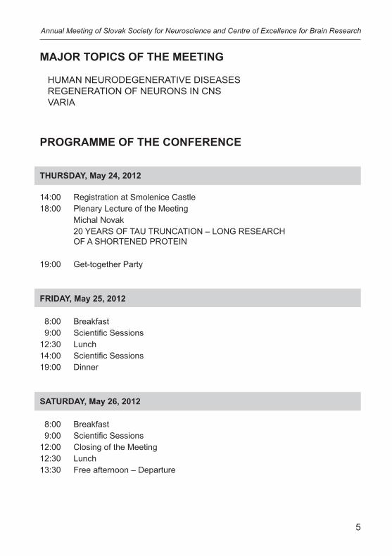
MAJOR TOPICS OF THE MEETING
HUMAN NEURODEGENERATIVE DISEASESREGENERATION OF NEURONS IN CNS VARIA
PROGRAMME OF THE CONFERENCE
THURSDAY, May 24, 2012
14:00 Registration at Smolenice Castle18:00 Plenary Lecture of the Meeting Michal Novak 20 YEARS OF TAU TRUNCATION – LONG RESEARCH OF A SHORTENED PROTEIN 19:00 Get-together Party
FRIDAY, May 25, 2012
8:00 Breakfast 9:00 Scientific Sessions12:30 Lunch14:00 Scientific Sessions19:00 Dinner
SATURDAY, May 26, 2012
8:00 Breakfast 9:00 Scientific Sessions12:00 Closing of the Meeting12:30 Lunch13:30 Free afternoon – Departure
Annual Meeting of Slovak Society for Neuroscience and Centre of Excellence for Brain Research
6
Annual Meeting of Slovak Society for Neuroscience and Centre of Excellence for Brain Research
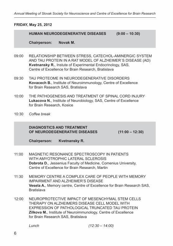
FRIDAY, May 25, 2012
HUMAN NEURODEGENERATIVE DISEASES (9:00 – 10:30)
Chairperson: Novak M.
09:00 RELATIONSHIP BETWEEN STRESS, CATECHOL-AMINERGIC SYSTEM AND TAU PROTEIN IN A RAT MODEL OF ALZHEIMER´S DISEASE (AD) Kvetnansky R., Instute of Experimental Endocrinology, SAS, Centre of Excellence for Brain Research, Bratislava
09:30 TAU PROTEOME IN NEURODEGENERATIVE DISORDERS Kovacech B., Institute of Neuroimmunology, Centre of Excellence for Brain Research SAS, Bratislava
10:00 THE PATHOGENESIS AND TREATMENT OF SPINAL CORD INJURY Lukacova N., Institute of Neurobiology, SAS, Centre of Excellence for Brain Research, Kosice
10:30 Coffee break
DIAGNOSTICS AND TREATMENT OF NEURODEGENERATIVE DISEASES (11:00 – 12:30) Chairperson: Kvetnansky R.
11:00 MAGNETIC RESONANCE SPECTROSCOPY IN PATIENTS WITH AMYOTROPHIC LATERAL SCLEROSIS Dobrota D., Jessenius Faculty of Medicine, Comenius University, Centre of Excellence for Brain Research, Martin
11:30 MEMORY CENTRE A COMPLEX CARE OF PEOPLE WITH MEMORY IMPAIRMENT AND ALZHEIMER’S DISEASE Vesela A., Memory centre, Centre of Excellence for Brain Research SAS, Bratislava
12:00 NEUROPROTECTIVE IMPACT OF MESENCHYMAL STEM CELLS THERAPY ON ALZHEIMERS DISEASE CELL MODEL WITH EXPRESSION OF PATHOLOGICAL TRUNCATED TAU PROTEIN Zilkova M., Institute of Neuroimmunology, Centre of Excellence for Brain Research SAS, Bratislava
Lunch (12:30 – 14:00)
7
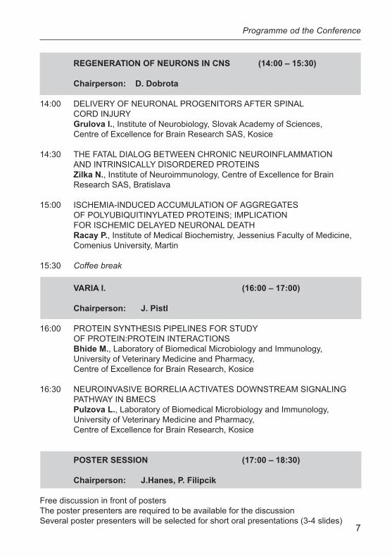
REGENERATION OF NEURONS IN CNS (14:00 – 15:30) Chairperson: D. Dobrota
14:00 DELIVERY OF NEURONAL PROGENITORS AFTER SPINAL CORD INJURY Grulova I., Institute of Neurobiology, Slovak Academy of Sciences, Centre of Excellence for Brain Research SAS, Kosice
14:30 THE FATAL DIALOG BETWEEN CHRONIC NEUROINFLAMMATION AND INTRINSICALLY DISORDERED PROTEINS Zilka N., Institute of Neuroimmunology, Centre of Excellence for Brain Research SAS, Bratislava
15:00 ISCHEMIA-INDUCED ACCUMULATION OF AGGREGATES OF POLYUBIQUITINYLATED PROTEINS; IMPLICATION FOR ISCHEMIC DELAYED NEURONAL DEATH Racay P., Institute of Medical Biochemistry, Jessenius Faculty of Medicine, Comenius University, Martin
15:30 Coffee break
VARIA I. (16:00 – 17:00)
Chairperson: J. Pistl
16:00 PROTEIN SYNTHESIS PIPELINES FOR STUDY OF PROTEIN:PROTEIN INTERACTIONS Bhide M., Laboratory of Biomedical Microbiology and Immunology, University of Veterinary Medicine and Pharmacy, Centre of Excellence for Brain Research, Kosice
16:30 NEUROINVASIVE BORRELIA ACTIVATES DOWNSTREAM SIGNALING PATHWAY IN BMECS Pulzova L., Laboratory of Biomedical Microbiology and Immunology, University of Veterinary Medicine and Pharmacy, Centre of Excellence for Brain Research, Kosice
POSTER SESSION (17:00 – 18:30)
Chairperson: J.Hanes, P. Filipcik
Free discussion in front of postersThe poster presenters are required to be available for the discussionSeveral poster presenters will be selected for short oral presentations (3-4 slides)
Programme od the Conference
8
Annual Meeting of Slovak Society for Neuroscience and Centre of Excellence for Brain Research
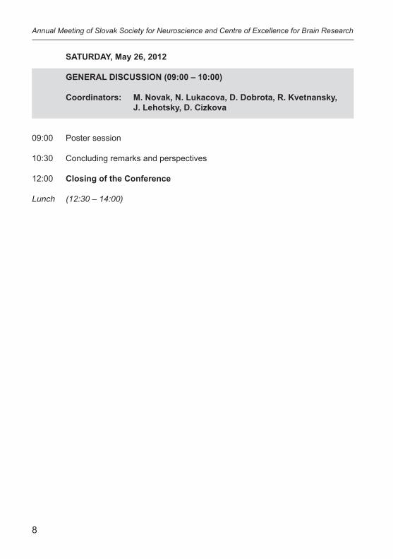
SATURDAY, May 26, 2012
GENERAL DISCUSSION (09:00 – 10:00)
Coordinators: M. Novak, N. Lukacova, D. Dobrota, R. Kvetnansky, J. Lehotsky, D. Cizkova
09:00 Poster session
10:30 Concluding remarks and perspectives
12:00 Closing of the Conference
Lunch (12:30 – 14:00)
9
Programme od the Conference
POSTERS
P1. Bosikova E., Niederova-Kubikova L., Jarvis E.D.STRIATAL RECOVERY AFTER NEUROTOXIC BILATERAL LESION
P2.Brecik M., Barath P., Kovacech B., Salingova B., Novak M.IDENTIFICATION AND ANALYSIS OF THE SOLUBLE TRUNCATED TAU SPECIES
P3.Bugos O., Zilka N., Kucerak J., Novak P., Stozicka Z.,Koson P., Filipcik P., Novak M.NOVEL TRANSGENIC RAT MODEL FOR HUMAN TAUOPATHY SHOWS PROGRESSIVE NEUROFIBRILLARY DEGENERATION IN THE CORTEX WITHOUT PROMINENT NEURONAL LOSS
P4.Bundzikova J., Majercikova Z., Mikkelsen J.D., Kiss A.DIVERSITIES IN THE STIMULATORY EFFECT OF ANTIPSYCHOTICS ON THE PARAVENTRICULAR NUCLEUS OXYTOCINERGIC NEURONS
P5.Cehlar O., Skrabana R., Kovac A., Kovacech B.,Novak M.STRUCTURAL INSIGHTS INTO THE MICROTUBULE-BINDING REGIONS OF THE INTRINSICALLY DISORDERED PROTEIN TAU
P6.Cente M., Filipcik P., Opattova A., Novak M.TRUNCATED HUMAN TAU PROTEIN INDUCES CELLULAR STRESS AND INFLAMMATORY PHENOTYPE IN A RAT MODEL OF TAUOPATHY
P7.Chomova M., Muchova J., Durackova Z.THE IMPACT OF UNCONTROLLED HYPERGLYCEMIA ON RAT BRAIN MITOCHONDRIA
P8.Durdiakova J., Kubranska A., Ostatnikova D., Celec P.ANDROGEN RECEPTOR POLYMORPHISM AFFECTS MENTAL ROTATION ABILITY IN INTELLECTUALLY GIFTED BOYS
10
Annual Meeting of Slovak Society for Neuroscience and Centre of Excellence for Brain Research
P9.Flachbartova Z., Kovacech B., Skrabana R., Novak M.IDENTIFICATION OF TAU INTERACTING PARTNERS IN RAT MODEL OF TAUOPATHY
P10.Hresko S., Natarajan S., Bhide M.RAPID PIPELINE FOR PROTEIN PRODUCTION IN LEISHMANIA CELL FREE EXPRESSION SYSTEM
P11.Husarova V., Bittsansky M., Ondrejka I.MEDICATION EFFECTS ON NEUROMETABOLITES IN ADHD: A 1H MAGNETIC RESONANCE SPECTROSCOPY STUDY
P12.Jadhav S., Zilka N., Marosova L., Neradil P., Bugos O., Novak M.DEREGULATION OF SYNAPTIC PROTEINS MARK TAU PATHOLOGY IN TRANSGENIC RAT MODEL OF TAUOPATHIES
P13.Kazmerova Z., Zilka N., Bugos O., Kovac A., Novak M.MISFOLDED TRUNCATED TAU INDUCES MICROGLIAL ACTIVATION THROUGH NF-ΚB AND MAPK PATHWAY
P14.Kovac A., Tantalo L., Sahi S.K., Marra C.M., Banks W.A.MIGRATION OF TREPONEMA PALLIDUM ACROSS HUMAN BLOOD-BRAINBARRIER MODEL IN VITRO
P15.Kucerak J., Zilka N., Bugos O., Kovacech B., Obetkova D., Novak M.CSF TAU CORRELATES WITH SOLUBLE BUT NOT WITH INSOLUBLE TAU IN THE RAT TAUOPATHY MODEL
P16.Kucharikova A., Hricova L., Schreiberova A., Lukacova N.BEHAVIORAL TESTING AND nNOS IMMUNO-HISTOCHEMISTRY OF SPASTIC RATS TREATED WITH ORAL BACLOFEN
P17.Lakatosova S., Durdiakova J., Kubranska A., Ostatnikova D.SPATIAL ABILITIES AND AUTISM TRAITS IN MATHEMATICIANS; A PILOT STUDY
11
Programme od the Conference
P18.Lejavova K., Ondicova K., Mravec B,, Filipcik P., Novak M., Kvetnansky R.RESPONSES OF THE SYMPATHOADRENAL SYSTEM AND HYPOTHALAMO-PITUITARY-ADRENOCORTICAL AXIS TO STRESSOR ARE NOT SIGNIFICANTLY AFFECTED BY TAU PATHOLOGY IN RAT BRAIN
P19.Malinova M., Filova B., Babickova J., Tothova L., Ostatnikova D., Celec P., Hodosy J.NON-GENOMIC BEHAVIORAL EFFECTS OF SEX HORMONES
P20.Mucha R., Madar M., Pulzova L., Hresko S., Bencurova E., Cepkova M., Mlynarcik P., Bhide M.STUDY OF ADHESION PROTEINS INVOLVED IN CROSSING OF BLOOD-BRAIN BARRIER BY NEUROINVASIVE FRANCISELLA TULARENSIS SUBSP. HOLARCTICA STRAIN
P21.Nagyova M., Grulova I., Slovinska L., Cizkova D.THE MESENCHYMAL STEM CELL LABELING WITH FLUORESCENT CELL LINKER DYE PKH-67
P22.Novak P., Mravec B., Lejavova K., Filipcik P., Kvetnansky R., Novak M.TAU-DRIVEN NEURODEGENERATION INDUCES CATECHOLAMINERGIC DYSFUNCTION BOTH AT REST AND UNDER STRESS IN A RAT MODEL OF ALZHEIMER’S DISEASE
P23.Opattova A., Filipcik P., Cente M., Nagyova E., Majerova P., Novak M.TAU PROTEIN ACCUMULATION AND CLEARANCE IN THE NEURON-LIKE MODEL OF TAUOPATHY: REGULATION VIA PROTEASOME
P24.Paholikova K., Kovacech B., Barath P., Majerova P., Salingova B., Brecik M., Novak M.TRUNCATION CHANGES SUBCELLULAR LOCALIZATION OF TAU PROTEINS
P25.Pirnik Z., Majercikova Z., Bundzikova J., Kiss A.EFFECT OF ALPHA-2 ADRENOCEPTORS STIMULATION OR INHIBITION ON THE ACTIVITY OF OXYTOCIN AND CO-LOCALIZED NEUROPEPTIDES IN BRATTLEBORO RATS

12
Annual Meeting of Slovak Society for Neuroscience and Centre of Excellence for Brain Research
P26.Prcina M., Kontsekova E., Novak M.PRION PROTEIN PREVENTS THE HEAVY METALS OVERLOAD AND PROTECTS CULTURED CELLS AGAINST HEAVY METALS TOXICITY
P27.Skovierova H., Blahovcova E., Straka S., Dobrota D., Lehotsky J., Murin R.EFFECT OF HOMOCYSTEINE ON NEURAL CELLS
13
ABSTRACTSPLENARY LECTURE
AND LECTURES
14
Annual Meeting of Slovak Society for Neuroscience and Centre of Excellence for Brain Research
PLENARY LECTURE
20 YEARS OF TAU TRUNCATION – LONG RESEARCH OF A SHORTENED PROTEIN
Novak M., Zilka N., Kovacech B., Barath P., Kontsekova E.
Institute of Neuroimmunology, Slovak Academy of Sciences, Centre of Excellence for Brain Research SAS, Bratislava, Slovakia
Pathological truncations of human brain proteins represent the common feature of many neurodegenerative disorders including Alzheimer‘s disease, Parkinson’s disease and Huntington’s disease. Protein truncations significantly change the structure and function of the proteins and thus can engender their pathological metamorphosis. We have previously shown that truncated forms of tau protein are comprised in the core of the paired helical filaments that represent the main constituent of neurofibrillary pathology. In the current study we have identified various truncated tau species of different molecular signature displaying distinct levels of phosphorylation and ubiquitination indicating their diverse degenerative potency. In order to characterize the pathophysiology of AD specific truncated tau species we have used transgenic rat model for AD expressing human truncated tau. Expression of the tau protein induces formation of novel truncated tau species that originate from both transgenic human tau and endogenous rat tau proteins. Moreover, these truncated tau proteins are found exclusively in the misfolded fraction of tau suggesting that they actively participate in the tau misfolding process. These results show that truncated tau species are not only the inducers of neurofibrillary degeneration but they serve as a driving force generating additional misfolded truncated forms that finally speed up the process of AD tau metamorphosis.
15
Abstracts – lectures
PROTEIN SYNTHESIS PIPELINES FOR STUDY OF PROTEIN:PROTEIN INTERACTIONS
Bhide M.1,2
1 Institute of Neuroimmunology, Slovak Academy of Sciences, Centre of Excellence for Brain Research SAS, Bratislava, Slovakia,
2 Laboratory of Biomedical Microbiology and Immunology, University of Veterinary Medicine and Pharmacy, Kosice, Slovakia
Discovery of the novel protein-protein interactions is a dream of many bi-omedical scientists. Unfolding the underlying molecular principles of biologi-cal processes like inter and intracellular events, host-pathogen interactions, cell signaling etc. needs precise benchmarking and experimental evidence of protein-protein interactions. Since last two decades recombinant proteins are used throughout biomedical sciences and have become an inevitable tool in the study of protein-protein interactions. Their production was once the task of experts, however the development of commercially available systems has made the technology simpler. Yet, researchers face many issues and ques-tions like – Which is the most suitable host for expression (bacteria, yeast, insect cells, human cells, plants or protozoa) and vector? Should the protein be tagged and which affinity tag is the best? What is a good protein purification strategy? Should one express the full-length protein or a fragment thereof? Should be protein overexpressed in secreted form or intracellular? Should pu-rified protein be in native or denatured state? And so on... These are the prima-ry questions from plethora of the difficulties that encounter in the production of recombinant proteins. Unfortunately, because every protein is different, there can be no right answer to any of these questions.
By the 1970s, researchers had developed the ability to isolate genes or a segment of DNA that contains enough information to make one protein. By the 1980s, scientists were able to move genes from one organism to another. The first commercial application of recombinant DNA technology was in 1982, when researchers produced human insulin for the treatment of diabetes. In the last decade scientists have changed the face of protein production, e.g. from macro scale to nano scale, in-vivo to in-vitro synthesis, time consuming (several days to months) to rapid (1-2 day) and from classical species depen-dent promoter system to designing of single universal promoter for most of the expression systems.
16
Annual Meeting of Slovak Society for Neuroscience and Centre of Excellence for Brain Research
To this background, rapid protein synthesis workflows for protein-protein interactions studies have been standardized and developed in various labora-tories. These strategies include novel and rapid methods in 1. cloning like liga-tion independent cloning or double overlap extension based cloning, 2. use of vivid fluorescent tags, 3. rapid on-line and off-line protein purification methods, 4. use of cost effective prokaryotic and eukaryotic expression hosts like E.coli and Leishmania in-vivo systems, etc. These points will be presented, step-by-step, in the first half of the presentation.
Another pipeline of recombinant protein production “in-vitro on chip techni-que” is one of the most robust, rapid techniques used so far in the protein-pro-tein interaction assays. The second part of the presentation will be dedicated for this technique.
Acknowledgements: Thanks are due to R. Mucha, S. Hresko, L. Pulzova, M. Madar, E. Bencurova, P. Mlynarcik and M. Cepkova for their immense help in experimental setup of some of the workflows to be presented in the lecture. Financial support to setup these pipelines was from APVV-0036-10, VEGA-1/0621/09, 2/0121/11.
17
MAGNETIC RESONANCE SPECTROSCOPY IN PATIENTS WITH AMYOTROPHIC LATERAL SCLEROSIS
Dobrota D.1, Bittsansky M.1, Sivak S.2, Kurca E.2
1 Department of Medical Biochemistry, Jessenius Faculty of Medicine, Comenius University, Martin, Slovakia
2 Clinic of Neurology, Jessenius Faculty of Medicine, Comenius University, Martin, Slovakia
Amyotrophic lateral sclerosis (ALS) is a progressive neurodegenerative disease caused by degeneration of motor neurons in motor cortex, brainstem and spinal cord. Proton magnetic resonance spectroscopy (1H-MRS) allows the quantitative assessment of neuronal integrity in chosen CNS regions. Eleven patients with clinical diagnosis of ALS underwent a single-voxel 1H-MRS of both precentral gyri, pons, medulla oblongata, and occipital lobe. The amplitudes of N-acetylaspartate (NAA), choline (Cho), creatine (Cr), and their ratios were compared between the patient and the control group. Patient clini-cal state was measured using the Amyotrophic Lateral Sclerosis Functional Rating Scale (ALSFRS) and correlated with 1H-MRS values. Significant differ-ences in the metabolite amplitudes between the patient and the control groups were found in both motor cortices, pons and medulla oblongata (p<0,05). No differences in occipital lobe were present (p>0,05). 1H-MRS is sensitive to detect CNS metabolite changes in ALS patients. There is some evidence of a significant correlation between 1H-MRS and clinical findings. However, more patients must be studied for more precise correlation between MRS and clini-cal state.
This work was supported by the Ministry of Health grant MZ 2007/57- UK-17 and by project “Center of translational medicine” co-financed from EC sources and European Regional Development Fund.
Abstracts – lectures
18
Annual Meeting of Slovak Society for Neuroscience and Centre of Excellence for Brain Research
ADELIVERY OF NEURONAL PROGENITORS AFTER SPINAL CORD INJURY
Grulova I., Slovinska L., Nagyova M., Cizkova D.
Institute of Neurobiology, Slovak Academy of Sciences, Centre of Excellence for Brain Research SAS, Kosice, Slovakia
It is well documented that spinal cord injury (SCI) initiates a chain of events that lead to the destruction of gray, white matter and widespread functional losses of sensory, motor and reflex activity. Numerous preclinical experimental studies use stem cells, which can offer the potential replacement of diseased or dysfunctional cells with healthy, functioning ones and specifically promote regeneration through the production of growth factors after SCI. Neural pro-genitor cells (NPCs) represent multipotent stem cells that differentiate into cells of the nervous system; neurons, astrocytes, oligodendrocytes. They are found in both embryonic and adult mammalian brain and spinal cord. NPCs play an important role in the neuroregenerative processes following SCI and have been explored as a potential therapy for SCI. In our study we have an-alyzed survival, distribution of PKH-67 (fluorescent cell linker dyes) labeled NPCs isolated from embryonic rat brain(E16) and their impact on regeneration after SCI. SCI was realized by 2-French Fogarty catheter inserted epidurally at TH8-9 level. After 7 days following SCI, laminectomy was performed and ani-mals were treated with NPCs PKH-67. Optimal dose of cells injection (3µl / 30. 10-3 of cells per injection) was delivered through the glass pipette intraspinally to the lesion site (tip of the pipette aiming 2 mm in the lesion).Seven injections of NPCs were applied to the right and left side of the spinal cord. Animals were sacrificed at 21 days after SCI. Analysis confirmed that implanted NPCs sur-vived two weeks after delivery. In addition, transplanted PKH-67 NPCs were able to migrate and incorporate into the central lesion and fill the cavity. Grafts in damaged tissue could also create appropriate environment enriched with growth factors, which promote outgrowth of damaged axons. In this study we didn’t recognize dedifferentiation of transplantation cells into neurons.
Supported by: VEGA 2-0114-11, MVTS-COST-BM-1002, APVV SK-FR 0019-11, Center of Excellence for Brain Research.
19
TAU PROTEOME IN NEURODEGENERATIVE DISORDERS
Kovacech B., Barath P., Novak M.
Institute of Neuroimmunology, Slovak Academy of Sciences, Centre of Excellence for Brain Research SAS, Bratislava, Slovakia
Physiological forms of tau are highly soluble, intrinsically disordered pro-teins (IDP), which lack stable tertiary and secondary structure. Paradoxically, this highly soluble protein becomes misfolded and forms insoluble deposits in the brains of patients suffering from neurodegenerative disorders called tauopathies.
The etiology of the transformation process of the intrinsically disordered soluble protein tau into the insoluble misfolded aggregate became the subject of intense research. Tau undergoes multiple modifications in neurodegenera-tive disorders, most notably hyperphosphorylation and proteolytic truncation, which are thought to induce the tau transformation process. However, the pre-cise molecular mechanism by which the transformation occurs is not yet un-derstood.
In order to uncover the molecular mechanism of tau metabolism and mis-folding in neurodegenerative disorders we set out to map the tau proteome in healthy and diseased brains. Recent technological advances in proteomics in combination with advantages of immunological methods allow identification of physiological and pathological tau modifications, their temporal and spatial distribution.
This analysis showed that tau undergoes complex metabolic changes in healthy and diseased brains and that truncation and phosphorylation of tau are results of both physiological and pathological metabolisms.
The work was supported by grants from Axon Neuroscience SE, interna-tional grant ICGEB CRP/SVK 10-01 and by grants from the Slovak grant agen-cy APVV 0399-10, VEGA 2/0162/10.
Abstracts – lectures
20
Annual Meeting of Slovak Society for Neuroscience and Centre of Excellence for Brain Research
RELATIONSHIP BETWEEN STRESS, CATECHOL-AMINERGIC SYSTEM AND TAU PROTEIN IN A RAT MODEL OF ALZHEIMER´S DISEASE (AD)
Kvetnansky R.1,3, Lejavova K.1,2, Novak P.3, Nagyova E.3, Mravec B.1,2, Filipcik P.3, Novak M.3
1 Instute of Experimental Endocrinology, SAS, Bratislava, Slovakia 2 Institute of Pathophysiology, Faculty of Medicine, Bratislava, Slovakia 3 Institute of Neuroimmunology, Centre of Excellence for Brain Research
SAS, Bratislava, Slovakia
The origins of the neurofibrillary degeneration in AD are not known. Stress is one of the factors suspected of promoting this degeneration. The aim of this study was to investigate the mutual influences between stress, brain catecho-lamines (CA) and pathological modifications of tau protein. Transgenic rats expressing truncated tau protein were used. CRH-knockout mice were utilized to elucidate the role of corticosteroids in an impact of stress on tau protein modifications. A total of 14 brain areas were analyzed for levels of pathological tau protein, CA, and expression of CA-biosynthetic enzyme – tyrosine hydrox-ylase (TH). We found significant hyperphosphorylation of several Alzheimer’s disease associated epitopes on tau proteins (pT181, AT8, PHF). Tauopathy induced altered noradrenaline levels in many investigated brain areas and re-duced expression of TH. In this model of AD we found a sexually dimorphic impairment of CA pathways at rest and under stress, especially in locus coeru-leus (LC). The HPA axis has been found to be an important mediator of the hyperphosphorylation response of tau proteins to stress. The phosphorylation response to stress was found to be biphasic and transient and attenuated after chronic stress. Our results suggest that pathological phosphorylation of tau proteins induced by stress represents one of the potential mechanisms, which can lead to misfolding of tau proteins and thus to acceleration of neuro-degeneration. The results suggest a close interaction between neurofibrillary degeneration and stress.
Supported by grants: APVV-0088-10 and 0148–06, and VEGA 2/0188/09 and 2/0036/11.
21
THE PATHOGENESIS AND TREATMENT OF SPINAL CORD INJURY
Lukacova N., Kisucka A., Hricova L., Kucharikova A.
Institute of Neurobiology, Slovak Academy of Sciences, Kosice, Slovak Republic
The loss of descending control after ischemic or traumatic spinal cord injury (SCI) and incessant stimulation of Ia monosynaptic pathway, carrying proprio-ceptive impulses from the muscles and tendons into the spinal cord (SC) lead to development of spasticity. Data from our laboratory have shown that this pathway is nitrergic. Baclofen (bac; GABAB receptor agonist) therapy is stand-ard anti-spasticity treatment in clinical practice. While effective when applied spinally of systematically, a rapid development of tolerance represents serious complication for its long-term management. Here we hypothetized that nitric oxide (NO) produced by neuronal NO synthase (nNOS) may play a key role in setting the excitability of the a-motoneurons after SCI followed by 7, 10 and 14 days of survival. The animals were treated with 1) bac (3 µg/ 2 x per day/ i.t.) applied 3-times from the 7th day after transection, 2) NNLA (nNOS blocator), applied first 3 days after SCI in dose 20 mg/kg per day, i.m., 3) NNLA/bac, or with 4) NNLA (60 mg/kg/day, single dose) applied 10th day after SCI. We de-tected the changes in the level of nNOS protein, nNOS mRNA and nNOS-im-munoreactivity (IR). Furthermore, the tail flick test was used in order to inves-tigate the reflex response to pain stimulus. The reduction of spasticity and the inhibition of nNOS-IR in motoneurons was more effective after bac than after NNLA/bac therapy or after NNLA (20 mg/kg per day, i.m) applied 3 days after SCI. NNLA (60 mg/kg/day, single dose), applied 10th days after SCI strongly reduced trauma-induced nNOS-IR in motoneurons. The results indicate that there is a clear need for the development of new drug treatments which would be effective as an alternative therapy in baclofen-tolerant patients.
Supported by VEGA 2/0168/11 and APVV-0314-06 projects.
Abstracts – lectures
22
Annual Meeting of Slovak Society for Neuroscience and Centre of Excellence for Brain Research
NEUROINVASIVE BORRELIA ACTIVATES DOWNSTREAM SIGNALING PATHWAY IN BMECS
Pulzova L.1,2, Kovac, A.1, Bhide M.1,2
1 Institute of Neuroimmunology, Slovak Academy of Sciences, Centre of Excellence for Brain Research SAS, Bratislava, Slovakia,
2 Laboratory of Biomedical Microbiology and Immunology, University of Veterinary Medicine and Pharmacy, Kosice, Slovakia
Neuroborreliosis is the serious sequel of Lyme disease and can arise at any time during the course of disease. Invasion of CNS are attributed to penetra-tion of the blood-brain barrier (BBB). The main prerequisite for successful BBB translocation is stationary adhesion to surface of brain microvascular endothe-lial cells (BMECs) mediated by ligand:receptor interactions, subsequent cell signaling events and cytoskeleton remodelation. Our previous studies showed that OspA:CD40 dyad plays an eminent role in this adhesion process (1). It can be hypothesized that OspA has a potential to activate endothelium and thus facilitate BBB translocation.
To elucidate borrelial potential to activate cell signaling in BMECs, primary culture of rat BMECs was infected with neuro and non-neuroinvasive borrelial strain (SKT-7.1 and SKT-2). After 12 hrs of co-incubation was total RNA iso-lated and reverse transcribed. Non-infected BMECs served as negative con-trol. Quantitative measurement of mRNA expression for CD40, CD80, ELAM, VCAM-1, PECAM-1, ICAM-1, IL-1, IL-6, IL-10, TNFα, VEGF, MMP-1, MMP-2, MMP-3, MMP-9 and thrombomodulin was done by real-time PCR. Expression was normalized (ΔΔCt) to the housekeeping gene β-actin with the help of IQ5 software (Bio-Rad).
We found that only neuroinvasive Borrelia has a potential to activate CD40 and evoke the upregulation of CD40 itself, other adhesive molecules ICAM-1, VCAM-1, PECAM, proteinases MMP3 and MMP9 and pro-inflammatory cy-tokine IL-1, IL-6 and TNFα. All these molecules may play a role in borrelial translocation across BBB. Upregulation of ICAM-1, VCAM-1 and PECAM can intimate borrelial contact with BMECs and enables borreliae to stationary ad-here. It is well known that matrix metalloproteinases are essential for success-ful borrelial penetration across EM in several tissues. Elevated level of protei-nases may help to disintegrate TJs.
23
Taken together, this study demonstrated that neuroinvasive Borrelia is able to activate endothelium and evoke its reorganization that allows Borrelia to traverse BBB without cell damage.
Work was supported by research grants: APVV-0036-10, VEGA -1/0054/12, 2/0121/11.
Reference1. Lucia Pulzova, Andrej Kovac, Rastislav Mucha, Patrik Mlynarcik, Ele-
na Bencurova, Marian Madar, Michal Novak & Mangesh Bhide. OspA-CD40 dyad: ligand-receptor interaction in the translocation of neuroinvasive Borrelia across the blood-brain barrier, Scientific Reports 1, 2011.
Abstracts – lectures
24
Annual Meeting of Slovak Society for Neuroscience and Centre of Excellence for Brain Research
ISCHEMIA-INDUCED ACCUMULATION OF AGGREGATES OF POLYUBIQUITINYLATED PROTEINS; IMPLICATION FOR ISCHEMIC DELAYED NEURONAL DEATH
Racay P., Pilchova I., Dobrota D.
Institute of Medical Biochemistry, Jessenius Faculty of Medicine, Comenius University, Martin, Slovakia
Transient global brain ischemia represents a form of severe metabolic stress that has impact on all principal cellular molecular pathways including both syn-thesis and post-translational modifications of proteins. Post-translational modi-fication of proteins by mono- or polyubiquitinylation is a central mechanism to modulate a wide range of cellular functions therefore an insufficient protea-some degradation capability to cope with overproduced abnormal proteins has been implicated in numerous neurodegenerative conditions including ischemic brain injury.
The aim of this study was to investigate effect of transient global brain ischemia, on accumulation of polyubiquitinylated protein aggregates and in-duction of stress/chaperone proteins. In addition, possible correlation be-tween stress response and ischemia-induced mitochondrial apoptosis was investigated. Rats were subjected to 15 minutes forebrain ischemia followed by 1, 3, 24 and 72 hours of reperfusion. Transient cerebral ischemia induced a massive accumulation of polyubiquitinylated protein aggregates in the hip-pocampus that was paralleled with transcriptional activation of hsp70.1 gene. However, HSP70 protein level was significantly elevated only 24 and 72 hours after ischemia. Neither ischemia nor ischemia followed by reperfusion was as-sociated with significant changes of HSP90 and GRP78. Polyubiquitinylated protein aggregates level was also elevated 1 and 48 hours after sub-lethal 5 minutes ischemia. Preconditioned ischemia (15 minutes ischemia followed 48 hours after sub-lethal ischemia) was associated with even enhanced ac-cumulation of ubiquitinylated proteins of molecular mass higher than 110 kDa. HSP70 protein was significantly elevated 48 hours after sub-lethal ischemia as well as after preconditioned ischemia and all investigated time intervals of reperfusion. The elevated level of HSP70 after preconditioned ischemia might represent plausible explanation of inhibition of ischemia-induced mitochondrial apoptosis observed after preconditioned ischemia.
25
MEMORY CENTRE A COMPLEX CARE OF PEOPLE WITH MEMORY IMPAIRMENT AND ALZHEIMER’S DISEASE
Vesela A.
Memory Centre, Centre of Excellence for Brain Research SAS, Bratislava, Slovakia
Memory Centre is a specialized preventive, diagnostic, therapeutic and educational centre for people with memory impairment. Memory Centre is a member of the Centre of Excellence for Brain Research SAS. Diagnostics and treatment of the memory impairment and Alzheimer’s disease are being per-formed in the psychiatry and therapeutical pedagogue facilities. Complex serv-ices incorporate daily care for people suffering of Alzheimer’s disease with mild to moderate manifestations of cognitive decline. During the daily care we focus to activation and stimulation of mental functions and elimination of undesirable behavior of patients.
Memory training is a preventive program for improvement of memory and vitality capabilities at active senior age. Its major purpose is to prevent the de-cline of cognitive, motor, communication functions, etc. Based on the cognitive abilities of the patients four different groups of training approaches are created. For evaluation of cognitive abilities of patients we use following examination tests: MMSE (Mini Mental State Examination), ACE (Addenbrooke‘s Cognitive Examination) a MOCA (Montreal Cognitive Assessment).
Besides the day care activities the Memory Centre is a training institution for dementia caregivers and personnel in old people’s homes and medical in-stitutions. Four different training programs are available: 1. Activation program and memory training for seniors, 2. Cognitive functions training and specialized communication with people with memory impairment, 3. Trainer of memory for seniors, 4. Caregiver of Alzheimer’s disease patients in social services.
Abstracts – lectures
26
Annual Meeting of Slovak Society for Neuroscience and Centre of Excellence for Brain Research
THE FATAL DIALOG BETWEEN CHRONIC NEUROINFLAMMATION AND INTRINSICALLY DISORDERED PROTEINS
Zilka N., Kovac A., Novak M.
Institute of Neuroimmunology, Slovak Academy of Sciences, Centre of Excellence for Brain Research SAS, Bratislava, Slovakia
Neurodegeneration, induced by misfolded tau protein and α-synuclein, and neuroinflammation, driven by glial cells, represent the salient features of Alzheimer’s disease (AD) and Parkinson’s disease (PD), respectively. While neurodegeneration significantly correlates with disease progression, brain inflammation is considered to be key factor in regulating the resistance or susceptibility to AD or PD neurodegeneration. Several independent studies showed that there is a mutual relationship between the neuroinflammation and neurofibrillary lesions in AD and Lewy body lesions in PD. Numerous inde-pendent studies have reported that inflammatory responses may contribute to the development of tau and α-synucleinpathology and thus accelerate the course of these disorders. The bouquet of different pro-inflammatory cytokines can significantly affect the functional and structural properties of intracellular tau and α-synuclein. We can conclude that misfolded proteins are located at the crossroad of the neurodegenerative and neuroinflammatory pathways. Therefore disease-modified proteins represent an important target for anti-inflammatory therapeutic strategies for patients with Alzheimer’s disease or Parkinson’s disease.
This work was supported by Axon Neuroscience SE and structural fund 26240220046.
27
NEUROPROTECTIVE IMPACT OF MESENCHYMAL STEM CELLS THERAPY ON ALZHEIMERS DISEASE CELL MODEL WITH EXPRESSION OF PATHOLOGICAL TRUNCATED TAU PROTEIN
Zilkova M., Zilka N., Kazmerova Z., Majerova P., Novak M.
Institute of Neuroimmunology, Slovak Academy of Sciences, Centre of Excellence for Brain Research SAS, Bratislava, Slovakia
We have developed an inducible cell model for Alzheimer’s disease (AD cells) expressing human misfolded truncated tau protein (AT tau). We have showed that truncated tau slowed down the cell proliferation, reduced the met-abolic activity and induced caspase-3-independent apoptosis-like programmed cell death, tauoptosis. The aim of this study was to test whether mesenchymal stem cells (MSCs) have the potency to prevent Alzheimer’s disease cell model from cell death induced by human truncated tau. We found that MSCs signifi-cantly promoted survival and increased the metabolic activity of the AD cells (p < 0.0001). Moreover stem cells induced cell differentiation and formation of AD cell neurites with numerous varicosities. These data clearly indicate that mesenchymal stem cell have significant impact on tau cell death cascade and can ameliorate toxic effect of misfolded truncated tau. We suggest that the cell neuroprotective therapy rather than cell replacement therapy represent prospective strategy for treatment of Alzheimer’s disease and related tauopa-thies.
This work was supported by Axon Neuroscience SE and research grants VEGA 2/0204/11, VEGA 2/0144/08, VEGA 2/0067/10, VEGA 2/0193/11, LPP-0039-09, APVV 0631-07 and structural fund 26240220046.
Abstracts – lectures

28
Annual Meeting of Slovak Society for Neuroscience and Centre of Excellence for Brain Research

29
ABSTRACTSPOSTER PRESENTATIONS
Abstracts – plenary lectures, lectures and short oral communications
30
Annual Meeting of Slovak Society for Neuroscience and Centre of Excellence for Brain Research
STRIATAL RECOVERY AFTER NEUROTOXIC BILATERAL LESION
Bosikova E.1, Niederova-Kubikova L.1, Jarvis E.D.2
1 Institute of Animal Biochemistry and Genetics, Slovak Academy of Sciences, Ivanka pri Dunaji, Slovakia
2 Duke University, Durham, NC, USA
Adult neurogenesis is considered to be a common phenomenon from the 60th. Newborn neurons migrate from neurogenic zone into the whole fore-brain, especially to the striatal vocal nucleus Area X that is important for song learning and we found that this nucleus recovers after neurotoxic damage. The aim of this study was to investigate time course of such recovery, its mecha-nisms (neurogenesis or migration of neurons from adjacent area), and deter-mine types of neurons renewed. We used 77 adult male zebra finches (Taen-iopygia guttata) 4 to 12 months old and preformed bilateral lesions of Area X using ibotenic acid. The newborn cells were labeled by BrdU applied before or after the injury to determine the time course of incorporation of newborn neurons and their survival. TUNEL assay confirmed that neurons in damaged area were undoubtedly dead. Next we found that 1 day after the injury the toxin destroyed up to 95% of Area X and the most intensive reduction of the lesion was up to 1 month. At6 months, the lesion size decreased to about 20%. The neuronal densities and number of BrdU+ cells showed that this recovery was not due to neuron migration from adjacent area but neurogenesis. The incor-poration of cells generated after the lesion was dominant, but also the cells born before the injury participated on the recovery. All types of neurons that generally occur in intact Area X were found in the regenerated area. However, only medium spiny neurons were colocalized with BrdU, indicating that they arose after the injury. Moreover, the new neurons expressed singing-induced gene. In summary our data show that unlike in mammals the striatal area in birds regenerates after the neurotoxic injury and that it is able to execute its functions. Therefore we suggest that the songbird model might be potentially interesting for investigating neuronal mechanisms of brain repair.
31
IDENTIFICATION AND ANALYSIS OF THE SOLUBLE TRUNCATED TAU SPECIES
Brecik M., Barath P., Kovacech B., Salingova B., Novak M.
Institute of Neuroimmunology, Slovak Academy of Sciences, Centre of Excellence for Brain Research SAS, Bratislava, Slovakia
Alzheimer’s disease is a progressive neurodegenerative disease, where ab-errantly modified cytoskeletal protein tau forms neurofibrillary lesions, namely neurofibrillary tangles and neuropil threads. Formation of this lesions results from transition of tau from its soluble intrinsically disordered form into an insol-uble misordered state. This transition is induced by several posttranslational modifications where the most critical are truncation and hyperphosphorylation. During a sequential process, termed tau transition cascade, the protein under-goes gradual truncation and phosphorylation modifications which alter its bio-physical properties and result in pathological conformational changes. In this altered state tau acquires pro-aggregatory properties and forms aggregates which can be isolated in the sarcosyl insoluble fraction. Thus the “productive” cleavage processes drive the pathological misfolding cascade of tau. On the other hand, it is possible to isolate truncated tau species from the sarcosyl soluble fraction. We presume that a competitive process of „inactivating“ tau cleveage generates tau species incapable of partaking in the transition cas-cade. By tandem immunopurification we have isolated N- and C-terminal tau species from the soluble fraction of AD brains and by LC-MALDI analysis we have identified a possible cleavage site. This site is located within the 3rd re-peat and thus could act as an aggregation-preventing truncation event as the repeat domain constitutes the core of the paired helical filaments.
This work was supported by Axon Neuroscience SE and research grants: APVV 0036-10 and ICGEB CRP SVK 10-01.
Abstracts – poster presentations
32
Annual Meeting of Slovak Society for Neuroscience and Centre of Excellence for Brain Research
NOVEL TRANSGENIC RAT MODEL FOR HUMAN TAUOPATHY SHOWS PROGRESSIVE NEUROFIBRILLARY DEGENERATION IN THE CORTEX WITHOUT PROMINENT NEURONAL LOSS
Bugos O., Zilka N., Kucerak J., Novak P., Stozicka Z., Koson P., Filipcik P., Novak M.
Institute of Neuroimmunology, Slovak Academy of Sciences, Centre of Excellence for Brain Research SAS, Bratislava, Slovakia
Neurofibrillary degeneration induced by misfolded protein tau and neuronal loss are considered as a major pathological hallmarks of Alzheimer disease (AD) and related human tauopathies. Both pathological features showed simi-lar spatio-temporal distribution, however the issue whether tau neurodegen-eration can induce neuronal death remains open.
These findings emphasize the need for analysis of neurofibrillary lesions in-duced by expressing human truncated tau with three repeat domains, in novel transgenic rat model.
In this work we analyzed transgenic males to elucidate impact of modified human truncated tau on central nervous system.
Transgenic rats developed progressive age-dependent neurofibrillary de-generation in the brain isocortex. Neurofibrillary tangles (NFTs) satisfied sev-eral key histological criteria used to identify neurofibrillary degeneration in the human AD including argyrophilia, Thioflavin S reactivity and Congo red bire-fringence. NFTs were also identified with antibodies used to detect pathologic tau in human brain, including DC11, recognizing conformational modified tau and antibodies that are specific for hyperphosphorylated tau protein. Moreover, transgenic rats developed extensive sarcosyl insoluble tau protein complexes consisting of hyperphoshorylated rat endogenous and truncated tau species. In spite of that, transgenic rats showed neuronal loss neither in the cortex nor in the hippocampus.
These results suggest that progressive neurofibrillary degeneration induced by misfolded truncated tau does not lead neuronal loss in the brain of the novel transgenic rat model for human tauopathy.
This work was supported by Axon Neuroscience SE and the research grants APVV 0631-07, LPP-0039-09, VEGA 2/0161/11, VEGA 2/0067/10.
33
DIVERSITIES IN THE STIMULATORY EFFECT OF ANTIPSYCHOTICS ON THE PARAVENTRICULAR NUCLEUS OXYTOCINERGIC NEURONS
Bundzikova J.1, Majercikova Z.1, Mikkelsen J.D.2, Kiss A.1
1 Laboratory of Functional Neuromorphology, Institute of Experimental Endocrinology SAS Bratislava, Slovakia
2 Department of Translational Neurobiology, NeuroSearch A/S, Ballerup, Denmark
Acute injection of antipsychotics induces regional differences in Fos ex-pression in rat forebrain. However, stimulation of oxytocin (OXY) release in hypothalamic paraventricular nucleus (PVN) by antipsychotics indicates that these drugs may play an important role in autonomic, neuroendocrine, and behavioural processes. This study was focused to reveal responsiveness of a single Fos and hypothalamic OXY-producing PVN magnocellular neurons, in terms of quantitative and topographical distinctions, to teatment with antip-sychotics (clozapine, olanzapine, risperidone, haloperidol) displaying different pharmacological profiles. Wistar male rats injected i.p. with haloperidol (1 mg/kg), clozapine (30 mg/kg), olanzapine (30 mg/kg), risperidone (2mg/kg), vehi-cle (5 % chremophor) or saline were 60 min later sacrificed by perfusion. Fos and Fos/OXY were visualized by a single or dual immunohistochemistry in 4 distinct PVN subdivisions (Dc, Mid, PeV, and Ant) using a computerized light microscope. Most apparent activation of Fos and Fos/OXY cells was induced by clozapine and olanzapine; effects of risperidone and haloperidol were sub-stantially lower; no Fos/OXY co-stainings revealed control rats. Data indicate existence of a substantial diversity in stimulatory effect of used antipsychotics on quantity of Fos and Fos/OXY immunostainings in PVN with preferential ac-tion of atypicals clozapine over olanzapine and little effect of risperidone and haloperidol. These data might be helpful to understand more precisely extend of the extra-forebrain actions of substances used with a possible presumption of their functional impact and side effects.
Abstracts – poster presentations
34
Annual Meeting of Slovak Society for Neuroscience and Centre of Excellence for Brain Research
STRUCTURAL INSIGHTS INTO THE MICROTUBULE-BINDING REGIONS OF THE INTRINSICALLY DISORDERED PROTEIN TAU
Cehlar O., Skrabana R., Kovac A., Kovacech B., Novak M.
Institute of Neuroimmunology, Slovak Academy of Sciences, Centre of Excellence for Brain Research SAS, Bratislava, Slovakia
Protein tau, a typical representative of intrinsically disordered proteins, is under physiological conditions an axonal microtubule-associated protein. In the course of neurodegeneration tau protein dissociates from microtubules (MTs), misfolds and creates highly insoluble paired helical filaments. The sites on tau responsible for its binding to microtubules have been mapped to the proline rich region, microtubule binding repeats and the region closely follow-ing the repeats. A complementary structural investigation of two regions of tau protein (epitopes of monoclonal antibodies DC25 and Tau5) involved in MT binding has been performed. The used approach consists of the thermody-namic characterization of the interaction between the full length and truncated tau proteins and mentioned antibody Fab fragments calculated from the kinetic data obtained with the surface plasmon resonance measurements. These data have been correlated with the atomic structure insight on tau in the hotspots of tau-MT interaction conferred by the X-ray crystallography methods. The DC25 and Tau5 Fab fragments have been crystallized alone and in complex with a tau peptide. The complete X-ray datasets have been collected for both Fab fragments and for the complex of Tau5 Fab with peptide tau201-230. The structure of DC25 Fab fragment has been solved by molecular replacement and partially refined to a 2.41 Å resolution. The structure solution of Tau5apo- and holo-forms is currently on the way.
This work was supported by Axon Neuroscience SE and the Slovak Re-search and Development Agency under the contract No. LPP-0038-09 and by the Slovak Grant Agency VEGA grants Nos. 2/0162/10, 2/0217/10.
35
TRUNCATED HUMAN TAU PROTEIN INDUCES CELLULAR STRESS AND INFLAMMATORY PHENOTYPE IN A RAT MODEL OF TAUOPATHY
Cente M., Filipcik P., Opattova A., Novak M.
Institute of Neuroimmunology, Slovak Academy of Sciences, Centre of Excellence for Brain Research SAS, Bratislava, Slovakia
Progression of neurodegenerative cascade and formation of neurofibrillary tangles is inevitably accompanied by elevated cellular stress and inflammation. In order to study molecular events associated with generation and/or elimina-tion of neurofibrillary tangles we have employed the transgenic rat model of tauopathy expressing human truncated tau protein (AlzTau 151-391,4R). In this study we have analyzed expression levels of Hsp27 with respect to the appearance and accumulation of insoluble tau in the brains of transgenic ani-mals. We observed significantly increased Hsp27 gene expression (2.1-fold) in brains of aged truncated tau expressing animals when compared to wild type controls. Interestingly, the level of Hsp27 mRNA strongly correlated with the amount of sarkosyl insoluble tau (cc = 0.72, P = 0.0003). Moreover, tran-scriptomic analysis revealed upregulation of inflammation associated genes such as complement component C3 (3.9-fold) and CD18 (2.8-fold). Correlation analysis between the amount of insoluble tau protein and expression levels of C3 and CD18 revealed a strong positive relationship between these two param-eters (CC(C3-NFT) = 0.760 and CC(CD18-NFT) = 0.790). The results suggest that misfolded tau protein induces cellular stress and prominent inflammatory phenotype. Hence modulation of expression or activity of heat shock proteins may hold therapeutic promises in the treatment of neurodegeneration.
This work was supported by Axon Neuroscience SE and research grants: LPP-0043-09, VEGA 2/0146/11, 2/0179/12, 2/0204/10 and APVV grant No. 0634-07.
Abstracts – poster presentations
36
Annual Meeting of Slovak Society for Neuroscience and Centre of Excellence for Brain Research
THE IMPACT OF UNCONTROLLED HYPERGLYCEMIA ON RAT BRAIN MITOCHONDRIA
Chomova M., Muchova J., Durackova Z.
Institute of Medical Chemistry, Biochemistry and Clinical Biochemistry, Faculty of Medicine, Comenius University, Bratislava, Slovakia
Diabetic encephalopathy is characterized by impaired cognitive functions that appear to underlie neuronal damage triggered by glucose driven oxidative stress. The objective of the study was to examine the impact of uncontrolled hyperglycemia and dietary ω-3 and ω-6 fatty acid (FA) intervention on function-ing of rat cortical and hippocampal mitochondria. Male Wistar rats were ren-dered diabetic by a single injection of streptozotocin (45 mg/kg body weight, v. caudalis). The animals were divided into control and diabetic groups without dietary intervention and control and diabetic groups fed for 7 weeks ω-3 (80 mg/kg or 400 mg/kg) and ω-6 (100 mg/kg or 500 mg/kg) FA diet. A significant decrease of respiratory complex I activity (CI) was observed both cortical (66,7 % of control) and hippocampal (48,4 % of control) diabetic mitochondria. While ω-3 or ω-6 FA administration fully resp. partially recovered CI activity to control levels in cortical mitochondria, hippocampal CI activities were inhibited in all investigated diabetic groups and FA concentrations. Fluorescence measure-ment of dityrosines and lysine conjugates with lipoperoxide-end products as markers of oxidative stress showed significantly increased levels in cortical mitochondria of diabetic groups. Similarly, binding of fluorescent probe ANS to mitochondrial membranes was significantly increased in diabetic groups fed ω-3 and ω-6 FA diet and suggests possible conformational changes in cortical mitochondrial membranes. Surprisingly, a significant decline in SOD activities both brain mitochondria in diabetic groups fed unsaturated ω-3 and ω-6 FA indicates more prooxidative than beneficial effect of unsaturated FA in diabetic brain.
This work was supported by the Ministry of Education of Slovak Republic, grant VEGA 1/1133/11 and the EU project Diaplant N 00039.
37
ANDROGEN RECEPTOR POLYMORPHISM AFFECTS MENTAL ROTATION ABILITY IN INTELLECTUALLY GIFTED BOYS
Durdiakova J.1,2,*, Kubranska A.1, Ostatnikova D.1, Celec P.2
1 Institute of Physiology, Comenius University, Bratislava, Slovakia2 Institute of Molecular Biomedicine, Comenius University, Bratislava, Slovakia * email: [email protected]
Low levels of testosterone are associated with higher scores in mental rota-tion tests in men but not in women. It is currently unknown whether mental ro-tation is also associated with prenatal testosterone or with testosterone-related genetic polymorphisms. The aim of our study was to analyze associations be-tween prenatal testosterone exposure or actual testosterone effect and mental rotation in intellectually gifted boys and girls. One hundred forty seven boys and eighty girls aged 10-18 years with IQ>130 were enrolled. Saliva samples were collected and used for ELISA of actual levels of salivary and estradiol and testosterone. The 2D/4D finger length ratio as an indicator of prenatal testo-sterone was measured on both hands and averaged. Amthauer mental rota-tion test was used for the assessment of this spatial ability. The CAG repeat polymorphism in exon 1 of the androgen receptor gene was analyzed using PCR and capillary electrophoresis. In boys, correlation analysis revealed that 2D/4D finger length ratio (r2=0.029; p<0.05) and the number of CAG repeats in the androgen receptor gene (r2=0.048; p<0.01) were positively associated with mental rotation. Actual levels of testosterone did not correlate significantly with mental rotation. However, MANCOVA revealed that after adjustment of age as a confounding variable, only the effect of the genetic polymorphism was significant (r2=0.046; p<0.02). In intellectually gifted boys mental rotation is affected by the genetic polymorphisms of the androgen receptor and not by prenatal or actual testosterone levels.
Abstracts – poster presentations
38
Annual Meeting of Slovak Society for Neuroscience and Centre of Excellence for Brain Research
IDENTIFICATION OF TAU INTERACTING PARTNERS IN RAT MODEL OF TAUOPATHY
Flachbartova Z., Kovacech B., Skrabana R., Novak M.
Institute of Neuroimmunology, Slovak Academy of Sciences, Centre of Excellence for Brain Research SAS, Bratislava, Slovakia
Alzheimer’s disease (AD) is the most common form of dementia in humans. Neuronal tau protein plays a central role in the pathogenesis of AD. Tau is heavily posttranslationally modified in AD, which suggests that several signal-ing pathways participate in its metamorphosis from a highly soluble protein to the insoluble misfolded aggregate. Therefore, the identification of the patho-logical signaling pathways is the key to the understanding of the molecular underpinnings of neurofibrillary tau pathology.
Here we focused on the identification of proteins interacting with patho-logical tau as a means for identification of signaling cascades that initiate and drive AD. We used rat animal model SHR72 expressing pathological tau AT4R to solve this question. We utilized several proteomic methods and identified: Hsc70, mortalin, amphiphysin, CRMP2 and Sec23A as potential tau interacting partners in the brain of SHR72 rat animal model.
This study showed a multifactorial function of tau, where it is included in many signalling processess during its transformation into a misfolded aggre-gate.
This work was partially supported by Axon Neuroscience SE and Develop-ment Agency under the contract No. APVV-0399-10, ICGEB CRP/SVK10-01, VEGA 2/0162/10 and LPP 0326-06.
39
RAPID PIPELINE FOR PROTEIN PRODUCTION IN LEISHMANIA CELL FREE EXPRESSION SYSTEM
Hresko S., Natarajan S., Bhide M.
Laboratory of Biomedical Microbiology and Immunology, University of Veterinary medicine and Pharmacy in Kosice, 04181, Kosice, Slovakia
Leishmania tarentolae, a unicellular protozoan, has been established as a new host for recombinant protein production in recent years. The proteins produced in L. tarentolae have their animal-like N-glycosylation pattern [1]. Ex-isting protocol for protein expression are however time consuming and require extensive lab work and costly methods. A cell-free expression system based on the lysate of Leishmania for protein expression has been developed for a rapid production of recombinant proteins. Here we reported an alternative pipeline for protein expression in the Leishmania cell-free expression system where protein can be yielded in two days, by means of integrating Overlap Extension -PCR (OE-PCR) directed system for template creation.
Two fragments F1 and F2 were amplified from template pLEXSY_invitro_2 vector (Jena Biosciences, Germany) for the purpose of OE-PCR. These frag-ments enclosed the gene of interest in the final translation template. Gene for the human complement regulatory protein C1 inhibitor was chosen for the OE-PCR translation template preparation. Primers used for the synthesis of all three amplicons had complementary overlaps, which allowed fragments F1 and F2 to connect to the C1 inhibitor without the use of restriction and liga-tion enzymes. These three amplicons were mixed in one reaction mixture and subjected to OE-PCR in two steps. In the first step all the three amplicons were amplified without oligos where the fragments overlap acts as an oligos, in the second step the final hybrid fragment where amplified with the end prim-ers. These OE-PCR generated fragments were used directly as a template for protein synthesis on the immobilon (PVDF) membrane. As the fragment F2 holds the gene for GFP (Green Fluorescent Protein), the final translation template allows to generate GFP fused proteins in cell free expression system. The GFP in the fused protein helps in capture and immobilization of protein to the membrane based on the hydrophobicity, e.g. in the NAPPA array for cross linking antibodies are used. The GFP also acts as an epitope to monitor protein production and folding.
Abstracts – poster presentations
40
Annual Meeting of Slovak Society for Neuroscience and Centre of Excellence for Brain Research
These features may prove to be essential for the systematic study of protein structure- function relations and a series of applications such as high-through-put enzymatic testing of a large number of genomic expression products, rapid evolutionary design of proteins, construction of protein–protein interaction sys-tems.
Work was supported by research grants: APVV-0036-10, VEGA -1/0054/12, 2/0121/11.
41
MEDICATION EFFECTS ON NEUROMETABOLITES IN ADHD: A 1H MAGNETIC RESONANCE SPECTROSCOPY STUDY
Husarova V., Bittsansky M., Ondrejka I.
Institute of Physiology, Comenius University Faculty of Medicine, Bratislava, Slovakia; Clinic of Psychiatry, Martin Faculty Hospital and Comenius University Jessenius Faculty of Medicine, Martin, Slovakia; Institute of Medical Biochemistry, Comenius University Jessenius Faculty of Medicine, Martin, Slovakia
The cortical-striatal pathway with the disturbances in catecholaminergic neurotransmission is the most discussed in ADHD ethiopatogenesis. The aim of our work was to find out the 1H MRS neurometabolite changes after medica-tion in children with ADHD.
21 children were examined by 1H MRS before and after two months of treatment with methylphenidate (n=10) or atomoxetine (n=11). The spectra were taken from the dorsolateral prefrontal cortex (DLPFC, 8 ml) and white matter behind the DLPFC (anteriorsemioval center, 7.5 ml), bilaterally.
NAA/Cr (N-acetylaspartate/creatine) decreased in the left and Cho/Cr (choline) increased in the right DLPFC after atomoxetine medication. After methylphenidate medication Glx/Cr (glutamate/glutamine/GABA) increased in the left white mater.
Psychopharmacotherapy affects the neurometabolite levels detected by 1H MRS in children with ADHD. Atomoxetine could decrease the cortical-striatal circuit hyperactivity, however the NAA/Cr decrease could indicate the decreased neural viability. Methylphenidate could increase the tonic dopamine release in mesocortical pathway, however the slight inflammatory processes and neurotoxicity must be discussed in the context of increased Glx/Cr after medication.
Abstracts – poster presentations
42
Annual Meeting of Slovak Society for Neuroscience and Centre of Excellence for Brain Research
DEREGULATION OF SYNAPTIC PROTEINS MARK TAU PATHOLOGY IN TRANSGENIC RAT MODEL OF TAUOPATHIES
Jadhav S., Zilka N., Marosova L., Neradil P., Bugos O., Novak M.
Institute of Neuroimmunology, Slovak Academy of Sciences, Centre of Excellence for Brain Research SAS, Bratislava, Slovakia
Synaptic deficit is considered the strong predictor of pathology and disease progression in Alzheimer’s disease (AD). Several synaptic proteins are de-regulated in AD and other neurological diseases. To address events which lead to synaptic alterations in tauopathies, we isolated pre and postsynaptic com-partments of transgenic rat model expressing misfolded tau. Western blotting analysis revealed distribution of truncated tau in both the compartments. Inter-estingly, the endogenous rat tau was significantly increased in both the com-partments. The presence of truncated tau and pathologically elevated levels of endogenous rat tau was associated with synaptic changes. This is reflected as significant loss of synaptophysin- a vesicle protein and considerable increase of bassoon – vesicle clustering protein in presynaptic compartments of trans-genic animals. In addition, the postsynaptic compartments, showed decreased drebrin levels– an actin binding protein. However there was no change in GAP 43 and PSD95 levels. Also, fyn kinase– a tyrosine kinase implicated in amy-loid mediated excitotoxicity did not show significant redistribution in the PSD despite the presence of endogenous tau and truncated tau. Taken together, these data indicate misfolded tau could modify endogenous tau levels, alter cytoskeletal dynamics and impair selective synaptic proteins in the synaptic compartments. Moreover, the changes in postsynaptic density are independ-ent of fyn kinase redistribution.
This work was supported by Axon Neuroscience SE and research grants VEGA 2/0067/10, VEGA 2/0193/11, APVV 0631-07 and structural fund 26240220046.
43
MISFOLDED TRUNCATED TAU INDUCES MICROGLIAL ACTIVATION THROUGH NF-ΚB AND MAPK PATHWAY
Kazmerova Z., Zilka N., Bugos O., Kovac A., Novak M.
Institute of Neuroimmunology, Slovak Academy of Sciences, Centre of Excellence for Brain Research SAS, Bratislava, Slovakia
Misfolded truncated tau protein plays crucial role in the pathogenesis of Alzheimer’s disease and related tauopathies. In our previous study we showed, that misfolded truncated tau (151-391, 4R) induced neurofibrillary degenera-tion accompanied by microglial and astroglial activation in the brain of AD transgenic rats. In this study we proved that also extracellular pathologically modified tau protein isa relevant inductor of microglial activation and take part in the progression of AD neurodegeneration by supporting of neuroinflamma-tion. Moreover, we showed that extracellular misfolded truncated tau protein induced morphological changes of microglia from their resting to highly activat-ed phenotype and led to production of specific proinflammatory cytokines, as IL-1β, IL-6, TNF-α, MCP-1 as well as NO in mixed glial culture as well as in pri-mary microglia cells what suggest that microglia play a key role in tau-mediat-ed inflammatory response. Real-time PCR analysis revealed that human trun-cated tau induced upregulation of mRNA expression of several MAPKs (JNK1, p38b, ERK1) and transcription factors (c-Jun, c-Fos, NFkB1, NFkB2) in rat primary microglia cells that further increased transcription of proinflammatory genes ultimately leading to the release of IL-1β, IL-6, TNF-α proinflammatory cytokines. On the basis of these results, we suggest that misfolded truncated protein tau is an important inflammatory stimulus and could represent relevant target for immunotherapy of Alzheimer’s disease and related tauopathies.
This work was supported by Axon Neuroscience SE and research grants VEGA 2/0067/10, VEGA 2/0193/11, APVV 0631-07 and structural fund 26240220046
.
Abstracts – poster presentations
44
Annual Meeting of Slovak Society for Neuroscience and Centre of Excellence for Brain Research
MIGRATION OF TREPONEMA PALLIDUM ACROSS HUMAN BLOOD-BRAIN BARRIER MODEL IN VITRO
Kovac A.1,3, Tantalo L.2, Sahi S.K.2, Marra C.M.2, Banks W.A.1
1 Geriatrics Research Education and Clinical Center, VA Puget Sound Health Care Systém, Seattle, USA
2 Deptartment of Neurology, Harborview Medical Centre, Seattle, USA 3 Institute of Neuroimmunology, Slovak Academy of Sciences,
Centre of Excellence for Brain Research SAS, Bratislava, Slovakia
Syphilis is an increasingly important health problem worldwide. The World Health Organization estimates that, 12 million people acquire syphilis every year. Neurosyphilis is usually judged as a rare, “tertiary,” complication of syphi-lis that causes dementia and gait instability decades after infection. In reality, Treponema pallidum spp pallidum (TP), the bacterium that causes syphilis, gains access to the central nervous system, likely via the blood, very early in the course of disease. The mechanisms of TP neuroinvasion are not known and, to date, have not been investigated. We used in-vitro blood-brain barrier (BBB) model to investigate the migration mechanism of TP across the brain endothelial cells. We demonstrated that TP (Nichols strain) is able to cross monolayer of human brain microvascular endothelial cell line (hCMEC/D3) in vitro. In contrast to live bacteria, transmigration of heat-killed TP through the monolayer was significantly lower. We also showed that pre-incubation of sys-tem on 4°C reduces the transmigration. Furthermore we found the difference in transmigration rates through the cells cultivated on two different extracellular matrixes suggesting the differential attachment of bacteria to basement mem-brane components. A better understanding of the interactions between the TP and BBB should contribute to the understanding of the pathogenic mechanism of neurosyphilis in humans.
45
CSF TAU CORRELATES WITH SOLUBLE BUT NOT WITH INSOLUBLE TAU IN THE RAT TAUOPATHY MODEL
Kucerak J., Zilka N., Bugos O., Kovacech B., Obetkova D., Novak M.
Institute of Neuroimmunology, Slovak Academy of Sciences, Centre of Excellence for Brain Research SAS, Bratislava, Slovakia
Currently, there are no sensitive diagnostic assays for preclinical and early clinical Alzheimer’s disease (AD). Several epidemiological studies from human patients have suggested that tau biomarkers in the cerebrospinal fluid (CSF) represent valuable tools for AD diagnostics. However, the question whether CSF tau reflects staging of neurofibrillary degeneration remains to be an-swered. Our study aimed to correlate the levels of brain soluble and insoluble tau with the tau levels in the CSF in different stages of neurofibrillary degen-eration. For this purpose we used rat tauopathy model developing progressive cortical neurofibrillary degeneration.
We showed that levels of sarkosyl-insoluble tau in the cortex increased gradually with age, but reached a saturated status in the brainstem throughout ageing of the animals. We did not find any correlation between development of tauopathy, characterized by sarkosyl insoluble tau levels and CSF tau biomar-kers (CSF total tau and p-tau181 levels). However, we found that the levels of brain soluble tau remained relatively stable throughout ageing and well corre-lated with CSF tau. This study showed that CSF tau biomarkers do not reflect the progressive cortical neurodegeneration of transgenic rats but well correlate with brain soluble tau levels.
This work was supported by Axon Neuroscience SE, Structural fund 26240220046 and Research grants: APVV 0399-10, ICGEB CRP/SVK 10-01, VEGA 2/0162/10 and VEGA 2/0161/11.
Abstracts – poster presentations
46
Annual Meeting of Slovak Society for Neuroscience and Centre of Excellence for Brain Research
BEHAVIORAL TESTING AND nNOS IMMUNO-HISTOCHEMISTRY OF SPASTIC RATS TREATED WITH ORAL BACLOFEN
Kucharikova A., Hricova L., Schreiberova A., Lukacova N.
Institute of Neurobiology, Slovak Academy of Sciences, Kosice, Slovak Republic
Spinal cord injury is a grave disease often manifested by impaired motor activity as a result of spasticity. In our study we examined the effect of oral baclofen application on changes in reflex activity, motor function and nNOS expression in lumbar spinal cord of animals transected on Th9 level. The experiment was performed on 17 male Wistar rats divided into 5 groups: 1) control (n=3); 2-4) transected animals without baclofen treatment surviving for 1 (n=3), 6 (n=3) and 9 weeks (n=3); and 5) transected animals surviving for 9 weeks, repeatedly treated with baclofen (n=3). Baclofen (30mg/b.w.) was administered daily for 6 days, starting firstly 1 week and secondly 4th week after injury. Animals were subjected to immunohistochemical and behavioral analyses by using tail –flick test and BBB-locomotor rating scale. We detected strong nNOS expression in α - motoneurons of lumbar spinal cord 6 and 9 weeks after transection. Baclofen, applied repeatedly significantly decreased nNOS expression in α – motoneurons at 9th week of animal’s survival. The results from BBB score showed significant improvement of motor function in baclofen treated animals 3-6 weeks postoperatively. The tail-flick test values did not reveal a significant decrease of reflex activity after the treatment. Our findings demonstrate that NO plays a key role in processes related with the increase of reflex activity, and that baclofen, applied repeatedly at dose 30mg/b.w., improved motor function.
Supported by VEGA grant 2/0168/11.
47
SPATIAL ABILITIES AND AUTISM TRAITS IN MATHEMATICIANS; A PILOT STUDY
Lakatosova S., Durdiakova J., Kubranska A., Ostatnikova D.
Institute of Physiology, Comenius university, Faculty of Medicine, Bratislava, Slovakia
Sex differences in spatial abilities are influenced by organisational and ac-tivation effect of testosterone. However, enhanced testosterone effects are linked to psychopathology. According to hyper-male brain theory of autism, increased foetal testosterone leads to development of autism traits.
Aim of the study was to test spatial abilities and autism traits in mathemati-cians in comparison to non-mathematicians and to elucidate the effect of tes-tosterone on these parameters. 15 pregradual students of mathematics were recruited into the study (15 males, age 23 ± 1,3). Students of medical faculty were recruited as a control group. Participants underwent Mental Rotation Test (MRT) (Vandenberg and Kuse, Percept Mot Skills. 1978), they filled Autism-Spectrum Quotient (AQ) Questionaire (Baron Cohen et al., J Autism Dev Dis-ord. 2001) and gave samples of saliva. We have shown that mathematicians have better spatial abilities than non-mathematicians, since they strongly out-performed non-mathematicians in MRT (P=0.0015). Autistic traits (AQ score) were more frequent in mathematicians, however not significantly (P=0.0997). It is remarkable that 40% of mathematicians scored higher than 25 in AQ, which indicates increased autistic traits in comparison to 10% of non-mathe-maticians. Moreover, 13% of mathematicians scored extremely high (over 34) in the range of Asperger syndrome/high functioning autism patients. None of non-mathematicians scored in this range. These findings suggest that math-ematicians seem to have higher autistic traits and the non-significant results were obtained due to small sample size. There was no correlation between ac-tual salivary testosterone levels and performance in MRT, neither between tes-tosterone levels and AQ score (R2=0.0608, P=0,3952; R2=0.0112, P=0.7076, respectively). Increasing of number of participants may reveal the possible correlations.
Abstracts – poster presentations
48
Annual Meeting of Slovak Society for Neuroscience and Centre of Excellence for Brain Research
RESPONSES OF THE SYMPATHOADRENAL SYSTEM AND HYPOTHALAMO-PITUITARY-ADRENOCORTICAL AXIS TO STRESSOR ARE NOT SIGNIFICANTLY AFFECTED BY TAU PATHOLOGY IN RAT BRAIN Lejavova K.1,2, Ondicova K.1, Mravec B.1,2, Filipcik P.3, Novak M.3, Kvetnansky R.2,3
1 Institute of Pathophysiology, Faculty of Medicine, Bratislava, Slovakia2 Instute of Experimental Endocrinology, SAS, Bratislava, Slovakia3 Institute of Neuroimmunology, Centre of Excellence for Brain Research
SAS, Bratislava, Slovakia
As a consequence of activation of sympathoadrenal system and hypotala-mo-pituitary-adrenocortical axis (HPA), the levels of catecholamines (CA) and glucocorticoids are increased during stress. Stress is one of the possible fac-tors, which could increase progression of Alzheimer’s disease (AD). Typical features of AD is hyperphosphorylation and truncation of tau protein. The aim of this study was to determine whether responses of sympathetic nervous sys-tem and HPA to stressors are influenced by developed neurofibrilary pathology in central nervous system in transgenic rats (TG). Using cannulation into the jugular vein we collected blood samples before and in various intervals during immobilization. Levels of epinephrine (EPI) and norepinephrine (NE) and cor-ticosterone in plasma were analyzed by ELISA and RIA kits. We have shown that levels of both CA and glucocorticoid were elevated to the same extent in TG and wild type (WT) animals. We also determined tyrosine hydroxylase expression in the adrenal medulla by RT-PCR, but there were either no signifi-cant changes between WT and TG. Despite vast AD pathology in brain areas, which are involved in the regulation of peripheral symphatetic nervous system and HPA axis, we didn’t find relevant differences in levels of plasma EPI, NE and corticosterone in TG compared to WT animals.
This work was supported by APVV-0088-10 and VEGA 2/0036/11 grants.
49
NON-GENOMIC BEHAVIORAL EFFECTS OF SEX HORMONES
Malinova M.1,2, Filova B.1, Babickova J.1, Tothova L.1, Ostatnikova D. 3, Celec P.1,4, Hodosy J.1,3
1 Institute of Molecular Biomedicine, Faculty of Medicine, Comenius University, Bratislava, Slovakia
2 Department of Animal Physiology and Ethology, Faculty of Natural Sciences, Comenius University, Bratislava, Slovakia;
3 Institute of Physiology, Faculty of Medicine, Comenius University, Bratislava, Slovakia;
4 Institute of Pathological Physiology, Faculty of Medicine, Comenius University, Bratislava, Slovakia
Sex hormones have well-described organizational and activational effects on behavior. The mechanism of action of these effects is supposed to involve activation of intracellular steroid receptors acting as transcription factors in the nucleus. Beyond these genomic effects rapid non-genomic effects have been described for both, testosterone and estradiol. Behavioral effects medi-ated by these rapid actions are largely unknown. The aim of our experiment was to describe rapid behavioral effects of testosterone and estradiol in male castrated rats. Adult male rats were either castrated or sham castrated. The castrated rats were injected with testosterone 5mg/kg, estradiol 0,5mg/kg or olive oil. Five minutes after injection animals were tested in the open field test (5 min), novel object recognition test (5 min), light-dark box (5 min) and forced swim test (3 min). The whole battery of tests was conducted during 30 minutes after injection. Testosterone (p=0,02) and partially also estradiol (p=0,09) de-creased the time spent in the light part of the light-dark box. In the open field estradiol increased time spent in the central square (p=0,02). Testosterone showed a subtle non-significant anti-depressive effect in the forced swim test measured as immobility time. No differences between the groups were found in the novel object recognition. Rapid behavioral effects of testosterone include an anxiogenic and a potential depressive effect. The rapid action of estradiol on anxiety differed according to the test used. Further studies using larger groups of animals to conquer interindividual variability should reproduce the results and analyze these effects in females.
Abstracts – poster presentations
50
Annual Meeting of Slovak Society for Neuroscience and Centre of Excellence for Brain Research
STUDY OF ADHESION PROTEINS INVOLVED IN CROSSING OF BLOOD-BRAIN BARRIER BY NEUROINVASIVE FRANCISELLA TULARENSIS SUBSP. HOLARCTICA STRAIN
Mucha R.1, Madar M.1,2, Pulzova L.1,2, Hresko S.2, Bencurova E.2, Cepkova M.2, Mlynarcik P.2, Bhide M.1,2
1 Institute of Neuroimmunology, Slovak Academy of Sciences, Centre of Excellence for Brain Research SAS, Bratislava, Slovakia,
2 Laboratory of Biomedical Microbiology and Immunology, University of Veterinary Medicine and Pharmacy, Kosice, Slovakia
Tularemia (rabbit fever) is a serious infectious zoonotic disease caused by Francisella tularensis. It is already known that Francisella readily adhere to various cells like macrophages, epithelial and endothelial cells to evoke self-internalization or crossing of various cell barriers. Underlying molecular prin-ciple of adhesion of Francisella to various cells as well as protein candidates, which play crucial role in the adhesion process need to be revealed.
To identify interacting proteins ligand capture assay was employed, wherein whole cell lysates of two Francisella tularensis subsp. holarctica strains (LVS and Tul4) were separated by SDS-PAGE, proteins were electro-transferred on nitrocellulose membrane. Non-specific sites were blocked with ultra-pure BSA fraction V and membrane was hybridized with whole cell lysate of brain microvascular endothelial cells (BMEC) isolated from rat. Non-interacting pro-teins were washed out, while interacting proteins were stripped with stripping buffer (patent pending, Slovak patenting agency). Stripped proteins were frac-tionated on SDS-PAGE and subjected for MALDI-TOF-MS peptide mass fin-gerprinting (PMF). MALDI-TOF based peptide mass fingerprinting of ~60kDa protein gave maximum identity with ICAM-1 protein. To confirm the interaction between ICAM-1 and Francisella surface proteins, His-tagged ICAM-1 was overexpressed in S. cerevisiae expression system, purified and immobilized on cobalt-magnetic beads (magnetic beads based immobilized metal ion affin-ity chromatography, Bruker Daltonics). Bound His-tagged ICAM-1 was hybrid-ized with Francisella LVS whole cell lysate, unbound proteins were washed and His-tag ICAM-1-LVS ligand assembly was eluted with elution buffer and separated on SDS-PAGE.
We found that Tul4 strain (Francisella tularensis subsp. holarctica) lacks proteins, which are able to interact with surface proteins of BMEC. On the
51
other hand, prominent protein of ~60 kDa was found interacting with proteins LVS strain. ~60kDa protein representing ICAM-1 and ~40 kDa protein, were observed. ICAM-1 seems to be an important binding partner for Francisella in CNS invasion. This is the first report where adhesion of Francisella to brain microvascular endothelial cells has been revealed at proteomic level.
This work was supported by research grants: APVV-0036-10, VEGA -1/0621/09, 1/0608/09, 2/0121/11.
Abstracts – poster presentations
52
Annual Meeting of Slovak Society for Neuroscience and Centre of Excellence for Brain Research
THE MESENCHYMAL STEM CELL LABELING WITH FLUORESCENT CELL LINKER DYE PKH-67
Nagyova M., Grulova I., Slovinska L., Cizkova D.
Institute of Neurobiology, Slovak Academy of Sciences, Centre of Excellence for Brain Research SAS, Kosice, Slovakia
Mesenchymal stem cells (MSCs) are multipotent cells with the potential to differentiate into various tissues. Because multipotent MSCs are easily expand-ed in culture, there has been much interest in their clinical potential for tissue repair and gene transfer for a variety of degenerative or malignant diseases. To trace the transplanted MSCs, their survival, migration, differentiation and engraftment potential, it is necessary to find an effective labeling method with low cellular toxicity.The objective of present study was to investigate the effi-cacy of rat MSCs (rMSCs) labeling procedure with green fluorescent cell linker dye PKH67 under in vitro conditions. The PKH67 has been found to be useful for in vitro cell labeling proliferation studies and for in vivo cell tracking appli-cations. The PKH67-GL cell linker kit uses membrane labeling technology to stably incorporate a fluorescent dye with long aliphatic tails (PKH67) into lipid regions of the cell membrane. Our results confirm that labeling of rMSCs with PKH67 is non-toxic, does not affect the main cell function, viability or prolifera-tion (cell viability 98%), is well suitable for noninvasive monitoring of in vitro cell dynamic analysis and suggests its application for in vivo MSCs tracking, particullary investigating survival, homing, migration and differentiation follow-ing transplantation.
This study was supported by: MVTS-COST BM1002, SK-FR-0019-11, VEGA 2/0114/11, VEGA 1/0945/12, BrainCentrum-Centre of Excellence for Brain Research SAS.
53
TAU-DRIVEN NEURODEGENERATION INDUCES CATECHOLAMINERGIC DYSFUNCTION BOTH AT REST AND UNDER STRESS IN A RAT MODEL OF ALZHEIMER’S DISEASE
Novak P.1, Mravec B.2,3, Lejavova K.2, Filipcik P.1, Kvetnansky R.1,2, Novak M.1
1 Institute of Neuroimmunology, Slovak Academy of Sciences, Centre of Excellence for Brain Research SAS, Bratislava, Slovakia
2 Institute of Experimental Endocrinology, Slovak Academy of Sciences, Centre of Excellence for Brain Research SAS, Bratislava, Slovakia
3 Faculty of Medicine, Comenius University, Centre of Excellence for Brain Research SAS, Bratislava, Slovakia
The locus coeruleus is affected early in the cause of Alzheimer’s disease, yet a profound morphological change and cell loss is seen only in later stages. We have investigated levels of norepinephrine and norepinephrine synthesis enzymes (tyrosine hydroxylase and dopamine beta hydroxylase) in male and female transgenic rats expressing human truncated tau protein derived from Alzheimer’s disease, both at rest and under stress.
We discovered a gender-dimorphic impairment in catecholamine synthesis and norepinephrine levels, with females suffering greater catecholaminergic deficit at rest. Physiological levels of norepinephrine were found to be dis-turbed in the locus coeruleus, hippocampus and nucleus basalisMeynerti.
Histological analysis of transgenic rat brains was performed with antibodies against tyrosine hydroxylase, neurofibrillary pathology and the human-derived pathological tau protein to detect the source of the impairment. The locus coer-uleus was found to contain neither neurofibrillary pathology nor transgenically expressed pathological tau, and tyrosine hydroxylase staining did not indicate cell loss. Several locus coeruleus afferents – lateral parabrachial nucleus, medial parabrachial nucleus, nucleus prepositushypoglossi and lateral paragi-gantocellular nucleus were found to suffer massive neurodegeneration.
Thus we conclude that neurofibrillary degeneration of locus coeruleus af-ferents can induce a noradrenergic deficit long before massive locus coeruleus degeneration is evident, that females seem to be more vulnerable to such a deficit, and that stress in combination with neurofibrillary degeneration may lead to a greater relative lack of noradrenalin. We show a functional impair-ment of Locus coeruleus function in early stages of neurofibrillary degenera-
Abstracts – posters presentations
54
Annual Meeting of Slovak Society for Neuroscience and Centre of Excellence for Brain Research
tion, which may play a permissive role in neuroinflammation and promote defi-cits in attention and cognition seen in AD.
This work was supported by Axon Neuroscience SE and research grant APVV-0088-10.
55
TAU PROTEIN ACCUMULATION AND CLEARANCE IN THE NEURON-LIKE MODEL OF TAUOPATHY: REGULATION VIA PROTEASOME
Opattova A., Filipcik P., Cente M., Nagyova E., Majerova P., Novak M.
Institute of Neuroimmunology, Slovak Academy of Sciences, Centre of Excellence for Brain Research SAS, Bratislava, Slovakia
Alzheimer’s disease (AD) is the most common neurodegenerative disorder in adulthood. Histopathologically it is characterized by extensive accumulation of neurofibrillary tangles (NFTs) and senile plaques, which are assembled of tau protein and beta amyloid, respectively. Neurofibrillary tangles are primarily composed of post-translationally modified forms of tau among which truncated and hyperphosphorylated forms are considered crucial for paired helical fila-ments formation. It has been shown that proteolytic truncation, as a unique posttranslational modification, causes neurofibrillary pathology in rat model of tauopathy.
In our study we characterized degradation pathways of physiological and pathologically truncated tau protein isoforms and determined the effect of pro-teasome impairment in context with the intracellular tau protein accumulation and clearance. Our study showed that pathologically truncated tau (T151-391) is degraded slower than physiological full length tau protein (T1-441). We have also shown that pathologically truncated tau is degraded preferentially by ubiquitin-proteasome complex in the cellular model. Interestingly the expression of T151-391 leads to significant inhibition of ubiquitin proteasome system. Further we found that inhibition of Hsp90 accelerates truncated tau protein degradation simulta-neously with the enhancement of proteasomal activity “in vitro”. These findings point to the essential role of chaperones in clearance of toxic tau protein spe-cies.
In conclusion our results showed that regulation of processes involved in the intracellular protein degradation pathways is critical for development and spreading of neurofibrillary pathology typical for AD and other tauopathies.
This study was supported by research grant of APVV No. 0634-07, LPP 0043-09 and VEGA 2/0204/10, 2/0204/11, 2/0146/11 and research grant from Axon Neuroscience SE and structural fund 26240220046.
Abstracts – posters presentations
56
Annual Meeting of Slovak Society for Neuroscience and Centre of Excellence for Brain Research
TRUNCATION CHANGES SUBCELLULAR LOCALIZATION OF TAU PROTEINS
Paholikova K., Kovacech B., Barath P., Majerova P., Salingova B., Brecik M., Novak M.
Institute of Neuroimmunology, Slovak Academy of Sciences, Centre of Excellence for Brain Research SAS, Bratislava, Slovakia
The microtubule associated protein tau is mainly expressed in neurons of the central nervous system where it interacts with microtubules and actin fila-ments to form highly dynamic cytoskeletal network. Main physiological func-tion of tau is the promotion of microtubule polymerization and stabilization of cytoskeletal network, which is essential for normal axonal transport of vesicles within the neuron. In human, abnormally phosphorylated and truncated tau is a core component of the PHFs, a major constituent of neurofibrillary tangles. The neurofibrillary tangles are directly linked to pathology of many neurodegenera-tive diseases, including Alzheimer’s disease. Recently, this typical cytosolic pro-tein, mainly localized in the axonal and somatodendritic compartments of neu-rons, has been shown to translocate to the nucleus where it participates in DNA protection upon stress conditions. In this work, we have examined subcellular distribution and shuttling mechanism of tau proteins in human neuroblastoma cell lines and primary rat neurons, some of which can cause development of AD-like symptoms in transgenic rat. We show that one of the major posttransla-tion tau modifications, truncation, results in different subcellular distribution of tau proteins. Using human cellular models expressing either longest (tau441) or pathologically truncated (151-391) tau protein forms, we have confirmed their differential localization within the cell. These data were further confirmed in pri-mary rat neurons originated from transgenic animals SHR318. This evidence provides a new perspective on the physiological and pathological roles of tau protein and its modified forms.
This work has been supported by Axon Neuroscience SE and Scientific Grant Agency of the Ministry of Education, science, research and sport of the Slovak Republic and the Slovak Academy of Sciences VEGA 2/0186/12, 2/0204/11.
57
EFFECT OF ALPHA-2 ADRENOCEPTORS STIMULATION OR INHIBITION ON THE ACTIVITY OF OXYTOCIN AND CO-LOCALIZED NEUROPEPTIDES IN BRATTLEBORO RATS
Pirnik Z., Majercikova Z., Bundzikova J., Kiss A.
Laboratory of Functional Neuromorphology, Institute of Experimental Endocrinology SAS Bratislava, Slovakia
The hypothalamic paraventricular (PVN) and supraoptic (SON) nuclei syn-thesize vasopressin (AVP) and oxytocin (OXY), two neuropeptides crucial for the regulation of water-salt balance. Besides AVP and OXY, SON and PVN magnocellular neurons produce also other biologically active substances which participate in the regulation of water-salt homeostasis, such as tyrosine hydrox-ylase (TH), neuropeptide Y (NPY), and corticotropin releasing hormone (CRH). Previous studies have shown that α2-adrenoceptors differently act on the activ-ity of OXY neurons under basal conditions and under persisting osmotic stress. The aim of the present study was to reveal the importance of α2-adrenoceptors in the regulation of magnocellular OXY, TH, NPY, and CRH neurons in Long Evans and vasopressin deficient Brattleboro rats which suffer from permanent osmotic stress. Treatments: saline (Sal, 0.1ml/100g), xylazine (Xyl, 10mg/kg), idazoxan (Idx, 10mg/kg), atipamezole (Atip, 1mg/kg) and Idx or Atip followed 5 min later by Xyl. The animals were sacrificed by transcardial perfusion with fixa-tive 90 min after i.p. injections. Neuropeptides OXY, TH, NPY, and CRH were visualized by immunohistochemistry using a single 3,3´- diaminobenzidine tetra-chloride (DAB) and Fos by DAB intensified with nickel. Our data indicate that the α2-adrenoceptors are differently involved in the regulation of OXY, TH, NPY, and CRH magnocellular neurons under physiological conditions and under persist-ing osmotic stress (Brattleboro rats) and it seems that they play only a minor role in the regulation of TH, NPY, and CRH producing magnocellular neurons.
Abstracts – posters presentations
58
Annual Meeting of Slovak Society for Neuroscience and Centre of Excellence for Brain Research
PRION PROTEIN PREVENTS THE HEAVY METALS OVERLOAD AND PROTECTS CULTURED CELLS AGAINST HEAVY METALS TOXICITY
Prcina M., Kontsekova E., Novak M.
Institute of Neuroimmunology, Slovak Academy of Sciences, Centre of Excellence for Brain Research SAS, Bratislava, Slovakia
Prion protein (PrP) is a protein normally expressed in neurons and glia of brain and spinal cord, however minor expression was recorded in peripheral cells and tissues, too. Physiological function of the PrP is not known yet. It is known that PrP is able to bind divalent ions of copper and other metals, like zinc, manganese and nickel in its octarepeats region. Uptake of copper to a cell was considered as one of the potential physiological functions for PrP, but this was not confirmed. Possible connection of PrP to a metabolism of trace metals is illustrated by a fact that PrP expression is upregulated in response to elevation of copper and manganese concentrations and cells expressing PrP are more resistant to oxidative stress caused by manganese and zinc.
Here we showed that cells expressing human full-length PrP (huPrP1) are more resistant to chronic overload with copper, manganese, zinc or nickel than PrP knockout cells (PrP0/0/1). ATP production was measured as cells viability marker after 4 days of incubation in medium with high heavy metals concentra-tions. The resistance is caused by a lower accumulation of these heavy metals when their concentration exceeds physiological level, as shown by measuring of intracellular content of copper using ICP-MS. The resistance and the differences in metals accumulation are caused solely by the presence of the PrP, since hu-PrP1 and PrP0/0/1 cells differ only by the expression of PrP. Moreover, the sen-sitivity to metals toxicity is reverted after adding anti-PrP monoclonal antibody PrP41 to the medium. These results indicate that one of the PrP functions can be a modulation of divalent trace metals concentrations in cells and protecting cells against heavy metals overload and subsequent oxidative stress.
The work of authors was supported by Axon Neuroscience SE and the Grant 26240220008 from the EU Structural Funds.
59
EFFECT OF HOMOCYSTEINE ON NEURAL CELLS
Skovierova H., Blahovcova E., Straka S., Dobrota D., Lehotsky J., Murin R.
Comenius University in Bratislava, Jessenius Faculty of Medicine in Martin, Department of Medical Biochemistry, Martin, Slovakia
Hyperhomocysteinemia is considered to be a risk factor for neurodegen-erative diseases. Recent studies have shown that the increased level of ho-mocysteine (Hcy) is associated with cognitive and memory decline as well as brain damage in humans. Several molecular mechanisms of Hcy detrimental effect on neural cells were suggested and their role in impairing the physiologi-cal functions of animal neural cells was tested. These results indicate that Hcy influences several processes in species-dependent manner. With the aim to identify a capability of Hcy to exert its effect on human neural cells, we used glioblastoma cell line. We have observed that the glioblastoma cells dispose Hcy from culture media. Furthermore, our results indicate that Hcy affects in a dose-dependent manner i) the specific activity of lactate dehydrogenase; ii) the capacity of cells to release lactate in culture medium; and iii) survival of cells. In addition, the presence of homocysteine in culture media is linked with increased oxidative stress. Even though the exact molecular mechanism(s) by which Hcy may exert its effect on human cells remains unclear, we assume that increased level of Hcy can induce alterations in i) protein expression, ii) cellular metabolism, iii) cellular antioxidative status and iv) initiation of cell death.
This work was supported by the project “IDENTIFICATION OF NOVEL MARKERS IN DIAGNOSTIC PANEL OF NEUROLOGICAL DISEASES”, code: 26220220114, co-financed from EU sources and European Regional Develop-ment Fund, and VEGA 213/12 and MZ UK 55 UK 16/07.
Abstracts – posters presentations
60
Annual Meeting of Slovak Society for Neuroscience and Centre of Excellence for Brain Research
AUTHOR INDEX
Babickova J. 49Banks W.A. 44Barath P. 14, 19, 31, 56Bencurova E. 50Bhide M. 15, 22, 39, 50Bittsansky M. 17, 41Blahovcova E. 59Bosikova E. 30Brecik M. 31, 56Bugos O. 32, 42, 43, 45Bundzikova J. 33, 57Cehlar O. 34Celec P. 37, 49Cente M. 35, 55Cepkova M. 50Cizkova D. 18, 52Dobrota D. 17, 24, 59Durackova Z. 36Durdiakova J. 37, 47Filipcik P. 20, 32, 35, 48, 53, 55Filova B. 49Flachbartova Z. 38Grulova I. 18, 52Hodosy J. 49Hresko S. 39, 50Hricova L. 21, 46Husarova V. 41Chomova M. 36Jadhav S. 42Jarvis E.D. 30Kazmerova Z. 27, 43Kiss A. 33, 57Kisucka A. 21Kontsekova E. 14, 58, 61Korenova M. 65Koson P. 32Kovac A. 22, 26, 34, 43, 44
Kovacech B. 14, 19, 31, 34, 38, 45, 56Kubranska A. 37, 47Kucerak J. 32, 45Kucharikova A. 21, 46Kurca E. 17Kvetnansky R. 20, 48, 53Lakatosova S. 47Lehotsky J. 59Lejavova K. 20, 48, 53Lukacova N. 21, 46Madar M. 50Majercikova Z. 33, 57Majerova P. 27, 55, 56Malinova M. 49Marosova L. 42Marra C.M. 44Mikkelsen J.D. 33Mlynarcik P. 50Mravec B. 20, 48, 53Mucha R. 50Muchova J. 36Murin R. 59Nagyova E. 20, 55Nagyova M. 18, 52Natarajan S. 39Neradil P. 42Niederova-Kubikova L. 30Novak M. 14, 19, 20, 26, 27, 31, 32, 34, 35, 38, 42, 43, 45, 48, 53, 55, 56, 58Novak P. 20, 32, 53Obetkova D. 45Ondicova K. 48Ondrejka I. 41
61
Author index
Opattova A. 35, 55Ostatnikova D. 37, 47, 49Paholikova K. 56Pilchova I. 24Pirnik Z. 57Prcina M. 58Pulzova L. 22, 50Racay P. 24Sahi S.K. 44Salingova B. 31, 56Schreiberova A. 46Sivak S. 17
Skovierova H. 59Skrabana R. 34, 38Slovinska L. 18, 52Stozicka Z. 32Straka S. 59Tantalo L. 44Tothova L. 49Vesela A. 25Zilka N. 14, 26, 27, 32, 42, 43, 45Zilkova M. 27

62
Annual Meeting of Slovak Society for Neuroscience and Centre of Excellence for Brain Research
NOTES

63
Notes

64
Annual Meeting of Slovak Society for Neuroscience and Centre of Excellence for Brain Research

65
Notes

66
Annual Meeting of Slovak Society for Neuroscience and Centre of Excellence for Brain Research

67
Notes

68
Annual Meeting of Slovak Society for Neuroscience and Centre of Excellence for Brain Research

69
Notes

70
Annual Meeting of Slovak Society for Neuroscience and Centre of Excellence for Brain Research

71
72
Annual Meeting of Slovak Society for Neuroscience and Centre of Excellence for Brain Research
ANNUAL MEETING OF SLOVAK SOCIETY FOR NEUROSCIENCE &CENTRE OF EXCELLENCE FOR BRAIN RESEARCH
Programme and Abstracts Book
Published by AHO3 Brezova 716, Dunajska Luzna, in May 2012 as 61st publication.Co-Publisher:Institute of Neuroimmunology, Slovak Academy of Sciencies
Executive editor: Peter Filipcik
Layout: Peter Blaho
ISBN 978-80-969931-7-8

ISBN 978-80-969931-7-8
9 788096 993178