Embed Size (px)
Citation preview

ANNOUNCEMENTS
Review Session this Friday: 12:20 Morrill 349
IMAGE523 Poster Team: Let’s Meet After Class
Write AbstractPoster Design
Discussion: Exam, Midsemester Review


Needs Improvement
Pace of class meeting (12)
More explanation/detail on ppt slides (5)
Lab/Lecture Linkage (4)
More dental coverage (2)
Many other thoughtful ideas (1)•limit reading•more previous exams•figures in lab guide•lab more hands-on•more graded assignments
•and others
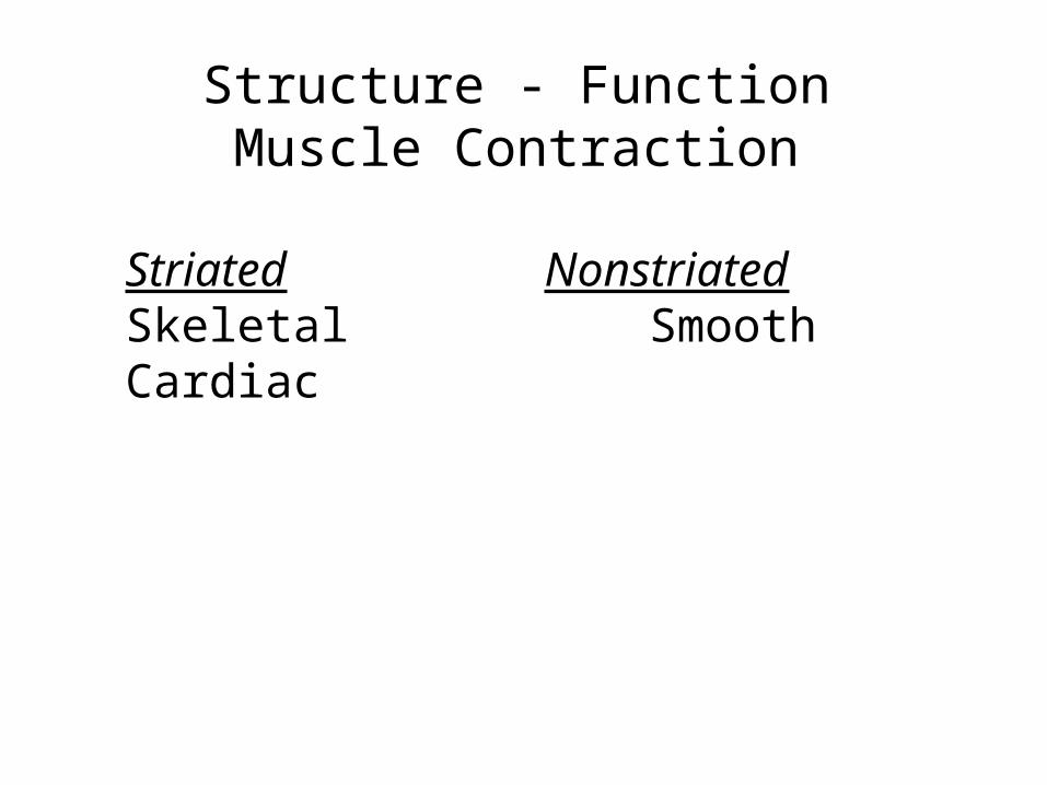
Structure - FunctionMuscle Contraction
Striated NonstriatedSkeletal SmoothCardiac

Striated Muscle: Sarcomere

Striated Muscle: Sarcomere- Z line to Z line Z lineZ line

SarcomereZ line to Z line
A bandI bandM lineH band
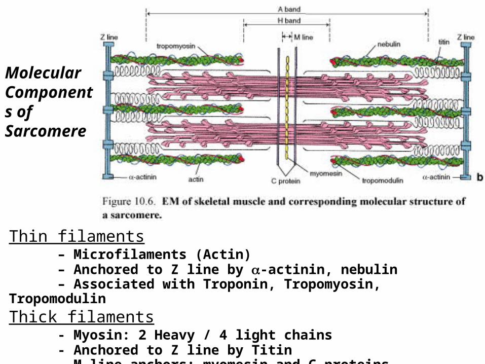
Thin filaments– Microfilaments (Actin)– Anchored to Z line by -actinin, nebulin– Associated with Troponin, Tropomyosin,
TropomodulinThick filaments
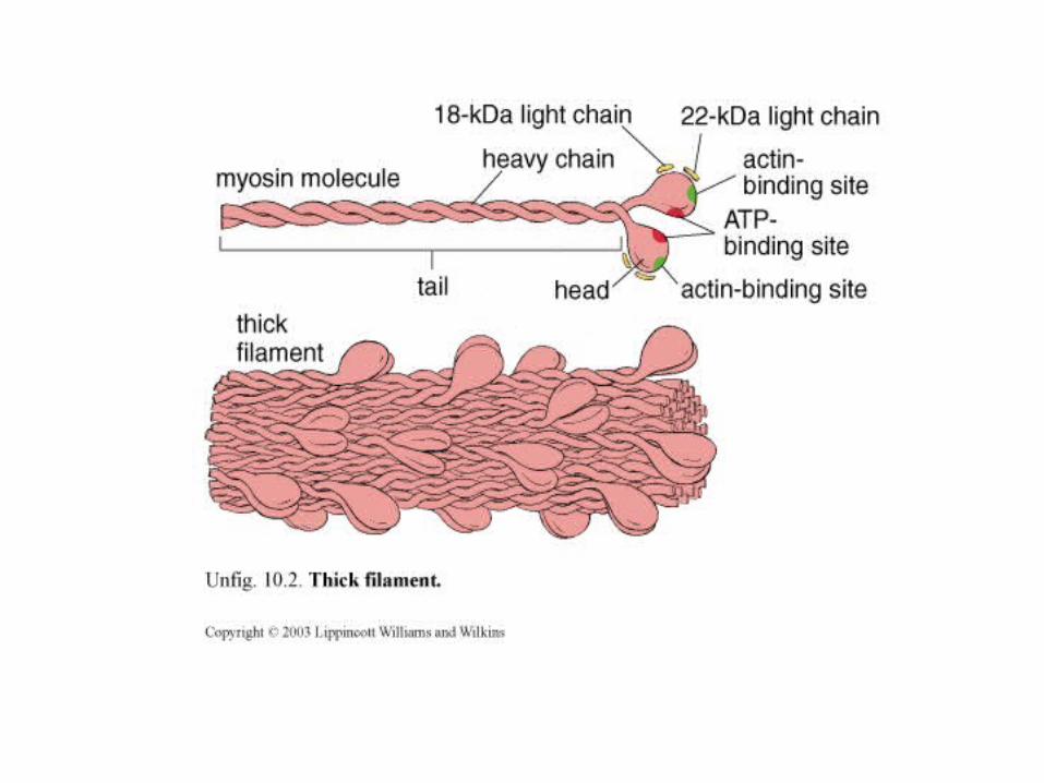
- Myosin: 2 Heavy / 4 light chains- Anchored to Z line by Titin- M line anchors: myomesin and C-proteins
Molecular Components of Sarcomere


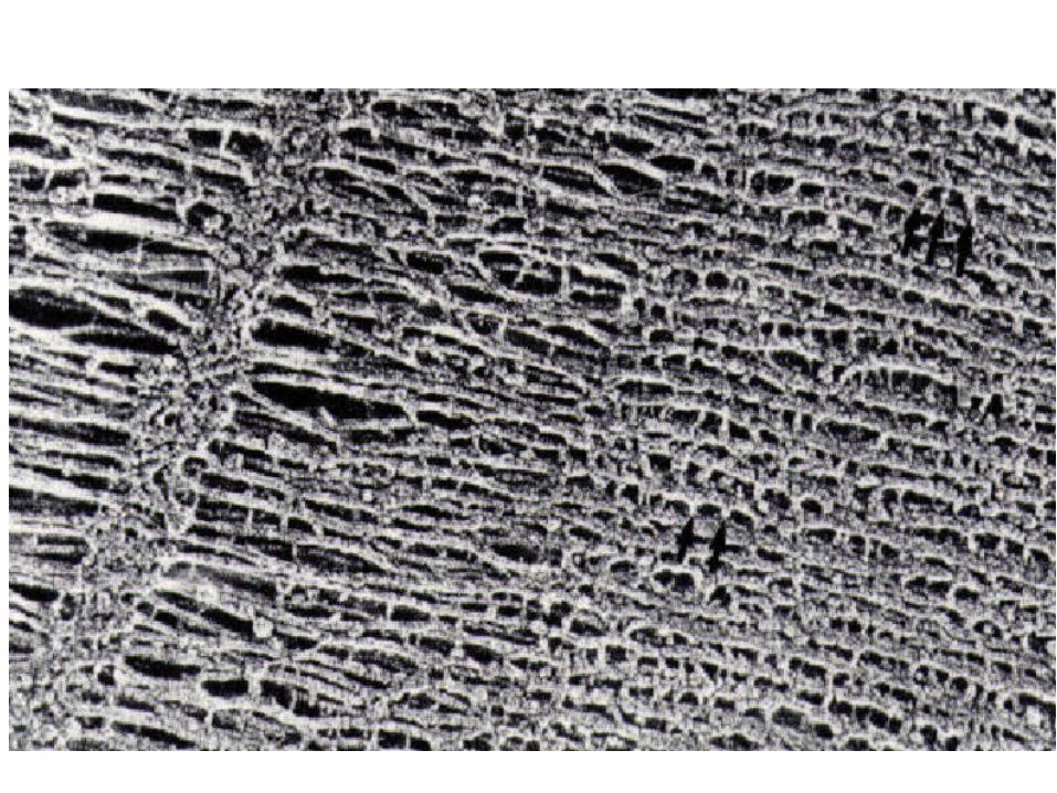
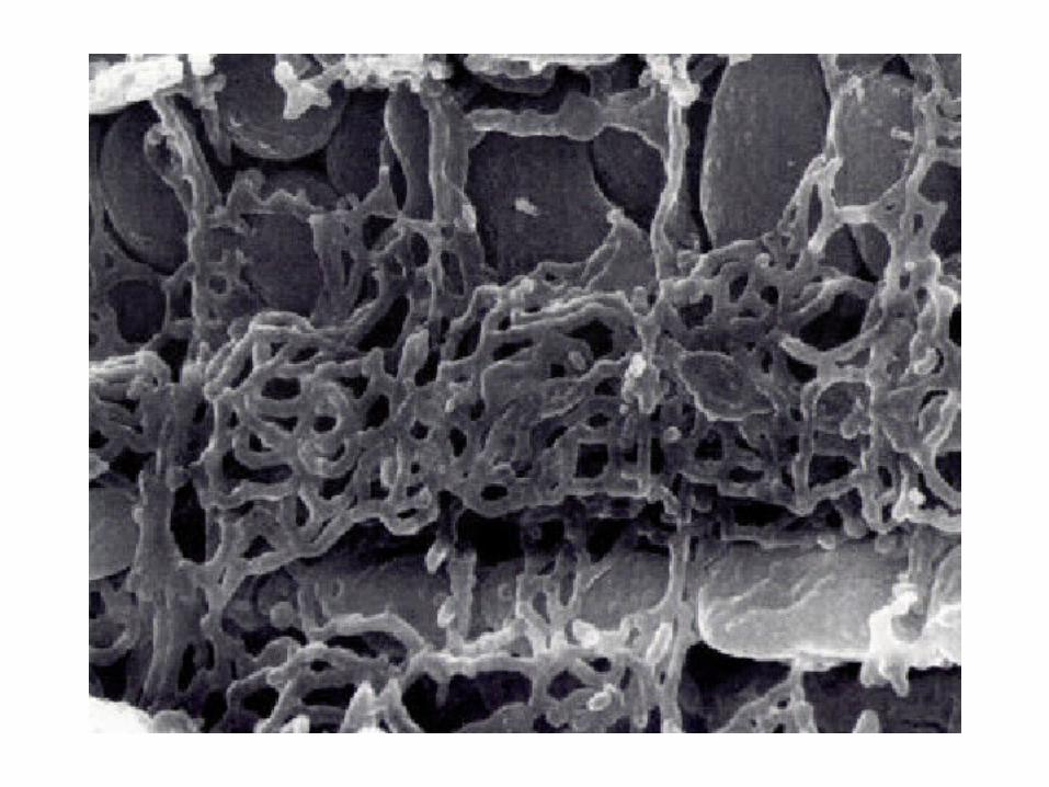
This is an electron micrograph of a section through a sarcomere of a skeletal muscle fiber.
Where in the sarcomere is the section taken?


Striated Muscle Contraction
Thin FilamentTropomyosin: – winds around actin – obscures actin-myosin binding site
Troponin Complex – Troponin T: interacts with Tropomyosin – Troponin I: prevents myosin-actin binding – Troponin C: binds Ca++, allowing myosin-actin binding


Striated Muscle Contraction
Thin FilamentTropomyosin Troponin – Troponin T – Troponin I – Troponin C
Ca+2
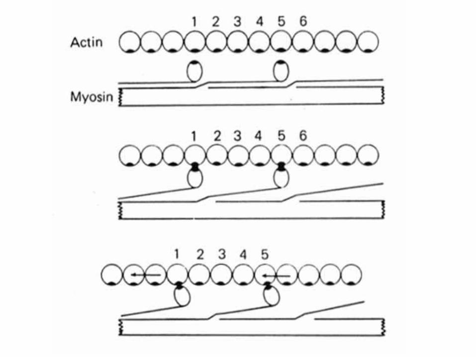
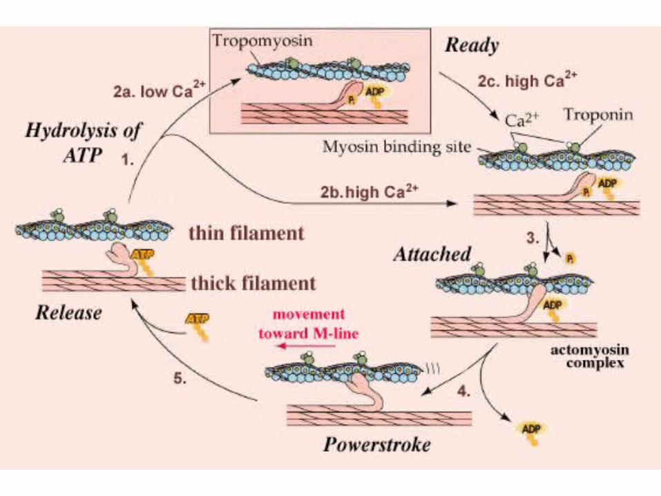

Contraction of Striated Muscle
- Release of Calcium from Sarcoplasmic Reticulum
- Calcium binds to Troponin
- Tropomyosin/Troponin shift to expose binding site
- Myosin head binds to Actin; Pi released
- Release of ADP from Myosin ---> POWERSTROKE
- Binding of ATP: Myosin detaches from Actin
- Splitting of ATP---> ADP + PI; Myosin head is reset

QuickTime™ and aAnimation decompressor
are needed to see this picture.
http://www.sci.sdsu.edu/movies/actin_myosin.html
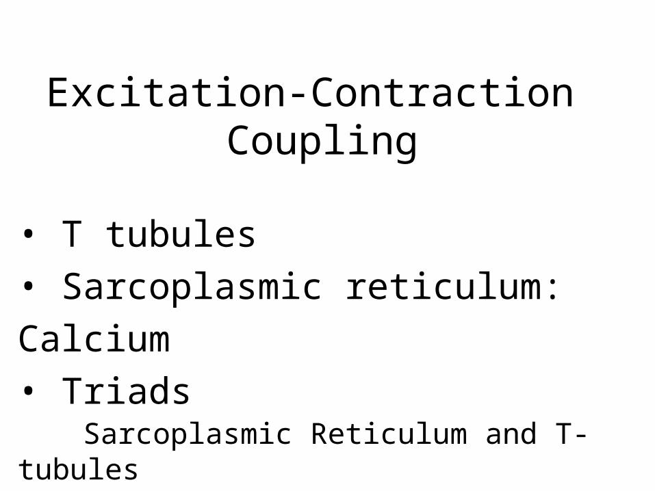
Excitation-Contraction Coupling
• T tubules• Sarcoplasmic reticulum: Calcium• Triads
Sarcoplasmic Reticulum and T-tubules

T-tubules
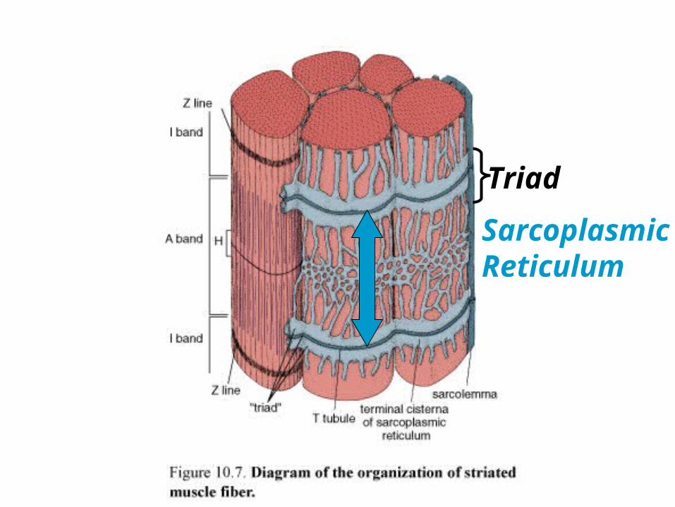
SarcoplasmicReticulum
Triad


Striated Muscle Structure
• T tubules• Sarcoplasmic reticulum: Calcium• Triads: juncture of T-tubules & SR
- Must release calcium from sarcoplasmic reticulum to initiate the contraction process.
Excitation-Contraction Coupling

Muscle Action Potential
Transverse Tubules
Sarcoplasmic Reticulum
Calcium release

Excitation-Contraction Coupling
Muscle Action Potential
Transverse Tubules
Sarcoplasmic Reticulum
Calcium release

T-tubule membrane

Smooth Muscle Contraction
- No organized sarcomere structure
- Actin filaments are associated with tropomyosin but
Not Troponin
What controls actin-myosin interaction?
Smooth Muscle Contraction
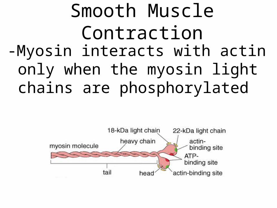
-Myosin interacts with actin only when the myosin light chains are phosphorylated

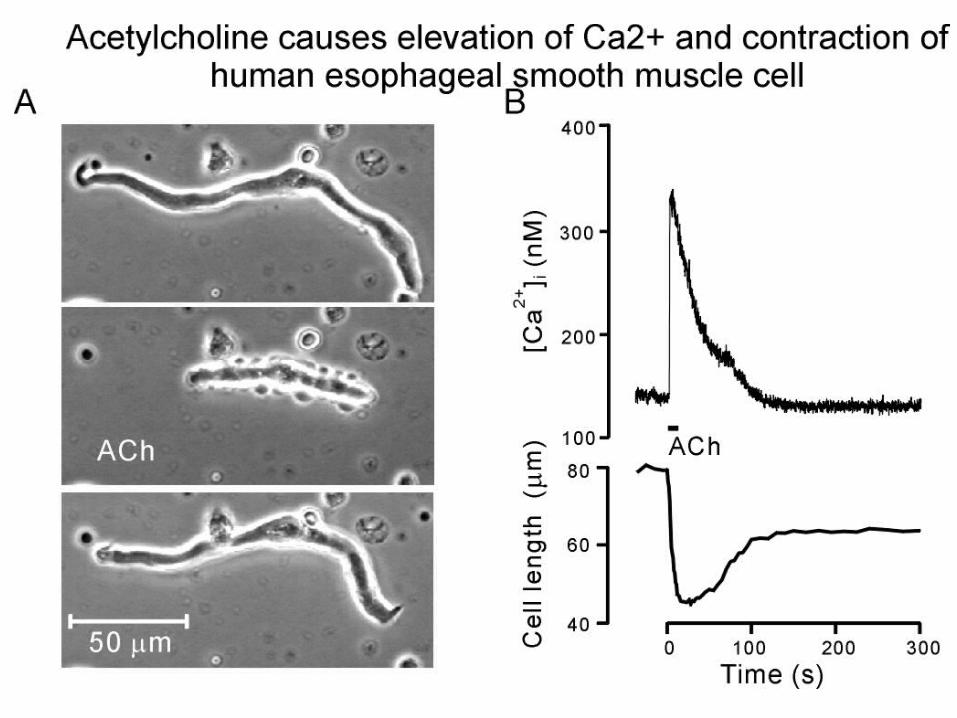
Smooth Muscle Excitation-Contraction1) Excitation=> Ca++ influx
2) Ca++ binds Calmodulin
3) Ca++-Calmodulinactivates myosin light chain kinase (MLCK )
4) MLCK phosphorylates myosin light chains
5) Myosin binds actin => contraction

Regulation of Smooth Muscle Contraction
Nonneural regulation
Hormonal: Oxytocin- uterine contraction
Nitric Oxide (NO):
Produced by endothelial cells of
arterioles
Relaxes smooth muscle

Mice have been produced whose eNOS (endothelial cell NO synthase) genes been "knocked out”. Predict the blood pressure levels of these mice.
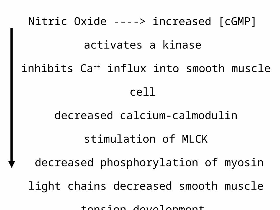
Nitric Oxide ----> increased [cGMP]
activates a kinase
inhibits Ca++ influx into smooth muscle cell
decreased calcium-calmodulin stimulation of
MLCK
decreased phosphorylation of myosin light
chains decreased smooth muscle tension
development
vasodilation (expansion of vessel lumen)
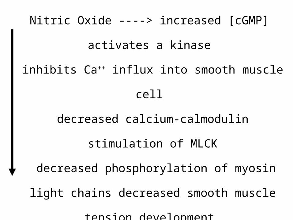
Nitric Oxide ----> increased [cGMP]
activates a kinase
inhibits Ca++ influx into smooth muscle cell
decreased calcium-calmodulin stimulation of
MLCK
decreased phosphorylation of myosin light
chains decreased smooth muscle tension
development
vasodilation (expansion of vessel lumen)
What would be the effect of drugs that inhibit the breakdown of cGMP ?
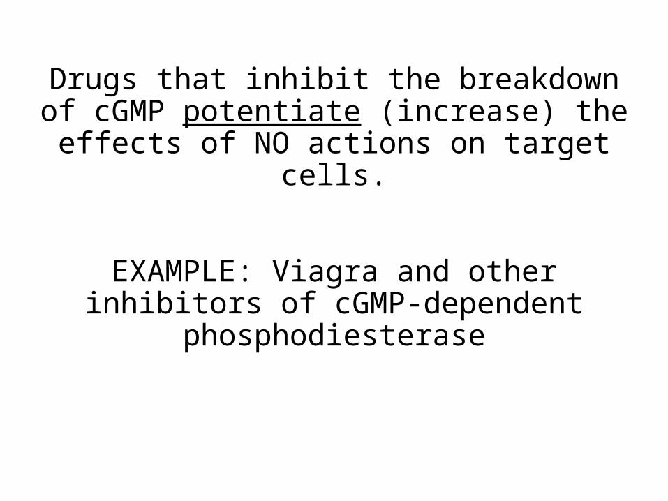
Drugs that inhibit the breakdown of cGMP potentiate (increase) the effects of
NO actions on target cells.
EXAMPLE: Viagra and other inhibitors of cGMP-dependent phosphodiesterase

Innervation of Muscle
Smooth Muscle
Innervation: boutons en passant acetylcholine
Stimulation spread by gap junctions

Innervation of Smooth Muscle
A multiunit system: fine innervation for regulation of individual cells; cells that control the iris opening
A single unit system:1 neuromuscularJunction serves asheet of muscle fibers; stimulus transmitted to otherMuscle cells via gapjunctions; wall of intestine
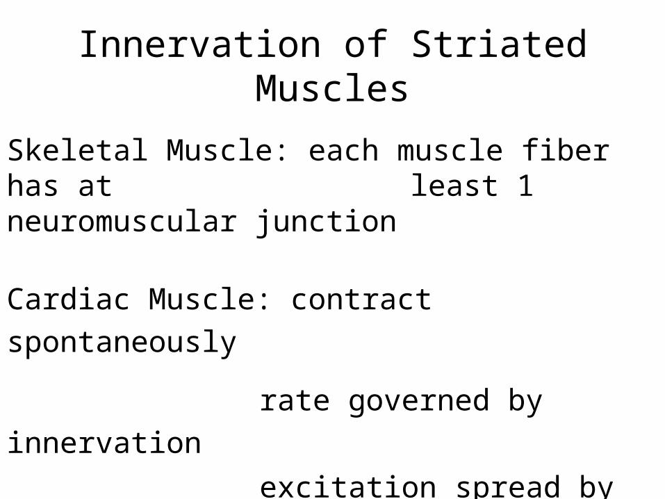
Innervation of Striated Muscles
Skeletal Muscle: each muscle fiber has at least 1 neuromuscular
junction
Cardiac Muscle: contract spontaneously
rate governed by innervation
excitation spread by gap
junctions

Motor Neuron
Nerve Action Potential
Synapse
Neuromuscular Junction
Neurotransmitter (ACh)
Receptors (AChR)
Muscle Action Potential
Transverse Tubules
Sarcoplasmic Reticulum
Calcium release

Neuromuscular Junction• Nerve stimulation
• Action potential
• Opening of calcium channels
• Exocytosis of synaptic vesicles
• Acetylcholine release
• Binding to Acetylcholine receptors
(AChR)
• Muscle depolarization including T-
tubules
• Opening of Calcium channels in SR

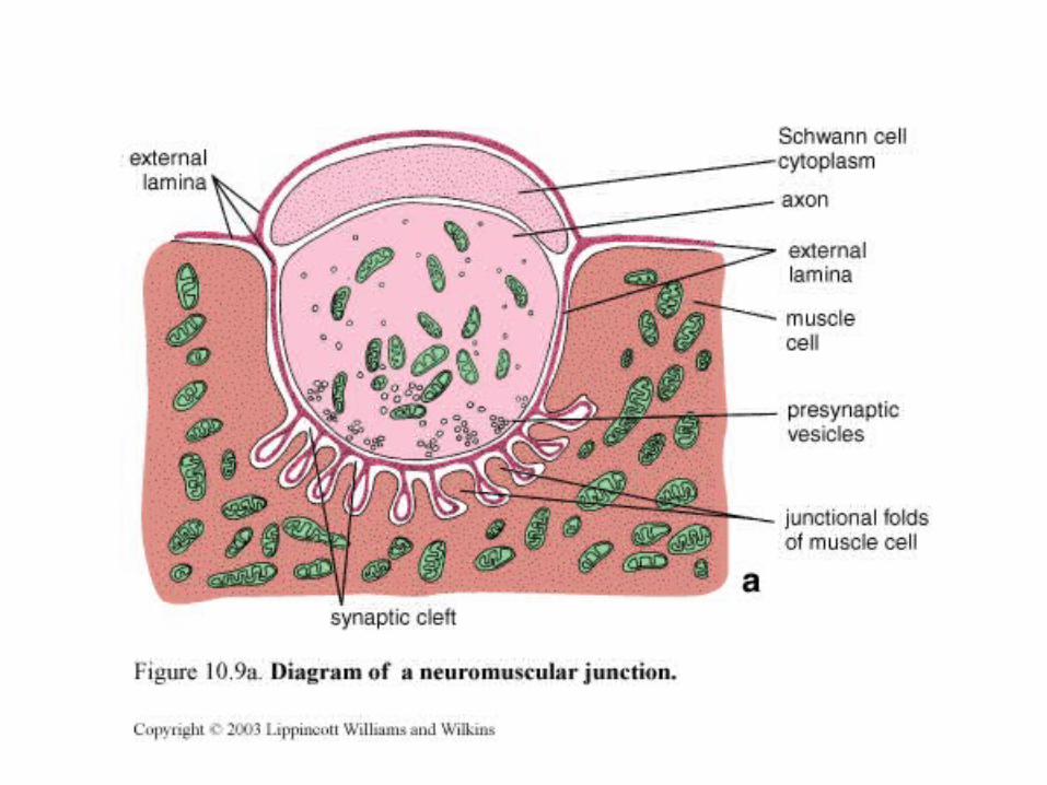
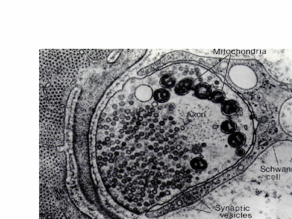
Motor endplate=Neuromuscular Junction
• Synaptic vesicles
• Active zones
• Junctional folds
• AChR (acetylcholine receptor)
clusters
• Schwann cell

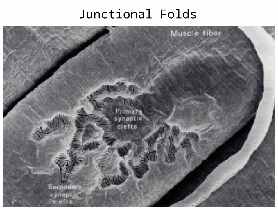
Junctional Folds

Types of skeletal muscles
Fast muscles– Strong and rapid contraction; rapid fatigue– ATP from anaerobic glycolysis– Phasic motor neuron
Slow muscles– Slower but sustained contraction– ATP from oxidative respiration (mitochondria)– Tonic motor neuron
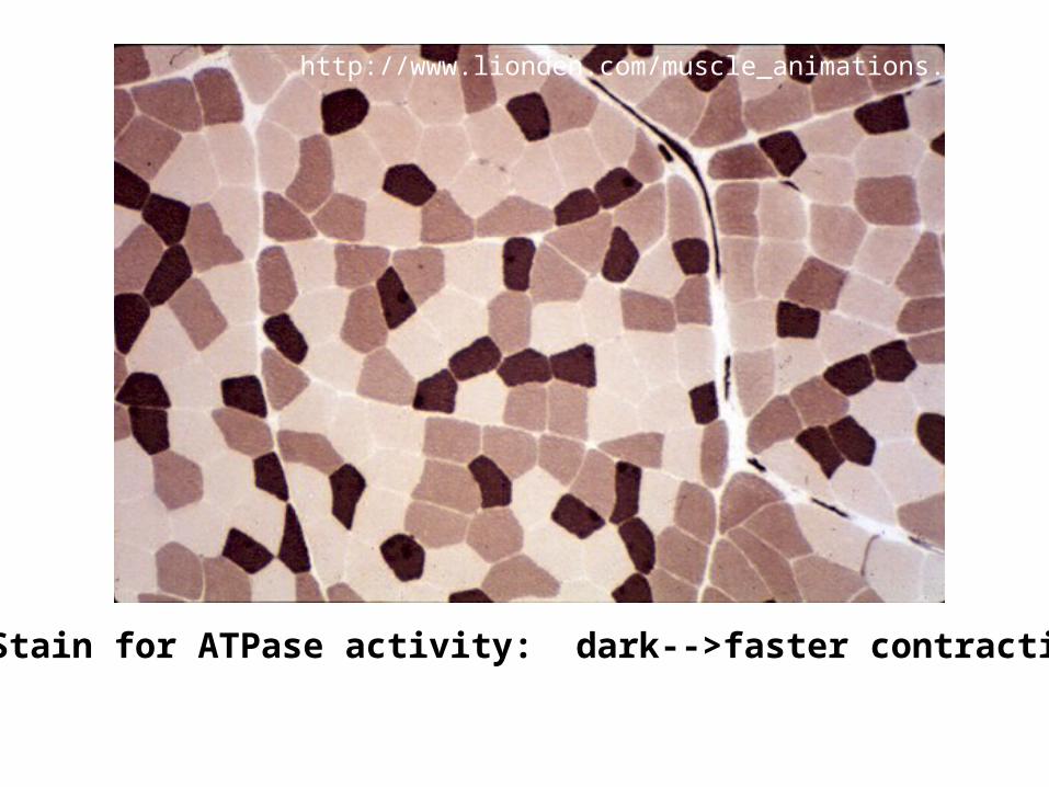
http://www.lionden.com/muscle_animations.htm
Stain for ATPase activity: dark-->faster contracting
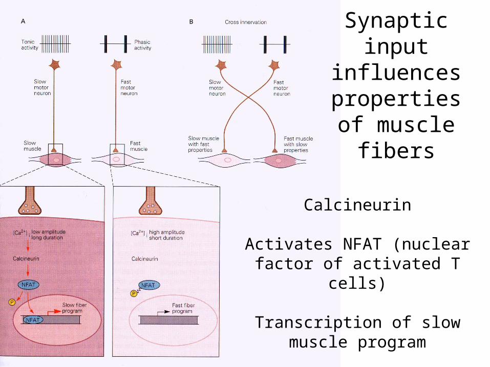
Synaptic input influences
properties of muscle fibers
Calcineurin
Activates NFAT (nuclear factor of activated T cells)
Transcription of slow muscle program