Embed Size (px)
Citation preview

Anjop J. Venker-van HaagenEar, Nose, Throat, and Tracheobronchial Diseases in Dogs and Cats
3-87706-635-6_00I-050 18.04.2005 12:03 Uhr Seite I

3-87706-635-6_00I-050 18.04.2005 12:03 Uhr Seite II
Anjop J. Venker-van Haagen
Ear, Nose, Throat, and Tracheobronchial Diseasesin Dogs and Cats
3-87706-635-6_00I-050 18.04.2005 12:03 Uhr Seite III
Anjop J. Venker-van Haagen, DVM, PhD, DECVS
Former Associate Professor of Veterinary Ear Nose and Throat Diseases
Faculty of Veterinary Medicine
Department of Clinical Sciences of Companion Animals
Utrecht University, The Netherlands
© 2005, Schlütersche Verlagsgesellschaft mbH & Co. KG, Hans-Böckler-Allee 7, 30173 Hannover
E-mail: [email protected]
Printed in Germany
ISBN 3-87706-635-6
Bibliographic information published by Die Deutsche Bibliothek
Die Deutsche Bibliothek lists this publication in the Deutsche Nationalbibliografie; detailed biblio-
graphic data are available in the Internet at http://dnb.ddb.de.
The author assumes no responsibility and make no guarantee for the use of drugs listed in this book.
The author/publisher shall not be held responsible for any damages that might be incurred by the
recommended use of drugs or dosages contained within this textbook. In many cases controlled
research concerning the use of a given drug in animals is lacking. This book makes no attempt to
validate claims made by authors of reports for off-label use of drugs. Practitioners are urged to follow
manufacturers’ recommendations for the use of any drug.
All rights reserved. The contents of this book, both photographic and textual, may not be reproduced in
any form, by print, photoprint, phototransparency, microfilm, video, video disc, microfiche, or any other
means, nor may it be included in any computer retrieval system, without written permission from the
publisher.
Any person who does any unauthorised act in relation to this publication may be liable to criminal
prosecution and civil claims for damages.
3-87706-635-6_00I-050 18.04.2005 12:03 Uhr Seite IV
V
Contents
Abbreviations. . . . . . . . . . . . . . . . . . . . . . . . . . VIII
Preface . . . . . . . . . . . . . . . . . . . . . . . . . . . . . . . IX
1 The Ear . . . . . . . . . . . . . . . . . . . . . . . . 1
1.1 Functional considerations . . . . . . . . . 1
1.1.1 The ear as sensory organ . . . . . . . . . 1
1.1.2. Middle ear matches differentimpedances . . . . . . . . . . . . . . . . . . . . 2
1.1.3 Organ of Corti, sensory organ for hearing . . . . . . . . . . . . . . . . . . . . . 3
1.1.4 Ascending and descending pathwaysfor hearing . . . . . . . . . . . . . . . . . . . . . 5
1.1.5 Vestibular organ, the key to posturalreflexes and eye movement . . . . . . . 5
1.2 History and clinical signs . . . . . . . . . 6
1.2.1 History . . . . . . . . . . . . . . . . . . . . . . . . 6
1.2.2 Clinical signs . . . . . . . . . . . . . . . . . . . 7
1.2.3 Physical examination . . . . . . . . . . . . . 9
1.3 Special diagnostic techniques . . . . . 9
1.3.1 Otoscopic examination . . . . . . . . . . . 10
1.3.2 Diagnostic imaging of the ear . . . . . . 12
1.3.3 Tympanometry . . . . . . . . . . . . . . . . . . 15
1.3.4 Neurological examination for vestibulardysfunction . . . . . . . . . . . . . . . . . . . . 16
1.3.5 Hearing tests . . . . . . . . . . . . . . . . . . . 16
1.4 Congenital diseases of the ear . . . . . 19
1.4.1 Congenital deformity of the external ear . . . . . . . . . . . . . . . . . . . . 19
1.4.2 Congenital deafness . . . . . . . . . . . . . 19
1.5 Inflammatory diseases of the ear . . . 21
1.5.1 Primary and secondary skin diseases ofthe auricle . . . . . . . . . . . . . . . . . . . . . 21
1.5.2 Perichondritis and chondritisof the auricle . . . . . . . . . . . . . . . . . . . 23
1.5.3 Cold agglutination and cutaneousvasculitis of the auricle . . . . . . . . . . . 24
1.5.4 Inflammatory diseases of the external earcanal . . . . . . . . . . . . . . . . . . . . . . . . . . 24
1.5.5 Inflammation of the tympanicmembrane . . . . . . . . . . . . . . . . . . . . . 30
1.5.6 Inflammatory disease of the middle ear . . . . . . . . . . . . . . . . . . . . . 30
1.5.7 Labyrinthitis . . . . . . . . . . . . . . . . . . . . 34
1.6 Tumors of the ear . . . . . . . . . . . . . . . . 35
1.6.1 Malignant tumors of the auricle . . . . 35
1.6.2 Malignant tumors of the externalear canal . . . . . . . . . . . . . . . . . . . . . . . 36
1.6.3 Tumors of the middle ear . . . . . . . . . 37
1.7 Trauma to the ear . . . . . . . . . . . . . . . 37
1.7.1 Trauma to the auricle . . . . . . . . . . . . . 37
1.7.2 Auricular hematoma . . . . . . . . . . . . . 38
1.7.3 Trauma to the external ear canal . . . 38
1.7.4 Trauma to the tympanicmembrane. . . . . . . . . . . . . . . . . . . . . . 39
1.7.5 Trauma to the temporal bone . . . . . . 40
1.8 Ototoxicity . . . . . . . . . . . . . . . . . . . . . 41
1.9 Hearing in dogs and cats . . . . . . . . . 42
1.9.1 Hearing and hearing loss in dogs . . . 43
1.9.2 Brain stem auditory evoked responses in dogs . . . . . . . . . . . . . . . 43
1.9.3 Hearing and hearing loss in cats . . . 46
1.9.4 Brain stem auditory evoked responsesin cats . . . . . . . . . . . . . . . . . . . . . . . . . 46
2 The Nose and Nasal Sinuses . . . . . . 51
2.1 Functional considerations . . . . . . . . . 51
2.1.1 Regulation and conditioning of theinspiratory and expiratory airflow . . . 51
2.1.2 Mucosal cleaning . . . . . . . . . . . . . . . . 52
2.1.3 Olfaction . . . . . . . . . . . . . . . . . . . . . . . 53
2.1.4 Specific functional systems . . . . . . . 53
2.2 History and clinical signs . . . . . . . . . 53
2.2.1 History . . . . . . . . . . . . . . . . . . . . . . . . 53
2.2.2 Clinical signs . . . . . . . . . . . . . . . . . . . 54
2.2.3 Physical examination . . . . . . . . . . . . 55
2.3 Special diagnostic techniques . . . . . 55
2.3.1 Diagnostic imaging . . . . . . . . . . . . . . 56
2.3.2 Rhinoscopy . . . . . . . . . . . . . . . . . . . . 57
2.3.3 Olfactory tests . . . . . . . . . . . . . . . . . . 59
2.4 Congenital diseases of the nose and nasal sinuses . . . . . . . . . . . . . . . 60
2.4.1 Congenital malformation of the nasal plane . . . . . . . . . . . . . . . . . . . . 60
2.4.2 Nasal dermoid sinus cysts . . . . . . . . 62
2.4.3 Congenital cerebrospinal fluid fistula 62
2.4.4 Congenital malformation of the frontal sinuses . . . . . . . . . . . . . . . . . . 62
2.4.5 Congenital ciliary dysfunction . . . . . . 62
2.5 Rhinitis and sinusitis . . . . . . . . . . . . . 63
2.5.1 Infectious rhinitis and sinusitis . . . . 63
2.5.2 Noninfectious rhinitis and sinusitis. . . . . . . . . . . . . . . . . . . . . . . . 68
2.6 Tumors of the nasal plane, the nasalcavity, and the frontal sinus . . . . . . . 72
Contents
3-87706-635-6_00I-050 18.04.2005 12:03 Uhr Seite V
VI
2.6.1 Tumors of the nasal plane . . . . . . . . . 72
2.6.2 Tumors in the nasal cavity . . . . . . . . . 74
2.6.3 Tumors in the frontal sinus . . . . . . . . 74
2.7 Trauma to the frontal sinus and the nose . . . . . . . . . . . . . . . . . . . . . . 75
2.7.1 Trauma to the frontal sinus . . . . . . . . 75
2.7.2 Trauma to the nose . . . . . . . . . . . . . . 76
2.8 Epistaxis . . . . . . . . . . . . . . . . . . . . . . . 77
2.8.1 Management of acute epistaxis . . . . 77
2.8.2 Causes of epistaxis . . . . . . . . . . . . . . 77
2.8.3 The diagnostic plan . . . . . . . . . . . . . . 78
2.8.4 Management of intermittent epistaxisof unknown origin . . . . . . . . . . . . . . . 79
3 The Pharynx . . . . . . . . . . . . . . . . . . . . 83
3.1 Functional considerations . . . . . . . . . 83
3.1.1 Auditory tube serves to equalizeatmospheric pressure . . . . . . . . . . . 83
3.1.2 Swallowing . . . . . . . . . . . . . . . . . . . . . 83
3.2 History and clinical signs . . . . . . . . . 88
3.2.1 History . . . . . . . . . . . . . . . . . . . . . . . . 88
3.2.2 Clinical signs . . . . . . . . . . . . . . . . . . . 88
3.3 Special diagnostic techniques . . . . . 89
3.3.1 Pharyngoscopy . . . . . . . . . . . . . . . . . 89
3.3.2 Diagnostic imaging of the pharynx . . 90
3.3.3 Electromyography of the pharyngealmuscles . . . . . . . . . . . . . . . . . . . . . . . 91
3.4 Congenital deformities and disordersof the pharynx . . . . . . . . . . . . . . . . . . 91
3.4.1 Hypoplasia of the soft palate . . . . . . 91
3.4.2 Congenital malformation of the softpalate . . . . . . . . . . . . . . . . . . . . . . . . 92
3.4.3 Hyperplasia of the soft palate . . . . . 92
3.4.4 Choanal atresia . . . . . . . . . . . . . . . . . 93
3.4.5 Craniopharyngioma (Rathke’s pouchtumor) . . . . . . . . . . . . . . . . . . . . . . . . . 94
3.5 Pharyngitis . . . . . . . . . . . . . . . . . . . . . 95
3.5.1 Nasopharyngitis . . . . . . . . . . . . . . . . . 95
3.5.2 Oropharyngitis and tonsillitis . . . . . . 99
3.5.3 Pharyngeal mucocele . . . . . . . . . . . . 101
3.6 Tumors of the pharynx . . . . . . . . . . . . 101
3.7 Blunt and penetrating injuries of thepharynx . . . . . . . . . . . . . . . . . . . . . . . 103
3.7.1 Blunt pharyngeal injuries . . . . . . . . . 103
3.7.2 Penetrating pharyngeal injuries . . . . 104
3.8 Dysphagia . . . . . . . . . . . . . . . . . . . . . 106
3.8.1 Causes of dysphagia . . . . . . . . . . . . . 106
3.8.2 Diagnosis in dysphagia . . . . . . . . . . . 113
3.8.3 Therapy in dysphagia . . . . . . . . . . . . 116
4 The Larynx . . . . . . . . . . . . . . . . . . . . . 121
4.1 Functional considerations . . . . . . . . . 121
4.1.1 The glottic closure reflex . . . . . . . . . . 121
4.1.2 Respiratory movements of the glottis . . . . . . . . . . . . . . . . . . . . . . . . . 122
4.1.3 Movements of the glottis in vocalization . . . . . . . . . . . . . . . . . . . . 122
4.1.4 Action of the glottis in coughing . . . . 122
4.1.5 Supplementary innervation of the dog’s intrinsic laryngeal muscles . . . 122
4.2 History and clinical signs . . . . . . . . . 124
4.2.1 History . . . . . . . . . . . . . . . . . . . . . . . . . 124
4.2.2 Clinical signs . . . . . . . . . . . . . . . . . . . 125
4.3 Special diagnostic techniques . . . . . 126
4.3.1 Laryngoscopy . . . . . . . . . . . . . . . . . . . 126
4.3.2. Diagnostic imaging of the larynx . . . 127
4.3.3 Electromyography of the intrinsiclaryngeal muscles . . . . . . . . . . . . . . . 127
4.4 Congenital deformities and disorders of the larynx . . . . . . . . . . . . 128
4.4.1 Congenital glottis stenosis . . . . . . . . 128
4.4.2 Congenital subglottic stenosis . . . . . 129
4.4.3 Laryngeal hypoplasia . . . . . . . . . . . . 130
4.5 Laryngitis . . . . . . . . . . . . . . . . . . . . . . 132
4.5.1 Benign laryngeal masses . . . . . . . . . 137
4.5.2 Ventral midline approach to the laryngeal cavities to expose large masses . . . . . . . . . . . . . . . . . . . . . . . . 137
4.6 Tumors of the larynx . . . . . . . . . . . . . 138
4.6.1 History and clinical signs oflaryngeal tumors . . . . . . . . . . . . . . . . 138
4.6.2 Imaging of laryngeal tumors . . . . . . . 139
4.6.3 Laryngoscopy for laryngeal tumors . . 139
4.6.4 Therapy for laryngeal tumors . . . . . . 140
4.7 Blunt and penetrating injuriesto the larynx . . . . . . . . . . . . . . . . . . . . 141
4.7.1 Blunt laryngeal injuries . . . . . . . . . . . 142
4.7.2 Penetrating laryngeal injuries . . . . . . 144
4.8 Laryngeal paralysis and functionaldisorders of the larynx . . . . . . . . . . . . 146
4.8.1 Neurogenic laryngeal paralysis . . . . . 147
4.8.2 Laryngeal spasm . . . . . . . . . . . . . . . . 160
4.8.3 Paradoxical vocal fold movement . . . 161
4.8.4 Sensory laryngeal paralysis and laryngeal dysfunction . . . . . . . . . . . . 161
Contents
3-87706-635-6_00I-050 18.04.2005 12:03 Uhr Seite VI
VII
5 The Trachea and Bronchi . . . . . . . . . . 167
5.1 Functional considerations . . . . . . . . . 167
5.1.1 Trachea and bronchi facilitate therespiratory airflow . . . . . . . . . . . . . . . 167
5.1.2 Trachea and bronchi condition therespiratory air . . . . . . . . . . . . . . . . . . . 167
5.2 History and clinical signs . . . . . . . . . 168
5.2.1 History . . . . . . . . . . . . . . . . . . . . . . . . 168
5.2.2 Clinical signs . . . . . . . . . . . . . . . . . . . 169
5.2.3 Physical examination . . . . . . . . . . . . 170
5.3 Special diagnostic techniques . . . . . 171
5.3.1 Diagnostic imaging . . . . . . . . . . . . . . 172
5.3.2 Bronchoscopy . . . . . . . . . . . . . . . . . . 172
5.4 Congenital diseases of the trachea and the bronchi . . . . . . . . . . . . . . . . . 175
5.4.1 Hypoplasia of the trachea . . . . . . . . . 175
5.4.2. Collapse of the trachea . . . . . . . . . . . 181
5.4.3 Segmental tracheal stenosis . . . . . . . 183
5.4.4 Congenital ciliary dysfunction . . . . . . 183
5.5 Tracheitis and bronchitis . . . . . . . . . . 184
5.5.1 Infectious tracheobronchitisin dogs . . . . . . . . . . . . . . . . . . . . . . . . 185
5.5.2 Infectious tracheobronchitisin cats . . . . . . . . . . . . . . . . . . . . . . . . . 187
5.5.3 Noninfectious tracheobronchitis . . . 187
5.5.4 Bronchiectasis . . . . . . . . . . . . . . . . . . 189
5.5.5 Prolapse of the dorsal ligament oftrachea and main stem bronchi . . . . 189
5.5.6 Foreign bodies in the tracheo-bronchial tree in dogs . . . . . . . . . . . . 191
5.5.7 Foreign bodies in the tracheo-bronchial tree in cats . . . . . . . . . . . . . 193
5.5.8 Tracheitis caused by aspiration . . . . 193
5.6 Tumors of the trachea and bronchi . . 194
5.7 Tracheal trauma . . . . . . . . . . . . . . . . . 195
5.7.1 Trauma to the cervical trachea . . . . . 196
5.7.2 Trauma to the thoracic trachea . . . . . 197
5.7.3 Tracheal stenosis . . . . . . . . . . . . . . . . 198
5.8 Airway management . . . . . . . . . . . . . 199
5.8.1 Endotracheal intubation . . . . . . . . . . 200
5.8.2 Cricothyroidotomy . . . . . . . . . . . . . . . 202
5.8.3 Tracheostomy . . . . . . . . . . . . . . . . . . . 202
5.8.4 Permanent tracheostoma . . . . . . . . . 203
5.8.5 Tracheal T-tube . . . . . . . . . . . . . . . . . . 205
6 Cranial Neuralgias and Facial andTrigeminal Paralysis . . . . . . . . . . . . . 209
6.1 Cranial neuralgias . . . . . . . . . . . . . . . 209
6.1.1 Glossopharyngeal neuralgia . . . . . . . 209
6.1.2 Trigeminal neuralgia . . . . . . . . . . . . . 209
6.2 Facial and trigeminal paralysis . . . . . 210
6.2.1 Facial paralysis . . . . . . . . . . . . . . . . . 210
6.2.2 Trigeminal paralysis . . . . . . . . . . . . . . 211
Contents
3-87706-635-6_00I-050 18.04.2005 12:03 Uhr Seite VII
VIII
Abbreviations
Abbreviations
ADC Analog-to-digital converter
B.O.S. Brachycephalic obstructivesyndrome
BAER Brain stem auditory evokedresponse
BERA Brain stem evoked responseaudiometry
CPG Central pattern generator
CRDs Complex repetitive discharges
CT Computed tomography
DAC Digital-to-analog converter
dB SPL Decibel sound pressure level
ECG Electrocardiogram
EEG Electroencephalogram
EMG Electromyogram/Electromy-ography
F generations Offspring generations
FISH and RH Methods for gene mapping usedmapping for association studies
Hz Hertz
i.d. Inside diameter
Ig Immunoglobulin
kHz Kilohertz
MRI Magnetic resonance imaging
Nd-YAG laser Laser using Yttrium-Aluminum-Garnet with Nd ions
NSAIDs Nonsteroidal anti-inflammatorydrugs
NTS Nucleus tractus solitarius
p < 0.01 The probability that the result isdue to chance is less than 1 in100 (highly significant)
P generation Parent generation
SLN Superior laryngeal nerve/Craniallaryngeal nerve
T-tube T-shaped tracheal tube
V Trigeminal nerve
VII Facial nerve
IX Glossopharyngeal nerve
X Vagus nerve
Xph Pharyngeal branch of the vagusnerve
XII Hypoglossal nerve
3-87706-635-6_00I-050 18.04.2005 12:03 Uhr Seite VIII
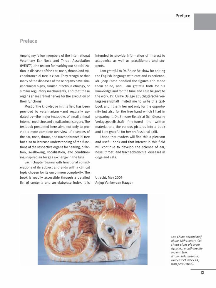
Cat. China, second half
of the 18th century. Cat
shows signs of severe
dyspnea: mouth breath-
ing and fear.
(From: Rijksmuseum,
Diary 1999, week 44,
with permission).
IX
Preface
Among my fellow members of the International
Veterinary Ear Nose and Throat Association
(IVENTA), the reason for marking out specializa-
tion in diseases of the ear, nose, throat, and tra-
cheobronchial tree is clear. They recognize that
many of the diseases of these organs have sim-
ilar clinical signs, similar infectious etiology, or
similar regulatory mechanisms, and that these
organs share cranial nerves for the execution of
their functions.
Most of the knowledge in this field has been
provided to veterinarians—and regularly up-
dated by—the major textbooks of small animal
internal medicine and small animal surgery. The
textbook presented here aims not only to pro-
vide a more complete overview of diseases of
the ear, nose, throat, and tracheobronchial tree
but also to increase understanding of the func-
tions of the respective organs for hearing, olfac-
tion, swallowing, vocalization, and condition-
ing inspired air for gas exchange in the lung.
Each chapter begins with functional consid-
erations of its subject and ends with a clinical
topic chosen for its uncommon complexity. The
book is readily accessible through a detailed
list of contents and an elaborate index. It is
intended to provide information of interest to
academics as well as practitioners and stu-
dents.
I am grateful to Dr. Bruce Belshaw for editing
the English language with care and experience.
Mr. Joop Fama handled the figures and made
them shine, and I am grateful both for his
knowledge and for the time and care he gave to
the work. Dr. Ulrike Oslage at Schlütersche Ver-
lagsgesellschaft invited me to write this text-
book and I thank her not only for the opportu-
nity but also for the free hand which I had in
preparing it. Dr. Simone Bellair at Schlütersche
Verlagsgesellschaft fine-tuned the written
material and the various pictures into a book
and I am grateful for her professional skill.
I hope that readers will find this a pleasant
and useful book and that interest in this field
will continue to develop the science of ear,
nose, throat, and tracheobronchial diseases in
dogs and cats.
Utrecht, May 2005
Anjop Venker-van Haagen
Preface
3-87706-635-6_00I-050 18.04.2005 12:03 Uhr Seite IX

3-87706-635-6_00I-050 18.04.2005 12:03 Uhr Seite X
1
1 The Ear
1.1 Functional considerations
1.1.1 The ear as sensory organ
The ear is a sensory organ that has evolved to re-
ceive and transform the air waves or vibrations
that we call sound into a code of neural impuls-
es to be conveyed to the brain. The resulting dis-
tinct patterns of neural activity in the brain are
then integrated with information from other sen-
sory systems to guide behavior.52 The first stage
of this transformation occurs in the external and
middle ear, which collect sound waves and am-
plify their pressure, so that the sound energy
can be successfully transmitted from air to the
fluid that fills the cochlea of the inner ear. In the
inner ear the signal is divided into simpler, sinu-
soidal components, with the result that the fre-
quency, amplitude, and phase of the original
signal are faithfully converted by the sensory
hair cells into encoded electrical activity in the
auditory nerve fibers.52 In the brain the earliest
stage of central processing occurs in the
cochlear nucleus, where the peripheral auditory
information diverges into a number of parallel
central pathways. These include the superior
olivary complex, where the information from the
two ears interacts to aid in localizing the sound
in space. The cochlear nucleus also projects to
the inferior colliculus of the midbrain, a major in-
tegrative center and the first place where audi-
tory information can interact with the motor sys-
tem. The inferior colliculus is an obligatory relay
for information traveling to the thalamus and
cortex, where more complex aspects of sound
are processed.52
External ear. The external ear is the portion lat-
eral to the tympanic membrane. It consists of
the external auditory canal and its cartilaginous
extension, the auricle. The medial part of the
auditory canal is surrounded and supported by
the temporal bone. The auricle is covered with
skin which continues as the lining of the audi-
tory canal. This skin is thin and in the medial
part of the auditory canal it has little subcuta-
neous tissue, but in the lateral part it bears
numerous hair follicles and ceruminous and
sebaceous glands. Both the bony and the carti-
laginous parts of the auditory canal provide an
open passageway for air to the tympanic mem-
brane. The tympanic membrane is the medial
boundary of the auditory canal and its lateral
component is formed by the epithelium of the
skin lining the auditory canal. In mammals the
auricle and the auditory canal are together
regarded as a simple funnel that collects and
crudely filters sound. In humans, however, the
auricle and auditory canal increase the acoustic
pressure at the tympanic membrane of sounds
in the 1.5 kHz to 5 kHz range, which is the fre-
quency range most important for speech per-
ception.37 In the dog and cat the auricle can be
turned toward the source of sound; right and
left auricles can move independently so that
each ear can focus on separate sounds. Hence
the animal does not have to turn its head to
localize sounds, as humans do. It is not clear to
what extent the shape of the auricle—large and
erect like that of the German shepherd or folded
like that of the cocker spaniel—influences hear-
ing capacity, but the latter might seem to be dis-
advantageous, at least in theory.
Tympanic membrane. The tympanic membrane
terminates the ear canal and covers the
entrance to the tympanic cavity, thereby sepa-
rating the external from the middle ear. The
membrane is composed of three layers, the
outer squamous cell epithelial layer being a
continuation of the epithelial layer of the skin of
the external ear canal, the inner mucosal layer
being a continuation of the mucosa of the mid-
dle ear or tympanic cavity, and the intervening
fibrous layer or tunica propria. The tympanic
membrane is thin, slightly oval, semitranspar-
ent, and concave, owing to traction on its
Functional considerations1
3-87706-635-6_00I-050 18.04.2005 12:03 Uhr Seite 1
2
1
medial side by the tensor tympani muscle.
There are three ossicles (malleus, incus,
stapes) in the middle ear, the manubrium of the
malleus being fixed in the tunica propria of the
tympanic membrane. The tensing of the tym-
panic membrane makes it ideal for the conver-
sion of sound waves into vibrations of the
malleus.
1.1.2. Middle ear matches different
impedances
The major function of the middle ear is to match
relatively low impedance airborne sounds to
the higher impedance fluid of the middle ear.
The term impedance in this context stands for a
medium’s resistance to movement. Because of
the difference in impedance of the two media,
99.9 % of the sound energy is reflected at the
interface between air and fluid and only 0.1 %
is converted into pressure changes in the fluid.
The middle ear overcomes this problem and
ensures transmission of the sound energy
across the air-fluid boundary. The first and
major boost is achieved by focusing the force
impinging on the relatively large diameter tym-
panic membrane onto the much smaller diame-
ter membrane of the oval window, where the
stapes, the last of the three ossicles, is
attached and where the vibration of the tym-
panic membrane is conveyed to the fluid of the
inner ear. A second and related process
involves the mechanical advantage gained by
the lever action of the three interconnected
ossicles which link the tympanic membrane to
the oval window.47, 52
Auditory ossicles. These are also attached to
the wall of the epitympanum or dorsal part of
the tympanic cavity by several ligaments. While
the manubrium of the malleus is embedded in
the tympanic membrane, the head is sus-
pended in the epitympanum and is fused with
the incus in a rigid joint. The long process of the
incus is then linked to the stapes by another
joint, one that is rigid in the direction of the pis-
ton-like movement of the stapes but flexible
perpendicular to this movement. The stapes is
suspended in the oval window of the cochlea by
two ligaments. The stapedius muscle—the
smallest striated muscle in the body—is
attached to the head of the stapes. It pulls the
stapes in a direction perpendicular to the pis-
ton-like motion and is innervated by the facial
nerve. The other muscle of the ossicles is the
tensor tympani muscle attached to the muscu-
lar process of the malleus. It pulls the
manubrium of the malleus inward, tensing the
tympanic membrane. This muscle is innervated
by the trigeminal nerve. One of the functions of
the two muscles of the middle ear is to support
and stiffen the ossicular chain. In addition,
because loud sounds are attenuated by the
actions of the acoustic reflex—the contraction
of both muscles in response to loud sounds—it
is likely that another function of the reflex is to
protect the inner ear from damage due to over-
exposure to excessive sounds. In addition to
their protective function, the two muscles may
attenuate low-frequency masking sounds that
might otherwise interfere with auditory func-
tion. Contraction of the muscles during chewing
would attenuate the associated sounds, which
are largely low frequency, while preserving sen-
sitivity to high-frequency external sounds.37, 47
Tympanic cavity. The ventral part of the tym-
panic cavity forms the tympanic bulla. Although
its function is not known with certainty, it may
be to improve the perception of sounds of very
high and very low frequencies.17 The middle
part of the tympanic cavity, the mesotympa-
num, includes the tympanic membrane in its
lateral wall and opens rostrally into the
nasopharynx via the auditory (eustachian)
tube. The auditory tube is short and its narrow
lumen is compressed laterally and usually not
open. The tube is confined by an inverted carti-
laginous trough except along its ventral border.
The Ear
3-87706-635-6_00I-050 18.04.2005 12:03 Uhr Seite 2
3
The pharyngeal openings of the left and right
auditory tubes are located in the lateral walls of
the nasopharynx and are marked by accumula-
tions of lymphoid tissue. The cartilage of the
auditory tube extends into the medial wall of
the pharyngeal opening and stiffens it. The
auditory tubes facilitate equalization of the
pressures on the opposite sides of the tym-
panic membrane. They open temporarily during
each swallow and yawn. This permits escape of
the slight secretion from the goblet cells and
the glands in the lining of the tympanic cavity.17
Inner ear. The inner ear is housed in a bony
labyrinth in the petrous portion of the temporal
bone. It contains the membranous labyrinth
with its sensory organs of hearing and balance.
The membranous labyrinth consists of an inter-
connecting series of epithelial-lined tubes and
spaces containing endolymph. There are three
functionally-related parts: (1) the semicircular
ducts, containing hair cells that detect acceler-
ation of the endolymph caused by rotation of
the head; (2) the utricle and saccule, containing
hair cells with a membrane, the macula, that
responds to linear acceleration of the head and
its static position; and (3) the cochlear duct,
which is the auditory portion of the labyrinth,
resembling a snail shell and containing the hair
cells involved in hearing, in the organ of Corti.
Cochlea. This is the bony shell surrounding the
cochlear duct in a spiral of 3 1/4 turns (in the
dog) around a hollow central core of bone, the
modiolus, which contains the cochlear nerve.
The osseous spiral lamina that winds around
the modiolus, much like the thread of a screw,
divides the lumen of the cochlea into the tym-
panic and vestibular canals, both containing
perilymph. The osseous spiral lamina begins
within the vestibule, the ovoid space that com-
municates with the cochlea rostrally and with
the semicircular canals caudally, and ends at
the apex. The vestibular canal communicates
with the vestibule and hence the fluid within,
the perilymph, is acted upon by the foot plate of
the stapes resting on the membrane in the oval
window. The round window is the opening, also
covered by a membrane, by which the tympanic
canal communicates with the middle ear. Both
windows are at the basal end of the cochlea.
The membranous cochlear duct completes the
separation of the two canals but they communi-
cate at the apex of the modiolus via a small
opening, the helicotrema. Perilymph gains
access from the subarachnoid space to the
vestibule, cochlea, and semicircular ducts via
the perilymphatic duct.19
1.1.3 Organ of Corti, sensory organ for hearing
The organ of Corti in the cochlear duct is the
sensory organ for hearing. It contains many dif-
ferent cells, of which the hair cells are the most
directly involved with hearing. The hair cells, so-
called because of the hair-like bundles of cilia
that project from their apex, are arranged in
rows along the basilar membrane, the connect-
ive tissue that forms the floor of the cochlear
duct. There are two main types of hair cells,
outer and inner. The outer hair cells—about
12,000 in the human cochlea—are arranged in
3 to 5 rows along the basilar membrane, while
the inner hair cells—about 3,500 in the human
cochlea—are arranged in a single row. The outer
hair cells are cylindrical and the inner hair cells
are shaped like a flask or pear. The outer hair
cells are incompletely surrounded by support-
ing cells (Deiter’s cells on the basilar membrane
side and Hensen’s cells laterally) and they lie
free in the perilymph covering the organ of
Corti. The inner hair cells are tightly surrounded
by supporting cells. The stereocilia of the outer
hair cells form an inverted »W« and a basal
body representing a rudimentary cilium
(kinocilium). The inner hair cells have stereo-
cilia arranged linearly and also a rudimentary
cilium.21, 47
Functional considerations1
3-87706-635-6_00I-050 18.04.2005 12:03 Uhr Seite 3
4
1
Stereocilia/hair cells. These are linked together
by specific structures. The tips of the tallest
outer hair cell stereocilia are embedded in the
overlying tectorial membrane, whereas the tips
of the inner hair cell stereocilia are free of the
membrane. The tectorial membrane is an-
chored medially at the limbus, medial to the
cochlear duct, and laterally to Hensen’s cells by
a fibrous net. The basilar membrane is attached
to the modiolus at a different site and when the
basilar membrane and the tectorial membrane
are displaced vertically by the traveling wave
created by sound energy delivered to the oval
window, the displacement of the basilar mem-
brane creates a shearing action between the
cuticular plate, the base of the stereocilia, and
the tectorial membrane. The stereocilia of the
outer hair cells which are attached to both
structures bend. The streaming movement of
the fluid between the cuticular plate and the
tectorial membrane may bend the inner hair cell
cilia which are not attached to the tectorial
membrane.37 It is the bending of the stereocilia
which initiates the electrical current in the hair
cells and the formation of the electrical poten-
tial in the fibers of the cochlear nerve.
The resting potential of the hair cell is
between –45 mV and –60 mV relative to the
fluid that bathes the basal end of the cell. At the
resting potential, only a small fraction of the
potassium-selective transduction channels at
the tip of the stereocilia are open. When the hair
bundle is displaced in the direction of the
tallest stereocilium, more transduction chan-
nels open, causing depolarization as K+ enters
the cell. Depolarization in turn opens voltage-
gated calcium channels in the hair cell mem-
brane, and the resultant Ca2+ influx causes
more transmitter release from the basal end of
the cell into the auditory nerve endings.
Because some of the transduction channels are
open at rest, the receptor potential is biphasic:
movement toward the tallest stereocilia de-
polarizes the cell, while movement in the oppo-
site direction leads to hyperpolarization. This
allows the hair cell to generate a sinusoidal
receptor potential in response to a sinusoidal
stimulus.52
The basal and apical surfaces of hair cells
are separated by tight junctions. The apical end
with stereocilia is exposed to the potassium-
rich, sodium-poor endolymph produced by the
stria vascularis. The basal end is bathed in peri-
lymph, the same fluid that fills the tympanic
canal, and is K+-poor and Na+-rich. The
endolymph is about 80 mV more positive than
the perilymph, while the inside of the hair cell is
about 45 mV more negative than the perilymph.
The resulting electrical gradient across the
membrane of the stereocilia (about 125 mV)
drives K+ through the open transduction chan-
nels into the hair cell.52
It is the inner hair cells that are the sensory
receptors and 95 % of the fibers in the auditory
nerve that project to the brain arise from this
subpopulation. The terminations of the outer
hair cells are almost all from axons that
descend from cells in the brain. The outer hair
cells have a function in changing the stiffness of
the tectorial membrane by actively contracting
and relaxing. In this way the outer hair cells
sharpen the frequency-resolving power of the
cochlea at particular locations, and thereby
account for the cochlea’s extreme sensitivity.52
The basilar membrane is stiffer at the basal end
than at the apex. The gradual change in stiff-
ness causes sounds reaching the ear to create a
wave on the basilar membrane that travels from
the base toward the apex of the cochlea. This
traveling motion is the basis for the frequency
separation that the basilar membrane provides,
higher frequencies activating sensory cells at
the base of the cochlea and lower frequencies
activating the sensory cells at the apex. The
outer hair cells interact actively with the motion
of the basilar membrane.
The Ear
3-87706-635-6_00I-050 18.04.2005 12:03 Uhr Seite 4
5
1.1.4 Ascending and descending pathways
for hearing
The auditory nervous system contains an
ascending and a descending pathway. The
ascending auditory nerve extends from the
organ of Corti to the cochlear nucleus in the
brain stem and its bipolar cell bodies are in the
spiral ganglion, located in the modiolar region
of the cochlea. Fibers cross over from the
cochlear nucleus to the contralateral superior
olivary complex and from there the bundle con-
tinues as the lateral lemnicus before ascending
to the inferior colliculus, in which most of its
fibers terminate. The bilateral inferior colliculi
are connected by commissural fibers and fibers
also project to the medial geniculate body.
From the medial geniculate body fibers project
to the primary auditory cortex.48, 49 Of the two
descending pathways, the corticocochlear sys-
tem connects the primary auditory cortex with
the inferior colliculus and the periolivary
nucleus, while the olivocochlear system con-
nects these pontine nuclei with hair cells of,
mainly, the contralateral cochlea, as described
in the cat.27
1.1.5 Vestibular organ, the key to postural
reflexes and eye movement
The vestibular organ and the cochlea are joined
and the common membranous labyrinth that
forms the auditory cochlea also comprises the
utricle, the sacculus, and the semicircular
canals of the vestibular organ. The vestibular
membranous labyrinth within the osseous
labyrinth is filled with endolymph. Like the
cochlear endolymph, it is high in K+ and low in
Na+. The space between the osseous labyrinth
and the membranous labyrinth is filled with
perilymph, similar in composition to that in the
vestibular and tympanic canals of the cochlea,
low in K+ and high in Na+. As in the cochlea, the
cell bodies of the vestibular hair cells are
embedded in perilymph and their stereocilia
are in endolymph. Depolarization of these cells
is similar to that of the cochlear hair cells (see
above). Movement of the endolymph in the
direction toward the tallest stereocilium caus-
ing an influx of K+ via the top of the stereocilia,
which in turn opens the voltage-gated calcium
channels. The calcium influx causes more
release of transmitter from the basal end of the
cell. Movement away from the tallest stereo-
cilium causes hyperpolarization of the hair cell
and thus reduces nerve transmission. The
vestibular hair cells are located in the utricle
and the sacculus and in the three ampullae at
the base of the semicircular canals.53
Vestibular hair cells. These hair cells provide
the basis for vestibular function. The hair bun-
dles have a specific orientation in each part of
the vestibular organ. The accelerating move-
ment of the endolymph in the semicircular
canals causes the cap of the ampullary crest,
the organ consisting of hair cells and their sup-
porting cells, to bend following the movement
of the fluid. Sensory receptors in the macule of
the saccule and the utriculus consist of hair
cells and associated supporting cells. Overlying
the hair cells is the otolithic membrane, in
which crystals are embedded. A shearing
motion between the macule and the otolithic
membrane occurs when the head undergoes
linear acceleration.
Vestibular function is a key component in
both postural reflexes and eye movements.
Damage to the system affects balance, the con-
trol of eye movements when the head is mov-
ing, and the sense of orientation in space. The
dysfunction of the vestibular system will be
illustrated in the section on ototoxicity.
Functional considerations1
3-87706-635-6_00I-050 18.04.2005 12:03 Uhr Seite 5
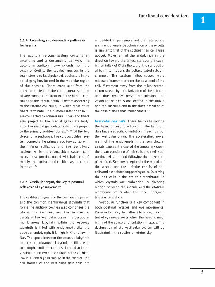
Figure 1.1 a–c:
This cocker spaniel was
shaking its head contin-
uously. Its ear canals
were clean and not
inflamed. (a) The aur-
icles are long but were
normal on visual inspec-
tion. (b) Palpation of the
auricles revealed several
heavy lumps of hair with
accumulated dirt and
food. (c) Clipping away
the hair stopped the
shaking.
6
1
1.2 History and clinical signs
1.2.1 History
The medical history in diseases of the ear is
characterized less by hearing disorders than by
pain. Pain can be caused by disease of both the
external ear and the middle ear, and can be uni-
lateral or bilateral. When the inner ear is
involved in the dog or the cat, vestibular dys-
function is more commonly mentioned in the
history than loss of hearing. Diseases of the ear
are usually presented as disorders affecting
one or both ears exclusively. However, ques-
tioning may reveal signs of a more generalized
skin disorder of which inflammation of the
external ear is a part, or recurrent periods of
fever and other signs of infection together with
middle ear disease, or other signs of neuro-
genic disease rather than a vestibular problem
alone. It is therefore essential that additional
questions be asked about the animal’s general
condition, appetite, drinking, and physical
activity; whether there have been changes in its
habits; and whether there have been similar
problems in the past, in either or both ears. The
onset of ear problems may be sudden or grad-
ual. The onset of signs caused by a foreign body
in the external ear is often sudden and recog-
nized by the owner. In contrast, inflammation of
the external ear often begins gradually but
becomes progressively worse; with such a his-
tory it is useful to ask what treatments have
been tried. There are many ways of »treating«
inflammation of the external ear which cause
the inflammation to persist or even to increase.
If parasitic infection is suspected, questions
should be asked about contacts with other ani-
mals, of the same or different species.79
Vestibular dysfunction is usually sudden in
onset and the signs are usually dramatic, but
hearing loss may go unnoticed, especially if
unrelated to a specific event. In some cases of
sudden deafness an associated event is men-
tioned by the owner, but it is not always easy to
find a logical relation between this and the
hearing loss. Unilateral hearing loss is often
masked by normal hearing in the other ear.
The Ear
a b
3-87706-635-6_00I-050 18.04.2005 12:03 Uhr Seite 6
7
1.2.2 Clinical signs
Pain is an »unpleasant sensory and emotional
experience associated with actual or potential
tissue damage«.1, 44 It is a complex subjective
experience, depending on the severity of the
noxious damage, but also on a variety of addi-
tional cognitive and emotional aspects, and is
therefore difficult to measure. It is even more
complicated when a dog or cat is in apparent
pain as described by the owner or caretaker. Ear
pain is usually recognized if the animal tries to
prevent handling of the ear, is less alert than
usual, and sometimes very carefully scratches
the ear or shakes the head. Pain and pruritus
are not easily distinguished by casual observa-
tion, and in dogs and cats the signs of both can
be suppressed by analgesic drugs. Pain caused
by external ear disease may be severe and may
change the dog’s or cat’s behavior, something
often better recognized in retrospect when the
pain disappears with successful treatment.
Signs of external ear disease. These are pre-
dominantly pain and pruritus, the pain some-
times causing the dog to turn its head slightly
with the painful ear downwards. In dogs the aur-
icle may be in an uncharacteristic position for
the breed, and in cats it may be folded and
turned backwards. The concave side of the aur-
icle is usually thinly haired and inflammation of
the external ear can be recognized by swelling
and lesions of the skin, often with excess ceru-
men and exudate. Long hair on the auricle can
become heavy with accumulated dirt and food,
also causing the dog to shake its head in the ab-
sence of ear inflammation (Figure 1.1 a–c). Rub-
bing or scratching of the ear which injures the
skin can lead to bacterial infection, increasing
the inflammation and pain. Other signs of in-
flammation include scaling, hyperpigmenta-
tion, and tissue proliferation, the latter particu-
larly at the base of the auricle on the concave
side and around the entrance to the ear canal.
Thickening of the auricle can result from acute or
chronic dermatitis or perichondritis. The auricle
can also be extremely thickened by a hematoma
within the cartilage layer, presenting as a bulge
on the concave side. In contrast to an abscess,
which occurs most often in cats, a hematoma
does not result in pain or general malaise.
Temperature of the auricle. The temperature of
the auricle varies with the flow of blood, best
appreciated on the concave side by the accom-
panying variation in its pink color. The color can
vary not only with the ambient temperature but
also with the balance between sympathetic and
parasympathetic influence on blood flow in the
auricle. A »red and warm« auricle may be a nor-
mal and transient finding, and is then usually
bilateral.
Cerumen, or ear wax, is formed on the concave
side of the base of the auricle and in the exter-
nal ear canal. It is a mixture of the secretions of
the sebaceous and the ceruminous glands. It
has a waxy consistency and varies in color from
History and clinical signs1
c
3-87706-635-6_00I-050 18.04.2005 12:03 Uhr Seite 7
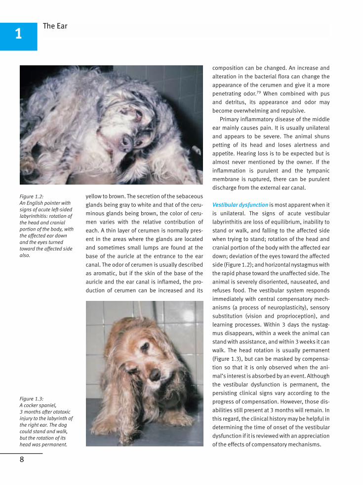
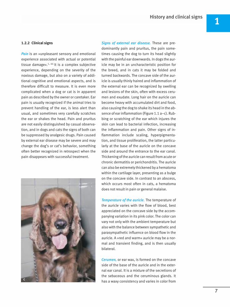
Figure 1.2:
An English pointer with
signs of acute left-sided
labyrinthitis: rotation of
the head and cranial
portion of the body, with
the affected ear down
and the eyes turned
toward the affected side
also.
Figure 1.3:
A cocker spaniel,
3 months after ototoxic
injury to the labyrinth of
the right ear. The dog
could stand and walk,
but the rotation of its
head was permanent.
8
1
yellow to brown. The secretion of the sebaceous
glands being gray to white and that of the ceru-
minous glands being brown, the color of ceru-
men varies with the relative contribution of
each. A thin layer of cerumen is normally pres-
ent in the areas where the glands are located
and sometimes small lumps are found at the
base of the auricle at the entrance to the ear
canal. The odor of cerumen is usually described
as aromatic, but if the skin of the base of the
auricle and the ear canal is inflamed, the pro-
duction of cerumen can be increased and its
composition can be changed. An increase and
alteration in the bacterial flora can change the
appearance of the cerumen and give it a more
penetrating odor.79 When combined with pus
and detritus, its appearance and odor may
become overwhelming and repulsive.
Primary inflammatory disease of the middle
ear mainly causes pain. It is usually unilateral
and appears to be severe. The animal shuns
petting of its head and loses alertness and
appetite. Hearing loss is to be expected but is
almost never mentioned by the owner. If the
inflammation is purulent and the tympanic
membrane is ruptured, there can be purulent
discharge from the external ear canal.
Vestibular dysfunction is most apparent when it
is unilateral. The signs of acute vestibular
labyrinthitis are loss of equilibrium, inability to
stand or walk, and falling to the affected side
when trying to stand; rotation of the head and
cranial portion of the body with the affected ear
down; deviation of the eyes toward the affected
side (Figure 1.2); and horizontal nystagmus with
the rapid phase toward the unaffected side. The
animal is severely disoriented, nauseated, and
refuses food. The vestibular system responds
immediately with central compensatory mech-
anisms (a process of neuroplasticity), sensory
substitution (vision and proprioception), and
learning processes. Within 3 days the nystag-
mus disappears, within a week the animal can
stand with assistance, and within 3 weeks it can
walk. The head rotation is usually permanent
(Figure 1.3), but can be masked by compensa-
tion so that it is only observed when the ani-
mal’s interest is absorbed by an event. Although
the vestibular dysfunction is permanent, the
persisting clinical signs vary according to the
progress of compensation. However, those dis-
abilities still present at 3 months will remain. In
this regard, the clinical history may be helpful in
determining the time of onset of the vestibular
dysfunction if it is reviewed with an appreciation
of the effects of compensatory mechanisms.
The Ear
3-87706-635-6_00I-050 18.04.2005 12:03 Uhr Seite 8
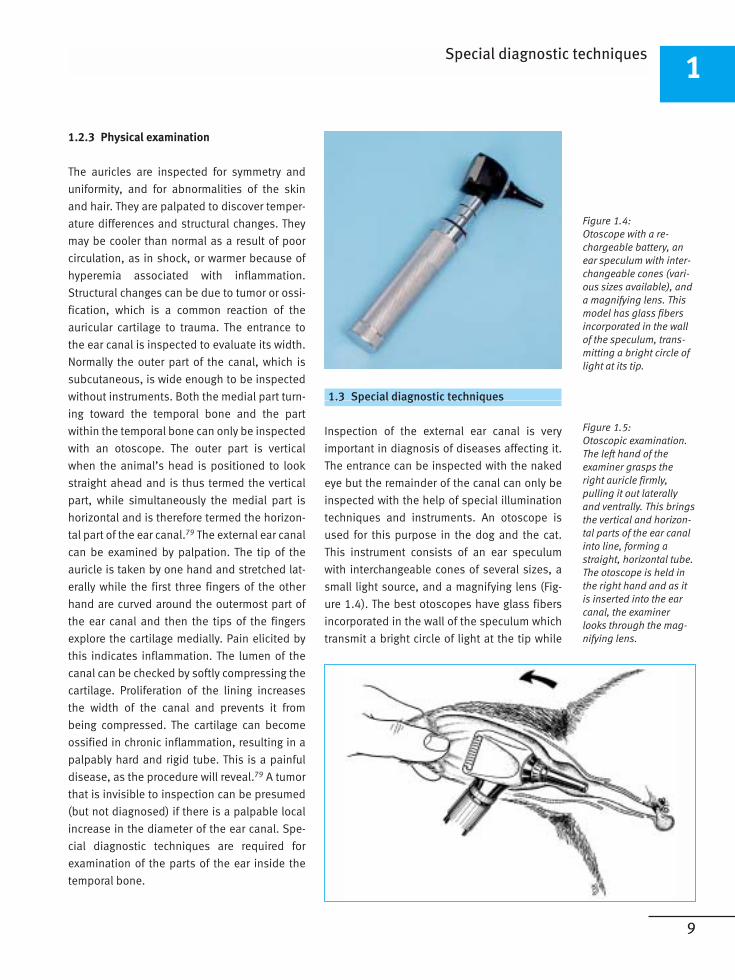
Figure 1.4:
Otoscope with a re-
chargeable battery, an
ear speculum with inter-
changeable cones (vari-
ous sizes available), and
a magnifying lens. This
model has glass fibers
incorporated in the wall
of the speculum, trans-
mitting a bright circle of
light at its tip.
Figure 1.5:
Otoscopic examination.
The left hand of the
examiner grasps the
right auricle firmly,
pulling it out laterally
and ventrally. This brings
the vertical and horizon-
tal parts of the ear canal
into line, forming a
straight, horizontal tube.
The otoscope is held in
the right hand and as it
is inserted into the ear
canal, the examiner
looks through the mag-
nifying lens.
9
1.2.3 Physical examination
The auricles are inspected for symmetry and
uniformity, and for abnormalities of the skin
and hair. They are palpated to discover temper-
ature differences and structural changes. They
may be cooler than normal as a result of poor
circulation, as in shock, or warmer because of
hyperemia associated with inflammation.
Structural changes can be due to tumor or ossi-
fication, which is a common reaction of the
auricular cartilage to trauma. The entrance to
the ear canal is inspected to evaluate its width.
Normally the outer part of the canal, which is
subcutaneous, is wide enough to be inspected
without instruments. Both the medial part turn-
ing toward the temporal bone and the part
within the temporal bone can only be inspected
with an otoscope. The outer part is vertical
when the animal’s head is positioned to look
straight ahead and is thus termed the vertical
part, while simultaneously the medial part is
horizontal and is therefore termed the horizon-
tal part of the ear canal.79 The external ear canal
can be examined by palpation. The tip of the
auricle is taken by one hand and stretched lat-
erally while the first three fingers of the other
hand are curved around the outermost part of
the ear canal and then the tips of the fingers
explore the cartilage medially. Pain elicited by
this indicates inflammation. The lumen of the
canal can be checked by softly compressing the
cartilage. Proliferation of the lining increases
the width of the canal and prevents it from
being compressed. The cartilage can become
ossified in chronic inflammation, resulting in a
palpably hard and rigid tube. This is a painful
disease, as the procedure will reveal.79 A tumor
that is invisible to inspection can be presumed
(but not diagnosed) if there is a palpable local
increase in the diameter of the ear canal. Spe-
cial diagnostic techniques are required for
examination of the parts of the ear inside the
temporal bone.
1.3 Special diagnostic techniques
Inspection of the external ear canal is very
important in diagnosis of diseases affecting it.
The entrance can be inspected with the naked
eye but the remainder of the canal can only be
inspected with the help of special illumination
techniques and instruments. An otoscope is
used for this purpose in the dog and the cat.
This instrument consists of an ear speculum
with interchangeable cones of several sizes, a
small light source, and a magnifying lens (Fig-
ure 1.4). The best otoscopes have glass fibers
incorporated in the wall of the speculum which
transmit a bright circle of light at the tip while
Special diagnostic techniques1
3-87706-635-6_00I-050 18.04.2005 12:03 Uhr Seite 9
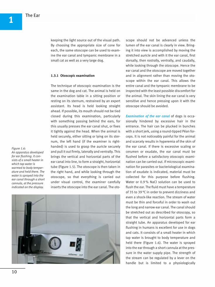
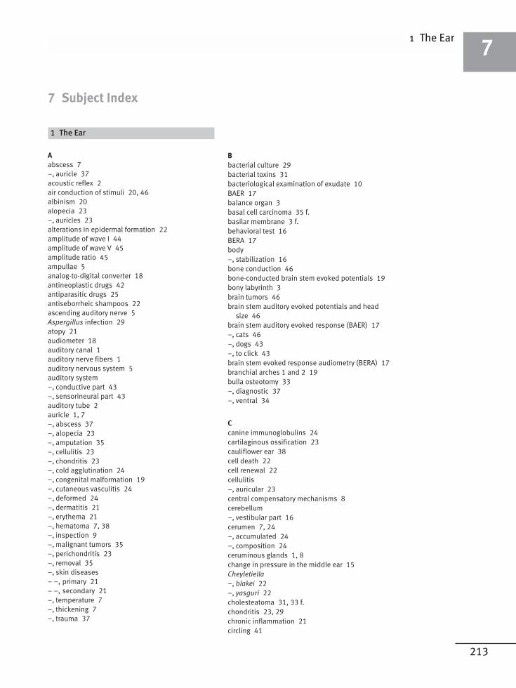
Figure 1.6:
An apparatus developed
for ear flushing. It con-
sists of a small heater in
which tap water is
warmed to body temper-
ature and held there. The
water is sprayed into the
ear canal through a short
cannula, at the pressure
indicated on the display.
10
1
keeping the light source out of the visual path.
By choosing the appropriate size of cone for
each, the same otoscope can be used to exam-
ine the ear canal and tympanic membrane in a
small cat as well as a very large dog.
1.3.1 Otoscopic examination
The technique of otoscopic examination is the
same in the dog and cat. The animal is held on
the examination table in a sitting position or
resting on its sternum, restrained by an expert
assistant. Its head is held looking straight
ahead. If possible, its mouth should not be tied
closed during this examination, particularly
with something passing behind the ears, for
this usually presses the ear canal shut, or fixes
it tightly against the head. When the animal is
held securely, either sitting or lying on its ster-
num, the left hand (if the examiner is right-
handed) is used to grasp the auricle securely
and pull it out firmly, laterally and ventrally. This
brings the vertical and horizontal parts of the
ear canal into line, to form a straight, horizontal
tube (Figure 1.5). The otoscope is then taken in
the right hand, and while looking through the
otoscope, so that everything is carried out
under visual control, the examiner carefully
inserts the otoscope into the ear canal. The oto-
scope should not be advanced unless the
lumen of the ear canal is clearly in view. Bring-
ing it into view is accomplished by moving the
stretched auricle and with it the ear canal, first
dorsally, then rostrally, ventrally, and caudally,
while looking through the otoscope. Hence the
ear canal and the otoscope are moved together
and in alignment rather than moving the oto-
scope within the ear canal. This allows the
entire canal and the tympanic membrane to be
inspected with the least possible discomfort for
the animal. The skin lining the ear canal is very
sensitive and hence pressing upon it with the
otoscope should be avoided.
Examination of the ear canal of dogs is occa-
sionally hindered by excessive hair in the
entrance. The hair can be plucked in bunches
with a short jerk, using a round-tipped Péan for-
ceps. It is not noticeably painful for the animal
and scarcely results in hyperemia of the skin of
the ear canal. If there is excessive scaling or
cerumen or exudate, the ear canal must be
flushed before a satisfactory otoscopic exami-
nation can be carried out. If microscopic exami-
nation for parasites or bacteriological examina-
tion of exudate is indicated, material must be
collected for this purpose before flushing.
Water or 0.9 % NaCl solution can be used to
flush the ear. The fluid must have a temperature
of 35 to 39 °C in order to prevent dizziness and
even a shock-like reaction. The stream of water
must be thin and forceful in order to wash out
the long and narrow ear canal. The canal should
be stretched out as described for otoscopy, so
that the vertical and horizontal parts form a
straight tube. An apparatus developed for ear
flushing in humans is excellent for use in dogs
and cats. It consists of a small heater in which
tap water is brought to body temperature and
held there (Figure 1.6). The water is sprayed
into the ear through a short cannula at the pres-
sure in the water supply pipe. The strength of
the stream can be regulated by a lever on the
handle but is limited to a physiologically
The Ear
3-87706-635-6_00I-050 18.04.2005 12:03 Uhr Seite 10
213
1 The Ear7
7 Subject Index
1 The Ear
A
abscess 7
–, auricle 37
acoustic reflex 2
air conduction of stimuli 20, 46
albinism 20
alopecia 23
–, auricles 23
alterations in epidermal formation 22
amplitude of wave I 44
amplitude of wave V 45
amplitude ratio 45
ampullae 5
analog-to-digital converter 18
antineoplastic drugs 42
antiparasitic drugs 25
antiseborrheic shampoos 22
ascending auditory nerve 5
Aspergillus infection 29
atopy 21
audiometer 18
auditory canal 1
auditory nerve fibers 1
auditory nervous system 5
auditory system
–, conductive part 43
–, sensorineural part 43
auditory tube 2
auricle 1, 7
–, abscess 37
–, alopecia 23
–, amputation 35
–, cellulitis 23
–, chondritis 23
–, cold agglutination 24
–, congenital malformation 19
–, cutaneous vasculitis 24
–, deformed 24
–, dermatitis 21
–, erythema 21
–, hematoma 7, 38
–, inspection 9
–, malignant tumors 35
–, perichondritis 23
–, removal 35
–, skin diseases
– –, primary 21
– –, secondary 21
–, temperature 7
–, thickening 7
–, trauma 37
B
bacterial culture 29
bacterial toxins 31
bacteriological examination of exudate 10
BAER 17
balance organ 3
basal cell carcinoma 35 f.
basilar membrane 3 f.
behavioral test 16
BERA 17
body
–, stabilization 16
bone conduction 46
bone-conducted brain stem evoked potentials 19
bony labyrinth 3
brain tumors 46
brain stem auditory evoked potentials and head
size 46
brain stem auditory evoked response (BAER) 17
–, cats 46
–, dogs 43
–, to click 43
brain stem evoked response audiometry (BERA) 17
branchial arches 1 and 2 19
bulla osteotomy 33
–, diagnostic 37
–, ventral 34
C
canine immunoglobulins 24
cartilaginous ossification 23
cauliflower ear 38
cell death 22
cell renewal 22
cellulitis
–, auricular 23
central compensatory mechanisms 8
cerebellum
–, vestibular part 16
cerumen 7, 24
–, accumulated 24
–, composition 24
ceruminous glands 1, 8
change in pressure in the middle ear 15
Cheyletiella
–, blakei 22
–, yasguri 22
cholesteatoma 31, 33 f.
chondritis 23, 29
chronic inflammation 21
circling 41
3-87706-635-6_213-238 18.04.2005 13:47 Uhr Seite 213
214
7
clear away debris 11
clearing mechanism 24
click stimuli 18, 47
cochlea 3, 5
–, histology of the development 20
–, inner ear 1
cochlear duct 3
–, membranous 3
cochlear microphonics 41
cochlear nerve 3
–, electrical potential in the fibers 4
cochlear nucleus 1, 5
compensatory mechanisms 35
compliance 15
computed tomography (CT) 12
contact hypersensitivity 21
–, caused by medications 22
contrast medium 14
coronal scan 13
cortex 1
Corti
–, organ of 3
crest
–, mesoderm 19
–, neural 19
crusting 23
CT 12
culture testing 30
cuticular plate 4
cystadenomas 36
D
Dalmatian dogs
–, deafness 20
dandruff 22
data acquisition software 18
deafness
–, acquired 19
–, congenital 19, 43, 46
– –, caused by a genetic defect 20
–, hereditary
– –, associated with disorders of pigmentation 20
–, unilateral 21
Deiter’s cells 3
demodicosis 22
densitometric reading 13
depigmentation 23
depolarization 4
dermatitis 22
–, secondary 22
deviation of the eyes 41
diagnostic imaging 12
different stimulations
–, threshold 45
drug allergy 24
E
ear canal
–, cleaning by flushing 25
–, congenital atresia 19
–, diameter
– –, palpable local increase 9
–, examination 10
–, external
– –, abscess around the distal part 39
– –, bleeding 39
– –, continuity 39
– –, diagnostic imaging 12
– –, foreign bodies 25
– –, inflammatory diseases 24
– –, inspection 9
– –, interruption 39
– –, malignant tumor 36
– –, massive purulent discharge 30
– –, ossification 28 f.
– –, polyp 32
– –, stenosis 39
– –, trauma 38
–, horizontal part 9
–, increase in the microflora 24
–, removal 27, 36
–, resection technique 28
–, vertical part 9
ear mite infestation 25
ectoderm 19
electrical potential in the fibers of the cochlear
nerve 4
endocrinopathies 22
endolymph 3 f.
epithelial elements
–, primary degeneration 20
epithelial layer
–, proliferating external
– –, vascularization 30
epithelial migration 40
epitympanum 2
equilibrium 16
erosion 28
eustachian tube 2
–, dysfunction 30
– –, major cause 31
–, function 15
–, obstruction
– –, major cause 31
excessive hair in the entrance 10
excessive scaling 10
external ear 1
–, congenital deformity 19
–, disease
– –, signs 7
–, inflammation 7
–, sensory innervation 24
exudate
–, bacteriological examination 10
Subject Index
3-87706-635-6_213-238 18.04.2005 13:47 Uhr Seite 214
215
eyes
–, abnormal positioning 16
F
facial nerve 32
–, motor innervation from 24
facial paralysis 41
fibrous layer 1
fine needle aspiration biopsies 35
first pharyngeal pouch 19
FISH mapping 21
flushing 10
–, apparatus 10
fly bites 22
food hypersensitivity 21
foreign body 11, 25
–, clinical signs 25
–, forceps 25
frequency 17
frequency following response 44
frequency specificity of hearing in cats 46
frostbite 24
funnel-shaped fibroelastic cartilage 24
G
ganglion
–, spiral 21
ganglion cells
–, loss 20
general anesthesia 12
gland carcinoma
–, sebaceous 35 f.
glands
–, ceruminous 1, 8
–, sebaceous 1, 8
glossopharyngeal nerve 24
H
hair cells 3 f.
–, inner 3 f.
–, outer 3 f.
–, vestibular 5
hair follicles 1
hair loss 22
hearing
–, capacity 1
–, cats 46
–, classification 17
–, dogs 43
–, frequency-specific 17
–, impairment 16
–, intensity-specific 17
–, sensorineural 46
–, test 16
– –, earliest discriminating 19
hearing loss
–, cats 46
–, cisplatin-induced 42
–, combined conductive and sensorineural 43, 46
–, conductive 43, 46
– –, causes 43
–, dogs 43
–, sensorineural 41, 43, 46
– –, causes 43
helicotrema 3
hemangiosarcoma 35 f.
hematoma
–, auricle 7, 38
Hensen’s cells 3
histiocytoma 35 f.
histological examination 32
horizontal nystagmus 8
Horner’s syndrome 31
hydrocephalus 43
hyperestrogenism 22
hyperpolarization 4
hypersensitivity 21, 25
hypopigmentation 20
hypothyroidism 22
I
immittance measurement 15
immune-mediated disorders 23
impedance 2
incus 2
inferior colliculus 5
–, midbrain 1
inflammation 9, 21
inner ear 3
–, cochlea 1
inner hair cells 3 f.
interpeak latencies 44
intraoral film 12
Ivermectin 22
K
keratin 34
keratinization
–, altered 22
–, disorders 22
kinocilium 3
L
labyrinth 3
–, vestibular membranous 5
labyrinthitis 21, 31, 34, 46
–, infectious 21
Larmour frequency 13
lateral lemnicus 5
lead poisoning 24
leishmaniasis 22
limbus 4
linear acceleration 5
linear filter setting 18
1 The Ear7
3-87706-635-6_213-238 18.04.2005 13:47 Uhr Seite 215
216
7
local spreading
–, evaluation 36
loss of transparency 30
lupus erythematosus 23 f.
lymphoplasmacytic inflammation 24
lymphoreticular neoplasms 24
M
macule 3
–, saccule 5
magnetic resonance imaging (MRI) 13
malleus 2
–, manubrium 2, 11
manubrium of the malleus 2, 11
mast cell tumor 35 f.
medial geniculate body 5
melanin
–, forming 20
melanoblasts 21
melanocytes 20
melanoma
–, malignant 35 f.
membrane of the oval window 2
membranous labyrinth 3, 5
meningitis
–, secondary 21
merle pigmentation 20
mesenchymal condensation 19
mesoderm crest 19
mesotympanum 2
microscopic examination for parasites 10
midbrain
–, inferior colliculus 1
middle ear 1
–, adhesion of the tympanic membrane 15
–, cavity
– –, density 33
–, change in pressure 15
–, cholesteatoma 34
–, fluid filling 15
–, impedance 15
–, inflammation 30
– –, acute 30
– –, primary 8
– –, purulent 30
–, major function 2
–, polyp in the mucosa 32
–, surgical exploration 33
–, tumor 37
modiolus 3
MRI 13
N
needle electrode 17
neomycin
–, systemically administered 42
neural crest 19, 21
neural impulses 1
neural structures
–, secondary degeneration 20
neurological examination
–, vestibular dysfunction 16
neuroplasticity 8, 41
noise burst stimuli 47
nonscreen film 12
Notoedres cati 22
noxious damage 7
nystagmus 16
–, head 41
–, horizontal 8
O
olivocochlear system 5
open-mouth projections
–, rostrocaudal 12
osseous spiral lamina 3
ossicles 2
ossicular chain 2
ossification
–, cartilaginous 23
otic capsule 19
otitis externa 21
–, acute
– –, caused by foreign bodies 25
– –, diffuse bacterial 25
–, chronic 26
– –, bacterial 29
– –, proliferative 27
–, complications 29
otitis media
–, chronic 31, 34
– –, causes 31 f.
–, development 30
–, effusion 30
–, unresolved 33
Otodectes cynotis 25
otolithic membrane 5
otoscope 9
otoscopy 10, 12, 30
ototoxic drugs
–, pathways 41
ototoxicity 21, 41
–, acute 41
–, certain systemically administered drugs 42
–, concentration of the agent 41
–, duration of its contact 41
outer hair cells 3 f.
oval window
–, membrane 2
P
pain 7, 9
–, caused by external ear disease 7
–, cognitive aspects 7
Subject Index
3-87706-635-6_213-238 18.04.2005 13:47 Uhr Seite 216
217
–, emotional aspects 7
palpation 9
parasites
–, causing dermatitis 22
–, microscopic examination 10
pars flaccida 11
pars tensa 11
Pasteurella multocida 37
peak latencies 44
pemphigus
–, foliaceus 23
–, vulgaris 23
perichondritis 23, 29
perilymph 4
perilymphatic duct 3
periolivary nucleus 5
petrous portion of the temporal bone 3
pharyngeal opening 3
physical examination 9
piebald gene 20 f.
piston-like motion 2
polychondritis 23
–, relapsing 23
pontine nuclei 5
positioning of the patient 13
postural reflexes 16
presbyacusis 43, 46
pressure to prevent reaccumulation of blood 38
primary auditory cortex 5
primordial membranous labyrinth 19
progress of compensation 41
Proteus mirabilus 26
pruritus 22
Pseudomonas 23, 42
–, aeruginosa 26
–, infection 29
R
Reissner’s membrane 20
resting potential 4
RH mapping 21
rostrocaudal open-mouth projections 12
rotation of the head 41
round window 3
–, membrane
– –, permeability 42
S
saccule 3, 5
–, macule 5
sagittal scan 13
sarcoidosis 23
Sarcoptes canis 22
screen film in a cassette 12
sebaceous glands 1, 8
seborrhea 22
–, primary idiopathic 22
secondary bacterial infections 21
sedation 12
semicircular canals 3, 5
semicircular ducts 3
sensitivity testing 29 f.
sensory elements
–, primary degeneration 20
sensory hair cells 1
sensory organ 1
sensory substitution 8, 41
shearing motion 5
signs of shock 39, 41
sinusoidal receptor potential 4
sinusoidal stimulus 4
skin
–, trauma
– –, caused by mechanical cleaning techniques 24
skull
–, radiographic examination 12
soft ointment
–, containing a broad-spectrum antibiotic 26
–, containing a glucocorticoid and a broad-spectrum
antibiotic 25
sound energy 1 f.
sound in space 1
sound localization 1
sound pressure level 17
speech perception 1
spiral ganglion 5
spotting 21
squamous cell carcinoma 35 f.
squamous cell epithelial layer 1
squamous epithelium 34
stapedius muscle 2
stapes 2
staphylococcal pyrogenic exotoxin 42
Staphylococcus aureus 26, 42
stereocilia 3 f.
stimuli from a vibrator 46
Streptococcus 42
stria vascularis 4
–, decrease in thickness 20
subarachnoid space 3
superimpositions 12
superior olivary complex 1, 5
sutures
–, through-and-through interrupted mattress 38
–, wound 37
sympathetic nerve
–, dysfunction 31
T
tectorial membrane 4
temporal bone 1, 9
–, destruction 37
–, fracture 39
–, masses within 14
–, petrous portion 3
1 The Ear7
3-87706-635-6_213-238 18.04.2005 13:47 Uhr Seite 217
218
7
–, trauma 40
tensor tympani muscle 2
thalamus 1
thresholds of hearing 46
toneburst stimuli 18
tonebursts 43, 46
transduction channels 4
transverse scan 13
trauma 37–39
trigeminal nerve 2, 24
tumor 35 f.
tunica propria 1
tympanic bulla 2
–, enlargement 34
tympanic canals 3
tympanic cavity 2
–, lining 3
–, ventral part 2
tympanic membrane 1, 11, 15
–, appearance of the opening of a fistula 33
–, experimentally perforated
– –, healing 40
–, fibrovascular repair 40
–, flaccid 15
–, healing 31, 40
–, inflammation 30
–, integrity 15
–, nonhealing 40
–, regrowth 33
–, rupture 12, 30 f., 39 f.
–, trauma 39
tympanometer 15
tympanometry 15
U
ulcer 28
ultrasound imaging 14
utricle 3, 5
V
vagus nerve 24
vestibular canals 3
vestibular dysfunction 8, 21
–, neurological examination 16
vestibular nerve 16
vestibular nuclei 16
vestibular organ 5
vestibular system 16
vestibule 3
visual disturbances 16
voltage-gated calcium channels 4 f.
W
Waardenburg syndrome 20
white cats
–, deafness 20
white spotting 20
Y
yeast infections 21
Subject Index
2 The Nose and Nasal Sinuses
A
adenocarcinoma 74
adnexal tissue 62
adrenalin solution 58
airflow
–, conditioning 51
–, regulation 51
airstream symmetry 55
Alternaria 65, 67
amygdala 53
anemia
–, aplastic 78
anesthesia 57
–, monitoring of the depth 58
–, risks 61
arc-like effective stroke 62
aspergillosis 77
–, clinical signs 59, 65, 77
–, disseminated 65
–, nasal
– –, systemic treatment 67
–, sinonasal
– –, treatment 66
Aspergillus 64
–, fumigates 65
–, infection 54
–, plaques 59, 65, 77
aspiration of brownish-red fluid
–, repeated 75
atrophy 59
–, conchae 59, 65
axonemal tubule 52
3-87706-635-6_213-238 18.04.2005 13:47 Uhr Seite 218
219
B
biopsy forceps 58
bird cages 65
blanket functions 52
blepharospasm 63
blood flow 52
bone resorption 69
Bordetella bronchiseptica 59, 63
brain damage 75
breed
–, brachycephalic 60, 62
–, dolichocephalic 62
bridge of the nose
–, fistula in the midline 62
C
calicivirus infection 64
canine distemper 64
Capillaria aerophilia 68
cardiomegaly 52
chemosis 63
choanae 51, 58
–, fractures 76
chondrosarcoma 74
chorda tympani 69
ciliary beat 52
–, velocity 63
ciliary dysfunction
–, congenital 62
ciliary dyskinesia
–, primary 62 f.
ciliated cell 52
clefts
–, repair 61
closure, airtight 79
clotrimazole 66
coagulation disorders 78
coagulopathy
–, acquired 78
computed tomography (CT) 56, 75
–, positioning of the patient 56
conchae 51, 58
–, atrophy 59, 65
– –, via bone resorption 63
–, ethmoidal 53
–, loss of normal maxillary and ethmoidal 74
–, minimal loss 68
congenital abnormalities 60, 62
congenital neural tube defect 62
conjunctivitis 63
contrast medium 56 f., 75
cor pulmonale 52
coumarin 78
cribriform plate 53
crust formation 64
cryptococcosis 67
Cryptococcus
–, staining 67
Cryptococcus neoformans 65
CT 56
cyst 62
–, nasal dermoid sinus 62
cytokines
–, proinflammatory 68
cytoreductive therapy 74
cytotoxic agents 71
D
dental abnormalities 55
dental root infections 77
depigmentation 55
depression 65
diagnostic imaging 56
diagnostic techniques
–, special 55
dog
–, brachycephalic 51
–, dolichocephalic 51
–, emaciated nose 60
–, sense of smell loss 55
dryness 69
–, pathological 55
dynein arms 52
–, defects 62
dyspnea 74
–, nasal obstruction 55
–, relief 61
E
ectoturbinate 51
emphysema
–, subcutaneous 75
endotracheal intubation 77
enilconazole 66
entorhinal cortex 53
epidermal tissue 62
epistaxis 54, 77
–, acute
– –, management 77
–, causes 77
–, diagnostic plan 78
–, intermittent of unknown origin 79
–, recurrent 68
epithelium
–, ciliated respiratory 68
estrogen 78
ethmoid 51
extent of the destruction 65
external nose
–, bony case 51
–, cartilaginous parts 51
exudate
–, mucopurulent 54
2 The Nose and Nasal Sinuses7
3-87706-635-6_213-238 18.04.2005 13:47 Uhr Seite 219
220
7
F
facial deformity 61
feline calicivirus 63
feline herpesvirus-1 63
fibroma 72
fibrosarcoma 72
fistula 69
–, congenital cerebrospinal fluid 62
–, nasal-cutaneous 62
–, oronasal 65
– –, result from extraction of a maxillary canine tooth
70
–, surgically closure 70
fistulogram 62
forebrain 53
foreign body 58
–, rhinitis 70
frontal bone
–, resorption 65
–, trauma 75
–, trephined opening 66
frontal sinus 51
G
gelatin sponge 79
glands
–, serous 52
glucocorticoids 71
goblet cells 52
granuloma
–, noncaseated epithelioid 70
–, sarcoidal 71
granulomatous infections 67
grayish-yellow material 66
H
hemophilia A and B 78
hemorrhagic discharge 74
–, intermittent 65
herpesvirus infection 64
hippocampus 53
histiocytosis 71
histological examination 73
homeostasis 79
host resistance 65
house dust 65
household plants 65
humidification 52
hypertrophy 59
hypothalamus 53
hypovolemic shock 76
I
incisive bones 51
infecting spores
–, number 65
inflammation 77
inflammatory tissue 68
intermittent rhinosinusitis 64
intracranial extensions 62
intranasal catheter 76
intraoral film 56
ipsilateral external carotid artery
–, ligation 77
K
Kartagener’s syndrome 62
keratitis 63
keratoconjunctivitis sicca
–, ipsilateral 69
L
lacrymal bones 51
lamina propria 52
Larmour frequency 57
leishmaniasis 78
Linguatula serrata 78
liver failure 78
lungs 61
lymphoma 78
lymphosarcoma 74
M
magnetic resonance imaging (MRI) 56 f., 75
maxillae 51
maxillary canine tooth 69
maxillary sinus 51
melena 77, 79
metachronous movement 52
microvilli 52
mouth inspection for abnormality 58
MRI 75
mucopurulent discharge 74
mucosa
–, destruction 65
–, focal proliferations 68
–, hypervascularization 72
–, nasal
– –, lesions 54
–, pale-pink 77
–, respiratory
– –, pseudostratified 52
–, thickening 72
–, underlying structures
– –, destruction 65
–, white 77
mucosal cleaning 52
mucus blanket
–, overlying 52
mucus clearance
–, velocity 63
myelosuppressive drugs 78
Subject Index
3-87706-635-6_213-238 18.04.2005 13:47 Uhr Seite 220
221
N
nasal alae 55
nasal bleeding 65, 74
nasal bone 51
–, rectangular opening 79
nasal cavity 51
–, abnormalities in morphology 59
–, caudal part 68
–, mucociliary clearance 63
–, mucosa 71
–, obstruction 55, 59
–, tumor 74
nasal discharge 54 f.
–, clear 62
–, profuse mucopurulent 65
–, serous 63
–, unilateral mucopurulent 54, 70
nasal passage
–, narrowing 70
nasal plane 51, 65
–, congenital malformation 60
–, depigmentation 71
–, surgical removal 73
–, swelling 71
–, tumor 72
nasal polyps 59
–, bilateral 68
–, clinical signs 68
–, extreme cases 68
–, multiple proliferations 69
–, rhinitis 68
–, surgical removal 68
–, unilateral 68
nasofrontal duct
–, obstruction 75
–, patency 75
nasolacrimal duct 53
neoplasma
–, hematopoietic 78
newborn puppies
–, herpes infection 64
nonscreen film 56
nostrils
–, narrow 55
–, narrowing 60
–, primary bacterial infection 64
–, widening 61
O
obstruction
–, dyspnea 55
–, temporary 76
–, upper airway 61
ocular discharge
–, serous 63
olfaction 51, 53
olfactory bulb 53
–, cavity 62
olfactory cilia 53
olfactory cortex
–, primary 53
olfactory epithelium
–, removal 69
olfactory information
–, processing 53
–, transduction 53
olfactory nerves 53
olfactory receptor cells 53
olfactory receptor neuron 53
open-mouth breathing 55
open-mouth projections 56
orbit 65
osteosarcoma 74
otoscope 58
oxygenation 76
P
pain 54, 57, 65
–, acute 54
–, chronic 54
painful infection 64
palatine bone 51
palliative therapy 64
paranasal sinus 51
parasympathetic system 51
Pasteurella multocida 63
periciliary layer 52
periodontitis 63, 69
periostitis 65
pharynx
–, nasal portion 51
phenobarbital 77
pheromones 53
physical examination 54 f.
Pneumonyssoides caninum 68
primary venereal tumors 74
proliferation of tissue around the root of the canine
tooth 70
protective reflex 54
pulmonary edema 52
R
radiation therapy 74
radiodense material 59
radiographic density 74
radiographic examination 61
–, skull 56
radiographs 55
–, rostrocaudal 56
regional lymph nodes 73
renal failure 78
respiratory activity
–, increased energy use 60
respiratory cilia
–, ultrastructural lesions 62
2 The Nose and Nasal Sinuses7
3-87706-635-6_213-238 18.04.2005 13:47 Uhr Seite 221
222
7
respiratory disease
–, viral upper
– –, cat 63
respiratory resistance 52
restlessness 77
rhinitis
–, allergic 72
–, causes
– –, dental disease 69
– –, foreign bodies 70
–, chronic 63
–, IgE-based 72
–, infectious 63
–, mycotic 64
–, nasal polyps 68
–, neurogenic 69
–, noninfectious 68
–, nonspecific chronic 72
–, obstructive 68
–, parasitic 68
–, primary bacterial 64
–, proliferative 59
–, viral
– –, dog 64
rhinoscopic removal 59
rhinoscopy 56 f., 64, 68, 70
–, otoscope 58
rhinosinusitis
–, intermittent 64
rhinosporidiosis 67
Rhinosporidium seeberi 67
rubbing of the nose 70
S
sagittal scan 57
sarcoidosis 70
sense of smell
–, diminished 60
–, loss 55
septal cartilage 51
septum 65
shearing force 52
shedding of virulent virus in carrier cats 64
shock
–, signs 75, 77
sinus
–, frontal 51
– –, congenital malformation 62
– –, fungus 65
– –, trauma 75
– –, tumor 74
–, maxillary 51
–, paranasal 51
–, wall
– –, thickening 66
sinusitis
–, infectious 63
–, noninfectious 68
situs inversus 62
skin depigmentation 54
skull
–, radiographic examination 56
sneezing 54, 74, 77
–, causes
– –, inflammation 54
– –, local drying 54
– –, mechanical stimuli 54
–, ferocious 70
–, paroxysmal 63
sniffing sound 60
snoring 55
soft palate
–, inspection 58
soft tissue contrast 57
soft tissue densities
–, irregular 66
specific functional systems 53
squamous cell carcinoma 72, 74
Staphylococcus aureus 64
sternal recumbency 58
stridor 55
suction cannula 58
superimposition of structures 56
supine position 57
supraorbital process 51
–, osseous septae 51
symblepharon 63
sympathetic nervous system 51
T
telescope 58
–, flexible fiberoptic cable 58
–, light source 58
–, suitable 58
thalamus 53
toxins 65
tracheostoma 55
tracheotomy 74, 76
trauma 76 f.
trigeminal chemosensory system 53
trigeminal system
–, efferents 54
tumor 74, 77
U
ulceration
–, gingival 63
ultrastructural abnormalities 62
V
vagal stimulation 58
vasoconstriction 51
vasodilatation 52
venous plexus 52
vidian nerve 52
Subject Index
3-87706-635-6_213-238 18.04.2005 13:47 Uhr Seite 222
223
viral culture 64
virus
–, immunization against 63
virus carriers 63
virus infection
–, cat-to-cat infection 63
–, chronic carriers 63
–, identification of carriers 64
virus vaccine
–, live, modified 63
visual guidance 57
vomeronasal organ 53
von Willebrand’s disease 78
3 The Pharynx
A
abscess 103
–, life-threatening 103
–, neck 105
ademantinoma 94
aerophagia 95, 110
airway
–, life-threatening obstruction 100
–, obstruction 103
–, protection 84
Alzheimer’s disease 112
ameloblastinoma 94
angioleiomyoma 102
anisocoria 94
arch
–, palatoglossal 83
–, palatopharyngeal 83
arthritic changes 114
aspergillosis
–, nasal 99
aspiration pneumonia 89
atresia
–, choanal 93
– –, bilateral 93
– –, unilateral 93
atrophy
–, bone 104
auditory tube 83
B
balloon catheter 116
biopsy forceps 89
blindness
–, sudden 94
blood loss
–, severe 105
bloody mucous
–, drooling 103
bolus formation 87, 91
bone atrophy 104
brain damage 103
brain injury
–, traumatic 112
3 The Pharynx7
brain stem 106
buccopharyngeal membrane 93
C
canine adenovirus-2 99
canine distemper 99
canine parainfluenza virus 99
choanae
–, bilateral atresia 93
–, nasal cavity 83
–, new
– –, mucosal lining 94
choking 89
cleft lip and palate 91
cleft palate
–, cats 103
closed-mouth breathing 95
complex repetitive discharges 91
computed tomography (CT) 90
congenital oropharyngeal cleft 98
contrast studies
–, preoperative 114
contrast videofluorography 91, 98, 110, 113 f.
coughing 89, 113
cranial laryngeal nerve 84, 106
cranial nerve
–, motor neurons 85
craniopharyngioma 91, 94
–, clinical signs 94
cricoid cartilage
–, caudal border 83
cricopharyngeal muscle 86, 110
–, relaxation 91
cricopharyngeal relaxation 84
Cryptococcus 99
–, staining 99
D
defense mechanism 113, 116
deglutition 83
depression 94
diagnostic imaging 90
3-87706-635-6_213-238 18.04.2005 13:47 Uhr Seite 223
224
7
diaphragm 111
dog
–, brachycephalic 93
dorsal region
–, including the NTS 85
dysphagia 88, 98, 106
–, causes
– –, central neurogenic lesions 112
– –, muscular disorders 106, 110
– –, neurogenic 106
– –, obstructive disorders 112
– –, pain 112
–, esophageal 116
–, management 116
–, manometry 116
–, myogenic 111
–, oropharyngeal 92
–, peripheral neurogenic 106 f.
–, pharyngeal disease 89
–, pharyngoscopy 116
–, specific diagnostic techniques 114
–, systemic diseases 113
dyspnea 93, 95, 104
–, pharyngeal disease 88
–, severe 95
dystrophy
–, muscular 91, 110
E
edema 103
electromyography (EMG) 86
–, continuous EMG recordings 87
emaciation 113
EMG 86
emphysema
–, mediastinal 104
–, subcutaneous 104
endoscope
–, capable of 180° 89
–, flexible with a working channel 106
–, retroflexion 97
endoscopic grasping forceps 106
endotracheal intubation 105
–, emergency 104
endotracheal tubes 89
epithelium
–, ciliated columnar 96
esophageal musculature
–, peristaltic contractions 84
esophagus 111
–, smooth muscles
– –, cat 85
eustachian tube 83
–, opening 89
F
facial nerves 85
fauces
–, pillars 84
faucial arch 84
feline calicivirus 95, 99
feline herpesvirus-1 95, 99
feline leukemia 101
fetid breath 97
fibrillation potential 91
fine needle aspiration biopsy 112
fistula formation 104
foreign body
–, nasopharynx 97
frontal cortex 84
G
gagging 89
gargling 88
gastroesophageal junction 115
glands
–, mucous
– –, distended mucosal 101
glossopharyngeal nerve 84, 86
–, transection 115
goblet cells 96
H
hand feeding 92
hematoma 103
hemostasis 100
hyoid bone 114
–, injury 105
hyopharyngeal muscle 86
hypopituitarism 94
I
inspection 89
interneuronal network 84
interneurons 85
intrapharyngeal opening 83
–, circular closure 83
–, closing 95
involuntary regulation 106
L
laryngeal injuries
–, penetrating 104
laryngeal muscle
–, intrinsic 111
laryngopharynx 83
–, caudal 105
larynx
–, leakage of liquid or food into 91
limb muscles 111
lingual nerve 84
loss of sensory innervation 106
Subject Index
3-87706-635-6_213-238 18.04.2005 13:47 Uhr Seite 224
225
lung metastasis 112
lupus erythematosus 113
lymph nodes
–, retropharyngeal 112
lymphoma 101
M
magnetic resonance imaging (MRI) 90
malignant melanoma 101
manometry
–, dysphagia 116
mast cell tumor 102
masticatory muscles 111
megaesophagus 111
membrane-formed bone
–, ossification 91
middle ear polyp 96
mononuclear cell inflammation 111
motor innervation
–, loss 110
motor neuron disease 112
motor paralysis
–, extraocular 94
mucosa
–, focal hypertrophy 96
–, lesions 116
–, nasopharyngeal
– –, inflammatory irritation 95
– –, sensitive 98
–, pharyngeal
– –, wounds 104
multiloculated cyst 94
muscle fibers
–, progressive loss 111
muscular dystrophy 106
myasthenia gravis 106, 111, 113
–, acquired 111
mylohyoid muscle 85
myopathy 111
N
narrow pharynx 93
nasal aspergillosis 99
nasal cavity
–, choanae 83
nasal discharge 92, 113
nasopharyngeal area
–, pain 95
nasopharyngeal carcinoma 90
nasopharyngeal obstruction 88
nasopharyngeal polyp 96
–, clinical signs 96
–, removal 96
nasopharyngeal stenosis 97
nasopharyngitis 95
–, primary bacterial 95
–, viral 95
nasopharynx 83
–, foreign body 97
–, fungal infection 98
–, inspection 90
–, obstruction 96
necrosis 111
needle penetration 106
neuralgia 117
–, glossopharyngeal 113
neuroendocrine carcinoma 102
nociception 113
noxious stimuli 113
NTS 84
nucleus ambiguus 85
O
oropharyngeal inspection under sedation 96
oropharyngeal obstruction 88
oropharyngitis 99
oropharynx 83
–, inspection 89
–, melanomas 102
–, receptors 84
P
palate
–, primary 91
–, rostral hard
– –, incomplete 92
–, secondary 91
–, soft 83
– –, cleft
– – –, surgical repair 92
– –, congenital malformation 92
– –, fresh lacerations 105
– –, hyperplasia 92
– –, hypoplasia 91 f., 98, 116
– –, overlong 93
– –, tensor muscle 83, 85
palatoglossal arch 83
palatopharyngeal arch 83
palatopharyngeal closure 84
Parkinson’s disease 112
pemphigoid
–, mucous membrane 113
peripharyngeal masses 112
peristalsis
–, secondary 115
phagocytosis 111
pharyngeal constrictor muscles
–, peristalsis 84
pharyngeal injury
–, blunt 103
pharyngeal isthmus 83
–, function 93
pharyngeal mucocele 101
pharyngeal muscle 111
3 The Pharynx7
3-87706-635-6_213-238 18.04.2005 13:47 Uhr Seite 225
226
7
–, contraction 113 f.
– –, timing 107
–, electromyography 91, 116
–, sequence of activity 107
pharyngeal pain 89
pharyngeal plexus 86
pharyngitis 95
–, acute 99
–, chronic 100
pharyngoscopy 89
–, dysphagia 116
physical examination 88
pica 100
pneumothorax 104
polymyositis 113
positive sharp waves 91
pressure transducers 116
pterygoid muscle 85
pyriform recess 105
R
radiographic examination 104, 112
radiographs 114
–, laterolateral 102
–, neck 104
Rathke’s pouch tumor 94
reflux of food 115
regurgitation 89, 93, 98, 113
reticular formation 85
–, lateral
– –, around the nucleus ambiguus 85
retrobulbar region
–, injury 105
retroflexion endoscope 97
rostral digastric muscle 85
S
salivation 95
seizures 94
sella turcica 94
Senn retractor with blunt prongs 89
sensory loss 106
sequential activity 106
signs of shock 103 f.
skull
–, base
– –, fracture 103
SLN 84
sneezing
–, reverse 83, 95, 97
snoring 92, 95, 97
soft tissue injuries 103
solitary nucleus 106
solitary system 84
solitary tract and nucleus (NTS) 84
somnolence 94
sound mimicing 95
sphincter
–, relaxed cranial esophageal 114
squamous cell carcinoma 101
stimulation via peripheral pathways versus central
pathways 106
strabismus 94
Streptococcus
–, beta-hemolytic 99
stridor
–, snoring 88
–, wheezing 88
stylohyoid muscle 85
stylopharyngeal muscle 86
superior laryngeal nerve (SLN) 84
–, transection 107
surgical exploration 104
swallowing 83
–, afferents 84
–, buccopharyngeal 85
–, central pattern generator 84
– –, master neurons 85
–, contributions of the glossopharyngeal nerve and
the pharyngeal branch of the vagus nerve 86
–, difficulties 106
–, dynamics
– –, classification 116
–, irrespective of eating 83
–, movements 114
–, painful 89
–, pharyngeal
– –, central pattern generator 107
– –, duration 107
–, pharyngeal pain 99
–, regulation 87
–, repeated 97
–, stages
– –, esophageal 84, 110
– –, oral 84, 110
– –, oral preparatory 84
– –, pharyngeal 84, 110
swallowing center 84, 112
T
thyroid tumor 112
thyropharyngeal muscle 86
tonsillar crypt 83
tonsillar fossa 83
tonsillectomy 100
tonsillitis 99
–, chronic 100
tonsils 84
–, enlarged 101
–, palatine 83
– –, cats 83
– –, tonsillectomy 100
tracheotomy 104
trauma
–, causes
Subject Index
3-87706-635-6_213-238 18.04.2005 13:47 Uhr Seite 226
227
– –, automobile accidents 103
– –, fights 103
– –, penetration by a stick 103
trigeminal nerve 84
–, spinal tract 84
tumor 101
V
vagus
–, dorsal nucleus 85
vagus nerve
–, pharyngeal branch 86
–, transection of the pharyngeal branch 115
velopharyngeal insufficiency 83
ventilation by hand 104
ventral midline incision 104
voluntary regulation 106
vomiting 89, 113
W
wound
–, internal penetrating 104
–, near the tonsil 105
–, under the tongue 105
4 The Larynx
A
abduction during expiration 161
abscess 134
adduction during inspiration 161
adenocarcinoma 138
administration of oxygen 160
afferent impulses 121
air passage
–, restoration 149
air pockets 127
airway obstruction
–, caused by vocal folds 148
–, management 141, 144
anemia 125
anesthetic spray 143
aspiration 152, 161
atrophy
–, denervation 154
–, neurogenic 157
axonal degeneration 153
–, acute 153
–, chronic 153
–, distal 153
B
biopsy material
–, examination 137
bipolar needle electrode 128
blood loss from the mouth 145
blunt injury 142
Bordetella bronchiseptica 133, 135
brachycephalic airway obstructive syndrome 131
brachycephalic breed 128
brachycephalic dogs 130
C
canine adenovirus-1 133
canine adenovirus-2 132 f.
canine herpesvirus 133
canine parainfluenza virus 132 f.
canine reovirus-1, -2, -3 133
carcinoma 138
cartilage 121
–, arytenoid lateralization 161
–, dorsolateral nasal
– –, flaccid 131
–, fractures 145
–, lack of sturdiness 152
–, laryngeal
– –, calcification 127, 135
–, mucosal coverage 145
–, rigidity 130
–, thyroid
– –, compressing 147
caustic agents 132
central nervous system
–, heritable and sporadic disorders 152
central respiratory centers 125
chemoreceptors
–, CO2 122
–, O2 122
–, peripheral 125
chemotherapy 140
choke chain 143, 147
choke collar 132
chondroma 138
chondrosarcoma 138
chromatolysis 157
circulation
–, insufficient 125
4 The Larynx7
3-87706-635-6_213-238 18.04.2005 13:47 Uhr Seite 227
228
7
closure
–, bilateral 121
–, glottic 121
–, reflex 161
complex repetitive discharges 128
computed tomography (CT) 127, 139
congenital deformities 128 f., 131
cough 125, 132, 142
–, action of the glottis 122
–, defense mechanism 122
–, dry 124, 133
–, frequency 125
–, onset 125
–, productivity 125
–, sound 125
cough receptors 122
–, external stimulation 125
cough reflex 122
cranial nerve receptors
–, stimulation 161
cranial tibial muscles 153
cricoarytenoid muscle
–, dorsal 154
cricothyroid muscles 149
cross innervation 123
cyanosis 160
cystic masses 127
cysts
–, bilateral 131
–, laryngeal 137
cytological diagnosis 140
D
defense mechanisms 122, 161
degenerative neurogenic diseases 147
denervation atrophy 154
denervation potentials 157
diagnostic imaging 127
digestive chambers 154 f.
diminished endurance 125
disappointing growth 128
dorsal recumbency 149
drainage 135
dysfunction 161
–, unilateral 146
dysphagia 143, 161
dyspnea 124 f., 132, 138, 141 f., 146–148
–, causes
– –, abnormal hemoglobin 125
– –, anemia 125
– –, hypercapnia 125
– –, hypoxemia 125
– –, insufficient circulation 125
– –, insufficient oxygen 125
– –, insufficient ventilation 125
–, extreme 160
E
ECG monitoring 144
edema 127, 133, 142
–, caused by irritation 152
–, glottis 132
–, laryngeal 133, 137
efferent innervation 121
electrical pulses 161
electromyography (EMG) 121, 127, 135, 153, 155,
157, 160
–, additional recordings 123
–, stimulation-evoked 122
emergency 145
–, intubation 133
EMG 121
emphysema
–, mediastinal 144
–, subcutaneous 143 f.
endotracheal intubation 144, 160
–, emergency 149
endotracheal tube
–, incautious insertion 143
–, replacement by a tracheal cannula 138
–, sterile 137
endurance
–, lack of 138
enlargement 139
epiglottis
–, flaccid 131
–, removal 140
–, tear 145
esophagus
–, integrity 144
extralaryngeal extension 139
F
feline calicivirus 132
–, vaccine 132
feline herpesvirus-1 132, 146
–, vaccine 132
fever 132
fibrillation potentials 128
fibropapilloma 138
fibrosarcoma 138
fine-needle aspiration biopsy 127, 140
fracture 142
–, cartilage 145
–, discontinuity of the laryngeal cartilages 143
–, hyoid bone 143
functional disorders 128, 146 f., 149, 151, 153,
155, 157, 159, 161
functions 121
G
glands
–, moisten the laryngeal mucosa 133
glomus
Subject Index
3-87706-635-6_213-238 18.04.2005 13:47 Uhr Seite 228
229
–, aorticum 122
–, caroticum 122
glottic closure reflex 121
glottis
–, action in coughing 122
–, closure 121
–, edema 132
–, inspection 127
–, insufficient abduction 146
–, insufficient opening 131
–, movements
– –, in vocalization 122
– –, respiratory 122
–, stenosis
– –, acquired
– – –, subglottic 129
– –, congenital 128
– – –, subglottic 129
granulation tissue 145
H
harness 142
heart failure 125
hematoma 141–142
hemilaryngectomy 140
hemoglobin
–, abnormal 125
higher central nervous systems 122
hoarseness 132 f., 136, 138, 147
hospitalization 145
hyoid bone
–, fracture 143
hypercapnia 125
hyperthermia 126
hypoplasia 130, 152
hypoxemia 125
I
impairment of motor function 146
incision on the midline through the thyroid cartilage
137
incision on the ventral midline 137
injuries
–, penetrating 141, 144
innervating nerves
–, one-by-one cutting 123
inspection 126
inspiration
–, forced 160
intrinsic laryngeal muscles
–, dog
– –, supplementary innervation 122
K
kennel cough 132
L
labored breathing 125
laryngeal chemoreflexes 121
laryngeal collapse 131
laryngeal inlet
–, widening 149
laryngeal innervation
–, neurophysiology 121
laryngeal masses
–, benign 137
laryngeal muscles
–, extrinsic 121
–, intrinsic 121
– –, electromyography 127
– –, innervation 147
– –, neurogenic atrophy 157
laryngeal nerve
–, cranial 121, 161
– –, electrical stimulation 161
– – –, bilateral 161
– – –, unilateral 161
– –, external branch 149
– –, interruption 146
–, recurrent 121, 146
– –, interruption 146
– –, progressive degeneration 157
– –, trauma 147
– –, wallerian degeneration 157
–, superior 146
–, unilateral recurrent
– –, neurectomy 122
laryngeal saccules 131
laryngeal ventricles 131
–, eversion 131
laryngectomy
–, total 140
laryngitis 132, 147, 161
–, acute infectious 132 f.
–, acute of non-infectious origin 133
–, chronic 134 f.
– –, granulomatous 134
– –, idiopathic 132
– –, in laryngeal paralysis 135
– –, unknown origin 134
–, irreversible damage 132
–, recurrent 152
–, therapy 132
laryngomalacia in infants 131
laryngoscope 127
laryngoscopy 126, 136, 139, 142, 153
lazy vocal fold 146
leakage of fluid and food
–, preventation 152
leiomyoma 138
lidocaine 161
lipoma 138
listlessness 138
4 The Larynx7
3-87706-635-6_213-238 18.04.2005 13:47 Uhr Seite 229
230
7
lymph nodes
–, enlarged 139
M
magnetic resonance imaging (MRI) 127, 139
mast cell tumor 138
mechanoreceptors 121
melanoma 138
mild dehydration 133
motor innervation 121
motor neurons
–, diseases 152
–, loss 157
mucosa
–, color 125
–, epiglottic
– –, thickened 134
–, erosions 132
–, laryngeal
– –, sensory branches 140
– –, sensory loss 146
– –, swelling 132
– –, thickening 134
mucosal continuity 144
mucosal coverage of the cartilage 145
muscles denervation 153
muscles involved in swallowing
–, contraction 161
muscular disease 127
muscular weakness syndrome 146
myoneural junction disease 146
myxochondroma 138
N
neck
–, radiography 144
necropsy 154
nerve paralysis
–, unilateral recurrent 143
nerve stimulation studies 123
nerves
–, ruptured 148
neurogenic degenerative diseases 152
neuromuscular system 121
nodular lesions 134
normal activity 127
nucleus
–, ambiguus 121, 146
– –, histological studies 156
–, solitarius 121
O
obstruction 128
–, airway
– –, caused by vocal folds 148
– –, management 141, 144
–, life-threatening 133
–, upper airways 125
oncocytoma 138
osteosarcoma 138
oxygen
–, insufficient 125
P
pain 132, 142 f., 145
palpation 135
panic 145
paralysis 122, 135, 143, 146 f., 149, 151, 153,
155 f., 159, 161
–, ankylotic 146
–, bilateral 146 f.
– –, caused by injury 148
– –, progressive 161
–, caused by muscular disease 146
–, caused by neuronal degeneration 152 f.
–, hereditary transmission 155
–, neurogenic 127, 147
–, sensory 161
–, sign of polyneuropathy 157
–, spontaneous 149
–, unilateral 146 f.
paramedian incision 149
paroxysms of a harsh 133
Pasteurella multocida 135
penetrating injury 141, 144
perikaryal degeneration 152
perikaryon lesion 155
pharyngeal plexus
–, bilateral 140
pharynx
–, integrity 144
pneumothorax 144
polymyositis 146
polyneuropathy 152, 161
primary viral disease 132
pulmonary reflexes 125
R
radiographic anatomy 127
radiography 137, 139, 144
–, lateral 139
–, neck 144
–, thorax 142, 144
rasping sound during panting 135
reattachment 152
regenerating axons
–, misdirection 149
reluctance to swallow 132
respiration
–, labial 125
respiratory center in the brain stem 122
respiratory sounds 124
reticular formation 121
–, interneurons 121
Subject Index
3-87706-635-6_213-238 18.04.2005 13:47 Uhr Seite 230
231
rhabdomyoma 138
rhabdomyosarcoma 138
rheumatoid arthritis 146
rima glottidis 126
S
scar tissue retraction 148
sedation 160
sensors
–, chemical 121
sensory nerves 152
signs of shock 141, 144
single granulomatous mass 134
spasm 160 f.
–, functional 160
– –, dogs being trained to attack a man 160
– –, dogs undergoing severe training 160
sphinx posture 126
stimulation
–, chemical 121
–, mechanical 121
sting of a bee 133
stoma 141
stress 126
stridor 125, 147, 153
–, expiratory 128
–, inspiratory 128
–, laryngeal 125
– –, changing 125
stridorous breathing 132, 135 f., 138
supplementary innervation 123
surgery complications 152
surgical lateralization 147
sutures
–, tearing 152
– –, prevention 152
swallowing 161
–, during purring 134
symmetry of the malformations 130
T
tachypnea 146
tear in epiglottis 145
thermal sensors 121
thorax
–, radiography 142, 144
tongue
–, laceration 142
trachea
–, integrity 144
tracheal cannula 133, 138, 142
tracheobronchitis
–, eosinophilic 135
tracheostoma 134
–, permanent 132, 149
–, standard permanent 141
tracheostomy 133, 137, 142, 144 f., 147, 149
–, permanent 140
–, temporary 140
trauma 141
–, articulations between the hyoid bones 143
tumor 138 f., 146
–, clinical signs 138
–, history 138
–, imaging 139
–, therapy 140
U
ultrasonography 127, 137, 139
upper airways
–, obstruction 125
V
ventilation
–, insufficient 125
ventral midline approach 137
vesicles 132
vocal fold
–, abuse 136
–, movement
– –, paradoxical 161
–, obstructing the airway 148
–, paralyzed
– –, lateralization 147
–, webbing 129
vocalization
–, impaired 135
–, movements of the glottis 122
voice
–, breaking 138
–, functional disorders 160
–, hoarse 133, 136, 138
–, loss 134, 146
–, no loss 147
W
wallerian degeneration 153, 155, 157
webbing 129, 137, 145
wheezing 125
wound
–, internal penetrating 145
–, near the tonsil 145
–, under the tongue 145
4 The Larynx7
3-87706-635-6_213-238 18.04.2005 13:47 Uhr Seite 231
232
7
5 The Trachea and Bronchi
A
accentuated expiration 188
adenocarcinoma 194
adequacy of oxygenation 173
afferent nerves 168
air bronchogram 172
airway management 199
airway pressure 169
alveolar washing 173
ammonium disinfectants
–, quaternary 185
anastomosis 195, 198
–, cartilage-to-cartilage 195
annular ligaments 167
anomalies in the lumen 171
anxiety 181
artificial ventilation 200
aspergillosis 186
asthma 188
atropine sulfate 183
B
bacteria 185
biopsy 194 f.
biopsy forceps 173
biopsy of elevations 187
blanket functions 168
Bordetella bronchiseptica 185 f.
bougienage 199
brachycephalic dogs 201
broad-spectrum antibiotic 203
bronchi
–, large
– –, structure 167
–, main stem 167
– –, cranial parts 186
– –, flushing 173
–, physical examination 171
–, prolapse
– –, main stem 189
–, smaller 167
– –, constriction 169
–, thickening 172
–, tumor 194
bronchial lavage fluid
–, culture 189
bronchial sounds
–, increased 190
bronchial walls
–, calcification 172
bronchiectasis 184, 189
–, focal 189
bronchioles 167
–, constriction 169
bronchitis
–, chronic 192
–, long-term 189
–, mild 184
–, recurrent 189
bronchodilator 189
bronchopneumonia 172, 184 f., 189, 193, 203
bronchoscope 174
–, flexible 173
–, rigid 173
bronchoscopy 171 f., 185–187, 191, 193 f.
–, anesthetic risks 173
–, oxygen administration 173
–, vagal stimulation 173
brushing 187
C
canine adenovirus-1, -2, -3 185
canine adenovirus-2 184
canine herpesvirus 185
canine parainfluenza virus 184
cannula
–, anchored with a soft cord around the neck 203
–, consisting of an outer and an inner part 203
–, rigid 173
–, sutured to the skin 203
–, tracheal 203
cartilage
–, destruction 201
–, hyaline
– –, C-shaped rings 167
cartilage-to-cartilage anastomosis 195
cartilaginous rings 191
catheter
–, flexible 173
cells
–, basal 167
–, ciliated 167
–, goblet 167
–, intermediate 167
–, squamous 193
chondroma 194
chondrosarcoma 194
cilia
–, admixed with microvilli 167
–, electron microscopy 184
–, motion 168
ciliary beat 167
ciliary dysfunction
–, congenital 183
ciliary dyskinesia
–, primary 183
ciliary lesions 184
computed tomography (CT) 194, 198
cough 168 f., 194, 206
–, caused by stimulation of the vagus 169
Subject Index
3-87706-635-6_213-238 18.04.2005 13:47 Uhr Seite 232
233
–, character 169
–, chronic 189
–, chronic dry 193
–, chronic frequent 187
–, dry 169
–, dry and harsh 184 f.
–, due to heart failure 169
–, frequent 180
–, honking 169, 181
–, intermittent bouts 183
–, longstanding 193
–, of acute onset 191
–, painful and nonproductive 169
–, paroxysmal 168 f.
–, productive 185
–, refractory to antibiotic therapy 187
–, single 168
–, soft and painful 169
–, sudden outburst 168
–, wet 169
cough receptors 168 f.
cough reflex 169
crackles 190
crackling sensation 196
crackling sounds 171
cricothyroid joint injury 201
cricothyroidotomy 171, 200, 202
cuff
–, leakage 201
culture examination 171
cytological culture 173
cytological examination 171, 173, 187, 189
D
defense mechanism 168
diagnostic imaging 172
diaphragm 170
–, paralysis 170
diphenoxylate hydrochloride 183
distress
–, severe 181
dorsal ligament
–, thickening 191
duration of inspiration:expiration 170
dyspnea 168 f., 183, 194, 196, 198, 201
–, caused by obstruction of the trachea 169
–, increasing expiratory 189
–, on exertion 180
–, progressive 181
–, recurrent attacks 188
–, young dog 180
E
edema 198, 200 f.
electrocardiogram (ECG) 173, 201
emergency surgical airway 172, 202
emphysema
–, subcutaneous 196
endoscopy 198
endotracheal intubation 200 f.
–, complications 201
–, long-term 196
–, prolonged
– –, laryngeal injury 201
endotracheal tube
–, Cole-type 200
–, length 200
–, Murphy-type 200
endotracheal tube cuff
–, hyperinflation 196
–, hypotension 196
eosinophils 187–189
epithelium lining
–, pseudostratified 167
Escherichia coli 186
extrathoracic restriction 170
F
feline calicivirus 184
feline herpesvirus-1 184
fibrosis
–, interstitial 172
Filaroides osleri 174, 186
–, treatment 187
flash generator for photography 173
flush and suction device 171
flush and suction system 173
foamy mucus in the pharynx 201
foreign body 191, 193
–, forceps 173, 192
–, removal 192
G
gagging 184
glands
–, bronchial 167
–, tracheal 167
glucocorticoids 183, 188, 191
granulation tissue 198
–, formation 183
gunshot wound 195
H
headrest
–, adjustable 173
hematoma 198
honking sound 191
humidification 167
hypercapnia during sleep 202
hyperinflation 188
hypervascularization 193
hypnosis 201
hypoplasia
–, larynx 203
5 The Trachea and Bronchi7
3-87706-635-6_213-238 18.04.2005 13:47 Uhr Seite 233
234
7
–, trachea 175, 180
hypoxia 196
–, during sleep 202
I
inbreeding 180
inspired air
–, conditioning 167
–, cooled 167
–, warmed 167
interaction of reflexes 168
internal stent 182
interneuronal network of the reticular formation 169
intrathoracic avulsion 197
irritants
–, chemical 168
–, continuous mechanical 168
ischemic injury 201
J
jugular vein
–, compressing 203
K
Kartagener’s syndrome 184
kennel cough 184
Klebsiella 186
L
lamina propria 167
laryngeal muscles 168
laryngeal nerve
–, recurrent 195, 197
– –, pressure 201
laryngoscopy 200
larynx
–, hypoplasia 203
–, physical examination 170
–, stenosis 201, 204
lavage
–, bronchial 171, 185, 187
–, bronchoalveolar 171
leakage of saliva 193
leiomyoma 194
levamisole 187
ligament
–, annular
– –, disruption 198
–, cricoid 202
–, tracheal 181
lipoma 194
lumen during inspiration
–, enlargement 167
lung
–, physical examination 171
lung edema 201
lung emphysema 188
lung failure
–, progressive 189
lung fibrosis 174
lung function failure 170
lung infiltrates 193
lung lobe consolidation 184
lung segment
–, consolidation 192
lung tissue
–, loss of elasticity 190
lymphoma
–, malignant 194
M
macroaggregated albumin
–, 99mtechnetium-labeled 168
magnetic resonance imaging (MRI) 198
mast cell tumor 194
medulla oblongata 168
mucociliary blanket 167
mucociliary dysfunction
–, prognosis 184
mucociliary transport rate 168
mucopurulent material 171
mucosa
–, disruption 198
–, laryngeal
– –, defective sensory innervation 194
– –, prone to develop edema 200
–, tracheal
– –, sensitivity 170
– –, swollen 193
mucosal cleaning 167
mucosal lining 171
mucus
–, bilayered blanket 168
–, excessive 201, 203
–, foamy in the pharynx 201
mycoplasma 185 f.
N
nasal obstruction
–, permanent 204
Nd-YAG laser 199
negative pressure in the trachea 203
normal respiratory sound 171
O
obstruction of the lumen 171
obstructive nodules 187
one lung ventilation 201
Oslerus osleri 186
osteochondroma 194
outer layer 168
oxygen administration 173, 196, 201 f.
Subject Index
3-87706-635-6_213-238 18.04.2005 13:47 Uhr Seite 234
235
P
palate
–, soft
– –, lifting 201
palliative measure 195
Pasteurella 187, 189
periciliary layer 168
phenobarbital 183, 191
pleura
–, physical examination 171
pneumomediastinum 196
pneumothorax 196
prednisolone 187, 189
pressure exerted by the wall of the tube 201
prolonged panting after exercise 180
–, without stridor 180
Pseudomonas 186
pulmonary gas exchange 201
pulse administration 173
punctures
–, small 196
R
radiography 172, 188, 191, 193 f., 196–198
–, dorsoventral view 172
–, lateral view 172
radiolucency of the lung field 188
rapid respiration 180
rescue management 200
respiratory movements 170
respiratory muscles 168, 170
respiratory sound 171
–, bronchial 171
–, moist 180
restlessness 191
rhonchi
–, musical 171
–, nonmusical 171, 192
ribs
–, fractured 196
round opening in the trachea 203
S
scar tissue formation 198
secondary healing 197
signs of shock 196
Simonsiella 193
situs inversus 184
skin incision
–, transverse 203
skin wound 196
squamous cell carcinoma 194
Staphylococcus 186
stenosis
–, laryngeal 201, 204
–, short 199
–, tracheal 183, 196–198
–, treatment 198
stimuli
–, chemical 169
–, tactile 169
–, variable threshold 169
Streptococcus 186 f.
stridor 170
–, expiratory 194
–, inspiratory 194
stridorous breathing 168 f.
submucosal nodules 186
suffocation 201
surfactant
–, measurement 173
T
T-tube 197 f., 200, 205
teeth penetrating the trachea 195
telescope 173 f.
temporary bypass 202
tension sutures 195
terbutaline 189
thoracic volume 170
thoracic wall
–, physical examination 170
thoracotomy
–, lateral 198
thorax
–, percussion 171
–, physical examination 170
–, radiographs 189
trachea
–, caudal 186
–, cervical
– –, radiographs 171
– –, trauma 195 f.
–, collapse 181
– –, evaluation 172
– –, surgical treatment 182
– –, videofluoroscopic diagnosis 182
–, construction 167
–, cranial 193
–, diameter 172, 180
–, hypoplasia 175, 180
–, lacerating 199
–, necrosis 183
–, neoplasia 169
–, obstruction 169
–, physical examination 170
–, prolapse of the dorsal ligament 189
–, radiography 180, 182
–, resection 196
– –, dehiscence 197
–, rupture 197
–, stenosis 196–198
– –, segmental 183
–, support rings sutured on the outside 183
–, thoracic 172
5 The Trachea and Bronchi7
3-87706-635-6_213-238 18.04.2005 13:47 Uhr Seite 235
236
7
– –, trauma 197
–, tumor 194
– –, primary 194
tracheal images 172
tracheal lumen
–, cytology of the material 193
tracheal membrane plication
–, dorsal 182
tracheal muscle 167
–, broad 181
tracheal opening
–, maintenance 203
tracheal rings 175
–, cleavage between two rings 196
–, congenital defect affects the shape 181
–, no collapse 190
tracheitis 184
–, causes
– –, acquired deformity 184
– –, allergy 184
– –, aspiration 193
– –, congenital deformity 184
– –, foreign body 184
– –, idiopathic 184
– –, trauma 184
–, therapy 184
–, viral
– –, treatment 185
– – –, antibiotic 186
tracheobronchial tree
–, foreign bodies 191, 193
tracheobronchitis
–, bacterial 186
–, eosinophilic 187 f.
– –, therapy 187, 189
–, fungal 186
–, infectious 187
–, noninfectious 187
–, parasitic 186
– –, diagnosis 186
–, primary 186
–, viral 185
tracheoscopy 197
tracheostoma
–, permanent 200, 203
– –, creation 204
tracheostomy 192, 200, 202
–, indications 202
trauma 195
–, blunt
– –, protection 196
tube displacement
–, complication 204
tube inserting
–, main stem bronchus 201
tumor 194
V
vagus nerve 168
Venturi effect 191
video-equipment 173
videoendoscopy 173
videofluoroscopic examination 172
vocal fold motion 169
vomitus 201
W
walls of the bronchi
–, thickening 187
wheezing 171, 188
wound dehiscence 197
Subject Index
A
Aujeszky’s disease 209
auricle
–, ipsilateral
– –, sagging 210
B
band around the muzzle 211
brain examination 209
C
canine dropped jaw syndrome 211
carbamazepine 209
–, causing heart block 209
–, combination with phenytoin 209
cheiloplasty technique 210
computed tomography (CT) 209, 211
cranial nerves with sensory functions 209
F
facial muscles
–, dysfunction 210
–, electromyographic examination 211
H
hyposensitivity of the face 209
6 Cranial Neuralgias and Facial and Trigeminal Paralysis
3-87706-635-6_213-238 18.04.2005 13:47 Uhr Seite 236
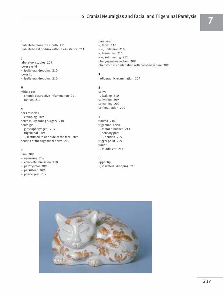
237
I
inability to close the mouth 211
inability to eat or drink without assistance 211
L
laboratory studies 209
lower eyelid
–, ipsilateral drooping 210
lower lip
–, ipsilateral drooping 210
M
middle ear
–, chronic destructive inflammation 211
–, tumors 211
N
neck muscles
–, cramping 209
nerve injury during surgery 210
neuralgia
–, glossopharyngeal 209
–, trigeminal 209
– –, restricted to one side of the face 209
neuritis of the trigeminal nerve 209
P
pain 209
–, agonizing 209
–, complete remission 210
–, paroxysmal 209
–, persistent 209
–, pharyngeal 209
paralysis
–, facial 210
– –, unilateral 210
–, trigeminal 211
– –, self-limiting 211
pharyngeal inspection 209
phenytoin in combination with carbamazepine 209
R
radiographic examination 209
S
saliva
–, leaking 210
salivation 209
screaming 209
self-mutilation 209
T
trauma 210
trigeminal nerve
–, motor branches 211
–, sensory part
– –, neuritis 209
trigger point 209
tumor
–, middle ear 211
U
upper lip
–, ipsilateral drooping 210
6 Cranial Neuralgias and Facial and Trigeminal Paralysis7
3-87706-635-6_213-238 18.04.2005 13:47 Uhr Seite 237