Embed Size (px)
Citation preview
Copyright © 2008 Pearson Education, Inc., publishing as Pearson Benjamin Cummings
PowerPoint® Lecture Presentations for
BiologyEighth Edition
Neil Campbell and Jane Reece
Lectures by Chris Romero, updated by Erin Barley with contributions from Joan Sharp
Chapter 46
Animal Reproduction
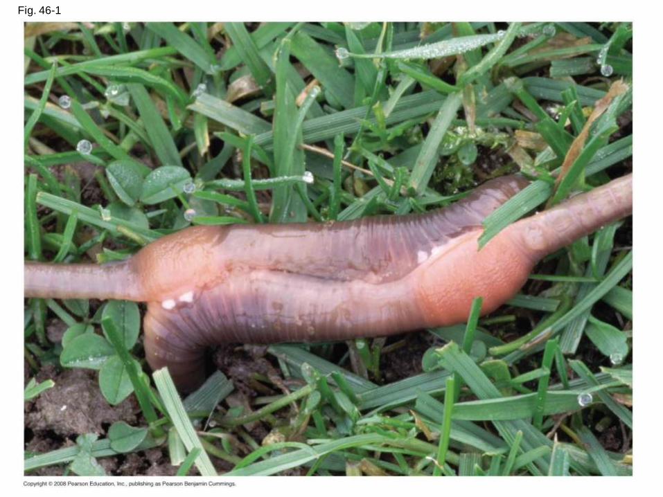
Fig. 46-1
Copyright © 2008 Pearson Education, Inc., publishing as Pearson Benjamin Cummings
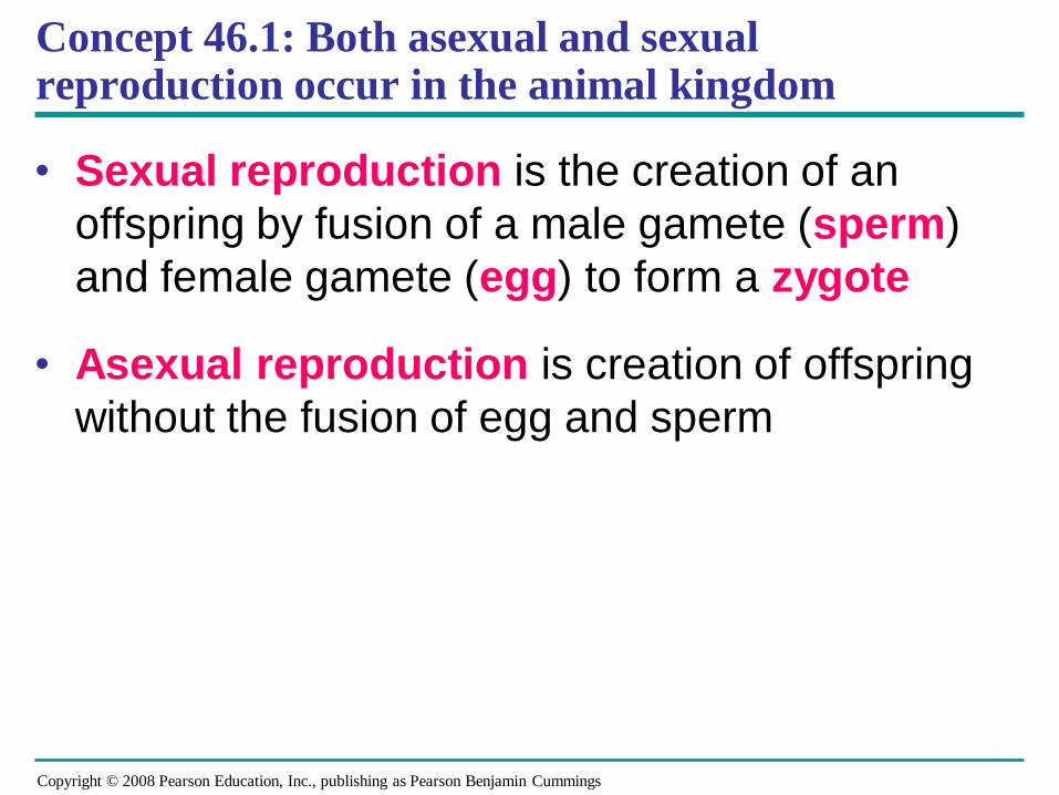
Concept 46.1: Both asexual and sexual reproduction occur in the animal kingdom
• Sexual reproduction is the creation of an
offspring by fusion of a male gamete (sperm)
and female gamete (egg) to form a zygote
• Asexual reproduction is creation of offspring
without the fusion of egg and sperm
Copyright © 2008 Pearson Education, Inc., publishing as Pearson Benjamin Cummings
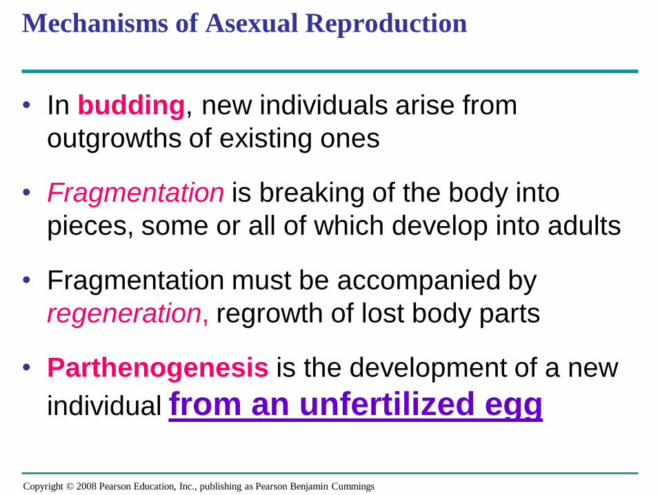
Mechanisms of Asexual Reproduction
• In budding, new individuals arise from
outgrowths of existing ones
• Fragmentation is breaking of the body into
pieces, some or all of which develop into adults
• Fragmentation must be accompanied by
regeneration, regrowth of lost body parts
• Parthenogenesis is the development of a new
individual from an unfertilized egg
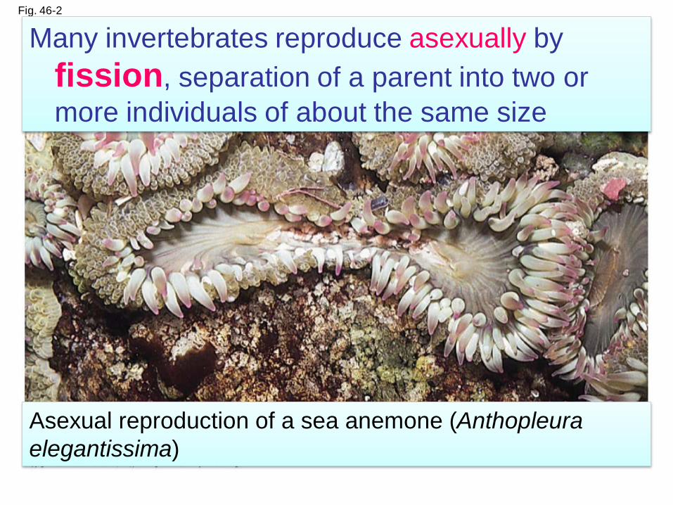
Fig. 46-2
Many invertebrates reproduce asexually by
fission, separation of a parent into two or
more individuals of about the same size
Asexual reproduction of a sea anemone (Anthopleura
elegantissima)
Copyright © 2008 Pearson Education, Inc., publishing as Pearson Benjamin Cummings
Sexual Reproduction
• Sexual reproduction results in genetic
recombination, which provides potential
advantages:
– An increase in variation in offspring, providing
an increase in the reproductive success of
parents in changing environments
– An increase in the rate of adaptation
– A shuffling of genes and the elimination of
harmful genes from a population
Copyright © 2008 Pearson Education, Inc., publishing as Pearson Benjamin Cummings
Sexual Reproduction
Sexual reproduction methods: fusion of
gametes produced by meiosis
• Hermaphroditism
• Separate sexes
• External fertilization
• Internal fertilization
Copyright © 2008 Pearson Education, Inc., publishing as Pearson Benjamin Cummings
• Individuals of some species undergo sex
reversals
• Some species exhibit male to female reversal
(for example, certain oysters), while others
exhibit female to male reversal (for example,
a coral reef fish)
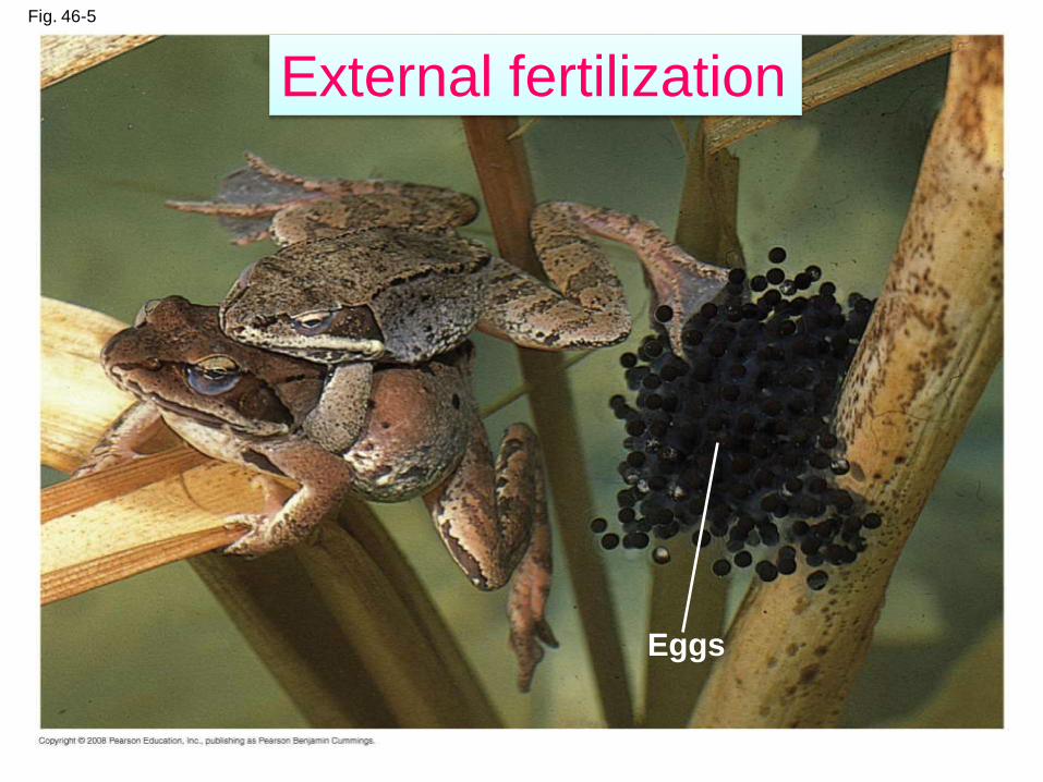
Fig. 46-5
Eggs
External fertilization
Copyright © 2008 Pearson Education, Inc., publishing as Pearson Benjamin Cummings
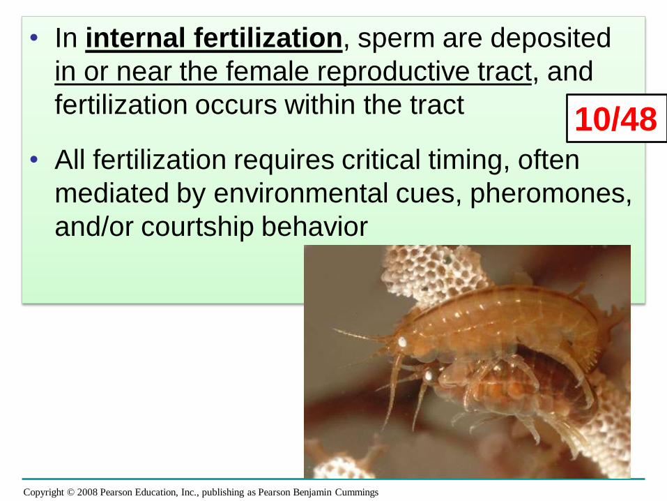
• In internal fertilization, sperm are deposited
in or near the female reproductive tract, and
fertilization occurs within the tract
• All fertilization requires critical timing, often
mediated by environmental cues, pheromones,
and/or courtship behavior
10/48
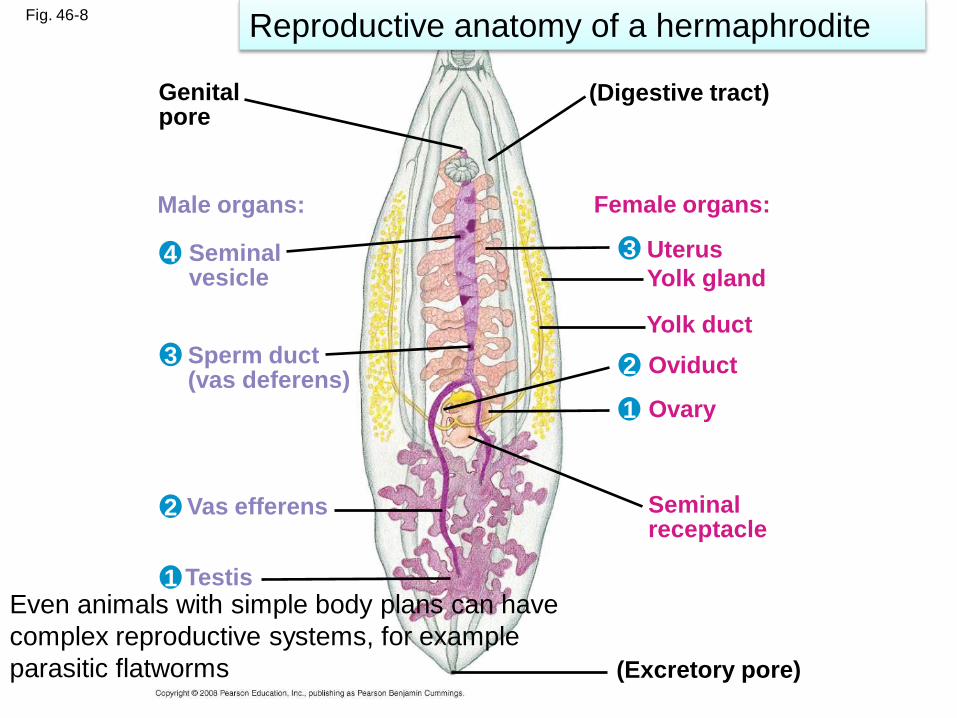
Fig. 46-8
Genitalpore
(Digestive tract)
Male organs:
Seminalvesicle
Sperm duct(vas deferens)
Vas efferens
Testis
Female organs:
Uterus
Yolk gland
Yolk duct
Oviduct
Ovary
Seminalreceptacle
(Excretory pore)
4
3
2
1
3
2
1
Even animals with simple body plans can have
complex reproductive systems, for example
parasitic flatworms
Reproductive anatomy of a hermaphrodite
Copyright © 2008 Pearson Education, Inc., publishing as Pearson Benjamin Cummings
Concept 46.3: Reproductive organs produce and transport gametes
• The following section focuses
on the human reproductive
system
Copyright © 2008 Pearson Education, Inc., publishing as Pearson Benjamin Cummings
Female Reproductive Anatomy
• The female external reproductive structures
include the clitoris and two sets of labia
• The internal organs are a pair of gonads and
a system of ducts and chambers that carry
gametes and house the embryo and fetus
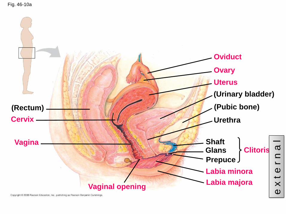
Fig. 46-10a
(Rectum)
Cervix
Vagina
Vaginal opening
Oviduct
Ovary
Uterus
(Urinary bladder)
(Pubic bone)
Urethra
ClitorisShaftGlans
Prepuce
Labia minora
Labia majora
e x
t e
r n
a l
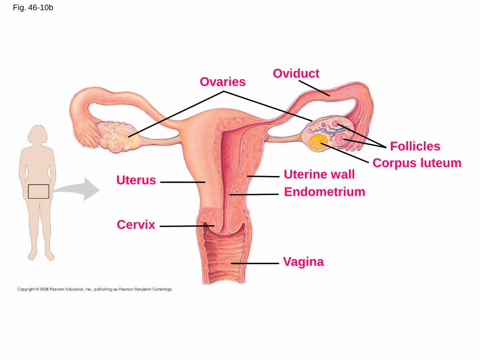
Fig. 46-10b
OvariesOviduct
Follicles
Corpus luteumUterine wallUterus
Cervix
Endometrium
Vagina
Copyright © 2008 Pearson Education, Inc., publishing as Pearson Benjamin Cummings
Female reproductive anatomy
• Ovaries: produce ova (eggs)
• Ovarian follicles: contain immature ova
• Uterine tube (oviduct): captures ovulated
ova; site of fertilization
• Uterus: site of embryo development
• Endometrium: lining of uterus
• Cervix: opens into vagina
Copyright © 2008 Pearson Education, Inc., publishing as Pearson Benjamin Cummings
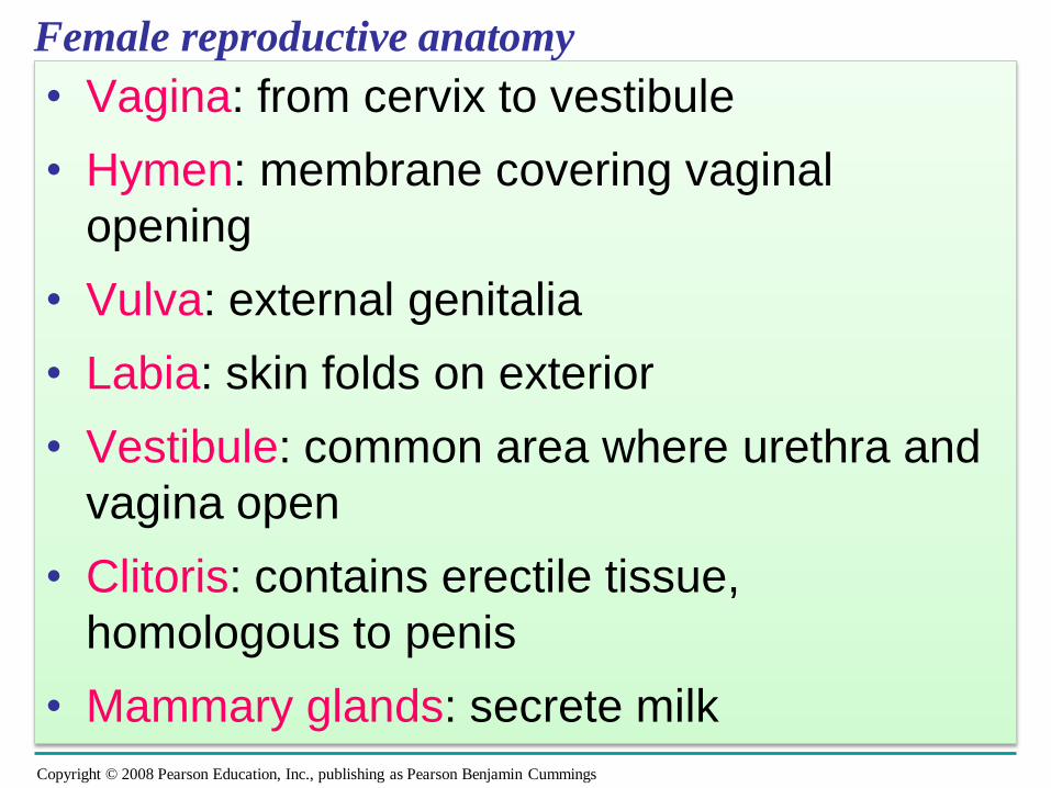
Female reproductive anatomy
• Vagina: from cervix to vestibule
• Hymen: membrane covering vaginal
opening
• Vulva: external genitalia
• Labia: skin folds on exterior
• Vestibule: common area where urethra and
vagina open
• Clitoris: contains erectile tissue,
homologous to penis
• Mammary glands: secrete milk
Copyright © 2008 Pearson Education, Inc., publishing as Pearson Benjamin Cummings
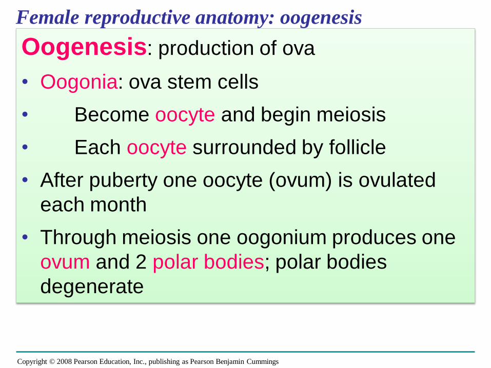
Female reproductive anatomy: oogenesis
Oogenesis: production of ova
• Oogonia: ova stem cells
• Become oocyte and begin meiosis
• Each oocyte surrounded by follicle
• After puberty one oocyte (ovum) is ovulated
each month
• Through meiosis one oogonium produces one
ovum and 2 polar bodies; polar bodies
degenerate
Copyright © 2008 Pearson Education, Inc., publishing as Pearson Benjamin Cummings
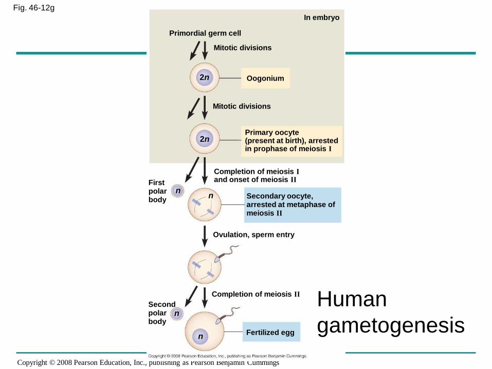
Fig. 46-12g
Primordial germ cell
Mitotic divisions
Oogonium
Mitotic divisions
Primary oocyte(present at birth), arrestedin prophase of meiosis I
Completion of meiosis Iand onset of meiosis II
Secondary oocyte,arrested at metaphase of meiosis II
Firstpolarbody
Ovulation, sperm entry
Completion of meiosis II
Secondpolarbody
Fertilized egg
2n
2n
nn
n
n
In embryo
Human
gametogenesis
Copyright © 2008 Pearson Education, Inc., publishing as Pearson Benjamin Cummings
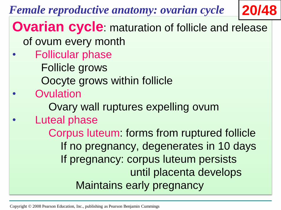
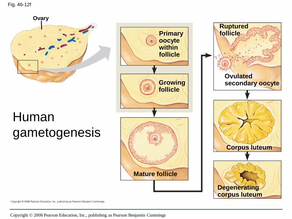
Female reproductive anatomy: ovarian cycle
Ovarian cycle: maturation of follicle and release
of ovum every month
• Follicular phase
Follicle grows
Oocyte grows within follicle
• Ovulation
Ovary wall ruptures expelling ovum
• Luteal phase
Corpus luteum: forms from ruptured follicle
If no pregnancy, degenerates in 10 days
If pregnancy: corpus luteum persists
until placenta develops
Maintains early pregnancy
20/48
Copyright © 2008 Pearson Education, Inc., publishing as Pearson Benjamin Cummings
Fig. 46-12f
Ovary
Primaryoocytewithinfollicle
Rupturedfollicle
Growingfollicle
Mature follicle
Ovulatedsecondary oocyte
Corpus luteum
Degeneratingcorpus luteum
Human
gametogenesis
Copyright © 2008 Pearson Education, Inc., publishing as Pearson Benjamin Cummings
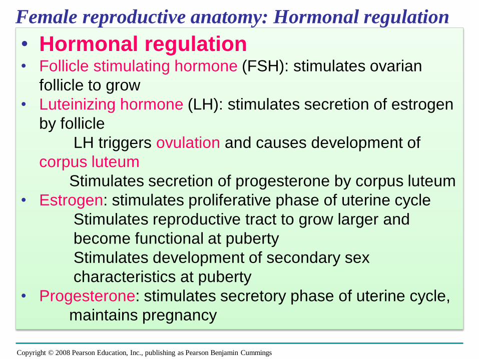
Female reproductive anatomy: Hormonal regulation
• Hormonal regulation• Follicle stimulating hormone (FSH): stimulates ovarian
follicle to grow
• Luteinizing hormone (LH): stimulates secretion of estrogen
by follicle
LH triggers ovulation and causes development of
corpus luteum
Stimulates secretion of progesterone by corpus luteum
• Estrogen: stimulates proliferative phase of uterine cycle
Stimulates reproductive tract to grow larger and
become functional at puberty
Stimulates development of secondary sex
characteristics at puberty
• Progesterone: stimulates secretory phase of uterine cycle,
maintains pregnancy
Copyright © 2008 Pearson Education, Inc., publishing as Pearson Benjamin Cummings
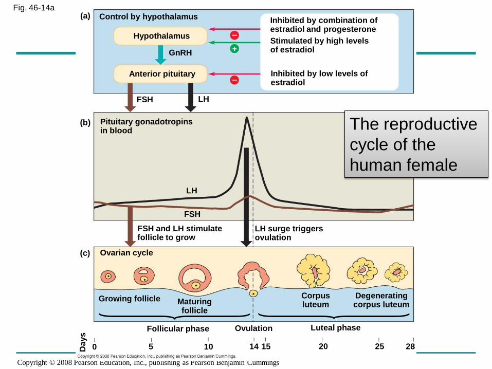
Fig. 46-14aControl by hypothalamus Inhibited by combination of
estradiol and progesterone
Stimulated by high levelsof estradiol
Inhibited by low levels of estradiol
Hypothalamus
GnRH
Anterior pituitary
FSH LH
Pituitary gonadotropinsin blood
LH
FSH
FSH and LH stimulatefollicle to grow
LH surge triggersovulation
Ovarian cycle
Growing follicle Maturingfollicle
Corpusluteum
Degeneratingcorpus luteum
Follicular phase Ovulation Luteal phase
(a)
(b)
(c)
Da
ys
0 5 10 14 15 20 25 28| | | | | | | |
–
–
+
The reproductive
cycle of the
human female
Copyright © 2008 Pearson Education, Inc., publishing as Pearson Benjamin Cummings
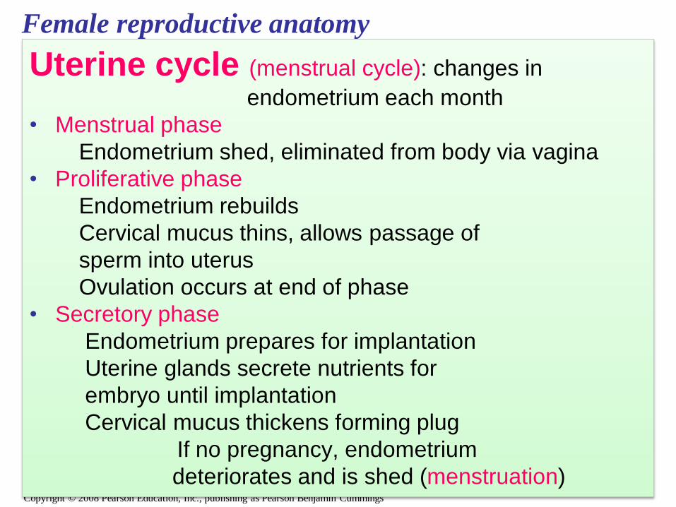
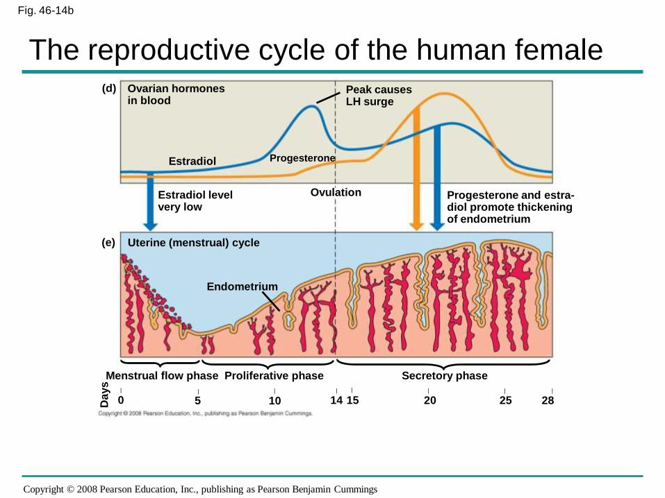
Female reproductive anatomy
Uterine cycle (menstrual cycle): changes in
endometrium each month
• Menstrual phase
Endometrium shed, eliminated from body via vagina
• Proliferative phase
Endometrium rebuilds
Cervical mucus thins, allows passage of
sperm into uterus
Ovulation occurs at end of phase
• Secretory phase
Endometrium prepares for implantation
Uterine glands secrete nutrients for
embryo until implantation
Cervical mucus thickens forming plug
If no pregnancy, endometrium
deteriorates and is shed (menstruation)
Copyright © 2008 Pearson Education, Inc., publishing as Pearson Benjamin Cummings
Fig. 46-14b
Ovarian hormones in blood
Peak causesLH surge
Estradiol level very low
Estradiol Progesterone
Ovulation Progesterone and estra-diol promote thickeningof endometrium
Uterine (menstrual) cycle
Endometrium
0 5 10 14 20 25 28| | | | | | | |
Days
15
Menstrual flow phase Proliferative phase Secretory phase
(d)
(e)
The reproductive cycle of the human female
Copyright © 2008 Pearson Education, Inc., publishing as Pearson Benjamin Cummings
Female reproductive anatomy: estrous cycle
• Estrous cycle: • Majority of mammals
In the absence of a pregnancy, the uterus
reabsorbs the endometrium and no extensive
fluid flow occurs
• Mammals with estrous cycle typically copulate
only during the period of estrus (sometimes
called heat), when female is receptive to
mating
Copyright © 2008 Pearson Education, Inc., publishing as Pearson Benjamin Cummings
Menstrual Versus Estrous Cycles
• Menstrual cycles are characteristic of humans
and some other primates:
– The endometrium is shed from the
uterus in a bleeding called
menstruation
– Sexual receptivity is not limited to a timeframe
Copyright © 2008 Pearson Education, Inc., publishing as Pearson Benjamin Cummings
• Estrous cycles are characteristic of most
mammals:
– The endometrium is reabsorbed by the
uterus
– Sexual receptivity is limited to a “heat” period
– The length and frequency of estrous cycles
varies from species to species
Copyright © 2008 Pearson Education, Inc., publishing as Pearson Benjamin Cummings
Menopause
• After about 500 cycles, human females
undergo menopause, the cessation of
ovulation and menstruation
• Menopause is very unusual among
animals
• Menopause might have evolved to allow a
mother to provide better care for her children
and grandchildren - ?
Copyright © 2008 Pearson Education, Inc., publishing as Pearson Benjamin Cummings
Male Reproductive Anatomy
• The male’s external reproductive organs are
the scrotum and penis
• Internal organs are the gonads, which produce
sperm and hormones, and accessory glands
30/48
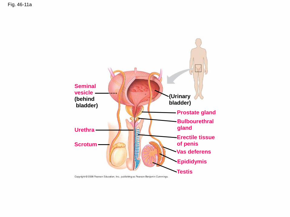
Fig. 46-11a
Seminalvesicle(behindbladder)
Urethra
Scrotum
(Urinarybladder)
Prostate gland
Bulbourethralgland
Erectile tissueof penis
Vas deferens
Epididymis
Testis
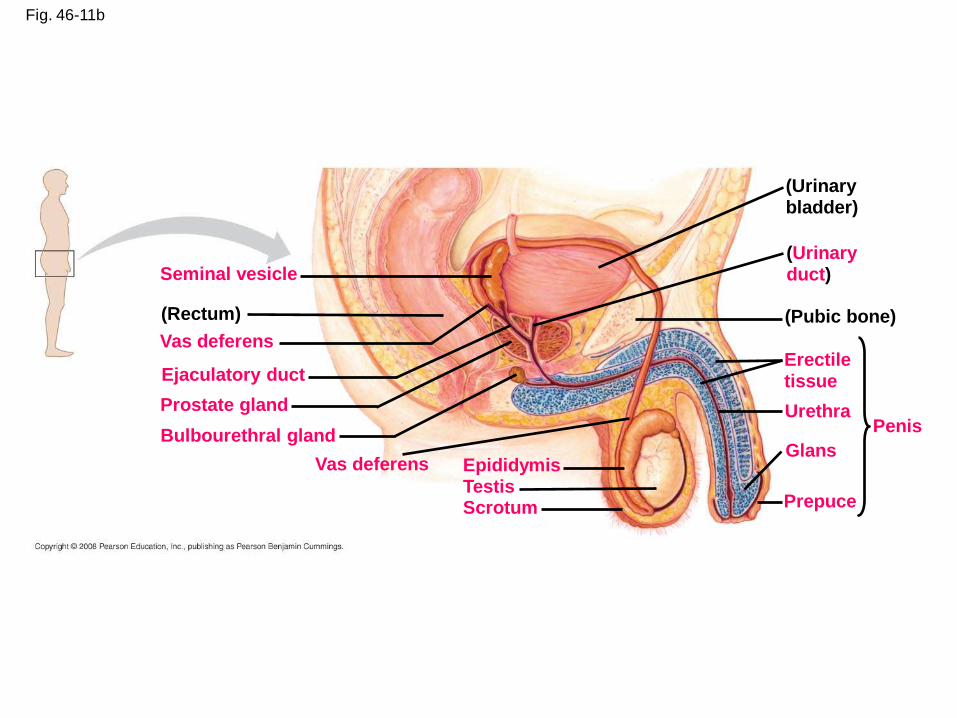
Fig. 46-11b
Seminal vesicle
(Rectum)
Vas deferens
Ejaculatory duct
Prostate gland
Bulbourethral gland
Vas deferens EpididymisTestisScrotum
(Urinarybladder)
(Urinaryduct)
(Pubic bone)
Erectiletissue
Urethra
Glans
Prepuce
Penis
Copyright © 2008 Pearson Education, Inc., publishing as Pearson Benjamin Cummings
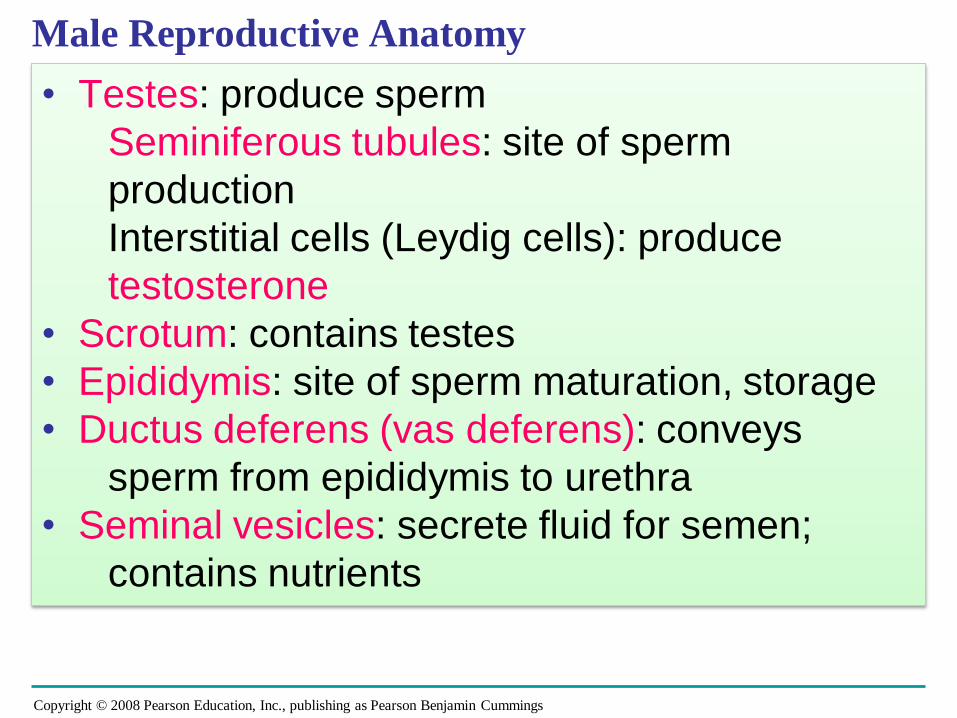
Male Reproductive Anatomy
• Testes: produce sperm
Seminiferous tubules: site of sperm
production
Interstitial cells (Leydig cells): produce
testosterone
• Scrotum: contains testes
• Epididymis: site of sperm maturation, storage
• Ductus deferens (vas deferens): conveys
sperm from epididymis to urethra
• Seminal vesicles: secrete fluid for semen;
contains nutrients
Copyright © 2008 Pearson Education, Inc., publishing as Pearson Benjamin Cummings
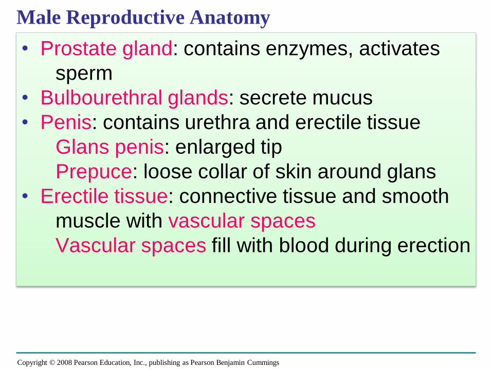
Male Reproductive Anatomy
• Prostate gland: contains enzymes, activates
sperm
• Bulbourethral glands: secrete mucus
• Penis: contains urethra and erectile tissue
Glans penis: enlarged tip
Prepuce: loose collar of skin around glans
• Erectile tissue: connective tissue and smooth
muscle with vascular spaces
Vascular spaces fill with blood during erection
Copyright © 2008 Pearson Education, Inc., publishing as Pearson Benjamin Cummings
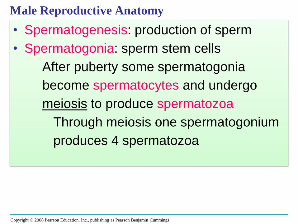
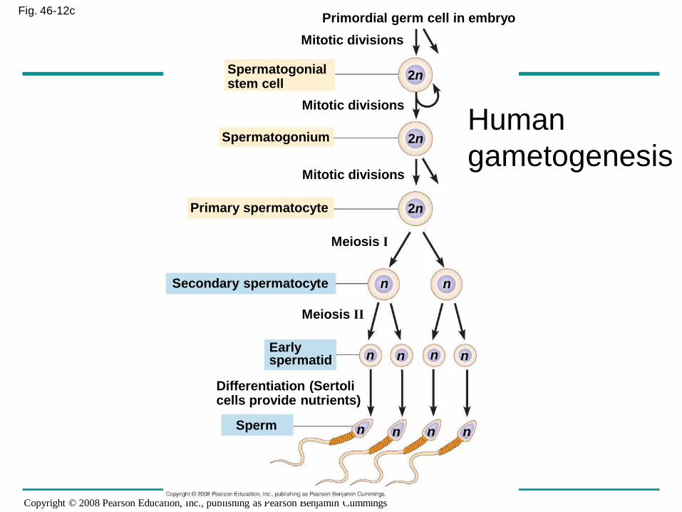
Male Reproductive Anatomy
• Spermatogenesis: production of sperm
• Spermatogonia: sperm stem cells
After puberty some spermatogonia
become spermatocytes and undergo
meiosis to produce spermatozoa
Through meiosis one spermatogonium
produces 4 spermatozoa
Copyright © 2008 Pearson Education, Inc., publishing as Pearson Benjamin Cummings
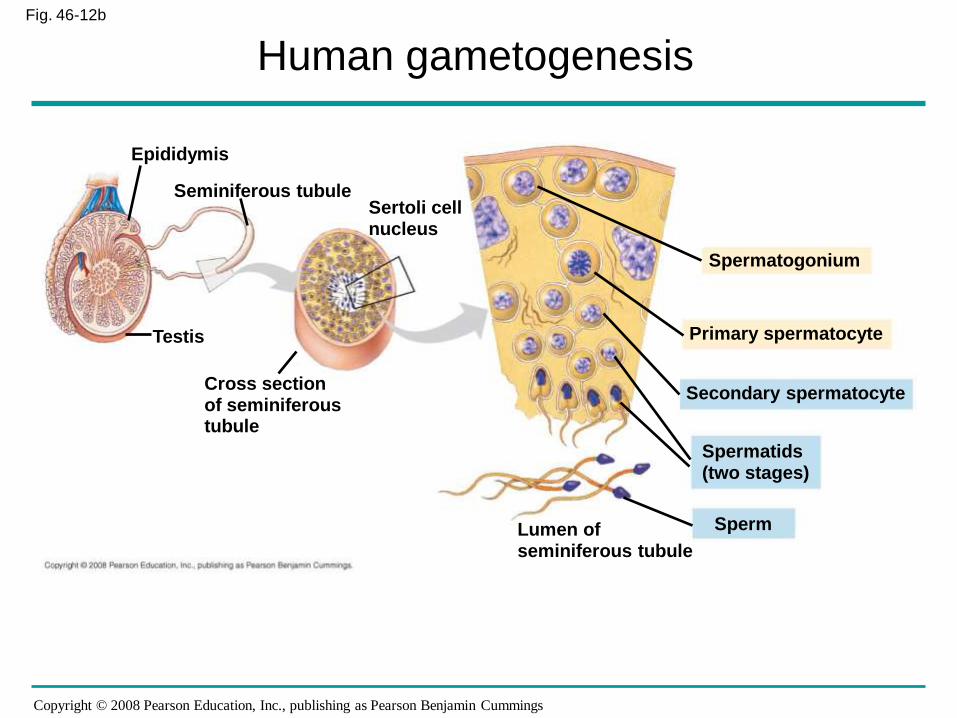
Fig. 46-12b
Epididymis
Seminiferous tubuleSertoli cellnucleus
Testis
Cross sectionof seminiferoustubule
Spermatogonium
Primary spermatocyte
Secondary spermatocyte
Spermatids(two stages)
SpermLumen ofseminiferous tubule
Human gametogenesis
Copyright © 2008 Pearson Education, Inc., publishing as Pearson Benjamin Cummings
Fig. 46-12cPrimordial germ cell in embryo
Mitotic divisions
Spermatogonialstem cell
Mitotic divisions
Spermatogonium
Mitotic divisions
Primary spermatocyte
Meiosis I
Secondary spermatocyte
Meiosis II
Earlyspermatid
Differentiation (Sertolicells provide nutrients)
Sperm
2n
2n
2n
n n
n n n n
n n n n
Human
gametogenesis
Copyright © 2008 Pearson Education, Inc., publishing as Pearson Benjamin Cummings
Male Reproductive Anatomy: Hormonal regulation
• Hormonal regulation
• Follicle-stimulating hormone: stimulates
spermatogenesis
• Luteinizing hormone: stimulates interstitial cells to
secrete testosterone
• Testosterone: stimulates spermatogenesis
Stimulates accessory organs to grow and
function
Stimulates development of secondary sex
characteristics at puberty
Responsible for sex drive in males and
females
Copyright © 2008 Pearson Education, Inc., publishing as Pearson Benjamin Cummings
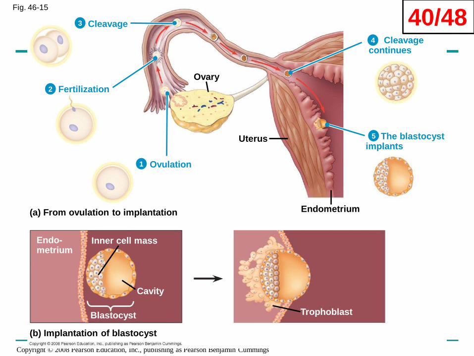
Fertilization, development, and parturition
• Fertilization: union of sperm and ova producing
zygote
• Cleavage: rapid mitosis following fertilization
Blastocyst formed
Trophoblasts: contribute to placenta formation
Inner cell mass: forms embryonic disc
• Implantation: when embryonic disc reaches
uterus
Trophoblasts adhere to endometrium
Blastocyst burrows in and becomes covered by
endometrial cells
Copyright © 2008 Pearson Education, Inc., publishing as Pearson Benjamin Cummings
Fig. 46-15
Ovary
Uterus
Endometrium(a) From ovulation to implantation
(b) Implantation of blastocyst
Cleavage
Fertilization
Ovulation
Cleavage continues
The blastocystimplants
Trophoblast
Inner cell mass
Cavity
Blastocyst
Endo-metrium
1
2
3
4
5
40/48
Copyright © 2008 Pearson Education, Inc., publishing as Pearson Benjamin Cummings
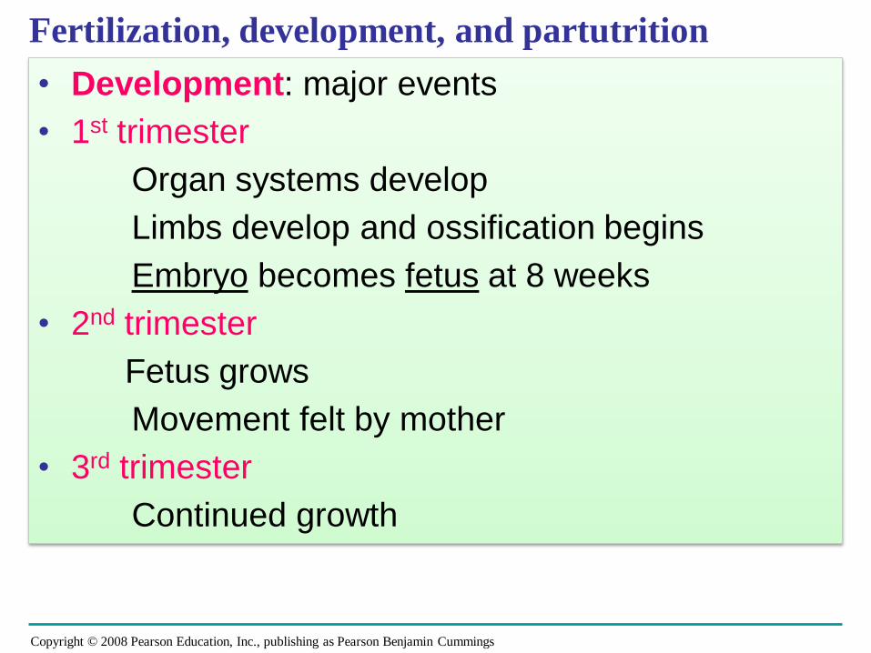
Fertilization, development, and partutrition
• Development: major events
• 1st trimester
Organ systems develop
Limbs develop and ossification begins
Embryo becomes fetus at 8 weeks
• 2nd trimester
Fetus grows
Movement felt by mother
• 3rd trimester
Continued growth
Copyright © 2008 Pearson Education, Inc., publishing as Pearson Benjamin Cummings
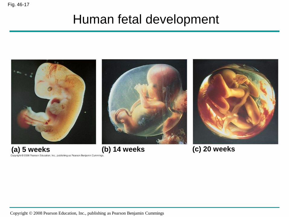
Fig. 46-17
(a) 5 weeks (b) 14 weeks (c) 20 weeks
Human fetal development
Copyright © 2008 Pearson Education, Inc., publishing as Pearson Benjamin Cummings
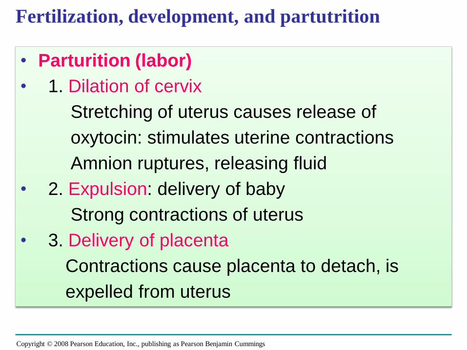
Fertilization, development, and partutrition
• Parturition (labor)
• 1. Dilation of cervix
Stretching of uterus causes release of
oxytocin: stimulates uterine contractions
Amnion ruptures, releasing fluid
• 2. Expulsion: delivery of baby
Strong contractions of uterus
• 3. Delivery of placenta
Contractions cause placenta to detach, is
expelled from uterus
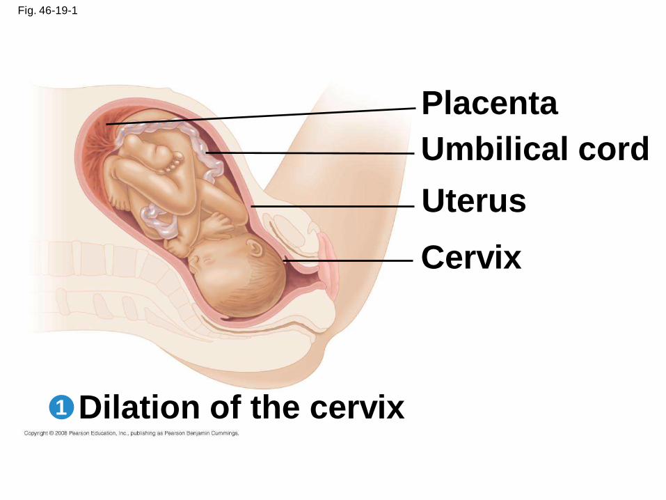
Fig. 46-19-1
Placenta
Umbilical cord
Uterus
Cervix
Dilation of the cervix1
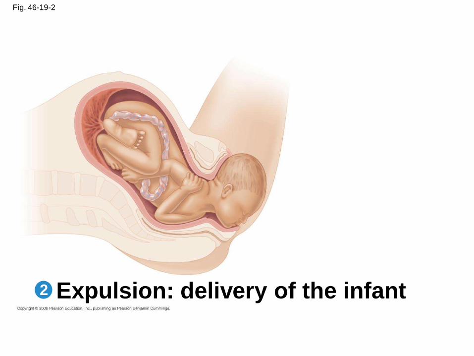
Fig. 46-19-2
Expulsion: delivery of the infant2
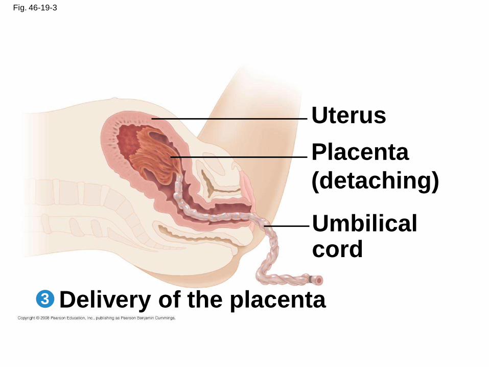
Fig. 46-19-3
Delivery of the placenta
Uterus
Placenta
(detaching)
Umbilicalcord
3
Copyright © 2008 Pearson Education, Inc., publishing as Pearson Benjamin Cummings
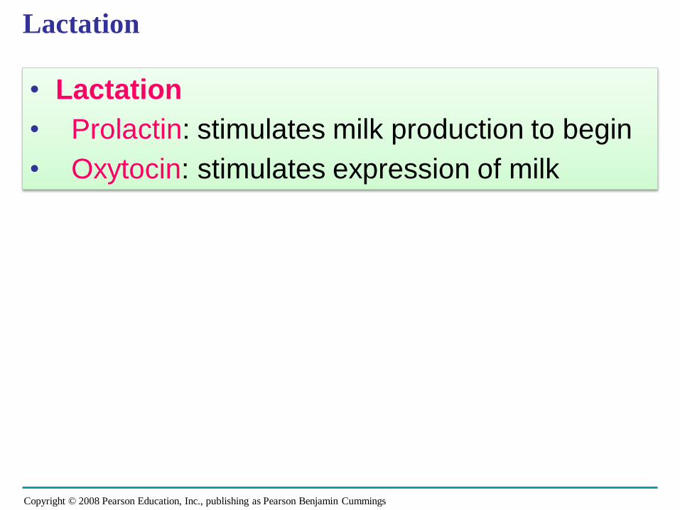
Lactation
• Lactation
• Prolactin: stimulates milk production to begin
• Oxytocin: stimulates expression of milk
Copyright © 2008 Pearson Education, Inc., publishing as Pearson Benjamin Cummings
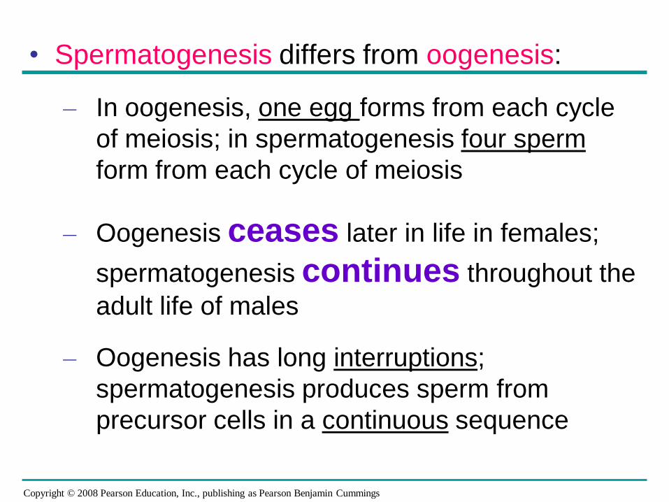
• Spermatogenesis differs from oogenesis:
– In oogenesis, one egg forms from each cycle
of meiosis; in spermatogenesis four sperm
form from each cycle of meiosis
– Oogenesis ceases later in life in females;
spermatogenesis continues throughout the
adult life of males
– Oogenesis has long interruptions;
spermatogenesis produces sperm from
precursor cells in a continuous sequence

Copyright © 2008 Pearson Education, Inc., publishing as Pearson Benjamin Cummings

Copyright © 2008 Pearson Education, Inc., publishing as Pearson Benjamin Cummings
Thank you for your attention and participation!
Copyright © 2008 Pearson Education, Inc., publishing as Pearson Benjamin Cummings
You should now be able to:
1. Distinguish between asexual and sexual reproduction
2. Explain how hermaphroditism may be advantageous to animals that have difficulty encountering a member of the opposite sex
3. Using diagrams, identify and state the function of each component of the male and female reproductive systems
4. Describe oogenesis and spermatogenesis; describe three major differences between them
Copyright © 2008 Pearson Education, Inc., publishing as Pearson Benjamin Cummings
5. Explain how the uterine and ovarian cycles are synchronized and describe the functions of the hormones involved


![Ocw [animal reproduction]](https://img.pdfslide.us/doc/110x75/58835b371a28ab42678b649f/ocw-animal-reproduction.jpg)