Embed Size (px)
Citation preview
Animal Reproduction
• a female is born with about 2 million primary oocytes (pre-eggs) in her ovaries
• by the time she is 7 years old, only approx. 300 thousand remain
• primary oocytes = cells that have started meiosis I, but have been halted until menstruation begins
• only about 400-500 oocytes will be released over the course of a woman’s reproductive years
• most menstrual cycles are about 28 days
Humans:
reproductive years begin around ages 10-16
end around late 40’s – mid-50’s (menopause)
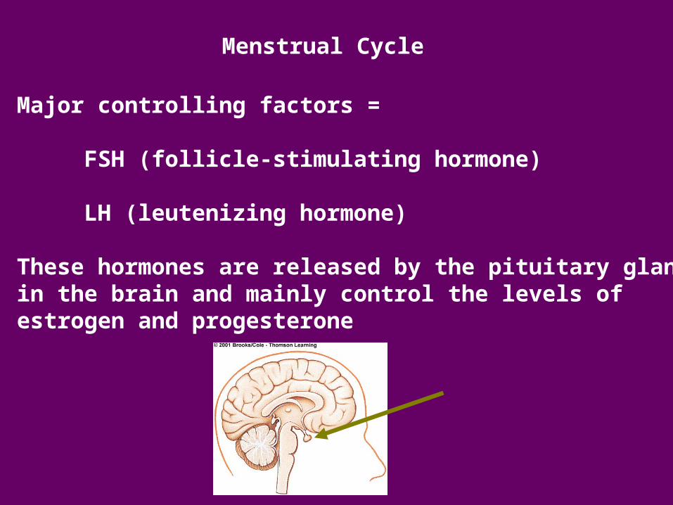
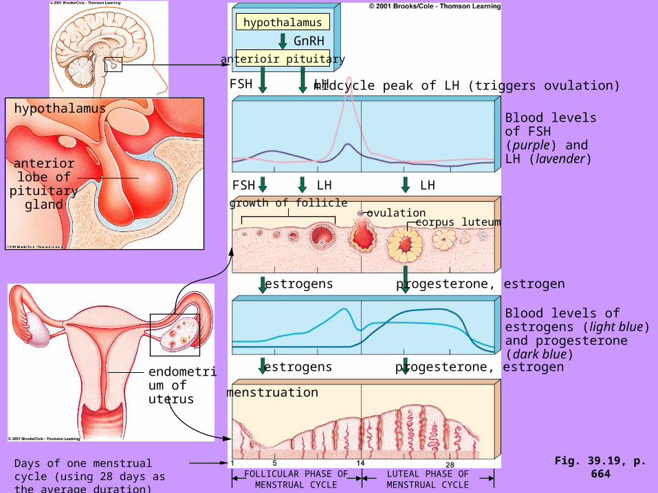
Menstrual Cycle
Major controlling factors =
FSH (follicle-stimulating hormone)
LH (leutenizing hormone)
These hormones are released by the pituitary glandin the brain and mainly control the levels of estrogen and progesterone
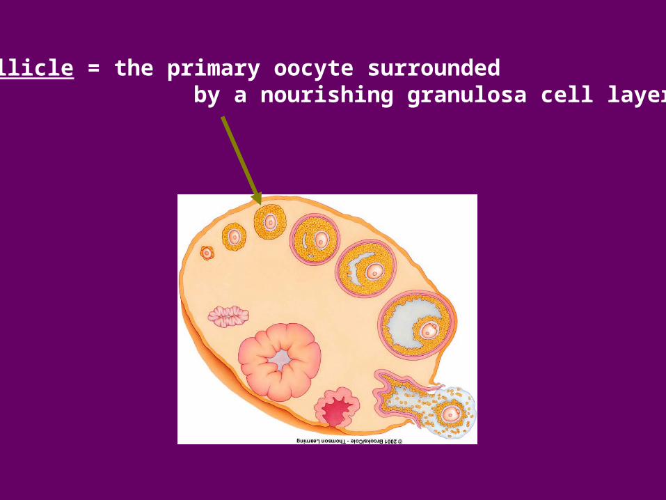
follicle = the primary oocyte surrounded by a nourishing granulosa cell layer
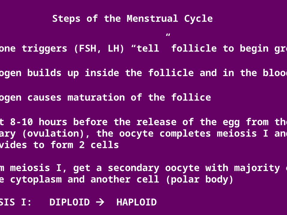
Steps of the Menstrual Cycle
1. hormone triggers (FSH, LH) “tell” follicle to begin growing
2. estrogen builds up inside the follicle and in the blood
3. estrogen causes maturation of the follice
4. about 8-10 hours before the release of the egg from the ovary (ovulation), the oocyte completes meiosis I and divides to form 2 cells
* from meiosis I, get a secondary oocyte with majority of the cytoplasm and another cell (polar body)
MEIOSIS I: DIPLOID HAPLOID
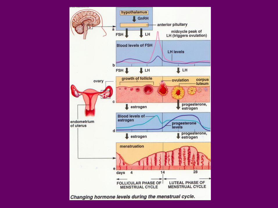
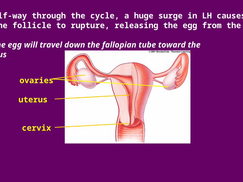
5. Half-way through the cycle, a huge surge in LH causes the follicle to rupture, releasing the egg from the ovary
the egg will travel down the fallopian tube toward the uterus
ovaries
uterus
cervix
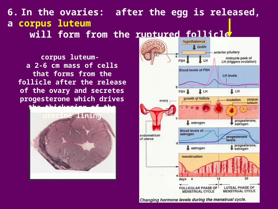
6. In the ovaries: after the egg is released, a corpus luteum will form from the ruptured follicle
corpus luteum- a 2-6 cm mass of cells that forms from the follicle after the release
of the ovary and secretes progesterone which drives the thickening of the uterine lining
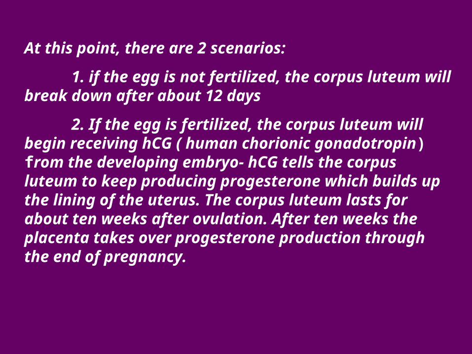
At this point, there are 2 scenarios:
1. if the egg is not fertilized, the corpus luteum will break down after about 12 days
2. If the egg is fertilized, the corpus luteum will begin receiving hCG ( human chorionic gonadotropin) from the developing embryo- hCG tells the corpus luteum to keep producing progesterone which builds up the lining of the uterus. The corpus luteum lasts for about ten weeks after ovulation. After ten weeks the placenta takes over progesterone production through the end of pregnancy.
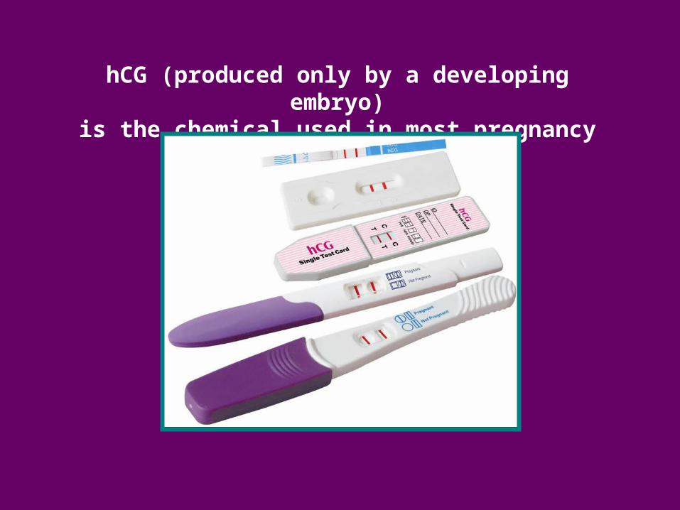
hCG (produced only by a developing embryo)is the chemical used in most pregnancy test kits
hypothalamus
anterioir pituitary
FSH LH midcycle peak of LH (triggers ovulation)
Blood levels of FSH (purple) and LH (lavender)
FSH LH LH
estrogens progesterone, estrogen
estrogens progesterone, estrogen
Blood levels of estrogens (light blue) and progesterone (dark blue)
growth of follicle
FOLLICULAR PHASE OF MENSTRUAL CYCLE
LUTEAL PHASE OF MENSTRUAL CYCLE
menstruation
endometrium of uterus
Days of one menstrual cycle (using 28 days as the average duration)
hypothalamus
anterior lobe of pituitary
gland
ovulationcorpus luteum
GnRH
Fig. 39.19, p. 664
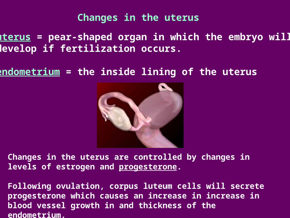
Changes in the uterus
uterus = pear-shaped organ in which the embryo will develop if fertilization occurs.
endometrium = the inside lining of the uterus
Changes in the uterus are controlled by changes in levels of estrogen and progesterone.
Following ovulation, corpus luteum cells will secrete progesterone which causes an increase in increase in blood vessel growth in and thickness of the endometrium.
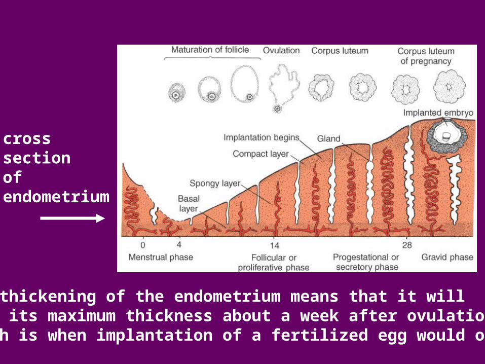
cross section of endometrium
this thickening of the endometrium means that it willbe at its maximum thickness about a week after ovulation(which is when implantation of a fertilized egg would occur)
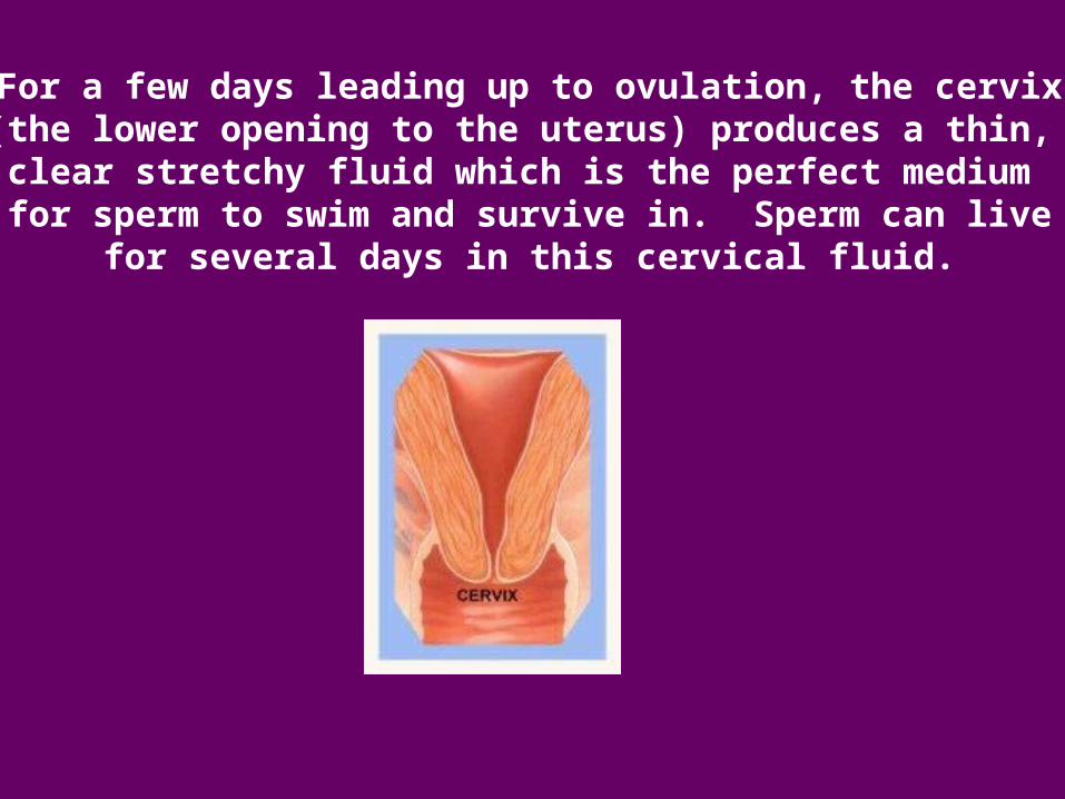
For a few days leading up to ovulation, the cervix(the lower opening to the uterus) produces a thin, clear stretchy fluid which is the perfect medium
for sperm to swim and survive in. Sperm can livefor several days in this cervical fluid.
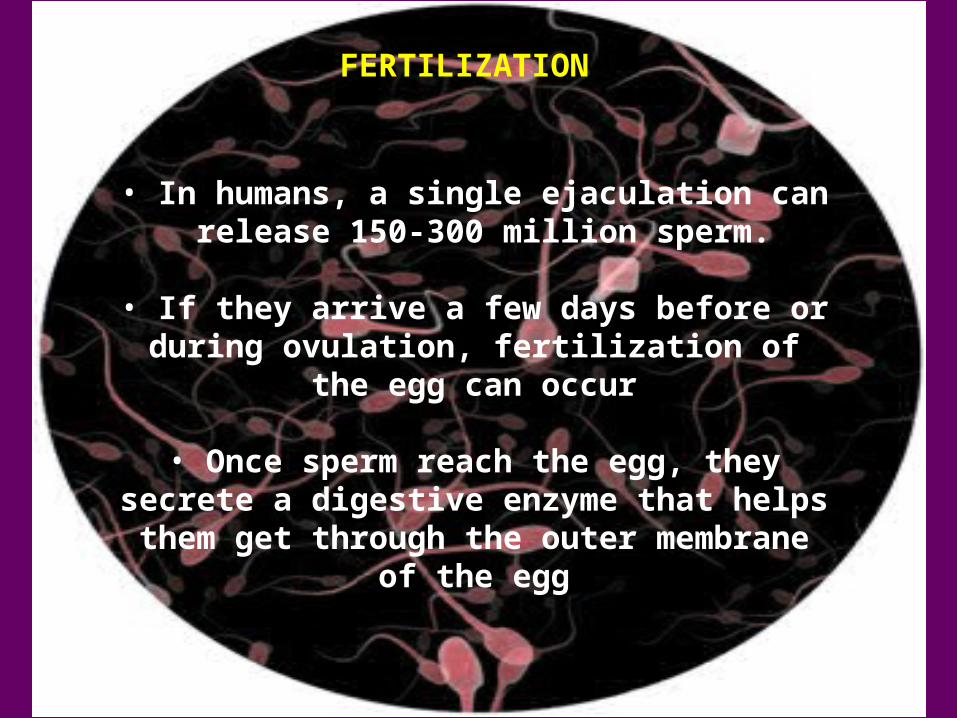
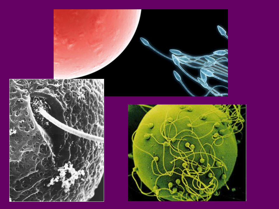
FERTILIZATION
• In humans, a single ejaculation can release 150-300 million sperm.
• If they arrive a few days before or during ovulation, fertilization of the egg can occur
• Once sperm reach the egg, they secrete a digestive enzyme that helps them get through
the outer membrane of the egg
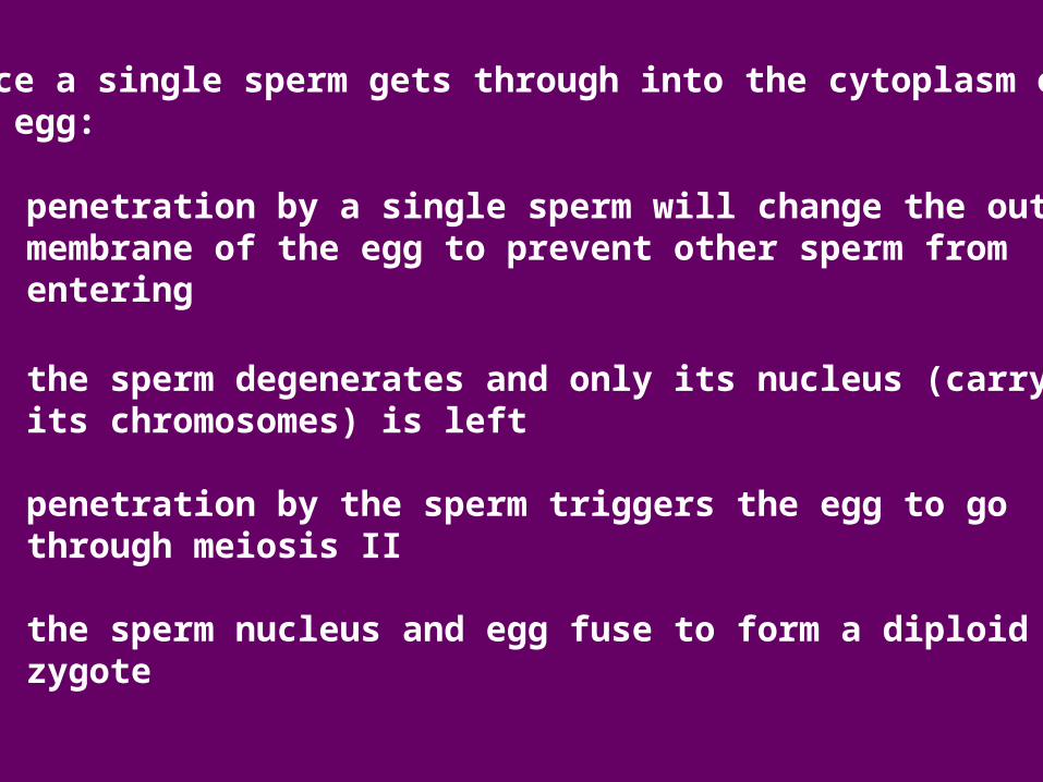
Once a single sperm gets through into the cytoplasm ofan egg:
• penetration by a single sperm will change the outer membrane of the egg to prevent other sperm from entering
• the sperm degenerates and only its nucleus (carrying its chromosomes) is left
• penetration by the sperm triggers the egg to go through meiosis II
• the sperm nucleus and egg fuse to form a diploid zygote
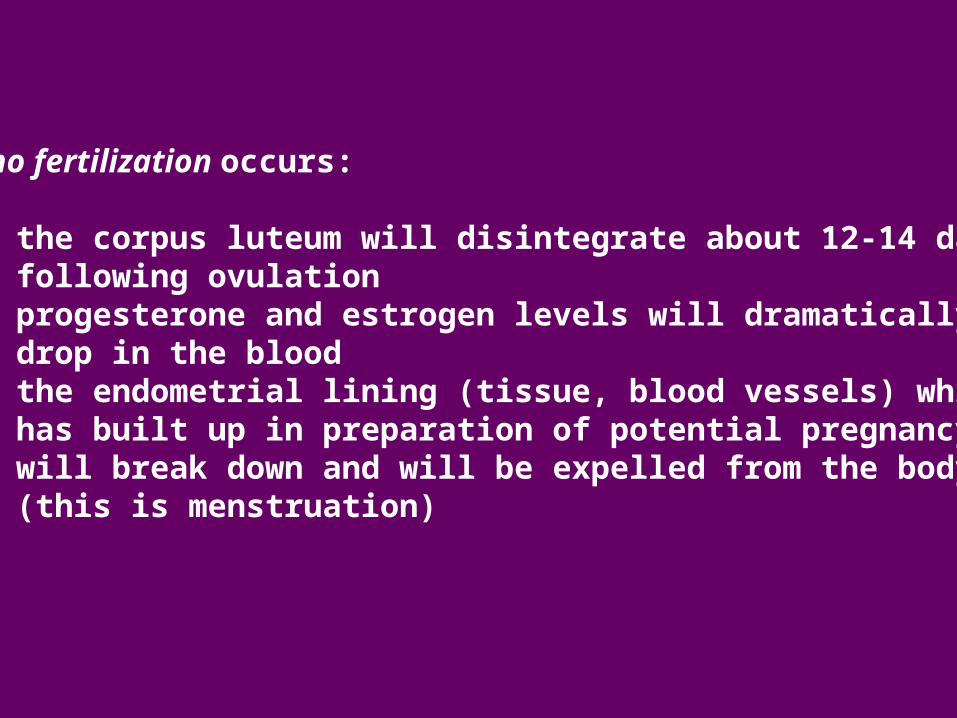
If no fertilization occurs:
• the corpus luteum will disintegrate about 12-14 days following ovulation• progesterone and estrogen levels will dramatically drop in the blood • the endometrial lining (tissue, blood vessels) which has built up in preparation of potential pregnancy will break down and will be expelled from the body (this is menstruation)
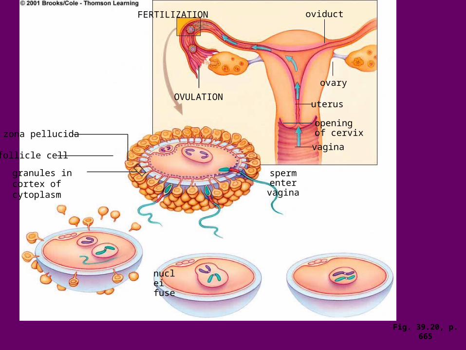
zona pellucida
follicle cell
granules in cortex of cytoplasm
nuclei fuse
FERTILIZATION
OVULATION
oviduct
ovary
uterus
opening of cervix
vagina
sperm enter
vagina
Fig. 39.20, p. 665
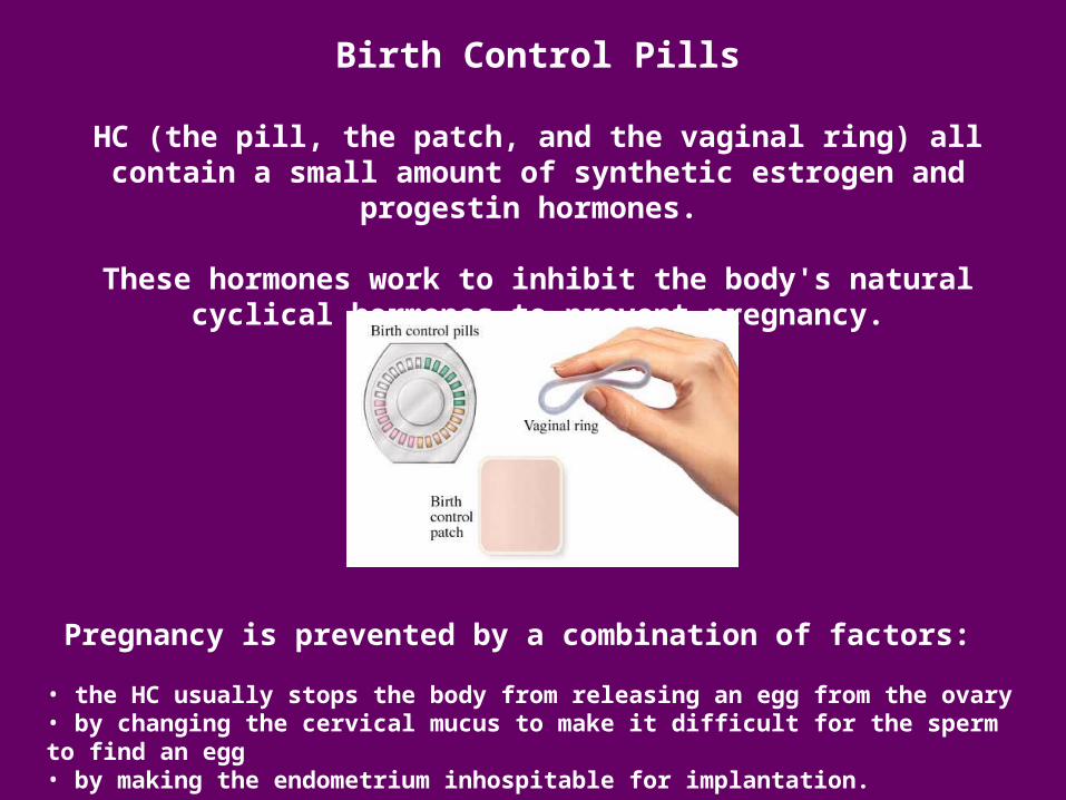
Birth Control Pills
HC (the pill, the patch, and the vaginal ring) all contain a small amount of synthetic estrogen and progestin hormones.
These hormones work to inhibit the body's natural cyclical hormones to prevent pregnancy.
Pregnancy is prevented by a combination of factors:
• the HC usually stops the body from releasing an egg from the ovary • by changing the cervical mucus to make it difficult for the sperm to find an egg• by making the endometrium inhospitable for implantation.
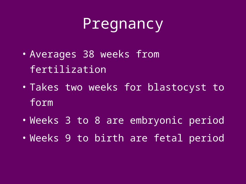
Pregnancy
• Averages 38 weeks from fertilization
• Takes two weeks for blastocyst to form
• Weeks 3 to 8 are embryonic period
• Weeks 9 to birth are fetal period
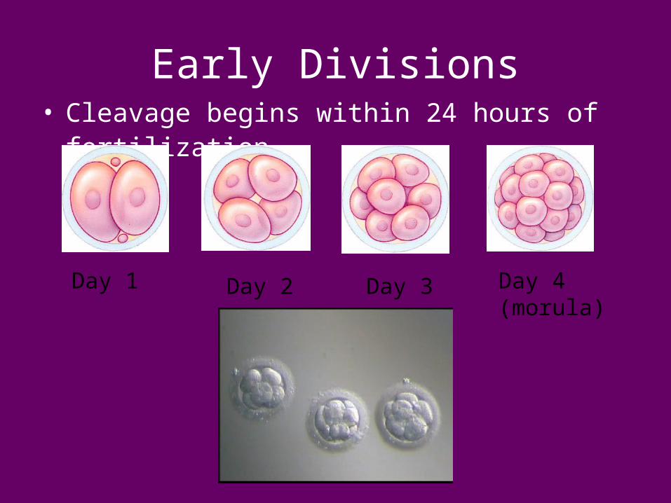
Early Divisions• Cleavage begins within 24 hours of fertilization
Day 1 Day 2 Day 3 Day 4(morula)
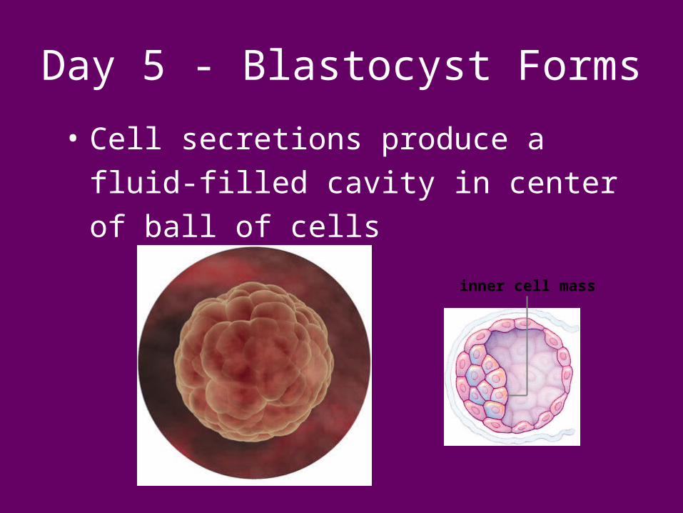
Day 5 - Blastocyst Forms
• Cell secretions produce a fluid-filled
cavity in center of ball of cells
inner cell mass
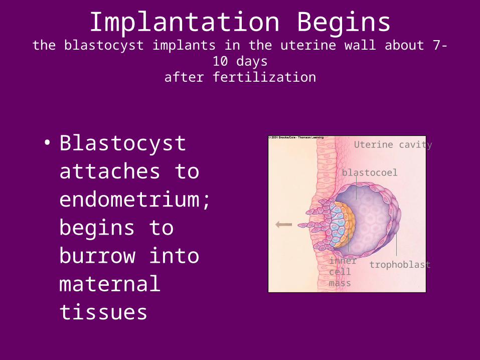
Implantation Beginsthe blastocyst implants in the uterine wall about 7-10 days
after fertilization
• Blastocyst attaches to endometrium; begins to burrow into maternal tissues
blastocoel
inner cell mass
trophoblast
Uterine cavity
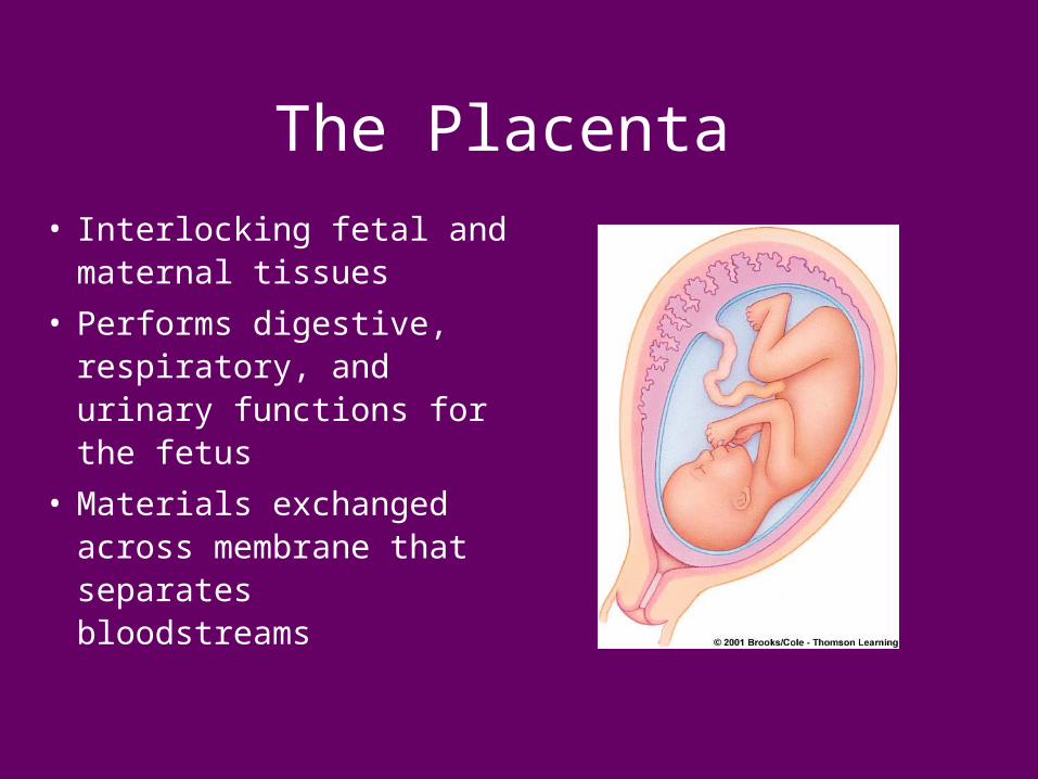
The Placenta
• Interlocking fetal and maternal tissues
• Performs digestive, respiratory, and urinary functions for the fetus
• Materials exchanged across membrane that separates bloodstreams
Nutrition and Risks
A well-balanced diet is extremely important because thedeveloping embryo needs a fell-range of nutrients:
• folic acid and b vitamins are particularly important in lowering chances of birth defects
On average, women need to eat enough to increase bodyweight by 20-25 lbs. to insure that there is no nutritional risk for the developing baby.
Risks:
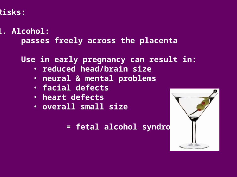
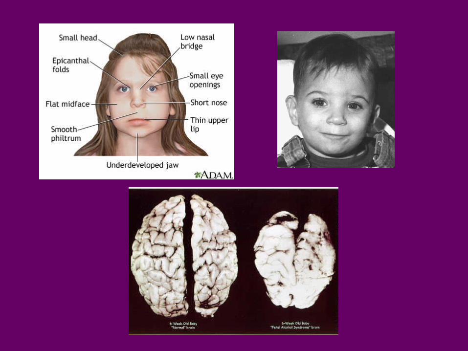
1. Alcohol:passes freely across the placenta
Use in early pregnancy can result in:• reduced head/brain size• neural & mental problems• facial defects• heart defects• overall small size
= fetal alcohol syndrome
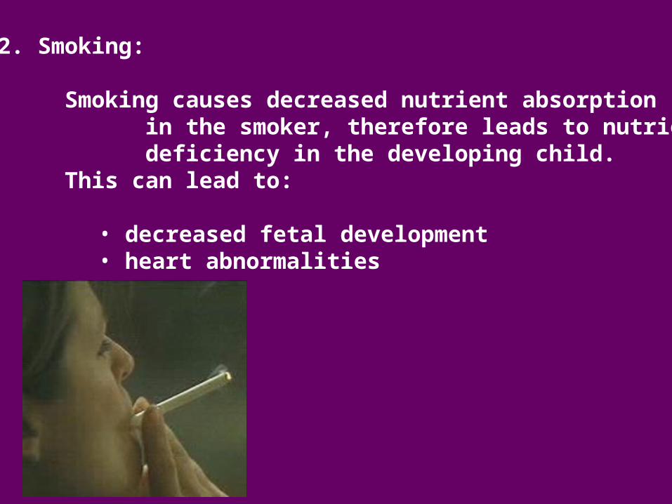
2. Smoking:
Smoking causes decreased nutrient absorption in the smoker, therefore leads to nutrient deficiency in the developing child.
This can lead to:
• decreased fetal development• heart abnormalities