Embed Size (px)
Citation preview
Accepted Manuscript
Animal NLRs continue to inform plant NLR structure and function
Hayden Burdett, Bostjan Kobe, Peter A. Anderson
PII: S0003-9861(19)30052-9
DOI: https://doi.org/10.1016/j.abb.2019.05.001
Reference: YABBI 7999
To appear in: Archives of Biochemistry and Biophysics
Received Date: 25 January 2019
Revised Date: 10 April 2019
Accepted Date: 1 May 2019
Please cite this article as: H. Burdett, B. Kobe, P.A. Anderson, Animal NLRs continue to inform plantNLR structure and function, Archives of Biochemistry and Biophysics (2019), doi: https://doi.org/10.1016/j.abb.2019.05.001.
This is a PDF file of an unedited manuscript that has been accepted for publication. As a service toour customers we are providing this early version of the manuscript. The manuscript will undergocopyediting, typesetting, and review of the resulting proof before it is published in its final form. Pleasenote that during the production process errors may be discovered which could affect the content, and alllegal disclaimers that apply to the journal pertain.
MANUSCRIP
T
ACCEPTED
ACCEPTED MANUSCRIPT
Animal NLRs continue to inform plant NLR structure and function Hayden Burdett1*, Bostjan Kobe1 and Peter A. Anderson2* 1School of Chemistry and Molecular Biosciences, Institute for Molecular Bioscience and Australian Infectious Diseases Research Centre, University of Queensland, Brisbane, QLD 4072, Australia 2College of Sciences, Flinders University, Adelaide, SA 5001, Australia *Corresponding authors – Email: [email protected] and [email protected]
Abstract Plant NLRs share many of the structural hallmarks of their animal counterparts. At a functional level, the central nucleotide-binding pocket appears to have binding and hydrolysis activities, similar to that of animal NLRs. The TIR domains of plant NLRs have been shown to self-associate, and there is emerging evidence that full-length plant NLRs may do so as well. It is therefore tempting to speculate that plant NLRs may form higher-order complexes similar to those of the mammalian inflammasome. Here we review the available knowledge on structure-function relationships in plant NLRs, focusing on how the information available on animal NLRs informs the mechanism of plant NLR function, and highlight the evidence that innate immunity signalling pathways in multicellular organisms often require the formation of higher-order protein complexes.
Introduction
Innate immunity
Given their sessile nature, plants must be able to combat any and all pathogens present in their ecological niche. Unlike mammals, plants do not have an adaptive, circulating immune system, meaning that each plant cell must be able to detect and respond to pathogenic invaders. The plant innate immune system is primed to deal with both general, non-host, and more specialised, host-adapted pathogens. General pathogens are detected through recognition of pathogen-associated molecular patterns (PAMPs) by transmembrane pattern-recognition receptors (PRRs), reminiscent of the Toll-like receptor (TLR) system in animals. Common PAMPs include, bacterial flagellin [1] and fungal chitin [2], and are typically components essential for pathogen survival or pathogenicity. More examples of PAMPs can be found in an extensive review by Boutrot and Zipfel [3]. The detection of PAMPs by PRRs results in PAMP-triggered immunity (PTI), comprised of a range of responses, including an increase in cytosolic Ca2+, plasma membrane depolarisation, a burst of reactive oxygen species (ROS) and stomatal closure [4], and can also include callose deposition [5] and other physiological changes. Many host-adapted pathogens are able to subvert the PTI response, through secretion of so-called effector proteins into the host cytosol [6, 7]. Effectors have a highly diverse range of biochemical functions [8], most to restore a more hospitable

MANUSCRIP
T
ACCEPTED
ACCEPTED MANUSCRIPT
environment for the pathogen, so it can continue to colonise the plant. However, plants have evolved receptors called resistance (R) proteins that detect and respond to these secreted effector proteins; the best studied group is the nucleotide-binding leucine-rich repeat receptor (NLR) class. NLR proteins can recognise effectors directly, or indirectly through detection of modified host proteins, and signal a strong immune response, known as effector-triggered immunity (ETI) [6, 7], which is often characterised by localised cell death around the site of infection. The localised cell death, known as the hypersensitive response (HR), is a controlled event that prevents specialised pathogens from further colonising neighbouring cells [9, 10]. While this resistance response is one of the most important traits bred into many of our crop plants, resistance to pathogens does not always require the cell death response [11]. How plant NLRs signal an immune response is not well understood, especially when compared to analogous animal systems involved in cell death and innate immunity. The animal receptors include apoptotic protease activating factor 1 (APAF-1) [12] and the mammalian nucleotide-binding oligomerisation domain (NOD)-like receptors (NLRs) [13]. Structural and biochemical data on plant NLRs is scarce. However, the available information has provided insights into how plant NLRs may function, especially when used in combination with structural and biochemical data from animal NLRs. This review will focus on the biochemical and structural information of plant NLRs, and how it compares to what is known about animal NLRs. The review will highlight gaps in our knowledge of the plant proteins, and what can be inferred from animal NLRs to speculate upon how plant NLRs may function.
NLRs: A diverse group of multi-domain proteins
Both animal and plant NLRs belong to the STAND super-family of proteins (signal transduction ATPases with numerous domains) [14]. STAND proteins are related to the AAA+ proteins (ATPases associated with diverse cellular activities) [15]. STAND proteins contain a central nucleotide-binding oligomerization domain (NOD). Humans have 22 defined members of the NLR family [16], whereas plants can have hundreds of different NLRs, depending on the species [17]. Apoptotic proteins similar to the plant NLRs include APAF-1 [18], Drosophila Apaf-1-related killer (DARK) from Drosophila melanogaster [19] and cell death protein 4 (CED-4) from Caenorhabditis elegans [20]. Animal NLRs are cytosolic, and expressed in a variety of different cell types, including immune and epithelial cells, with some members expressed primarily in phagocytes [21]. Animal NLRs are involved in the recognition of PAMPs and other non-microbial signals, resulting in the activation of NF-κB [21, 22], MAPKs [21] and caspase-1 [23]. APAF-1 is the central executioner of mitochondria-dependent apoptosis in animal cells [12], recruiting caspases in response to the release of cytochrome c [18]. A summary of the domain architecture of NLRs from plants and animals is presented in Figure 1. Currently, there are no structures of a full-length plant NLR; however, there are several crystal structures of the N-terminal signalling domains. This is in contrast to animal NLRs, and to APAF-1, with a number of crystal and cryoEM (cryo-

MANUSCRIP
T
ACCEPTED
ACCEPTED MANUSCRIPT
electron microscopy) structures available, with APAF-1 having multiple structures of both active [24-27] and inactive [28, 29] conformations. All structures of plant and animal NLRs, and structurally related apoptosis regulators, are listed in Table 1. [INSERT Figure 1]
Figure 1. Domain architecture of NLRs and members of the ARC family. All have a central nucleotide-binding domain, with multiple subdomains. The signalling domains of NLRs are highly variable. NLRs possess a C-terminal LRR domain, while APAF-1 and DARK have WD40-repeat domains. Some NLRs have additional non-canonical domains; for example, Pikp-1 has an integrated HMA-domain between the CC domain and NB domain. NLR - nucleotide-binding oligomerisation domain (NOD)-like receptor and nucleotide-binding leucine-rich repeat receptor; NLRC – CARD containing NLR; NLRP – pyrin containing NLR; TIR domain - Toll/interleukin-1 receptor domain; PYD – pyrin domain; CC – coiled coil; CARD - caspase activation and recruitment domain; AD - activation domain; BIR - baculovirus IAP-repeat; NBD – nucleotide-binding domain; NOD – NB-oligomerisation domain; HD – helical domain; ARC - shared between APAF-1, some R proteins and CED-4; WHD – winged helical domain; LRR – leucine-rich repeat; WD40 – 40 amino acid motif terminating in tryptophan (W) and aspartate (D); WRKY - tryptophan-arginine-lysine-tyrosine domain, HMA – heavy metal-associated domain; FIIND - function-to-find domain.
[INSERT Table 1]
Table 1. Summary of all currently available structures of plant and animal NLRs in the Protein Data Bank (PDB), listing the domains included in each structure, resolution (Å) and the structure-determination technique. Colouring of domains is consistent with [INSERT Figure 1]
Figure 1.
NLRs typically contain a sensor domain (LRR or WD40 (a 40 amino acid motif terminating in tryptophan (W) and aspartate (D)), or an integrated decoy domain, (discussed below), a nucleotide-binding domain (comprised of an NBD/NOD and helical domains), and a signalling domain. Animal NLRs are often divided based on their N-terminal signalling domain, with the NLRP (pyrin domain-containing) and NLRC (caspase recruitment domain-containing) families comprising the two largest groups. Plant NLRs are also divided into two classes, defined by either a CC or TIR N-terminal domain (both discussed later). CC-NLRs (or CNLs) are found throughout the plant kingdom, while TIR-NLRs (or TNLs) are found exclusively in dicotyledonous plants, with no TIR-NLRs discovered in monocot species (containing all the food crop grass species) [30-33]. Recently, a new but evolutionarily distinct type of N-terminal domain was discovered in plants, a TIR2 domain, with sequence similarity to TIR domains [33]. The TIR, CC, NB-ARC and LRR domains of plant NLRs will now be discussed, with comparisons made to their animal counterparts.
Plant NLRs: The N-terminal signalling domains
TIR domain
The TIR domain was named after the cytoplasmic region of the D. melanogaster Toll protein, and the human interleukin-1 receptor [34]. TIR domains can be found across all kingdoms of life, and while there are examples of TIR-only proteins, TIR domains are often found linked with other domains. In plants, TIR domains are typically found at the N-termini of intracellular NLRs, while in the animal kingdom, TIR domains can be found at the C-termini of transmembrane receptors (TLRs and IL-1Rs), and in

MANUSCRIP
T
ACCEPTED
ACCEPTED MANUSCRIPT
various cytosolic TLR adaptor proteins. Numerous crystal structures of plant TIRs have been solved [35-39], each sharing a flavodoxin-like fold, with a β-sheet core surrounded by α-helices. Numerous in planta expression studies have shown TIRs to be the signalling domains in plant TIR-NLRs, as they are both necessary and sufficient to stimulate HR in a variety of dicotyledonous plants. Frost, et al. [40] demonstrated that the TIR domain of the flax rust resistance allele, L10, was sufficient to signal effector-independent HR in planta. Since then, similar observations have been made for TIR domains from numerous NLRs, including L6 [35], RPS4 [38, 41], RPP1 [42], RPV1 [39] and SNC1 [37]. How the TIR domains induce HR in plants is as yet unknown. From in planta and structural analyses, it is clear that TIR self-association is a vital part of this ability. Studies by Bernoux, et al. [35], Williams, et al. [38] and Schreiber, et al. [43] showed that TIR domains can self-associate in solution. This self-association, although not well understood, is mediated by two interfaces. Mutations in either interface can abrogate TIR-induced HR in planta, suggesting that both are important for TIR domain signalling. The interfaces are named for the secondary structure elements predominantly involved in the interaction, and hence are called the AE interface, comprised predominantly of residues from the αA and αE helices; and the DE interface, comprised of residues from the αD and αE helices. [INSERT Figure 2]
Figure 2. Plant TIR domain interfaces. (A) RPS4 (PDB ID 4C6R) TIR domains, in orange, feature the AE interface, comprising residues in the αA and αE helices. (B) L6 (PDB ID 3OZI) TIR domains in blue feature the DE interface, comprising residues in the αD and αE helices, and the βE sheet. Both interfaces are symmetric. The blue circles and yellow triangles are to help orient the αA and αE helices between the two interfaces.
The AE interface was first characterised by Williams, et al. [38], identifying this interface in RPS4 and RRS1 TIR domain homodimers, and in RPS4:RRS1 TIR domain heterodimers, in three separate crystal structures. A highly-conserved intercalating histidine pair forms the core of this interface. Mutations of residues in this interface impair self-association, autoactivity and effector-dependent activity in L6 [37], RPS4 [38] and SNC1 [37]. The DE interface was first characterised by Bernoux, et al. [35] in the L6 TIR domain crystal structure. It comprises an extensive network of hydrogen bonds and electrostatic interactions with residues from the αD and αE helices, as well as residues from the βE strand and DE and EE loops. Zhang, et al. [37] further demonstrated the importance of both the AE and DE interfaces by introducing mutations in both interfaces into full-length L6. Mutations in both the AE interface and DE interfaces knocked out effector-dependent HR in L6 [35, 37], SNC1 [37], RPP1 [43] and RPS4 [37]. TIR:TIR interactions are thought to result in scaffold-like platforms, enabling interaction with other proteins involved in downstream signalling [44]. The mammalian TLR-adaptor, MAL, has recently been shown to form open-ended
MANUSCRIP
T
ACCEPTED
ACCEPTED MANUSCRIPT
filamentous assemblies, and the interfaces of these assemblies have been shown to be crucial for function [45]. These interfaces differ from the symmetric interfaces found in plant TIRs, with the intra-strand interface forming an asymmetric interaction between the BB and EE regions of the MAL TIR domains, and the inter-strand asymmetric interactions between the BC and CD regions. Like plant TIRs, both interfaces are important for function. The proposed model [45] of signalling by TLR4, involves the PAMP lypopolysaccharide-induced interaction of two or more TLR4 TIR domains through intra-strand contacts, recruitment of MAL TIR domain to the extended surface created by the TLR4 TIR dimer through inter-strand contacts, and elongation of the trimer through recruiting additional MAL subunits, and subsequently the TIR domains of another TLR adaptor, MyD88, into a higher-order complex. The assembly of MyD88 recruits the IRAK kinases, leading to their proximity-based activation and signalling by phosphorylation. The formation of oligomeric platforms for the recruitment of signalling partners, leading to proximity-based activation of effector enzymes, is common to a number of innate immunity pathways in animals, and has been termed SCAF (signalling by cooperative assembly formation) [46-50]. The CARD, PYD and other death-fold domains from various mammalian NLRs can form higher-order signalling complexes that lead to SCAF [48]. SCAF may also apply to TIR-NLRs from plants, but in this case involving finite assemblies similar to the ring-like NLR inflammasomes, rather than the filamentous open-ended assembly observed for MAL. Ring-like structures would also be consistent with the presence of the NB-ARC and LRR domains in plant NLRs, which are difficult to accommodate in a MAL-like filament assembly. However, it is certainly possible that plant TIR-only proteins may form elongated open-ended structures. Recently, a TIR domain from the mammalian TLR adaptor family, SARM1 (sterile alpha and TIR motif-containing 1), was shown to possess NADase activity [51]. This is the first example of a TIR domain displaying enzymatic activity. Consistent with SCAF, the activity is dependent on self-association [51, 52]. The NADase activity of SARM1 was shown to promote Wallerian degeneration of axons, with SARM1-induced depletion of NAD+ leading to axon degeneration [51, 52]. Recently, we have demonstrated NADase activity in several plant TIRs in vitro, using recombinantly purified proteins (Burdett et al, unpublished data). Given these structural and functional similarities, it is tempting to speculate that plant TIRs may also assemble into higher-order structures with similar signalling properties to those of animal TIRs.
CC domain
The CC (coiled-coil) domains of plant NLRs are not as well structurally characterised as the TIR domains. To date, there is structural data available for three truncated forms of plant NLR CC domains, Sr33 from wheat [53], Rx from potato [54], and MLA10 from barley [55]. Like TIR domains, CC domains act as cell-death signalling modules; however, like TIR domains, not all CC domains are capable of inducing cell death by themselves. There is currently no structure available for the minimum fragment required for CC-induced cell-death signalling. Like TIR domains, some CC domains require self-association to signal cell death, and some CC domains bind co-factors in order to perform their function (for more detail on plant CC domains, please refer to the review by Bentham, et al. [56]).
MANUSCRIP
T
ACCEPTED
ACCEPTED MANUSCRIPT
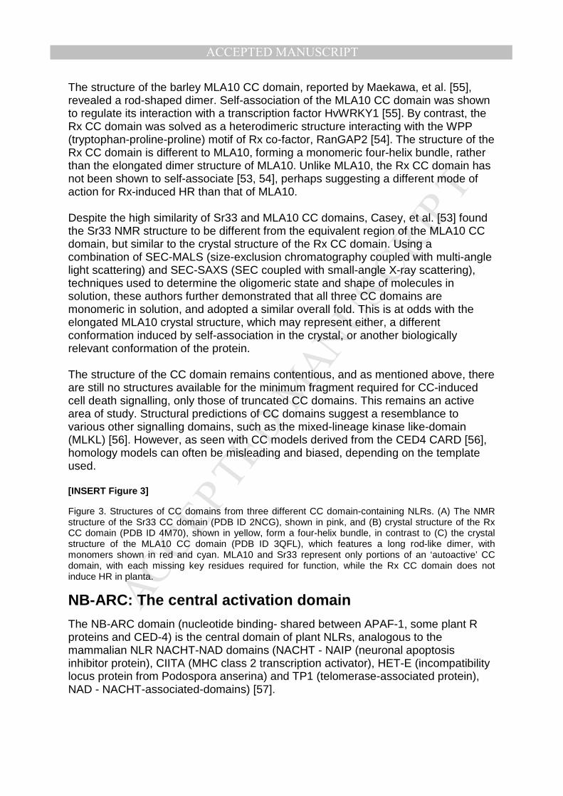
The structure of the barley MLA10 CC domain, reported by Maekawa, et al. [55], revealed a rod-shaped dimer. Self-association of the MLA10 CC domain was shown to regulate its interaction with a transcription factor HvWRKY1 [55]. By contrast, the Rx CC domain was solved as a heterodimeric structure interacting with the WPP (tryptophan-proline-proline) motif of Rx co-factor, RanGAP2 [54]. The structure of the Rx CC domain is different to MLA10, forming a monomeric four-helix bundle, rather than the elongated dimer structure of MLA10. Unlike MLA10, the Rx CC domain has not been shown to self-associate [53, 54], perhaps suggesting a different mode of action for Rx-induced HR than that of MLA10. Despite the high similarity of Sr33 and MLA10 CC domains, Casey, et al. [53] found the Sr33 NMR structure to be different from the equivalent region of the MLA10 CC domain, but similar to the crystal structure of the Rx CC domain. Using a combination of SEC-MALS (size-exclusion chromatography coupled with multi-angle light scattering) and SEC-SAXS (SEC coupled with small-angle X-ray scattering), techniques used to determine the oligomeric state and shape of molecules in solution, these authors further demonstrated that all three CC domains are monomeric in solution, and adopted a similar overall fold. This is at odds with the elongated MLA10 crystal structure, which may represent either, a different conformation induced by self-association in the crystal, or another biologically relevant conformation of the protein. The structure of the CC domain remains contentious, and as mentioned above, there are still no structures available for the minimum fragment required for CC-induced cell death signalling, only those of truncated CC domains. This remains an active area of study. Structural predictions of CC domains suggest a resemblance to various other signalling domains, such as the mixed-lineage kinase like-domain (MLKL) [56]. However, as seen with CC models derived from the CED4 CARD [56], homology models can often be misleading and biased, depending on the template used. [INSERT Figure 3]
Figure 3. Structures of CC domains from three different CC domain-containing NLRs. (A) The NMR structure of the Sr33 CC domain (PDB ID 2NCG), shown in pink, and (B) crystal structure of the Rx CC domain (PDB ID 4M70), shown in yellow, form a four-helix bundle, in contrast to (C) the crystal structure of the MLA10 CC domain (PDB ID 3QFL), which features a long rod-like dimer, with monomers shown in red and cyan. MLA10 and Sr33 represent only portions of an ‘autoactive’ CC domain, with each missing key residues required for function, while the Rx CC domain does not induce HR in planta.
NB-ARC: The central activation domain
The NB-ARC domain (nucleotide binding- shared between APAF-1, some plant R proteins and CED-4) is the central domain of plant NLRs, analogous to the mammalian NLR NACHT-NAD domains (NACHT - NAIP (neuronal apoptosis inhibitor protein), CIITA (MHC class 2 transcription activator), HET-E (incompatibility locus protein from Podospora anserina) and TP1 (telomerase-associated protein), NAD - NACHT-associated-domains) [57].

MANUSCRIP
T
ACCEPTED
ACCEPTED MANUSCRIPT
Regulation and activation of plant NLR immune signalling is controlled through the NB-ARC domain, largely through its interactions with other domains of the NLR, which are influenced by the binding of ADP and ATP ([58-64]. Three distinct subdomains comprise the NB-ARC domain of plant NLRs: the nucleotide-binding domain (NBD), and the ARC1 and ARC2 subdomains, which share similarity with NAD1/HD1 and NAD2/WHD domains in mammalian NLRs [65]. Mammalian NLRs have an additional domain in the NACHT region, called the NAD3/HD2 (or ARC3 domain in APAF-1 and DARK1). As yet there is no known structure of a plant NB-ARC domain, however there are numerous reported crystal and cryoEM structures of animal NLRs, including APAF-1 [26, 28, 29, 66], CED-4 [67, 68] and NLRC4 [69-71], and these structures have been used extensively to guide both mutagenesis and biochemical studies of plant NLRs. The APAF-1 structure solved by Riedl, et al. [28] was the first to include any NB-ARC domain. It showed that the CARD, NB, ARC1 and ARC2 domains all arrange around an ADP molecule. When comparing the APAF-1 structures of the inactive, ADP-bound [28], and the active [26] forms, there are significant conformational differences in the NB-ARC domain. The NBD and ARC1 subdomains remain relatively stationary, while the ARC2 and HD2 subdomains shift significantly between the two forms. Along with the conformational changes, there is an exchange of nucleotide, with ADP being replaced with ATP, or in this instance, a dATP molecule. The additional phosphate of ATP is thought to drive some of these conformational changes, through interactions with highly conserved motifs in the NBD, ARC1 and ARC2 subdomains.
[INSERT Figure 4]
Figure 4. Domain organisation and conformational changes in the NB-ARC domain of APAF-1. The ARC2 and ARC3 subdomains undergo large conformational changes upon nucleotide exchange and cytochrome C binding. The CARD also moves away from the NBD, facilitating nucleotide exchange. These conformational changes, and exchange of nucleotide allow oligomerisation. (A) The ADP-bound structure is from [28] (PDB ID 1Z6T) , and (B) the ATP-bound from [26] (PDB ID 5JUY). The CARD is shown in purple, NBD in red, ARC1 in dark orange, ARC2 in pale orange and ARC3 domain in yellow. (C) Positions of key nucleotide-binding motifs in APAF-1 in ADP and ATP-bound forms. Significant movement is observed for the LHD motif, analogous to the MHD motif in plant NLRs, upon nucleotide binding. The additional phosphate of ATP brings the LHD motif closer to the sensor-1 motif. The P-loop motif is shown in red, the Walker B motif in orange, the sensor-1 motif in yellow, the GLPL motif in green and the LHD motif in blue. ADP and ATP are shown in cyan.
The interactions of the NBD with the other subdomains, and with the bound nucleotide, are also thought to play important roles in plant NLR activation. Mutations within the highly conserved motifs of the NBD often change the activity of the NLR, either inactivating it (mutations in the P-loop, for example), or causing it to become constitutively active (usually termed autoactive). Reminiscent of animal NLR and APAF-1 NBDs, the NB-ARC domains of various plant NLRs have also been demonstrated to possess both nucleotide-binding, and in some cases, ATP-hydrolysis activities [55, 72-74]. Nucleotide binding and hydrolysis in plant NLRs are regulated by the same highly conserved motifs that are involved in interaction with the ADP and ATP molecules in APAF-1, which is illustrated in Figure 4C. The P-loop motif corresponds to a flexible glycine-rich loop containing a highly conserved lysine that coordinates the β-phosphate of ADP. Mutation of the lysine

MANUSCRIP
T
ACCEPTED
ACCEPTED MANUSCRIPT
results in loss of function in many plant NLRs [35, 72, 74-78] and can even perturb the autoactivity observed in gain-of-function mutants [58, 60, 73, 76, 79-81]. Downstream of the P-loop motif is the sensor-1 motif, which interacts with an aspartate in the L/MHD motif (discussed below). In the APAF-1 ADP bound state, the sensor-1 motif moves to directly interact with the γ-phosphate of the bound dATP/ATP in the apoptosome complex. The Walker B Motif is thought to be involved in the ATPase activity of plant NLRs, through coordination of the Mg2+ ion required for hydrolysis. The GLPL motif, located in the ARC1 subdomain, is thought to act as a hinge, allowing conformation changes to occur depending on the bound nucleotide. The MHD motif is one of the most important and best characterized motifs in plant NLRs. Mutations of the conserved histidine or aspartate generally result in a gain-of-function phenotype in planta [73, 74, 76, 77, 79, 82]. The histidine in the analogous LHD motif of APAF-1 binds the β-phosphate of ADP and brings the ARC2 domain into contact with the NB domain [28], stabilizing the closed inactive conformation of APAF-1. Upon dATP binding, the LHD motif moves away from the bound nucleotide, causing large conformational changes that allow formation of the oligomeric apoptosome. It is postulated that LHD/MHD mutations release important autoinhibitory interactions between the NB and ARC2 sub-domains through reduced affinities with ADP, and in doing so facilitate a preference for ATP binding. Thus, in plant NLRs, the NB-ARC domain may, beyond a role in activation, act as a bridge between the detection of the pathogen and signalling though the N-terminal domain. Following its role in oligomerization in animal NLRs, a similar role may also be expected for the NB-ARC domain in plant NLRs.
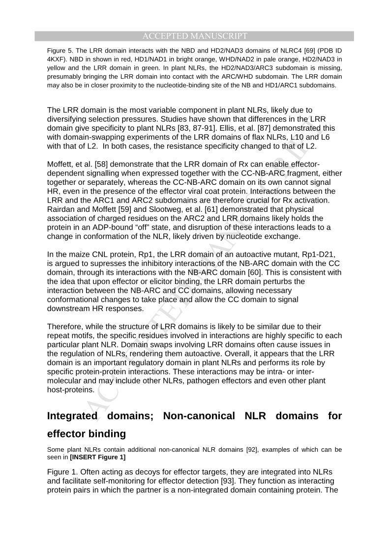
LRR: The C terminal sensor and regulatory domain The LRR does not have a singular defined role throughout all plant NLRs; it has been shown to be involved in effector detection [83], autoinhibition [58] and even activation of NLRs [58, 60]. As with the plant NB-ARC domain, there is currently no structure available for a plant NLR LRR domain. The most relevant structure is from the mammalian protein NLRC4 (Hu, et al. [69]), where the horseshoe-shaped LRR domain makes extensive contacts with the NBD, as well as with key residues in the NAD2/WHD, which is equivalent to the plant ARC2 subdomain. These contacts are important for the regulation and autoinhibition of NLRC4 signalling and can be seen in [INSERT Figure 5]
Figure 5. Furthermore, the LRR domain of NAIP5, involved in detection of an active flagellin derivative, acts in concert with the N-terminal BIR1, HD1 and LRR domains [84, 85]. It is plausible, therefore, that the related domains of plant NLRs may also be involved in effector detection. Found at the C-terminus of the canonical plant NLRs, the LRR domain consists of a series of LxxLxLxxNxL motif repeats (L=leucine, N=asparagine, x=any other amino acid). The LxxLxLxxNxL motif forms a β-strand, and is followed by a varied domain, before repeating again. These repeats form the horseshoe-like structure, with loops and α-helices exposed on the convex surface, and the parallel β-sheet located on the concave surface of the structure [86]. This structure has a high surface-area-to-volume ratio, making it an ideal platform suitable for protein-protein interactions. [INSERT Figure 5]
MANUSCRIP
T
ACCEPTED
ACCEPTED MANUSCRIPTFigure 5. The LRR domain interacts with the NBD and HD2/NAD3 domains of NLRC4 [69] (PDB ID 4KXF). NBD in shown in red, HD1/NAD1 in bright orange, WHD/NAD2 in pale orange, HD2/NAD3 in yellow and the LRR domain in green. In plant NLRs, the HD2/NAD3/ARC3 subdomain is missing, presumably bringing the LRR domain into contact with the ARC/WHD subdomain. The LRR domain may also be in closer proximity to the nucleotide-binding site of the NB and HD1/ARC1 subdomains.
The LRR domain is the most variable component in plant NLRs, likely due to diversifying selection pressures. Studies have shown that differences in the LRR domain give specificity to plant NLRs [83, 87-91]. Ellis, et al. [87] demonstrated this with domain-swapping experiments of the LRR domains of flax NLRs, L10 and L6 with that of L2. In both cases, the resistance specificity changed to that of L2. Moffett, et al. [58] demonstrate that the LRR domain of Rx can enable effector-dependent signalling when expressed together with the CC-NB-ARC fragment, either together or separately, whereas the CC-NB-ARC domain on its own cannot signal HR, even in the presence of the effector viral coat protein. Interactions between the LRR and the ARC1 and ARC2 subdomains are therefore crucial for Rx activation. Rairdan and Moffett [59] and Slootweg, et al. [61] demonstrated that physical association of charged residues on the ARC2 and LRR domains likely holds the protein in an ADP-bound “off” state, and disruption of these interactions leads to a change in conformation of the NLR, likely driven by nucleotide exchange. In the maize CNL protein, Rp1, the LRR domain of an autoactive mutant, Rp1-D21, is argued to supresses the inhibitory interactions of the NB-ARC domain with the CC domain, through its interactions with the NB-ARC domain [60]. This is consistent with the idea that upon effector or elicitor binding, the LRR domain perturbs the interaction between the NB-ARC and CC domains, allowing necessary conformational changes to take place and allow the CC domain to signal downstream HR responses. Therefore, while the structure of LRR domains is likely to be similar due to their repeat motifs, the specific residues involved in interactions are highly specific to each particular plant NLR. Domain swaps involving LRR domains often cause issues in the regulation of NLRs, rendering them autoactive. Overall, it appears that the LRR domain is an important regulatory domain in plant NLRs and performs its role by specific protein-protein interactions. These interactions may be intra- or inter-molecular and may include other NLRs, pathogen effectors and even other plant host-proteins.
Integrated domains; Non-canonical NLR domains for
effector binding Some plant NLRs contain additional non-canonical NLR domains [92], examples of which can be seen in [INSERT Figure 1]
Figure 1. Often acting as decoys for effector targets, they are integrated into NLRs and facilitate self-monitoring for effector detection [93]. They function as interacting protein pairs in which the partner is a non-integrated domain containing protein. The

MANUSCRIP
T
ACCEPTED
ACCEPTED MANUSCRIPT
genes coding for these proteins are often closely linked in the genome such that their co-expression ensures the appropriate stoichiometry of protein levels between the two members of the pair [94]. There are currently structures available for four different integrated domains from plant NLRs, the WRKY domain from RRS1 [95], and the HMA domains from Pikp-1 [96], Pikm-1 [97] and RGA5 [98], all in complex with their cognate effectors. Binding of the effector to integrated decoy domain is thought to facilitate some conformational changes in the NLR, initiating downstream defence signalling. This is distinct from of cytochrome c binding to APAF-1 WD40 domains, and flagellin to multiple domains in NAIP5, with each integrated decoy domain allowing for different effector targets. Integrated domains represent a unique feature of some plant NLRs and a further indication of the diversification of plant NLRs to combat a rapidly changing pathogen load.
Regulation of NLR activity by nucleotide binding and
hydrolysis
As the name suggests, plant NLRs have been shown to bind, and in some cases hydrolyse, ATP and other nucleotides [55, 72-74, 77]. The identity and location of these nucleotides within animal NLRs [28, 29, 66, 69, 99-106] is better understood than for plant NLRs. In APAF-1, exchange of nucleotides is crucial for activation, with the formation of the oligomeric apoptosome requiring the presence of dATP or ATP [66]. Although it is still unclear how nucleotide exchange occurs, some kinetic studies suggest that APAF-1 has a higher affinity for ATP, and higher cellular ATP/ADP ratios may mean that ATP outcompetes ADP for binding to the APAF-1 monomer. Unlike APAF-1, no ATP was observed in the active NLRC4 complex [71]. This could be explained by ADP release rather than ATP binding being sufficient for NLRC4 signalling; however, further study is required to confirm this. The NB-ARC domains of the tomato NLRs, I-2 and Mi-1, were the first to have their nucleotide-binding properties assessed [72, 73], and both were shown to preferentially bind adenosine nucleotides, with a preference for ATP over ADP. The first full-length NLR to be assessed for its nucleotide-binding properties was the barley CNL, MLA27. Purified MLA27 was shown to be predominately in an ADP-bound state [55]. The finding that NLRs purify with bound ADP, despite a preference for ATP, indicates the likelihood of ATPase activity. Binding of ADP and ATP has also been measured in the flax NLRs, M and L6 [74]. Different nucleotide-bound states of the protein were found to correlate with their HR phenotype. The wild-type M and L6 proteins were purified bound to ADP, and were in an inactive state in planta, unless challenged by the effector, AvrM. P-loop mutations of M that were found purified with negligible nucleotide were non-functional in planta, and a mutation in the MHD motif, found to be bound to ATP, was active in planta, even in the absence of the effector AvrM [74]. A study by Bernoux, et al. [77] of two flax NLRs, L6 and L7, revealed that differences in nucleotide-binding capabilities were the likely cause of different effector-dependent HR phenotypes observed for the two proteins. L6, which produces a

MANUSCRIP
T
ACCEPTED
ACCEPTED MANUSCRIPT
strong HR in response to the effector AvrL567, was purified with low levels of bound ADP, while L7, which induces a much weaker HR in response to the same effector, was found purified with much higher levels of ADP. Reciprocal mutations in the TIR domain where found to effectively reverse the nucleotide-binding and HR phenotypes of the two proteins. This observation led to the balanced equilibrium model of plant NLR activation, whereby the cycling of ADP and ATP binding oscillates the protein between inactive and active forms. The arrival of the effector protein then shifts the equilibrium towards a more active ATP-bound form [77]. Few studies have focused on the ATPase activity of plant NLRs. Tameling, et al. [72] observed that the plant NLRs I-2 and Mi-1 possessed ATPase activity, and that this activity was dependent on Mg2+, and that mutations to the P-loop prevented nucleotide binding and therefore ATP hydrolysis from occurring. Mutations in the Walker B motif that impaired ATP hydrolysis activity created autoactive phenotypes of the I-2 protein [73]. This suggests that hydrolysis, as proposed for APAF-1, may play a role in maintaining plant NLRs in an ADP bound off-state. It would then be the ATP-bound state that is crucial for downstream signalling. So although the role of ATP hydrolysis in plant NLR activation is still unclear and certainly not universal, it is well established in APAF-1 that hydrolysis functions to regulate activity [66]. Given that plant NLRs are found bound to ADP in inactive forms, and ATP in active forms, consistent with APAF-1’s nucleotide exchange, it is tempting to speculate that hydrolysis of ATP in plant NLRs also functions as a regulatory mechanism.
NLR oligomerisation
A feature common in animal NLRs is the requirement of protein oligomerisation in order to signal downstream inflammatory and immune responses. These large oligomeric structures, controlled by reorganisation of domains in the NLR monomers, provide a platform for protein-protein interactions. In plant NLRs, N terminal signalling domain dimerization has been demonstrated for a number of NLRs, and the full length tobacco N protein has been shown to form at least dimers in vivo [107]. As for the formation of large plant NLR oligomers, this is still speculative, however the structures of the NLRC4 inflammasome and APAF-1 apoptosome provide a model for how this may occur. The cryoEM structure of the APAF-1 apoptosome by Zhou, et al. [25] shows the formation of a heptameric complex ([INSERT Figure 6]
Figure 6A), in which the NBDs pack together side-by-side to form a ring, and outside of those, the ARC1/HD1 and ARC2/WHD subdomains alternate in a ring around the central NBD ring. The ARC3/HD2 subdomain then links the two WD40 propellers to the ARC2/WHD domain ([INSERT Figure 6]
Figure 6B). CARDs arrange together in the middle and on top of the APAF-1 NBDs, and are able to recruit procaspase-9, thus activating apoptosis. To switch from an inactive monomer to an active apoptosome complex requires extensive conformational changes in the APAF-1 monomer, driven by cytochrome c binding to the WD40 domains, and by nucleotide exchange [24, 29, 66]. The most

MANUSCRIP
T
ACCEPTED
ACCEPTED MANUSCRIPT
prominent change is the position of the ARC2/WHD domain relative to the NBD and ARC1/HD1 domains, as seen in [INSERT Figure 4] Figure 4. The change involves repositioning the ARC2/WHD away from the buried ADP molecule, exposing it to the solvent, allowing nucleotide exchange to occur. This shift of the ARC2/WHD moves the histidine in the LHD motif from direct contact with the ADP in the autoinhibited state, to a position distant from the nucleotide-binding pocket in the active apoptosome ([INSERT Figure 4]
Figure 4C). In the NLRC4 inflammasome [69-71], the NBDs pack side-by-side to form a ring, with the HD1 and WHD domains alternating outside the NBD ring. Like APAF-1, the HD2 links the LRR domain to the WHD. Unlike APAF-1 though, the NLRC4 inflammasome forms after being activated by NAIP2/5, with the activated NLRC4 recruiting other NLRC4 protomers until the ring is closed [71]. The number of subunits in the ring varies, with the NAIP2 nucleated inflammasome forming various complete and incomplete rings. Zhang, et al. [71] generated three reconstructions of the NAIP2/NLRC4 inflammasome with different symmetry imposed (C10, C11 and C12 symmetry), with their C11 eleven bladed disk refined to the highest resolution of 4.7 Å ([INSERT Figure 6]
Figure 6C). Because the subunits were averaged, the position of NAIP2 in the ring could not be determined. By contrast, Tenthorey, et al. [84] did not average the subunits and were able to distinguish NAIP5 in their structure of the NAIP5:NLRC4 complex. As seen in [INSERT Figure 6]
Figure 6D, NAIP5 adopts a similar overall conformation to the NLRC4 monomers. Rings of varying sizes were seen for the NAIP5-nucleated inflammasome disc, though few closed rings were observed. Overall, NLRC4 oligomerisation is distinct from APAF-1 oligomerisation, with each APAF-1 monomer requiring activation, while NLRC4 can be activated by a single NAIP2 or NAIP5 monomer. Unlike APAF-1 and NLRC4, plant NLRs do not have a HD2 domain. Therefore, any oligomer would likely involve interactions of the ARC1 and ARC2 subdomains with the LRR domain. In a TIR-NLR oligomer, the TIRs may be brought together in a fashion similar to that of the CARDs in APAF-1, allowing them to self-associate and signal HR through an, as yet, unknown mechanism. In a CC-NLR, how the CC domains arrange would ultimately be dictated by the structure of the full-length CC-domain, which may differ from the four-helix bundle observed for Sr33 and Rx CC domains, or the rod-like dimer observed in the crystals of MLA10 CC domains. There is evidence that some plant NLRs need to oligomerise to induce HR [43, 107]; however, the stoichiometry of the oligomer and the structural arrangement of domains in the NLR is unknown. It has been difficult to determine the oligomeric state of any plant NLR due to the low yield and low purity of recombinantly produced protein. [INSERT Figure 6]
Figure 6. The APAF-1 apoptosome and the NLRC4 inflammasome. (A) The APAF-1 apoptosome (PDB ID 5JUY) is comprised of seven subunits. Each of the domains of one subunit is displayed in (B). Cyan – cytochrome C, green - WD40 propellers, yellow – ARC3/HD2, pale orange – ARC2/WHD, bright orange – ARC1/HD1, red - NBD, purple - CARD, dark purple – procaspase-9. (C) The NLRC4 inflammasome with eleven-fold symmetry imposed (PDB ID 3JBL). Due to averaging, it is not possible to distinguish the subunit corresponding to NAIP2. Green - LRR, yellow - HD2, pale orange - WHD, bright orange - HD1, red - NBD. (D) NAIP5 and NLRC4 subunits of the NAIP5/NLRC4 inflammasome (PDB ID 6B5B). NAIP5 is bound to flagellin, shown in purple. Green - LRR, yellow - HD2, pale orange

MANUSCRIP
T
ACCEPTED
ACCEPTED MANUSCRIPT- WHD, bright orange - HD1, red – NBD, brown – BIR, purple – flagellin. Only three subunits are shown, but rings of different sizes were observed, with most not closed.
From the various structures of these related NLRs, a basic model of plant NLR activation can be hypothesised. In this model, the signalling component is moderated by interactions with the NB and ARC subdomains, as well as the LRR domain, and an ADP molecule coordinates these interactions. The LRR domain, in combination with other domains (including the non-canonical integrated domains) detects a signal, for example direct effector binding. The direct or indirect detection of the effector either induces conformational changes in the NLR, enabling nucleotide exchange, or locks the protein in an active ATP-bound state. This activated protein is then able to oligomerise and recruit the binding of downstream signalling molecules.
Conclusions and future perspectives While there are several structures available for the CC and TIR signalling components of plant NLRs, and several for the non-canonical domains [95-97, 108], there is still none for the NB-ARC or LRR, let alone a full-length protein. Determining how these domains interact in context of the full-length NLR is essential if we are to fully understand how plant NLRs are activated. To conclude, there is still little experimental evidence that plant NLRs can form oligomers analogous to the ring-like structures observed for APAF-1 and NLRC4. Although the formation of effector-activated oligomeric plant NLR rings is a tempting hypothesis, and would provide a complex on which to initiate SCAF similar to animal NLRs and TLRs, we still await the structure of a full-length plant NLR. Methods from immuno-precipitation and size exclusion chromatography to cryo-electron microscopy can be employed to investigate the universality of self-association and higher-order complex formation that lead to plant NLR signalling. Note add in proof The first structures of a full-length plant NLR, the Arabidopsis coiled-coil NLR ZAR1, have just been reported. Both the structures of the auto-inhibited ADP-bound ZAR1, and the active oligomeric form have been determined by cryo-electron microscopy, and they provide unprecedented new detail as well as surprises about the molecular mechanisms of plant NLR function, and confirm the key suggestions put forward in our review [109, 110]. ACKNOWLEDGEMENTS The work was supported by the Australian Research Council (ARC; DP160102244 and DP190102526). BK is an ARC Laureate Fellow FL180100109 and NHMRC Principal Research Fellow (1110971). Bibliography [1] C. Zipfel, S. Robatzek, L. Navarro, E.J. Oakeley, J.D. Jones, G. Felix, T. Boller, Bacterial disease resistance in Arabidopsis through flagellin perception, Nature 428(6984) (2004) pp. 764-7.

MANUSCRIP
T
ACCEPTED
ACCEPTED MANUSCRIPT
[2] A. Miya, P. Albert, T. Shinya, Y. Desaki, K. Ichimura, K. Shirasu, Y. Narusaka, N. Kawakami, H. Kaku, N. Shibuya, CERK1, a LysM receptor kinase, is essential for chitin elicitor signaling in Arabidopsis, Proc. Natl. Acad. Sci. U. S. A. 104(49) (2007) pp. 19613-19618. [3] F. Boutrot, C. Zipfel, Function, discovery, and exploitation of plant pattern recognition receptors for broad-spectrum disease resistance, Annu. Rev. Phytopathol. 55 (2017) pp. 257-286. [4] X. Yu, B. Feng, P. He, L. Shan, From chaos to harmony: responses and signaling upon microbial pattern recognition, Annu. Rev. Phytopathol. 55(1) (2017) pp. 109-137. [5] E. Luna, V. Pastor, J. Robert, V. Flors, B. Mauch-Mani, J. Ton, Callose deposition: a multifaceted plant defense response, Mol. Plant-Microbe Interact. 24(2) (2010) pp. 183-193. [6] J.L. Dangl, J.D. Jones, Plant pathogens and integrated defence responses to infection, Nature 411(6839) (2001) pp. 826-33. [7] P.N. Dodds, J.P. Rathjen, Plant immunity: Towards an integrated view of plant-pathogen interactions, Nat. Rev. Genet. 11(8) (2010) pp. 539-548. [8] M. Khan, D. Seto, R. Subramaniam, D. Desveaux, Oh, the places they'll go! A survey of phytopathogen effectors and their host targets, The Plant Journal 93(4) (2018) pp. 651-663. [9] J.B. Morel, J.L. Dangl, The hypersensitive response and the induction of cell death in plants, Cell Death Differ. 4(8) (1997) pp. 671-683. [10] M. Kabbage, R. Kessens, L.C. Bartholomay, B. Williams, The life and death of a plant cell, Annu. Rev. Plant Biol. 68(1) (2017) pp. 375-404. [11] A. Künstler, R. Bacsó, G. Gullner, Y.M. Hafez, L. Király, Staying alive – is cell death dispensable for plant disease resistance during the hypersensitive response?, Physiol. Mol. Plant Pathol. 93 (2016) pp. 75-84. [12] H. Yoshida, Y.-Y. Kong, R. Yoshida, A.J. Elia, A. Hakem, R. Hakem, J.M. Penninger, T.W. Mak, Apaf1 Is Required for Mitochondrial Pathways of Apoptosis and Brain Development, Cell 94(6) (1998) pp. 739-750. [13] N. Inohara, T. Koseki, L. del Peso, Y. Hu, C. Yee, S. Chen, R. Carrio, J. Merino, D. Liu, J. Ni, G. Nunez, Nod1, an Apaf-1-like activator of caspase-9 and nuclear factor-kappaB, J. Biol. Chem. 274(21) (1999) pp. 14560-7. [14] D.D. Leipe, E.V. Koonin, L. Aravind, STAND, a class of P-loop NTPases including animal and plant regulators of programmed cell death: multiple, complex domain architectures, unusual phyletic patterns, and evolution by horizontal gene transfer, J. Mol. Biol. 343(1) (2004) pp. 1-28. [15] A.F. Neuwald, L. Aravind, J.L. Spouge, E.V. Koonin, AAA+: A class of chaperone-like ATPases associated with the assembly, operation, and disassembly of protein complexes, Genome Res. 9(1) (1999) pp. 27-43. [16] M. Proell, S.J. Riedl, J.H. Fritz, A.M. Rojas, R. Schwarzenbacher, The Nod-like receptor (NLR) family: A tale of similarities and differences, PLoS One 3(4) (2008) pp. e2119. [17] J. Li, J. Ding, W. Zhang, Y. Zhang, P. Tang, J.Q. Chen, D. Tian, S. Yang, Unique evolutionary pattern of numbers of gramineous NBS-LRR genes, Mol. Genet. Genomics 283(5) (2010) pp. 427-38. [18] H. Zou, W.J. Henzel, X. Liu, A. Lutschg, X. Wang, Apaf-1, a Human Protein Homologous to C. elegans CED-4, Participates in Cytochrome c–Dependent Activation of Caspase-3, Cell 90(3) (1997) pp. 405-413.

MANUSCRIP
T
ACCEPTED
ACCEPTED MANUSCRIPT
[19] A. Rodriguez, H. Oliver, H. Zou, P. Chen, X. Wang, J.M. Abrams, Dark is a Drosophila homologue of Apaf-1/CED-4 and functions in an evolutionarily conserved death pathway, Nat. Cell Biol. 1 (1999) pp. 272. [20] J. Yuan, H.R. Horvitz, The Caenorhabditis elegans genes ced-3 and ced-4 act cell autonomously to cause programmed cell death, Dev. Biol. 138(1) (1990) pp. 33-41. [21] L. Franchi, N. Warner, K. Viani, G. Nuñez, Function of Nod-like receptors in microbial recognition and host defense, Immunol. Rev. 227(1) (2009) pp. 106-28. [22] J. Bertin, W.-J. Nir, C.M. Fischer, O.V. Tayber, P.R. Errada, J.R. Grant, J.J. Keilty, M.L. Gosselin, K.E. Robison, G.H.W. Wong, M.A. Glucksmann, P.S. DiStefano, Human CARD4 protein is a novel CED-4/Apaf-1 cell death family member that activates NF-κB, J. Biol. Chem. 274(19) (1999) pp. 12955-12958. [23] F. Martinon, K. Burns, J. Tschopp, The inflammasome: a molecular platform triggering activation of inflammatory caspases and processing of proIL-β, Mol. Cell 10(2) (2002) pp. 417-426. [24] S. Yuan, M. Topf, T.F. Reubold, S. Eschenburg, C.W. Akey, Changes in Apaf-1 conformation that drive apoptosome assembly, Biochemistry 52(13) (2013) pp. 2319-27. [25] M. Zhou, Y. Li, Q. Hu, X.C. Bai, W. Huang, C. Yan, S.H. Scheres, Y. Shi, Atomic structure of the apoptosome: Mechanism of cytochrome c- and dATP-mediated activation of APAF-1, Genes Dev. 29(22) (2015) pp. 2349-2361. [26] T.C. Cheng, C. Hong, I.V. Akey, S. Yuan, C.W. Akey, A near atomic structure of the active human apoptosome, eLife 5 (2016) pp. e17755. [27] Y. Li, M. Zhou, Q. Hu, X.C. Bai, W. Huang, S.H. Scheres, Y. Shi, Mechanistic insights into caspase-9 activation by the structure of the apoptosome holoenzyme, Proc. Natl. Acad. Sci. U. S. A. 114(7) (2017) pp. 1542-1547. [28] S.J. Riedl, W. Li, Y. Chao, R. Schwarzenbacher, Y. Shi, Structure of the apoptotic protease-activating factor 1 bound to ADP, Nature 434(7035) (2005) pp. 926-933. [29] T.F. Reubold, S. Wohlgemuth, S. Eschenburg, Crystal structure of full-length APAF-1: How the death signal is relayed in the mitochondrial pathway of apoptosis, Structure 19(8) (2011) pp. 1074-1083. [30] S.M. Collier, L.P. Hamel, P. Moffett, Cell death mediated by the N-terminal domains of a unique and highly conserved class of NB-LRR protein, Mol. Plant-Microbe Interact. 24(8) (2011) pp. 918-931. [31] J.X. Yue, B.C. Meyers, J.Q. Chen, D. Tian, S. Yang, Tracing the origin and evolutionary history of plant nucleotide-binding site-leucine-rich repeat (NBS-LRR) genes, New Phytol. 193(4) (2012) pp. 1049-63. [32] F. Jacob, S. Vernaldi, T. Maekawa, Evolution and conservation of plant NLR functions, Front. Immunol. 4 (2013) pp. 297. [33] P.F. Sarris, V. Cevik, G. Dagdas, J.D.G. Jones, K.V. Krasileva, Comparative analysis of plant immune receptor architectures uncovers host proteins likely targeted by pathogens, BMC Biol. 14(1) (2016) pp. 8. [34] N.J. Gay, F.J. Keith, Drosophila Toll and IL-1 receptor, Nature 351(6325) (1991) pp. 355-6. [35] M. Bernoux, T. Ve, S.J. Williams, C. Warren, D. Hatters, E. Valkov, X. Zhang, J.G. Ellis, B. Kobe, P.N. Dodds, Structural and functional analysis of a plant resistance protein TIR domain reveals interfaces for self-association, signaling, and autoregulation, Cell Host Microbe 9(3) (2011) pp. 200-211.

MANUSCRIP
T
ACCEPTED
ACCEPTED MANUSCRIPT
[36] K.G. Hyun, Y. Lee, J. Yoon, H. Yi, J.J. Song, Crystal structure of Arabidopsis thaliana SNC1 TIR domain, Biochem. Biophys. Res. Commun. 481(1-2) (2016) pp. 146-152. [37] X. Zhang, M. Bernoux, A.R. Bentham, T.E. Newman, T. Ve, L.W. Casey, T.M. Raaymakers, J. Hu, T.I. Croll, K.J. Schreiber, B.J. Staskawicz, P.A. Anderson, K.H. Sohn, S.J. Williams, P.N. Dodds, B. Kobe, Multiple functional self-association interfaces in plant TIR domains, Proc. Natl. Acad. Sci. U. S. A. 114(10) (2017) pp. E2046-E2052. [38] S.J. Williams, K.H. Sohn, L. Wan, M. Bernoux, P.F. Sarris, C. Segonzac, T. Ve, Y. Ma, S.B. Saucet, D.J. Ericsson, L.W. Casey, T. Lonhienne, D.J. Winzor, X. Zhang, A. Coerdt, J.E. Parker, P.N. Dodds, B. Kobe, J.D. Jones, Structural basis for assembly and function of a heterodimeric plant immune receptor, Science 344(6181) (2014) pp. 299-303. [39] S.J. Williams, L. Yin, G. Foley, L.W. Casey, M.A. Outram, D.J. Ericsson, J. Lu, M. Boden, I.B. Dry, B. Kobe, Structure and function of the TIR domain from the grape NLR protein RPV1, Front Plant Sci 7 (2016) pp. 1850. [40] D. Frost, H. Way, P. Howles, J. Luck, J. Manners, A. Hardham, J. Finnegan, J. Ellis, Tobacco transgenic for the flax rust resistance gene L expresses allele-specific activation of defense responses, Mol. Plant-Microbe Interact. 17(2) (2004) pp. 224-232. [41] M.R. Swiderski, D. Birker, J.D. Jones, The TIR domain of TIR-NB-LRR resistance proteins is a signaling domain involved in cell death induction, Mol. Plant-Microbe Interact. 22(2) (2009) pp. 157-165. [42] K.V. Krasileva, D. Dahlbeck, B.J. Staskawicz, Activation of an Arabidopsis resistance protein is specified by the in planta association of its leucine-rich repeat domain with the cognate oomycete effector, Plant Cell 22(7) (2010) pp. 2444-2458. [43] K.J. Schreiber, A. Bentham, S.J. Williams, B. Kobe, B.J. Staskawicz, Multiple domain associations within the Arabidopsis immune receptor RPP1 regulate the activation of programmed cell death, PLoS Path. 12(7) (2016) pp. e1005769. [44] T. Ve, S.J. Williams, B. Kobe, Structure and function of Toll/Interleukin-1 receptor/resistance protein (TIR) domains, Apoptosis 20(2) (2015) pp. 250-261. [45] T. Ve, P.R. Vajjhala, A. Hedger, T. Croll, F. DiMaio, S. Horsefield, X. Yu, P. Lavrencic, Z. Hassan, G.P. Morgan, A. Mansell, M. Mobli, A. O'Carroll, B. Chauvin, Y. Gambin, E. Sierecki, M.J. Landsberg, K.J. Stacey, E.H. Egelman, B. Kobe, Structural basis of TIR-domain-assembly formation in MAL- and MyD88-dependent TLR4 signaling, Nat. Struct. Mol. Biol. 24 (2017) pp. 743. [46] S. Nimma, T. Ve, S.J. Williams, B. Kobe, Towards the structure of the TIR-domain signalosome, Curr. Opin. Struct. Biol. 43 (2017) pp. 122-130. [47] A.R. Bentham, H. Burdett, P.A. Anderson, S.J. Williams, B. Kobe, Animal NLRs provide structural insights into plant NLR function, Ann. Bot. 119(5) (2017) pp. 689-702. [48] J.D. Nanson, M.H. Rahaman, T. Ve, B. Kobe, Regulation of signaling by cooperative assembly formation in mammalian innate immunity signalosomes by molecular mimics, Semin. Cell Dev. Biol. (2018). [49] J.D. Nanson, B. Kobe, T. Ve, Death, TIR, and RHIM: Self-assembling domains involved in innate immunity and cell-death signaling, J. Leukoc. Biol. (2018). [50] P.R. Vajjhala, T. Ve, A. Bentham, K.J. Stacey, B. Kobe, The molecular mechanisms of signaling by cooperative assembly formation in innate immunity pathways, Mol. Immunol. 86 (2017) pp. 23-37.
MANUSCRIP
T
ACCEPTED
ACCEPTED MANUSCRIPT
[51] K. Essuman, D.W. Summers, Y. Sasaki, X. Mao, A. DiAntonio, J. Milbrandt, The SARM1 Toll/interleukin-1 receptor domain possesses intrinsic NAD+ cleavage activity that promotes pathological axonal degeneration, Neuron 93(6) (2017) pp. 1334-1343.e5. [52] D.W. Summers, D.A. Gibson, A. DiAntonio, J. Milbrandt, SARM1-specific motifs in the TIR domain enable NAD(+) loss and regulate injury-induced SARM1 activation, Proc. Natl. Acad. Sci. U. S. A. 113(41) (2016) pp. E6271-E6280. [53] L.W. Casey, P. Lavrencic, A.R. Bentham, S. Cesari, D.J. Ericsson, T. Croll, D. Turk, P.A. Anderson, A.E. Mark, P.N. Dodds, M. Mobli, B. Kobe, S.J. Williams, The CC domain structure from the wheat stem rust resistance protein Sr33 challenges paradigms for dimerization in plant NLR proteins, Proc. Natl. Acad. Sci. U. S. A. 113(45) (2016) pp. 12856-12861. [54] W. Hao, S.M. Collier, P. Moffett, J. Chai, Structural basis for the interaction between the potato virus X resistance protein (Rx) and its cofactor Ran GTPase-activating protein 2 (RanGAP2), J. Biol. Chem. 288(50) (2013) pp. 35868-76. [55] T. Maekawa, W. Cheng, L.N. Spiridon, A. Toller, E. Lukasik, Y. Saijo, P. Liu, Q.H. Shen, M.A. Micluta, I.E. Somssich, F.L.W. Takken, A.J. Petrescu, J. Chai, P. Schulze-Lefert, Coiled-coil domain-dependent homodimerization of intracellular barley immune receptors defines a minimal functional module for triggering cell death, Cell Host Microbe 9(3) (2011) pp. 187-199. [56] A.R. Bentham, R. Zdrzalek, J.C. De la Concepcion, M.J. Banfield, Uncoiling CNLs: Structure/function approaches to understanding CC domain function in plant NLRs, Plant Cell Physiol. (2018). [57] E.V. Koonin, L. Aravind, The NACHT family - a new group of predicted NTPases implicated in apoptosis and MHC transcription activation, Trends Biochem. Sci. 25(5) (2000) pp. 223-224. [58] P. Moffett, G. Farnham, J. Peart, D.C. Baulcombe, Interaction between domains of a plant NBS-LRR protein in disease resistance-related cell death, EMBO J. 21(17) (2002) pp. 4511-4519. [59] G.J. Rairdan, P. Moffett, Distinct domains in the ARC region of the potato resistance protein Rx mediate LRR binding and inhibition of activation, Plant Cell 18(8) (2006) pp. 2082-93. [60] G.F. Wang, J. Ji, F. El-Kasmi, J.L. Dangl, G. Johal, P.J. Balint-Kurti, Molecular and functional analyses of a maize autoactive NB-LRR protein identify precise structural requirements for activity, PLoS Path. 11(2) (2015) pp. e1004674. [61] E.J. Slootweg, L.N. Spiridon, J. Roosien, P. Butterbach, R. Pomp, L. Westerhof, R. Wilbers, E. Bakker, J. Bakker, A.J. Petrescu, G. Smant, A. Goverse, Structural determinants at the interface of the ARC2 and leucine-rich repeat domains control the activation of the plant immune receptors Rx1 and Gpa2, Plant Physiol. 162(3) (2013) pp. 1510-1528. [62] S. Fenyk, S. Campillo Ade, E. Pohl, P.J. Hussey, M.J. Cann, A nucleotide phosphatase activity in the nucleotide binding domain of an orphan resistance protein from rice, J. Biol. Chem. 287(6) (2012) pp. 4023-4032. [63] J. Ade, B.J. DeYoung, C. Golstein, R.W. Innes, Indirect activation of a plant nucleotide binding site-leucine-rich repeat protein by a bacterial protease, Proc. Natl. Acad. Sci. U. S. A. 104(7) (2007) pp. 2531-2536. [64] R.T. Leister, D. Dahlbeck, B. Day, Y. Li, O. Chesnokova, B.J. Staskawicz, Molecular genetic evidence for the role of SGT1 in the intramolecular complementation of Bs2 protein activity in Nicotiana benthamiana, Plant Cell 17(4) (2005) pp. 1268-1278.

MANUSCRIP
T
ACCEPTED
ACCEPTED MANUSCRIPT
[65] F.L.W. Takken, A. Goverse, How to build a pathogen detector: Structural basis of NB-LRR function, Curr. Opin. Plant Biol. 15(4) (2012) pp. 375-384. [66] T.F. Reubold, S. Wohlgemuth, S. Eschenburg, A new model for the transition of APAF-1 from inactive monomer to caspase-activating apoptosome, J. Biol. Chem. 284(47) (2009) pp. 32717-32724. [67] N. Yan, J.J. Chai, E.S. Lee, L.C. Gu, Q. Liu, J.Q. He, J.W. Wu, D. Kokel, H.L. Li, Q. Hao, D. Xue, Y.G. Shi, Structure of the CED-4-CED-9 complex provides insights into programmed cell death in Caenorhabditis elegans, Nature 437(7060) (2005) pp. 831-837. [68] S. Qi, Y. Pang, Q. Hu, Q. Liu, H. Li, Y. Zhou, T. He, Q. Liang, Y. Liu, X. Yuan, G. Luo, H. Li, J. Wang, N. Yan, Y. Shi, Crystal structure of the caenorhabditis elegans apoptosome reveals an octameric assembly of CED-4, Cell 141(3) (2010) pp. 446-457. [69] Z.H. Hu, C.Y. Yan, P.Y. Liu, Z.W. Huang, R. Ma, C.L. Zhang, R.Y. Wang, Y.T. Zhang, F. Martinon, D. Miao, H.T. Deng, J.W. Wang, J.B. Chang, J.J. Chai, Crystal Structure of NLRC4 Reveals Its Autoinhibition Mechanism, Science 341(6142) (2013) pp. 172-175. [70] Z. Hu, Q. Zhou, C. Zhang, S. Fan, W. Cheng, Y. Zhao, F. Shao, H.W. Wang, S.F. Sui, J. Chai, Structural and biochemical basis for induced self-propagation of NLRC4, Science 350(6259) (2015) pp. 399-404. [71] L.M. Zhang, S.B. Chen, J.B. Ruan, J.Y. Wu, A.B. Tong, Q. Yin, Y. Li, L. David, A. Lu, W.L. Wang, C. Marks, Q. Ouyang, X.Z. Zhang, Y.D. Mao, H. Wu, Cryo-EM structure of the activated NAIP2-NLRC4 inflammasome reveals nucleated polymerization, Science 350(6259) (2015) pp. 404-409. [72] W.I. Tameling, S.D. Elzinga, P.S. Darmin, J.H. Vossen, F.L. Takken, M.A. Haring, B.J. Cornelissen, The tomato R gene products I-2 and MI-1 are functional ATP binding proteins with ATPase activity, Plant Cell 14(11) (2002) pp. 2929-2939. [73] W.I.L. Tameling, J.H. Vossen, M. Albrecht, T. Lengauer, J.A. Berden, M.A. Haring, B.J.C. Cornelissen, F.L.W. Takken, Mutations in the NB-ARC domain of I-2 that impair ATP hydrolysis cause autoactivation, Plant Physiol. 140(4) (2006) pp. 1233-1245. [74] S.J. Williams, P. Sornaraj, E. deCourcy-Ireland, R.I. Menz, B. Kobe, J.G. Ellis, P.N. Dodds, P.A. Anderson, An autoactive mutant of the M flax rust resistance protein has a preference for binding ATP, whereas wild-type M protein binds ADP, Mol. Plant-Microbe Interact. 24(8) (2011) pp. 897-906. [75] S.P. Dinesh-Kumar, W.H. Tham, B.J. Baker, Structure-function analysis of the tobacco mosaic virus resistance gene N, Proc. Natl. Acad. Sci. U. S. A. 97(26) (2000) pp. 14789-14794. [76] A. Bendahmane, G. Farnham, P. Moffett, D.C. Baulcombe, Constitutive gain-of-function mutants in a nucleotide binding site-leucine rich repeat protein encoded at the Rx locus of potato, Plant J. 32(2) (2002) pp. 195-204. [77] M. Bernoux, H. Burdett, S.J. Williams, X. Zhang, C. Chen, K. Newell, G.J. Lawrence, B. Kobe, J.G. Ellis, P.A. Anderson, P.N. Dodds, Comparative analysis of the flax immune receptors L6 and L7 suggests an equilibrium-based switch activation model, Plant Cell 28(1) (2016) pp. 146-159. [78] T. Qi, K. Seong, D.P.T. Thomazella, J.R. Kim, J. Pham, E. Seo, M.J. Cho, A. Schultink, B.J. Staskawicz, NRG1 functions downstream of EDS1 to regulate TIR-NLR-mediated plant immunity in Nicotiana benthamiana, Proc. Natl. Acad. Sci. U. S. A. 115(46) (2018) pp. E10979-e10987.
MANUSCRIP
T
ACCEPTED
ACCEPTED MANUSCRIPT
[79] P. Howles, G. Lawrence, J. Finnegan, H. McFadden, M. Ayliffe, P. Dodds, J. Ellis, Autoactive alleles of the flax L6 rust resistance gene induce non-race-specific rust resistance associated with the hypersensitive response, Mol. Plant-Microbe Interact. 18(6) (2005) pp. 570-582. [80] S.H. Gabriels, J.H. Vossen, S.K. Ekengren, G. van Ooijen, A.M. Abd-El-Haliem, G.C. van den Berg, D.Y. Rainey, G.B. Martin, F.L. Takken, P.J. de Wit, M.H. Joosten, An NB-LRR protein required for HR signalling mediated by both extra- and intracellular resistance proteins, Plant J. 50(1) (2007) pp. 14-28. [81] D.J. Sueldo, M. Shimels, L.N. Spiridon, O. Caldararu, A.J. Petrescu, M.H. Joosten, W.I. Tameling, Random mutagenesis of the nucleotide-binding domain of NRC1 (NB-LRR required for Hypersensitive Response-Associated Cell Death-1), a downstream signalling nucleotide-binding, leucine-rich repeat (NB-LRR) protein, identifies gain-of-function mutations in the nucleotide-binding pocket, New Phytol. 208(1) (2015) pp. 210-223. [82] S. de la Fuente van Bentem, J.H. Vossen, K.J. de Vries, S. van Wees, W.I. Tameling, H.L. Dekker, C.G. de Koster, M.A. Haring, F.L. Takken, B.J. Cornelissen, Heat shock protein 90 and its co-chaperone protein phosphatase 5 interact with distinct regions of the tomato I-2 disease resistance protein, Plant J. 43(2) (2005) pp. 284-298. [83] P.N. Dodds, G.J. Lawrence, A.M. Catanzariti, T. Teh, C.I. Wang, M.A. Ayliffe, B. Kobe, J.G. Ellis, Direct protein interaction underlies gene-for-gene specificity and coevolution of the flax resistance genes and flax rust avirulence genes, Proc. Natl. Acad. Sci. U. S. A. 103(23) (2006) pp. 8888-8893. [84] J.L. Tenthorey, N. Haloupek, J.R. Lopez-Blanco, P. Grob, E. Adamson, E. Hartenian, N.A. Lind, N.M. Bourgeois, P. Chacon, E. Nogales, R.E. Vance, The structural basis of flagellin detection by NAIP5: A strategy to limit pathogen immune evasion, Science 358(6365) (2017) pp. 888-893. [85] X. Yang, F. Yang, W. Wang, G. Lin, Z. Hu, Z. Han, Y. Qi, L. Zhang, J. Wang, S.F. Sui, J. Chai, Structural basis for specific flagellin recognition by the NLR protein NAIP5, Cell Res. 28(1) (2018) pp. 35-47. [86] B. Kobe, A.V. Kajava, The leucine-rich repeat as a protein recognition motif, Curr. Opin. Struct. Biol. 11(6) (2001) pp. 725-732. [87] J.G. Ellis, G.J. Lawrence, J.E. Luck, P.N. Dodds, Identification of regions in alleles of the flax rust resistance gene L that determine differences in gene-for-gene specificity, Plant Cell 11(3) (1999) pp. 495-506. [88] P.N. Dodds, G.J. Lawrence, J.G. Ellis, Six amino acid changes confined to the leucine-rich repeat beta-strand/beta-turn motif determine the difference between the P and P2 rust resistance specificities in flax, Plant Cell 13(1) (2001) pp. 163-178. [89] C.I. Wang, G. Guncar, J.K. Forwood, T. Teh, A.M. Catanzariti, G.J. Lawrence, F.E. Loughlin, J.P. Mackay, H.J. Schirra, P.A. Anderson, J.G. Ellis, P.N. Dodds, B. Kobe, Crystal structures of flax rust avirulence proteins AvrL567-A and -D reveal details of the structural basis for flax disease resistance specificity, Plant Cell 19(9) (2007) pp. 2898-2912. [90] B.B. Wulff, A. Heese, L. Tomlinson-Buhot, D.A. Jones, M. de la Pena, J.D. Jones, The major specificity-determining amino acids of the tomato Cf-9 disease resistance protein are at hypervariable solvent-exposed positions in the central leucine-rich repeats, Mol. Plant-Microbe Interact. 22(10) (2009) pp. 1203-1213. [91] M. Ravensdale, M. Bernoux, T. Ve, B. Kobe, P.H. Thrall, J.G. Ellis, P.N. Dodds, Intramolecular interaction influences binding of the flax L5 and L6 resistance proteins to their AvrL567 ligands, PLoS Path. 8(11) (2012) pp. e1003004.

MANUSCRIP
T
ACCEPTED
ACCEPTED MANUSCRIPT
[92] B.C. Meyers, A. Kozik, A. Griego, H. Kuang, R.W. Michelmore, Genome-wide analysis of NBS-LRR-encoding genes in Arabidopsis, Plant Cell 15(4) (2003) pp. 809-34. [93] S. Cesari, Multiple strategies for pathogen perception by plant immune receptors, New Phytol. 219(1) (2018) pp. 17-24. [94] E. Baggs, G. Dagdas, K.V. Krasileva, NLR diversity, helpers and integrated domains: making sense of the NLR IDentity, Curr. Opin. Plant Biol. 38 (2017) pp. 59-67. [95] Z.-M. Zhang, K.-W. Ma, L. Gao, Z. Hu, S. Schwizer, W. Ma, J. Song, Mechanism of host substrate acetylation by a YopJ family effector, Nat. Plants 3 (2017) pp. 17115. [96] A. Maqbool, H. Saitoh, M. Franceschetti, C.E. Stevenson, A. Uemura, H. Kanzaki, S. Kamoun, R. Terauchi, M.J. Banfield, Structural basis of pathogen recognition by an integrated HMA domain in a plant NLR immune receptor, eLife 4 (2015) pp. e08709. [97] J.C. De la Concepcion, M. Franceschetti, A. Maqbool, H. Saitoh, R. Terauchi, S. Kamoun, M.J. Banfield, Polymorphic residues in rice NLRs expand binding and response to effectors of the blast pathogen, Nat. Plants 4(8) (2018) pp. 576-585. [98] L. Guo, S. Cesari, K. de Guillen, V. Chalvon, L. Mammri, M. Ma, I. Meusnier, F. Bonnot, A. Padilla, Y.L. Peng, J. Liu, T. Kroj, Specific recognition of two MAX effectors by integrated HMA domains in plant immune receptors involves distinct binding surfaces, Proc. Natl. Acad. Sci. U. S. A. 115(45) (2018) pp. 11637-11642. [99] R. Parkhouse, J.P. Boyle, T.P. Monie, Blau syndrome polymorphisms in NOD2 identify nucleotide hydrolysis and helical domain 1 as signalling regulators, FEBS Lett. 588(18) (2014) pp. 3382-3389. [100] J.A. MacDonald, C.P. Wijekoon, K.-C. Liao, D.A. Muruve, Biochemical and structural aspects of the ATP-binding domain in inflammasome-forming human NLRP proteins, IUBMB Life 65(10) (2013) pp. 851-862. [101] A.D. Radian, S. Khare, L.H. Chu, A. Dorfleutner, C. Stehlik, ATP binding by NLRP7 is required for inflammasome activation in response to bacterial lipopeptides, Mol. Immunol. 67(2) (2015) pp. 294-302. [102] J.A. Duncan, D.T. Bergstralh, Y. Wang, S.B. Willingham, Z. Ye, A.G. Zimmermann, J.P. Ting, Cryopyrin/NALP3 binds ATP/dATP, is an ATPase, and requires ATP binding to mediate inflammatory signaling, Proc. Natl. Acad. Sci. U. S. A. 104(19) (2007) pp. 8041-6. [103] B. Faustin, L. Lartigue, J.M. Bruey, F. Luciano, E. Sergienko, B. Bailly-Maitre, N. Volkmann, D. Hanein, I. Rouiller, J.C. Reed, Reconstituted NALP1 inflammasome reveals two-step mechanism of caspase-1 activation, Mol. Cell 25(5) (2007) pp. 713-24. [104] Z. Ye, J.D. Lich, C.B. Moore, J.A. Duncan, K.L. Williams, J.P.Y. Ting, ATP binding by monarch-1/NLRP12 is critical for its inhibitory function, Mol. Cell. Biol. 28(5) (2008) pp. 1841-1850. [105] N. Askari, R.G. Correa, D. Zhai, J.C. Reed, Expression, purification, and characterization of recombinant NOD1 (NLRC1): A NLR family member, J. Biotechnol. 157(1) (2012) pp. 75-81. [106] J. Mo, J.P. Boyle, C.B. Howard, T.P. Monie, B.K. Davis, J.A. Duncan, Pathogen sensing by nucleotide-binding oligomerization domain-containing protein 2 (NOD2) is mediated by direct binding to muramyl dipeptide and ATP, J. Biol. Chem. 287(27) (2012) pp. 23057-23067.

MANUSCRIP
T
ACCEPTED
ACCEPTED MANUSCRIPT
[107] P. Mestre, D.C. Baulcombe, Elicitor-mediated oligomerization of the tobacco N disease resistance protein, Plant Cell 18(2) (2006) pp. 491-501. [108] Z.Q. Fu, M. Guo, B.-r. Jeong, F. Tian, T.E. Elthon, R.L. Cerny, D. Staiger, J.R. Alfano, A type III effector ADP-ribosylates RNA-binding proteins and quells plant immunity, Nature 447(7142) (2007) pp. 284-288. [109] J. Wang, J. Wang, M. Hu, S. Wu, J. Qi, G. Wang, Z. Han, Y. Qi, N. Gao, H.-W. Wang, J.-M. Zhou, J. Chai, Ligand-triggered allosteric ADP release primes a plant NLR complex, Science 364(6435) (2019) pp. eaav5868. [110] J. Wang, M. Hu, J. Wang, J. Qi, Z. Han, G. Wang, Y. Qi, H.-W. Wang, J.-M. Zhou, J. Chai, Reconstitution and structure of a plant NLR resistosome conferring immunity, Science 364(6435) (2019) pp. eaav5870. [111] M.D. Herman, M. Moche, S. Flodin, M. Welin, L. Tresaugues, I. Johansson, M. Nilsson, P. Nordlund, T. Nyman, Structures of BIR domains from human NAIP and cIAP2, Acta Crystallogr. Sect. F Struct. Biol. Cryst. Commun. 65(Pt 11) (2009) pp. 1091-6. [112] M. Hong, S.I. Yoon, I.A. Wilson, Structure and functional characterization of the RNA-binding element of the NLRX1 innate immune modulator, Immunity 36(3) (2012) pp. 337-47. [113] D.E. Vaughn, J. Rodriguez, Y. Lazebnik, L. Joshua-Tor, Crystal structure of Apaf-1 caspase recruitment domain: an alpha-helical Greek key fold for apoptotic signaling, J. Mol. Biol. 293(3) (1999) pp. 439-47. [114] P. Zhou, J. Chou, R.S. Olea, J. Yuan, G. Wagner, Solution structure of Apaf-1 CARD and its interaction with caspase-9 CARD: a structural basis for specific adaptor/caspase interaction, Proc. Natl. Acad. Sci. U. S. A. 96(20) (1999) pp. 11265-70. [115] C.L. Day, C. Dupont, M. Lackmann, D.L. Vaux, M.G. Hinds, Solution structure and mutagenesis of the caspase recruitment domain (CARD) from Apaf-1, Cell Death Differ. 6(11) (1999) pp. 1125-32. [116] H. Qin, S.M. Srinivasula, G. Wu, T. Fernandes-Alnemri, E.S. Alnemri, Y. Shi, Structural basis of procaspase-9 recruitment by the apoptotic protease-activating factor 1, Nature 399(6736) (1999) pp. 549-57. [117] S.L. Milam, N.I. Nicely, B. Feeney, C. Mattos, A.C. Clark, Rapid folding and unfolding of Apaf-1 CARD, J. Mol. Biol. 369(1) (2007) pp. 290-304. [118] Q. Hu, D. Wu, W. Chen, Z. Yan, C. Yan, T. He, Q. Liang, Y. Shi, Molecular determinants of caspase-9 activation by the Apaf-1 apoptosome, Proc. Natl. Acad. Sci. U. S. A. 111(46) (2014) pp. 16254-61. [119] T.W. Su, C.Y. Yang, W.P. Kao, B.J. Kuo, S.M. Lin, J.Y. Lin, Y.C. Lo, S.C. Lin, Structural insights into DD-fold assembly and caspase-9 activation by the Apaf-1 apoptosome, Structure 25(3) (2017) pp. 407-420. [120] S. Yuan, X. Yu, M. Topf, L. Dorstyn, S. Kumar, S.J. Ludtke, C.W. Akey, Structure of the Drosophila apoptosome at 6.9 Å resolution, Structure 19(1) (2011) pp. 128-140. [121] Y. Pang, X.C. Bai, C. Yan, Q. Hao, Z. Chen, J.W. Wang, S.H. Scheres, Y. Shi, Structure of the apoptosome: mechanistic insights into activation of an initiator caspase from Drosophila, Genes Dev. 29(3) (2015) pp. 277-87. [122] T.C. Cheng, I.V. Akey, S. Yuan, Z. Yu, S.J. Ludtke, C.W. Akey, A near-atomic structure of the Dark apoptosome provides insight into assembly and activation, Structure 25(1) (2017) pp. 40-52.

MANUSCRIP
T
ACCEPTED
ACCEPTED MANUSCRIPT
[123] W. Huang, T. Jiang, W. Choi, S. Qi, Y. Pang, Q. Hu, Y. Xu, X. Gong, P.D. Jeffrey, J. Wang, Y. Shi, Mechanistic insights into CED-4-mediated activation of CED-3, Genes Dev. 27(18) (2013) pp. 2039-48. [124] T. Jin, J. Curry, P. Smith, J. Jiang, T.S. Xiao, Structure of the NLRP1 caspase recruitment domain suggests potential mechanisms for its association with procaspase-1, Proteins 81(7) (2013) pp. 1266-70. [125] J. Oroz, S. Barrera-Vilarmau, C. Alfonso, G. Rivas, E. de Alba, ASC pyrin domain self-associates and binds NLRP3 protein using equivalent binding interfaces, J. Biol. Chem. 291(37) (2016) pp. 19487-501. [126] C. Eibl, S. Grigoriu, M. Hessenberger, J. Wenger, S. Puehringer, A.S. Pinheiro, R.N. Wagner, M. Proell, J.C. Reed, R. Page, K. Diederichs, W. Peti, Structural and functional analysis of the NLRP4 pyrin domain, Biochemistry 51(37) (2012) pp. 7330-41. [127] C. Shen, A. Lu, W.J. Xie, J. Ruan, R. Negro, E.H. Egelman, T.-M. Fu, H. Wu, Molecular mechanism for NLRP6 inflammasome assembly and activation, Proc. Natl. Acad. Sci. U. S. A. (2019) pp. 201817221. [128] A.S. Pinheiro, M. Proell, C. Eibl, R. Page, R. Schwarzenbacher, W. Peti, Three-dimensional structure of the NLRP7 pyrin domain: insight into pyrin-pyrin-mediated effector domain signaling in innate immunity, J. Biol. Chem. 285(35) (2010) pp. 27402-10. [129] M.Y. Su, C.I. Kuo, C.F. Chang, C.I. Chang, Three-dimensional structure of human NLRP10/PYNOD pyrin domain reveals a homotypic interaction site distinct from its mouse homologue, PLoS One 8(7) (2013) pp. e67843. [130] A.S. Pinheiro, C. Eibl, Z. Ekman-Vural, R. Schwarzenbacher, W. Peti, The NLRP12 pyrin domain: structure, dynamics, and functional insights, J. Mol. Biol. 413(4) (2011) pp. 790-803. [131] T. Jin, W. Chuenchor, J. Jiang, J. Cheng, Y. Li, K. Fang, M. Huang, P. Smith, T.S. Xiao, Design of an expression system to enhance MBP-mediated crystallization, Sci. Rep. 7 (2017) pp. 40991. [132] C. Eibl, M. Hessenberger, J. Wenger, H. Brandstetter, Structures of the NLRP14 pyrin domain reveal a conformational switch mechanism regulating its molecular interactions, Acta Crystallogr. D Biol. Crystallogr. 70(Pt 7) (2014) pp. 2007-18. [133] F. Manon, A. Favier, G. Nunez, J.P. Simorre, S. Cusack, Solution structure of NOD1 CARD and mutational analysis of its interaction with the CARD of downstream kinase RICK, J. Mol. Biol. 365(1) (2007) pp. 160-74. [134] N.P. Coussens, J.C. Mowers, C. McDonald, G. Nunez, S. Ramaswamy, Crystal structure of the Nod1 caspase activation and recruitment domain, Biochem. Biophys. Res. Commun. 353(1) (2007) pp. 1-5. [135] T. Srimathi, S.L. Robbins, R.L. Dubas, M. Hasegawa, N. Inohara, Y.C. Park, Monomer/dimer transition of the caspase-recruitment domain of human Nod1, Biochemistry 47(5) (2008) pp. 1319-25. [136] A.M. Ver Heul, L. Gakhar, R.C. Piper, R. Subramanian, Crystal structure of a complex of NOD1 CARD and ubiquitin, PLoS One 9(8) (2014) pp. e104017. [137] S. Maekawa, U. Ohto, T. Shibata, K. Miyake, T. Shimizu, Crystal structure of NOD2 and its implications in human disease, Nat Commun 7 (2016) pp. 11813. [138] C.A. Diebolder, E.F. Halff, A.J. Koster, E.G. Huizinga, R.I. Koning, Cryoelectron tomography of the NAIP5/NLRC4 inflammasome: implications for NLR activation, Structure 23(12) (2015) pp. 2349-2357.

MANUSCRIP
T
ACCEPTED
ACCEPTED MANUSCRIPT
[139] Y. Li, T.M. Fu, A. Lu, K. Witt, J. Ruan, C. Shen, H. Wu, Cryo-EM structures of ASC and NLRC4 CARD filaments reveal a unified mechanism of nucleation and activation of caspase-1, Proc. Natl. Acad. Sci. U. S. A. 115(43) (2018) pp. 10845-10852. [140] P.G. Gutte, S. Jurt, M.G. Grutter, O. Zerbe, Unusual structural features revealed by the solution NMR structure of the NLRC5 caspase recruitment domain, Biochemistry 53(19) (2014) pp. 3106-17.

MANUSCRIP
T
ACCEPTED
ACCEPTED MANUSCRIPTNLR Domain Note Species Resolution (Å) Technique PDB ID Reference L6 TIR Linum usitatissimum 2.3 X-Ray Diffraction 3OZI Bernoux, et al. [35]SNC1 TIR Arabidopsis thaliana 2.6 X-Ray Diffraction 5H3C Hyun, et al. [36]
TIR Arabidopsis thaliana 2.2 X-Ray Diffraction 5TEC Zhang, et al. [37]RPS4 TIR Heterodimer with RRS1 Arabidopsis thaliana 2.65 X-Ray Diffraction 4C6T Williams, et al. [38]
TIR Arabidopsis thaliana 2.05 X-Ray Diffraction 4C6R Williams, et al. [38]RPP1 TIR Arabidopsis thaliana 2.8 X-Ray Diffraction 5TEB Zhang, et al. [37]RPV1 TIR Vitis rotundifolia 2.3 X-Ray Diffraction 5KU7 Williams, et al. [39]RRS1 TIR Heterodimer with RPS4 Arabidopsis thaliana 2.65 X-Ray Diffraction 4C6T Williams, et al. [38]
TIR Arabidopsis thaliana 1.75 X-Ray Diffraction 4C6S Williams, et al. [38] WRKY Complex with PopP2, IP6, AcCoA and WRKY Arabidopsis thaliana 2.0 X-Ray Diffraction 5W3X Zhang, et al. [95]MLA10 CC Hordeum vulgare 2.05 X-Ray Diffraction 5T1Y Casey, et al. [53]
CC Hordeum vulgare 1.99 X-Ray Diffraction 3QFL Maekawa, et al. [55]Sr33 CC Aegilops tauschii N/A Solution NMR 2NCG Casey, et al. [53]Rx CC Complex with RanGAP2-WPP Solanum tuberosum 2.1 X-Ray Diffraction 4M70 Hao, et al. [54] Pikp-1 HMA Oryza sativa ssp. japonica 2.1 X-Ray Diffraction 5A6P Maqbool, et al. [96]
HMA Complex with AVR-PikD Oryza sativa ssp. japonica 1.6 X-Ray Diffraction 5A6W Maqbool, et al. [96]HMA Complex with AVR-PikD Oryza sativa ssp. japonica 1.35 X-Ray Diffraction 6G10 De la Concepcion, et al. [97]HMA Complex with AVR-PikD Oryza sativa ssp. japonica 1.9 X-Ray Diffraction 6G11 De la Concepcion, et al. [97]
Pikm-1 HMA Complex with AVR-PikA Oryza sativa ssp. japonica 1.3 X-Ray Diffraction 6FUD De la Concepcion, et al. [97]HMA Complex with AVR-PikD Oryza sativa ssp. japonica 1.2 X-Ray Diffraction 6FU9 De la Concepcion, et al. [97]HMA Complex with AVR-PikE Oryza sativa ssp. japonica 1.3 X-Ray Diffraction 6FUB De la Concepcion, et al. [97]
RGA5 HMA Oryza sativa ssp. japonica 1.78 X-Ray Diffraction 5ZNE Guo, et al. [98] HMA Complex with AVR1-CO39 Oryza sativa ssp. japonica 2.19 X-Ray Diffraction 5ZNG Guo, et al. [98] NAIP BIR BIR2 Homo sapiens 1.8 X-Ray Diffraction 2VM5 Herman, et al. [111]
Full-Length NAIP5 with flagellin derivative, Missing BIR3 Mus musculus 4.28 Electron Microscopy 5YUD Yang, et al. [85]NLRX1 LRR Homo sapiens 2.65 X-Ray Diffraction 3UN9 Hong, et al. [112]APAF-1 CARD Homo sapiens 1.3 X-Ray Diffraction 1CY5 Vaughn, et al. [113]
CARD Homo sapiens N/A Solution NMR 1C15 Zhou, et al. [114]CARD Homo sapiens N/A Solution NMR 1CWW Day, et al. [115]CARD Homo sapiens 1.6 X-Ray Diffraction 2YGS Qin, et al. [116] CARD Complex with pro-domain of procaspase-9 Homo sapiens 2.5 X-Ray Diffraction 3YGS Qin, et al. [116] CARD-NB-ARC Bound to ADP Homo sapiens 2.21 X-Ray Diffraction 1Z6T Riedl, et al. [28]CARD Folding and Unfolding Homo sapiens 1.59 X-Ray Diffraction 2P1H Milam, et al. [117]Full Length Murine APAF-1 Mus musculus 3 X-Ray Diffraction 3SFZ Reubold, et al. [29]Full Length APAF-1 R265S mutant Mus musculus 3.55 X-Ray Diffraction 3SHF Reubold, et al. [29]Full Length Apoptosome with cytochrome C Homo sapiens 9.5 Electron Microscopy 3J2T Yuan, et al. [24]CARD Complex with caspase-9 CARD Homo sapiens 2.1 X-Ray Diffraction 4RHW Hu, et al. [118] Full Length Apoptosome with cytochrome C Homo sapiens 3.8 Electron Microscopy 3JBT Zhou, et al. [25]Full Length Apoptosome with procaspoase-9 Homo sapiens 4.1 Electron Microscopy 5JUY Cheng, et al. [26]Full Length Apoptosome Apaf-1-Caspase-9 holoenzyme Homo sapiens 4.4 Electron Microscopy 5WVE Li, et al. [27] CARD CARD-CARD disk of APAF-1 and procaspase-9 Homo sapiens 2.99 X-Ray Diffraction 5WVC Su, et al. [119]
DARK Full Length Apoptosome Drosophila melanogaster 6.9 Electron Microscopy 4V4L Yuan, et al. [120]Full Length Apoptosome with Dronc CARD Drosophila melanogaster 4.1 Electron Microscopy 3J9K Pang, et al. [121]Full Length Apoptosome Drosophila melanogaster 4 Electron Microscopy 3J9L Pang, et al. [121]Full Length Apoptosome Drosophila melanogaster 4.4 Electron Microscopy 5JUL Cheng, et al. [122]
CED-4 Full Length Complex with CED-9 Caenorhabditis elegans 2.6 X-Ray Diffraction 2A5Y Yan, et al. [67] Full Length Apoptosome Caenorhabditis elegans 3.53 X-Ray Diffraction 3LQQ Qi, et al. [68] Full Length Apoptosome with CED-3 Caenorhabditis elegans 3.9 X-Ray Diffraction 3LQR Qi, et al. [68] Full Length Fusion protein with CED-3 Caenorhabditis elegans 3.21 X-Ray Diffraction 4M9S Huang, et al. [123]Full Length Apoptosome with CED-3 fragment Caenorhabditis elegans 3.34 X-Ray Diffraction 4M9X Huang, et al. [123]Full Length Apoptosome with CED-3 fragment Caenorhabditis elegans 4.2 X-Ray Diffraction 4M9Y Huang, et al. [123]Full Length Apoptosome with CED-3 fragment Caenorhabditis elegans 3.4 X-Ray Diffraction 4M9Z Huang, et al. [123]
NLRP1 CARD Homo sapiens 3.1 X-Ray Diffraction 3KAT unpublished CARD MBP-tag fusion Homo sapiens 1.99 X-Ray Diffraction 4IFP Jin, et al. [124] LRR Homo sapiens 1.65 X-Ray Diffraction 4IM6 unpublished LRR Homo sapiens 2.45 X-Ray Diffraction 5Y3S unpublished
NLRP3 PYD Homo sapiens N/A Solution NMR 2NAQ Oroz, et al. [125]NLRP4 PYD Homo sapiens 2.28 X-Ray Diffraction 4EWI Eibl, et al. [126] NLRP6 PYD MBP-tag fusion Homo sapiens 2.27 X-Ray Diffraction 6NDJ Shen, et al. [127] PYD NLRP6 PYD filament Homo sapiens 3.7 Electron Microscopy 6NCV Shen, et al. [127]NLRP7 PYD Homo sapiens N/A Solution NMR 2KM6 Pinheiro, et al. [128]NLRP10 PYD Homo sapiens N/A Solution NMR 2M5V Su, et al. [129] NLRP12 PYD Homo sapiens N/A Solution NMR 2L6A Pinheiro, et al. [130]
PYD MBP-tag fusion Homo sapiens 1.7 X-Ray Diffraction 4XHS unpublished PYD MBP tag fusion Homo sapiens 1.85 X-Ray Diffraction 5H7N Jin, et al. [131]
NLRP14 PYD Homo sapiens 2.6 X-Ray Diffraction 4N1J Eibl, et al. [132] PYD Homo sapiens 3 X-Ray Diffraction 4N1K Eibl, et al. [132] PYD Homo sapiens 1.99 X-Ray Diffraction 4N1L Eibl, et al. [132]
NOD1 CARD Homo sapiens N/A Solution NMR 2B1W Manon, et al. [133]CARD Homo sapiens N/A Solution NMR 2DBD unpublished CARD Homo sapiens 2 X-Ray Diffraction 2NSN Coussens, et al. [134]CARD Homo sapiens 1.9 X-Ray Diffraction 2NZ7 Srimathi, et al. [135]CARD Homo sapiens 2.15 X-Ray Diffraction 4E9M unpublished CARD Complex with ubiquitin Homo sapiens 2.9 X-Ray Diffraction 4JQW Ver Heul, et al. [136]
NOD2 NACHT-NAD-LRR Rabbit NOD2 serine mutant with ADP Oryctolagus cuniculus 3.09 X-Ray Diffraction 5IRL Maekawa, et al. [137]NACHT-NAD-LRR Rabbit NOD2 with ADP Oryctolagus cuniculus 3.31 X-Ray Diffraction 5IRM Maekawa, et al. [137]NACHT-NAD-LRR Rabbit NOD2 with ADP Oryctolagus cuniculus 2.34 X-Ray Diffraction 5IRN Maekawa, et al. [137]
NLRC4 NACHT-NAD-LRR ADP-bound autoinhibited Mus musculus 3.2 X-Ray Diffraction 4KXF Hu, et al. [69] NACHT-NAD-LRR NAIP5/NLRC4 inflammasome Mus musculus 40 Electron Microscopy 5AJ2 Diebolder, et al. [138]NACHT-NAD-LRR NAIP5/NLRC4/flagellin inflammasome Mus musculus 5.2 Electron Microscopy 6B5B Tenthorey, et al. [84]NACHT-NAD-LRR NAIP2/NLRC4 inflammasome Mus musculus 4.7 Electron Microscopy 3JBL Zhang, et al. [71]CARD NLRC4 CARD filament Homo sapiens 3.58 Electron Microscopy 6N1I Li, et al. [139]
NLRC5 CARD Mus musculus N/A Solution NMR 2MJM Gutte, et al. [140]