Embed Size (px)
Citation preview

Animal models for panic disorder.
Rhayra Xavier do Carmo Silva1, 2, 3, Sueslene Prado Rocha4, Anderson ManoelHerculano2, Monica Gomes Lima-Maximino4, Caio Maximino3,*
1 Programa de Pos-graduacao em Neurociencias e Biologia Celular,Universidade Federal do Para – Maraba/PA, Brazil2 Laboratorio de Neurofarmacologia Experimental, Universidade Federaldo Para – Belem/PA, Brazil3 Laboratorio de Neurociencias e Comportamento “Frederico GuilhermeGraeff”, Universidade Federal do Sul e Sudeste do Para – Maraba/PA,Brazil4 Laboratorio de Neurofarmacologia e Biofısica, Universidade do Estado doPara, Campus VIII – Maraba/PA, Brazil
Abstract
Panic disorder (PD) is characterized by recurrent and uncontrollable panic attacksassociated with behavioral changes and/or persistent anxiety due to the attacks. Thedevelopment of behavioral models in animals is important for the understanding of thepsychobiological and behavioral bases of PD. The present article reviews the mainmodels used in the current literature. Biobehavioral assays used in rats and miceinclude fear conditioning (which presents moderate predictive, face, and constructvalidities); the elevated T-maze (which presents good predictive validity, but low faceand construct validities); electrical stimulation of the periaqueductal gray (whichpresents good face validity, but moderate construct validity); predator exposure models(which present good predictive and moderate construct validity); andhypercapnia-induced responses (which present moderate construct validity). Thesethree approaches seek coherence with theories on fear as a way to increase itstranslational potential; thus, while the elevated T-maze is supported by theDeakin/Graeff theory, the mouse defense test battery relies on the concept of defensivedistance, and periaqueductal gray stimulation is based on the functional neuroanatomyof fear. Moreover, to higher or lower degree the three models are supported by an“etho-experimental” approach, with careful observation of animal behavior as a way ofdiscriminating different defensive strategies that model different aspects of anxiety, fear,and panic. These assays can be used, in conjunction with independent variables thatattempt to simulate the vulnerabilities and stressors which lead to panic attacks, toproduce true models of PD. Finally, an alternative/complementary model is proposedthat uses zebrafish alarm reaction to study this disorder.
Keywords: Panic disorder; Animal experimentation; Defensive behavior (Animal);Escape behavior (Animal)
1/21
Preprints (www.preprints.org) | NOT PEER-REVIEWED | Posted: 22 April 2019 doi:10.20944/preprints201808.0494.v2
© 2019 by the author(s). Distributed under a Creative Commons CC BY license.
1 Introduction 1
Panic disorder (PD) is characterized by recurrent and unexpected panic attacks that are 2
associated with behavioral changes and/or persistent anticipatory anxiety due to these 3
attacks (American Psychiatric Association, 2013). Panic attacks occur involuntarily and, 4
although they have unknown causes, there is evidence that, for the majority of patients, 5
moderate phobic or hypochondriac symptoms precede the attacks (Craske, Miller, 6
Rotunda, & Barlow, 1990; Fava, Grandi, & Canestrari, 1988); after the first attack 7
occurs, the individual often begins to worry about the possibility of the occurrence of 8
new attacks, and this recurrent worry or anticipatory anxiety increases avoidance 9
strategies, but the timing of its occurrence remains unpredictable. It was proposed, 10
then, that panic attacks are “unpredictable” rather than “unexpected” (Barlow, 2002). 11
Besides symptom phenomenology, PD differs from other anxiety disorders due to its 12
particular pharmacological response (Klein, 1964, 1981) and, partially, due to the 13
independent structure of genetic and environmental risk factors (Gratacos et al., 2007). 14
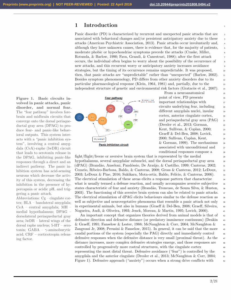
Figure 1. Basic circuits in-volved in panic attacks, panicdisorder, and normal fear.The “fear pathway” involves fore-brain and midbrain circuits thatconverge onto the dorsal periaque-ductal gray area (DPAG) to pro-duce fear- and panic-like behav-ioral outputs. This system inter-acts with a “panic inhibition sys-tem”, involving a central amyg-dala (CeA)-raphe (lwDR) circuitthat leads to serotonin release inthe DPAG, inhibiting panic-likeresponses through a direct and anindirect pathway. The panic in-hibition system has acid-sensingneurons which decrease the activ-ity of this system, decreasing theinhibition in the presence of hy-percapnia or acidic pH, and trig-gering a panic attack.Abbreviations: Cg – cingulate cor-tex; BLA – basolateral amygdala;CeA – central amygdala; MH –medial hypothalamus; DPAG –dorsolateral periaqueductal grayarea; lwDR – lateral wings of thedorsal raphe nucleus; 5-HT – sero-tonin; GABA – γ-aminoburyticacid; CRF – corticotropin releas-ing factor.
From a neuroanatomical 15
point of view, PD presents 16
important relationships with 17
circuits underlying fear, including 18
different amygdala nuclei, insular 19
cortex, anterior cingulate cortex, 20
and periaqueductal gray area (PAG) 21
(Dresler et al., 2013; Gorman, 22
Kent, Sullivan, & Coplan, 2000; 23
Graeff & Del-Ben, 2008; Lovick, 24
2000; Sullivan, Coplan, Kent, 25
& Gorman, 1999). The mechanisms 26
associated with unconditional and 27
conditional responses compose a 28
fight/flight/freeze or aversive brain system that is represented by the medial 29
hypothalamus, several amygdalar subnuclei, and the dorsal periaqueductal gray area 30
(dPAG) (Brandao, Anseloni, Pandossio, De Araujo, & Castilho, 1999; Canteras, 2002; 31
Cezario, Ribeiro-Barbosa, Baldo, & Canteras, 2008; Gross & Canteras, 2012; LeDoux, 32
2003; LeDoux & Pine, 2016; Sukikara, Mota-ortiz, Baldo, Felıcio, & Canteras, 2006). 33
The electrical stimulation of these areas elicits a response pattern that characterize 34
what is usually termed a defense reaction, and usually accompanies aversive subjective 35
states characteristic of fear and anxiety (Brandao, Troncoso, de Souza Silva, & Huston, 36
2003). The functioning of this aversive brain system can also be related to panic attacks. 37
The electrical stimulation of dPAG elicits behaviours similar to the defense reaction as 38
well as subjective and neurovegetative phenomena that resemble a panic attack not only 39
in experimental animals, but also in humans (Graeff & Del-Ben, 2008; Graeff, Silveira, 40
Nogueira, Audi, & Oliveira, 1993; Jenck, Moreau, & Martin, 1995; Lovick, 2000). 41
An important concept that organizes theories derived from animal models is that of 42
defensive direction and defensive distance (or predatory imminence continuum) (Deakin 43
& Graeff, 1991; Fanselow & Lester, 1988; McNaughton & Corr, 2004; McNaughton & 44
Zangrossi Jr, 2008; Perusini & Fanselow, 2015). In general, it can be said that the more 45
caudal portions of the system (especially the PAG) directly and immediately control 46
defensive responses when the defensive distance is very small (proximal threat). As the 47
distance increases, more complex defensive strategies emerge, and those responses are 48
controlled by progressively more rostral structures, with the cingulate cortex 49
representing the most distal threat. Defensive avoidance (“fear”) is controlled by the 50
amygdala and the anterior cingulate (Dresler et al., 2013; McNaughton & Corr, 2004; 51
Figure 1). Defensive approach (“anxiety”) occurs when a strong drive conflicts with 52
2/21
Preprints (www.preprints.org) | NOT PEER-REVIEWED | Posted: 22 April 2019 doi:10.20944/preprints201808.0494.v2

avoidance, triggering risk assessment behavior; these patterns are controlled by 53
septo-hippocampal system and posterior cingulate (R. J. Blanchard & Blanchard, 1988; 54
Deakin & Graeff, 1991; Gray & McNaughton, 2003; LeDoux, 1998). 55
2 Model validity and theories of panic disorder 56
The representational nature of animal models demands that its adequacy as an 57
hypothesis-generating device is satisfactory (Belzung & Lemoine, 2011; Maximino & van 58
der Staay, 2019; van der Staay, 2006; Willner, 1986, 1991). This is the issue of validity. 59
The act of modeling is the application of an independent variable – the simulation of a 60
disorder in non-human animals based on knowledge of the etiology and mechanisms of 61
the disease in humans – and the observation of a series of endpoints (dependent 62
variables)(Maximino & van der Staay, 2019; van der Staay, 2006; van der Staay & 63
Steckler, 2001). In the literature, sometimes tests are referred to as models, but most of 64
the behavioral tests that are used in the field represent endpoints that are not 65
model-specific. While screenings tests and biobehavioral assay need adequate validity at 66
the predictive and face levels, ”true” models rely on good construct validity (Maximino 67
& van der Staay, 2019). While many definitions of construct validity have been offered, 68
its translational relevance is at least dependent on whether the model has a sound 69
theoretical basis (Maximino & van der Staay, 2019; McNaughton & Zangrossi Jr, 2008; 70
van der Staay, 2006; van der Staay, Arndt, & Nordquist, 2009; van der Staay, Nordquist, 71
& Arndt, 2017). As a result, a model is as good as is the theory underlying the disease 72
it is targeting, and a true behavioral model is dependent on knowledge regarding 73
etiology and on the neurobehavioral mechanisms underlying the disorder. 74
Predictive validity is usually reduced to pharmacological isomorphism, the 75
congruence between clinical effects and drug effects in the animal tests. In the 76
pharmacotherapy of PD, drugs with clinical efficacy include triazolobenzodiazepines, 77
monoamine oxidase inhibitor, tricyclic antidepressants, and selective serotonin reuptake 78
inhibitor (SSRIs)(Mitte, 2005). The triazolobenzodiazepines belong to the group of 79
drugs called benzodiazepines that act by binding at the interface of the α and γ 80
subunits of the GABAA receptor. They are efficacious in the short-term treatment of 81
panic disorder, but have largely been replaced by SSRIs as the first-line 82
pharmacotherapy and are generally not regarded as appropriate for long-term therapy. 83
The SSRIs were developed in the early 1970s. All SSRIs possess relatively high affinity 84
for serotonin uptake sites. Imipramine is an antidepressant drug that can be used in the 85
treatment of panic disorder, and belongs to the group of tricyclic antidepressants that 86
act blocking both the serotonin transporter and the norepinephrine transporter. The 87
role of imipramine was initially evidenced during a psychopharmacological experiment 88
in which one group of patients referred for “tranquilizers”, as they were very “anxious” 89
and unable to respond to treatment with phenothiazines and sedatives, had the panic 90
attacks ceased by tricyclic antidepressant imipramine treatment (Klein, 1964). 91
Moclobemide is a antidepressant drug that act as a reversible inhibitor of 92
monoamine-oxidase-A, two comparative trials demonstrated moclobemide to be as 93
efficacious as fluoxetine in patients suffering from panic disorder (Mitte, 2005). 94
Pharmacologically isomorphic tests and models of PD, then, need to be sensitive to 95
treatment with SSRIs, MAOis, tricyclic antidepressants, and triazolobenzodiazepines. 96
Models of PD, as will be shown, are associated with construct validity on at least 97
two points: the first is the relationship between PD and theories of fear, and the second 98
to specific theories of disordered fear responses in PD. Neurobehavioral theories of PD 99
ans its relations to fear suggest that the disorder is due to disfunctions in circuits that 100
control normal fear (Coplan & Lydiard, 1998; Santos, D’Amico, & Dierssen, 2015). Fear 101
is thought to be organized at different levels of a circuit, organized in a rostrocaudal 102
3/21
Preprints (www.preprints.org) | NOT PEER-REVIEWED | Posted: 22 April 2019 doi:10.20944/preprints201808.0494.v2

pathway, that includes amygdaloid subnuclei, the medial hypothalamus, and portions of 103
the periaqueductal gray area (PAG)(Gross & Canteras, 2012), receives inhibitory 104
control from regions of the prefrontal cortex (Jovanovic & Norrholm, 2011), and receives 105
modulatory influence from monoamines such as serotonergic neurons from the raphe 106
(Abrams, Johnson, Hollis, & Lowry, 2004; Hale & Lowry, 2011). Fear is qualitatively 107
different from anxiety, as anxiety is oriented towards a potential threat, while fear is 108
oriented towards a real (distal or proximal) threat; as a result, behavioral manifestations 109
of fear tend towards escaping or avoiding the threat, while behavioral manifestations of 110
anxiety tend towards cautious investigation and risk assessment (Corr, 2011; Gray & 111
McNaughton, 2003; McNaughton & Corr, 2004; Perkins, Kemp, & Corr, 2007; Perusini 112
& Fanselow, 2015). Consistent with these theoretical propositions, generalized anxiety 113
disorder is distinct from, e.g., panic disorder in phenomenology (worry vs. sense of 114
imminent danger), neural bases, and neurochemistry – including responsiveness to 115
serotonergic drugs (Barlow, 2002; Barlow, Chorpita, & Turovsky, 1996; Klein, 1981). 116
Specific theories of PD have been proposed which include behavioral and neuronal 117
events (Barlow, 2002; Fava & Morton, 2009; Pilecki, Arentoft, & McKay, 2011; Roth, 118
Wilhelm, & Pettit, 2005). The suffocation false alarm theory, first championed by 119
Donald Klein (1993), proposes that spontaneous or unexpected panic attacks are 120
triggered by a malfunctioning “monitor” in the brain stem that detects conditions that 121
may lead to suffocation, including increased levels of carbon dioxide (CO2). Using 122
exploratory factor analysis, Meuret et al. (2006) showed that PD symptoms can be 123
classified in dimensions, one of which includes cardio-respiratory symptoms 124
(palpitations, shortness of breath, choking, chest pain, and numbness), somatic 125
symptoms (sweating, trembling, nausea, chills/hot flashes, and dizziness), and cognitive 126
symptoms (feelings of unreality, fear of going crazy, fear of losing control). Importantly, 127
hierarchical multiple regression suggested that cardio-respiratory symptoms are the 128
strongest predictors of panic severity, frequency of panic attacks, and agoraphobic 129
avoidance, while cognitive symptoms are predictors of worry due to panic. It has been 130
criticized because of its putative failure to identify abnormal physiological mechanisms 131
underlying dyspnea during panic attacks (Roth et al., 2005). However, there has been 132
considerable progress in that area, as physiological and psychobiological research 133
advances. The ventral respiratory group of the brainstem has been identified as a site 134
that detects CO2-associated pH changes (Mulkey et al., 2004; Nattie & Li, 1996; Paul, 135
Johnson, Shekhar & Lowry, 2014), but responses in more rostral regions, including 136
regions in the midbrain (tegmentum, parabrachial nucleus, and periaqueductal gray), 137
hypothalamus, as well as limbic and paralimbic areas (amygdala and periamygdalar 138
nuclei, cingulate, parahippocampal, and fusiform gyri, anterior insula) are reported in 139
neuroimaging studies with healthy humans (Brannan et al., 2001), suggesting a 140
distributed network involved in the affective representation and/or emotional regulation 141
of breathing. In addition to this ventral hindbrain-limbic circuit, the role of other 142
regions in pH-sensing has been suggested as well. A ”panic inhibition system” in the 143
lateral wings of the serotonergic dorsal raphe nucleus (that projects to the dorsal PAG) 144
has been described (Paul et al., 2014) that is activated by hypercapnic stimuli (Johnson, 145
Hollis, Moratalla, Lightman, & Lowry, 2005). Using dynamic T1ρ relaxation imaging, 146
which corresponds to an acidosis in the regions assessed, Magnotta et al. (2014) found a 147
greater activity-evoked T1ρ response in the visual cortex of PD patients. From a 148
molecular point of view, acidosis-related responses have been implicated as risk factors 149
for PD (Quagliato, Freire, & Nardi, 2018). The rs9915774 SNP of the ACCN1 gene (the 150
human homologue of acid-sensing ion channel 2) has been found to be associated with 151
PD in genome-wide association analysis (Gregersen et al., 2012). In a metanalysis of 152
four studies on the relationship between rs685012 SNP of the ACCN2 gene, which codes 153
the neuronal amiloride-sensitive cation channel 2, Quagliato et al. (2018) found that the 154
4/21
Preprints (www.preprints.org) | NOT PEER-REVIEWED | Posted: 22 April 2019 doi:10.20944/preprints201808.0494.v2

C allele increases the risk of PD, with a moderate effect. 155
Originally a modification of Klein’s model, Gorman’s neuroanatomical theory 156
(Dresler et al., 2013; Gorman et al., 2000; Gorman, Liebowitz, Fyer, & Stein, 1989) has 157
high heuristic value for the development of translationally relevant animal models, and 158
considerably good construct validity. The ”revised model” postulates that panic attacks 159
are analogous to conditioned fear responses, and are mediated by a fear network that is 160
centered in the amygdaloid nuclei and its connections to the hippocampus, medial 161
prefrontal cortex, hypothalamus, and periaqueductal gray area (Gorman et al., 2000). 162
Increasing overall serotonergic neurotransmission would normalize the ”abnormal 163
sensitivity” of these circuits in patients with PD by inhibiting noradrenergic neurons in 164
the locus coeruleus and reducing transmission to the PAG. Panic attacks can be 165
triggered either by misinterpretation of visceral afferents (including CO2-associated pH 166
changes) or by association of context with previously occurring panic attacks. 167
In parallel to this panic-eliciting circuit that is superimposed on the ”fear” circuit, a 168
”panic inhibition circuit” has been proposed (Paul et al., 2014). Normal functioning of 169
this circuit leads to a ”return to normal” when the threatening stimulus is no longer 170
present, initiating cautious exploration and anxiety-like responses that inform the 171
organism of the actual level of risk. Thus, hyperosmotic or hypercapnic stimuli activate 172
acid-sensing channels in the basolateral amygdala, which leads to the release of 173
corticotropin-releasing factor (CRF) in the lateral wings of the dorsal raphe nucleus and 174
in the ventrolateral PAG. The activation of CRF2 receptors in these regions elicits the 175
release of serotonin in the dorsal PAG, which inhibit the fight/flight/freeze response 176
system via a direct or an indirect pathway (Figure 1). Moreover, the serotonergic 177
neurons of the dorsal raphe are also acid-sensitive, and its activation decreases PAG 178
output (Johnson et al., 2005). In PD, this circuit is dysfunctional, and is no longer able 179
to counteract the action of the fear circuit (Paul et al., 2014). This model relies on the 180
Deakin/Graeff theory of the role of serotonin in anxiety and fear (Deakin & Graeff, 181
1991; Graeff, Guimaraes, de Andrade, & Deakin, 1996). 182
Figure 2. Evidence-basedcausal model of PD. Early lifeexperiences and anxious traitscombine to cause Anxiety Sen-sitivity, a trait that describesa vulnerability to aversive bod-ily sensations, including the aver-sive respiratory experiences thatlead to hypercapnia. Whenthe individual experiences hy-percapnia, an abnormal activa-tion of the fear circuitry leadsto catastrophic cognitions regard-ing threat and the imminence ofdeath, as well as to the activationof the hypothalamus-pituitary-adrenal (HPA) axis. These catas-trophic cognitions establish theoccasion for strategic avoidancestrategies that reduce the acti-vation of the fear circuitry, butperpetuate PD by inducing recur-rent worry or anticipatory anxiety,as well as agoraphobic avoidance.Based on Pilecki et al. (2011).
Pilecki et al. 183
(2011) proposed a ”causal model” 184
of PD that is empirically supported 185
by experimental evidence with 186
PD patients, and is translationally 187
relevant to animal models. In 188
that model (Figure 2), predisposing 189
factors for PD, including early 190
life adverse experiences (Battaglia 191
et al., 2009; Ogliari et al., 2010) and 192
anxious temperament, are causally 193
associated with susceptibility 194
to panicogenic factors – Anxiety 195
Sensitivity, the fear of bodily 196
sensations (especially associated 197
with CO2-associated pH changes), 198
and the occurrence of aversive 199
respiratory experiences that lead to hypercapnia. The model is a diathesis-stress theory 200
that attempts to explain why not all individuals panic when exposed to aversive 201
respiratory experiences. Under these conditions, a panic attack occurs, leading to 202
catastrophic cognitions regarding physical sensations as well as to the activation of the 203
hypothalamus-pituitary-adrenal axis and of the fear circuit. Panic attacks cease when 204
the individual successfully escapes or avoids the aversive situation, decreasing the 205
activation of the fear circuit, which is accompanied by a reduction in anxious/fearful 206
5/21
Preprints (www.preprints.org) | NOT PEER-REVIEWED | Posted: 22 April 2019 doi:10.20944/preprints201808.0494.v2

cognitions. Avoidance learning, however, can be an important factor in the development 207
of the other symptoms of PD, including agoraphobia and/or other avoidance strategies. 208
This model does not make direct reference to the neurocircuitry that is involved in 209
responses, but tries to explain the role of early life adverse experiences in the 210
development of PD; however, there is no reason to discard the circuitry that was 211
described above. 212
3 Endpoints for modeling panic disorder. 213
The development of animal models for panic disorder is needed to establish the 214
psychological and biological bases of this disorder, as well as to allow the development 215
of novel therapeutics. Differently from models for generalized anxiety and biobehavioral 216
tests for anxiety – that present a myriad of formats and exploit a diversity of different 217
behaviors (Belzung & Lemoine, 2011; Griebel & Holmes, 2013) –, few paradigms 218
explicitly model PD. Considering the behavioral difference between fear and anxiety, 219
one should expect that appropriate endpoints for PD models would preferentially 220
involve fear-like responses, and not anxiety. This distinction is based mainly on 221
reference to the theoretical constructs that support the models (McNaughton & 222
Zangrossi Jr, 2008): while tests for anxiety use situations that impose 223
approach/avoidance conflicts to animals – generating behavioral inhibition and risk 224
assessment responses –, endpoints that are relevant for PD are based on defensive 225
responses elicited by aversive stimuli or by the stimulation of specific brain regions 226
(Moreira et al., 2013). In rodents, five main strategies have be used as endpoints to 227
model panic attacks and/or PD: conditional fear responses (fear conditioning), escape in 228
the elevated T-maze, antipredator behavior in the mouse defense test battery, escape 229
and freezing responses elicited by the stimulation of the PAG or the medial 230
hypothalamus, and hypercapnia-induced responses (Battaglia & Ogliari, 2005; Moreira 231
et al., 2013). The use of these endpoints is useful in itself – as biobehavioral assays to 232
study the mechanisms of normal fear –, or as independent variables in a true model, in 233
which specific manipulations (early life stress, genetic engineering) create ”vulnerable 234
organisms” (Belzung & Lemoine, 2011) that show increased responses in these assays. 235
3.1 Fear conditioning. 236
Conditional fear responses have been widely used to establish fear-like responses and to 237
elucidate the neural bases of these responses (LeDoux, 1998; Schafe, Nader, Blair, & 238
LeDoux, 2001). In fear conditioning, animals learn to associate a previously neutral 239
stimulus (CS) with an aversive unconditioned stimulus (US, such as an electrical shock 240
(LeDoux, 2003; Pavlov, 1927). In cued fear conditioning, the CS is explicit and reliably 241
predicts the US, acting as a cue or signal; in contextual fear conditioning, animals 242
display a defensive reaction (usually freezing) when introduced to a context (e.g., 243
apparatus or chamber) in which they have been previously exposed to the US, and 244
therefore the context represents a complex CS (Davis, Walker, Miles, & Grillon, 2009). 245
Typically, rats and mice display a initial activity burst to the footshock followed by 246
freezing (LeDoux, 1998); reexposure to the CS in the absence of the US triggers memory 247
retrieval, which initiates reconsolidation and extinction processes. 248
While fear conditioning is adaptive, and although conditioning accounts of panic 249
disorder have been widely criticized, this process is an integral part of learning 250
explanations of PD that attempt to incorporate some complexities of contemporary 251
learning theory. Moreover, these learning accounts of PD provide a compelling 252
explanation for the persistence of PD, in spite of not always being able to explain the 253
etiology of these disorders. Bouton et al. (2001) have argued that panic attacks are 254
6/21
Preprints (www.preprints.org) | NOT PEER-REVIEWED | Posted: 22 April 2019 doi:10.20944/preprints201808.0494.v2

salient aversive USs during which feelings of anxiety and fear can be associated with 255
external and/or interoceptive cues that are present during the attacks; subsequent 256
exposure to these cues can trigger feelings of anticipatory anxiety, which in its turn can 257
exacerbate the next panic attack. 258
Enhanced fear conditioning has been associated with gene variants in rodent 259
orthologues of human PD risk genes. For example, Tmem132d, the human orthologue of 260
which has been associated with PD in genome-wide association studies (A Erhardt et al., 261
2012; Angelika Erhardt et al., 2011; Howe et al., 2015; Mors et al., 2012; Otowa et al., 262
2012; Otowa, Tanii, et al., 2009; Otowa, Yoshida, et al., 2009), has been recently found 263
to be associated in murine fear conditioning in a quantitative trait loci panel (Knoll, 264
Halladay, Holmes, & Levitt, 2016), and female mice with humanized COMT Val158Met 265
polymorphism show reduced contextual fear, increased cued fear, and reduced 266
extinction recall (Risbrough, Ji, Hauger, & Zhou, 2014). A role for serotonin (5-HT) has 267
also been suggested in conditional fear processes (Homberg, 2012): acute treatment 268
with selective 5-HT reuptake inhibitors (SSRIs) facilitates fear conditioning, reduces 269
contextual fear, and increases cued fear; 5-HT1A receptors inhibit the acquisition and 270
expression of contextual fear; 5-HT2A receptors facilitate the consolidation of cued and 271
contextual fear, while 5-HT2C receptors inhibit the retrieval of cued fear. Therefore, 272
5-HT appears to exert opposite effects on contextual and cued fear (Homberg, 2012). 273
While consistent with the idea that contextual fear is more related to anxiety than fear, 274
these results are actually opposed to what is observed in other models (Table 1). 275
Table 1. Effect of serotonergic drugs on putative models of fear and panic-like behavior.Abbreviations: ETM: Elevated T-mazed; MDTB: mouse defense test battery; PAG: periaqueductal gray area;SSRIs: selective serotonin reuptake inhibitors.
SSRIs (acute) 5-HT1AR 5HT2AR 5-HT2CR
Cued fear ↑ ? ↑ ↓Contextual fear ↓ ↓ ↑ ?ETM escape ↓ 0 0 0MDTB 0 0 0 0PAG stimulation ↓ ↓ (local injection) ↓ (local injection) 0 (injection)Zebrafish alarm reaction ↓ 0 ↓ ?
3.2 Elevated T-maze. 276
The elevated T-maze (ETM) is derived from the biobehavioral assay for anxiety that is 277
most widely used contemporaneously – the elevated plus-maze –, and is explicitly based 278
on the Deakin/Greaff hypothesis on the role of 5-HT on defensive behavior (Graeff, 279
Ferreira Netto, & Zangrossi Jr, 1998; Guimaraes, Carobrez, & Graeff, 2008; Zangrossi Jr 280
& Graeff, 2014). The ETM attempts to explicitly separate defensive behaviors that are 281
evoked by motivational conflict (i.e., behavioral inhibition and risk assessment) from 282
behaviors associated with fear states (i.e., escape). The apparatus consists in two open 283
arms and one closed arm in a perpendicular configuration, elevated 50 cm from the floor 284
(Viana, Tomaz, & Graeff, 1994). In an ETM session, the rat executes two tasks: 285
inhibitory avoidance and escape. First, the animal is transferred to the end of the closed 286
arm, and the latency to leave this arm is measured; after 30 s intervals, the task is 287
repeated twice, and increased latency to leave the closed arm is interpreted as the 288
acquisition of inhibitory avoidance (Graeff et al., 1998). After this inhibitory avoidance 289
task, the animal is positioned at the end of one of the open arms to measure the escape 290
response (Viana et al., 1994). These two responses – inhibitory avoidance and escape – 291
recruit different neuronal circuits (Silveira, Zangrossi Jr, Viana, Silveira, & Graeff, 2001) 292
7/21
Preprints (www.preprints.org) | NOT PEER-REVIEWED | Posted: 22 April 2019 doi:10.20944/preprints201808.0494.v2
and present opposite 5-HTergic modulation (Graeff, Viana, & Mora, 1996; Sena et al., 293
2003; Zangrossi Jr & Graeff, 2014; Zangrossi Jr et al., 2001; Table 1). Inhibitory 294
avoidance is interpreted as modeling anxiety-like responses, while escape is interpreted 295
as modeling panic attacks (Graeff et al., 1998). Moreover, the assay appears to present 296
good predictive validity for anxiolytic and panicolytic drugs (Graeff et al., 1998). An 297
important critique to the model, however, is its low face validity: the association of 298
escape from the open arm to panic attacks is tenuous, given that the escape observed in 299
the ETM is slow, directed, and controlled – very different from escape responses which 300
are observed in other models (Moreira et al., 2013). Moreover, while escape in the ETM 301
appears to work as a biobehavioral assay – that is, as a tool to investigate normal fear 302
responses, considering the involvement of structures which are associated with fear, as 303
well as the predictive validity of the method –, its construct validity for PD is low, as it 304
makes no explicit reference to etiology of to neurobehavioral mechanisms of panic (as 305
opposed to normal fear). 306
3.3 Mouse defense test battery. 307
The mouse defense test battery (MDTB) evaluates the behavior of mice exposed to a 308
natural predator, the rat, at different defensive distances (D. C. Blanchard, Griebel, & 309
Blanchard, 2001; Yang et al., 2004). The mouse is positioned in an oval runway, and an 310
anesthetized rat is manipulated by the experimenter so that its approaches the mouse at 311
a fixed velocity; when the rat approaches the mouse by about 1 m, the mouse usually 312
starts running, attempting to escape the aversive stimulus This assay explicitly assumes 313
defensive distance (or predatory imminence continuum) – the psychological distance 314
from the threat (R. J. Blanchard & D. C. Blanchard, 1988; Fanselow & Lester, 1988) – 315
as a central variable in decision making by the mouse. The model presents excellent 316
predictive validity, since acute treatment with triazolobenzodiazepines and chronic 317
treatment with imipramine, fluoxetine, and moclobemide decrease escape responses, 318
while acute treatment with imipramine and fluoxetine or with panicogenic agents 319
increase escape (D. C. Blanchard et al., 2001; R. J. Blanchard, Griebel, Henrie, & D. C. 320
Blanchard, 1997). Moreover, facilitating 5-HTergic neurotransmission in the 321
periaqueductal gray inhibits escape in the MDTB (Pobbe, Zangrossi Jr, Blanchard, & 322
Blanchard, 2011), pointing to a participation of the ”panic inhibition system” (Paul et 323
al., 2014) in the control of normal responses in the assay. The assay presents as 324
advantages the high ethological relevance, because it uses escape responses in an 325
ecologically valid context and avoids using artificial stimuli such as electric shocks; 326
moreover, the model allows the evaluation of two distinct responses – approach or 327
escape the stimulus – that are predictive of anxiolytic or panicolytic effects, respectively 328
(Moreira et al., 2013). Thus, as a biobehavioral assay, the MDTB presents good face 329
and predictive validity. 330
3.4 Stimulation of the dlPAG. 331
The electrical or chemical stimulation of the dorsal periaqueductal grey (dPAG) evokes 332
abrupt escape and/or freezing behavior and cardiovascular responses that is reminiscent 333
of a panic attack (Beckett & Marsden, 1995; Brandao, Zanoveli, Ruiz-Martinez, Oliveira, 334
& Landeira-Fernandez, 2008; Schenberg, 2010; Schenberg et al., 2014). Electrical 335
currents with different amplitude, or different concentrations of glutamatergic or 336
nitrergic drugs, are needed to induce freezing and escape (running and jumping) 337
responses (de Oliveira, Del Bel, & Guimaraes, 2001; Schenberg, Bittencourt, Sudre, & 338
Vargas, 2002; Vianna, Graeff, Brandao, & Landeira-Fernandez, 2001). Usually, lower 339
concentrations or currents elicit alertness and escape, while higher concentrations or 340
currents elicit a sequence that is characterized by alertness, freezing, and escape 341
8/21
Preprints (www.preprints.org) | NOT PEER-REVIEWED | Posted: 22 April 2019 doi:10.20944/preprints201808.0494.v2

behavior interspersed with periods of tense immobility (Brandao et al., 2005, 2003, 342
2008). Similarly, disinhibiting the medial hypothalamus – a diencephalic structure that 343
composes, along with the periaqueductal gray, the encephalic circuits which mediate 344
fear responses and panic attacks (Canteras, 2002) – with GABAergic antagonists 345
promote escape responses, hypertension, and tachycardia (DiMicco, Samuels, Zaretskaia, 346
& Zaretsky, 2002; Johnson, Lowry, Truitt, & Shekhar, 2008; Johnson, Truitt, Fitz, 347
Lowry, & Shekhar, 2008). Both methods are based on observations on the effects of 348
electrically stimulating these regions in humans, placing its construct validity in the 349
similarity of neuronal circuits (Moreira et al., 2013). These tests also appear to present 350
a good predictive validity, since panicolytic, but not anxiolytic drugs, alter behavior 351
after stimulation (Johnson, Lowry, et al., 2008; Schenberg et al., 2002). However, from 352
an etiological point of view, the induction of responses by direct stimulation of brain 353
regions has a tenuous relation to spontaneous panic attacks observed in humans, and no 354
references are made to how dysfunctions in these circuits lead to PD, or on the role of 355
predisposing factors such as anxiety sensitivity, reducing construct validity. 356
In addition to freezing that is observed during dPAG stimulation, post-stimulation 357
freezing is also observed (Brandao et al., 2008). Interrupting the electrical stimulation 358
of the dPAG at the escape threshold elicits this freezing behavior, that is accompanied 359
by activation of the laterodorsal nucleus of the thalamus, suggesting transfer of 360
information to more rostral structures (Brandao et al., 2008). This led Brandao et al. 361
(2008) to speculate that post-stimulation freezing is more related to risk assessment, as 362
the animal freezes in order to better accumulate information on threat levels (see also 363
Hagenaars, Oitzl, & Roelofs, 2014); as a result, while stimulation-elicited freezing should 364
be treated as a model for a panic attack, post-stimulation freezing would be a better 365
model for anticipatory anxiety in PD (Brandao et al., 2008). 366
3.5 Hypercapnia-induced behavioral and physiological 367
responses. 368
Hypersensitivity to CO2 shows considerable heuristic value for PD models (Battaglia, 369
Ogliari, D’Amato, & Kinkead, 2014), and is able to identify people at heightened risk 370
for PD before the onset of the disorder (Coryell, Pine, Fyer, & Klein, 2006). If these 371
responses are predictive of the “core” symptoms of PD (panic attacks and avoidance), 372
they can be understood as endophenotypes (Meuret et al., 2006). Using this 373
information, methods were developed in which hypercapnia-induced behavioral and 374
physiological responses are used in animals (Battaglia & Ogliari, 2005; Battaglia et al., 375
2014; Schenberg et al., 2014). For example, slow infusion of potassium cyanide (KCN), 376
an agent that induces cytotoxic hypoxia of the arterial chemoreceptor cells (Barros et 377
al., 2002), induces defensive responses in rats that are similar to that observed with 378
electrical or chemical stimulations of the dPAG (Schimitel et al., 2012), and 379
sub-threshold doses of KCN facilitate responses elicited by electrical stimulation of the 380
dPAG (Schimitel et al., 2012). Moreover, unilateral electrolytic lesions of the dPAG 381
inhibit this response, suggesting that this structure is necessary for KCN-elicited 382
responses. Unexpectedly, exposure to either 8% or 12% CO2 induced respiratory 383
alterations and producing signs of increased arousal in the open field (less exploratory 384
behavior and appearance of exophthalmus), but actually attenuated dPAG-evoked 385
responses, including freezing and flight (Schimitel et al., 2012). Since the KCN-evoked 386
defensive behaviors were markedly facilitated by the previous exposure to 8% or 12% 387
CO2, Schimitel et al. (2012) suggested the existence, in the PAG, of hypoxia-sensitive 388
neurons that, when activated, could both precipitate a spontaneous panic attack and 389
render the subject hyper-responsive to hypercapnia. c-Fos-like immunoreactivity 390
experiments suggested a role for the dorsolateral and ventrolateral PAG, as well as of 391
9/21
Preprints (www.preprints.org) | NOT PEER-REVIEWED | Posted: 22 April 2019 doi:10.20944/preprints201808.0494.v2

the lateral wings of the dorsal raphe nucleus (Berquin, Bodineau, Gros, & Larnicol, 392
2000; Casanova, Contreras, Moya, Torrealba, & Iturriaga, 2013; Hayward & Von 393
Reitzenstein, 2002; Johnson et al., 2011, 2005). 394
Interesting advances include modeling the differential sensitivity to hypoxia- or 395
hypercapnia-induced responses in animals. This can be done by using sub-threshold 396
concentrations; for example, rats injected with a sub-threshold concentration of lactate 397
infusions – a substance which in trigger panic attacks in PD patients (Bourin, Malinge, 398
& Guitton, 1995) – show no overt behavioral changes, but display tachycardia, 399
tachypnea, hypertension, and increased anxiety when GABA synthesis in the 400
dorsomedial hypothalamus is chronically inhibited (Johnson, Lowry, et al., 2008; 401
Johnson, Truitt, et al., 2008). These animals also show attenuated c-Fos-like responses 402
in the lateral wings of the dorsal raphe nucleus (lwDRN) and the VLPAG (Johnson, 403
Lowry, et al., 2008). Similarly, different rat strains show different sensitivities to CO2 404
inhalation (Winter, Ahlbrand, Naik, & Sah, 2017), and behavioral extremes are an 405
interesting source of variation to investigate vulnerabilities in disordered animals (van 406
der Staay, 2006). Interestingly, CO2-sensitive strains show more serotonergic neurons in 407
the lwDRN and the VLPAG and more adrenergic neurons in the locus coeruleus 408
(Winter et al., 2017). 409
3.6 Summary of behavioral endpoints in rodent tests. 410
All five approaches are heavily based on face and predictive validity and its relations to 411
theories on non-pathological fear, looking for coherence with theories on PD as a way to 412
elevate the translational potential of the model; as such, while the ETM is grounded on 413
the Deakin/Graeff theory, the MDTB is grounded mainly on the concept of defensive 414
distance. Moreover, to a greater or lesser degree, the five approaches are sustained by 415
an “etho-experimental” approach (R. J. Blanchard & D. C. Blanchard, 1988), based on 416
the careful observation of animal behavior as a way to discriminate between different 417
defensive strategies that model different aspects of anxiety, fear, and panic. The five 418
approaches present important advantages and disadvantages in modeling PD. Given the 419
complexity of PD, it is to be expected that the use of a single test will not mimic all its 420
aspects, considering the limitations related to the subjective aspects of the disorder 421
(LeDoux & Pine, 2016). Using different endpoints can mimic different aspects of the 422
disorder, allowing better elucidation of the behavioral and neuropathological 423
underpinnings of the disease. Ideally, various assays should be used to study the same 424
disorder, including the use of different species (de Mooij-van Malsen, Vinkers, Peterse, 425
Olivier, & Kas, 2011; Kalueff, Ren-Patterson, LaPorte, & Murphy, 2008; Kas, Gelegen, 426
Schalkwyk, & Collier, 2009; Keifer & Summers, 2016; van der Staay, 2006). 427
3.7 Complementary tests: The alarm response of zebrafish. 428
Aiming to enrich the breadth of etho-experimental approaches traditionally made with 429
rodents, the use of non-mammalian species has been proposed (Maximino et al., 2015). 430
Among these, zebrafish (Danio rerio Hamilton 1822), a cyprinid fish widely used in 431
genetics and developmental biology, has gained traction as a model organism in 432
behavioral research (Gerlai, 2010; Kalueff et al., 2012; Norton & Bally-Cuif, 2010; 433
Rinkwitz, Mourrain, & Becker, 2011; Shams, Rihel, Ortiz, & Gerlai, 2018). Importantly 434
for the study of fear and panic is the range of defensive responses that are observed in 435
this species, among which the alarm reaction is relatively well-characterized and 436
relevant for modeling PD (Maximino et al., 2018). This is a response that is initiated in 437
the olfactory system by substances that are released by the damaged skin of 438
conspecifics, and is characterized by dramatic and measurable changes in swimming 439
patterns, as well as in well-defined physiological responses. The ”alarm substance” 440
10/21
Preprints (www.preprints.org) | NOT PEER-REVIEWED | Posted: 22 April 2019 doi:10.20944/preprints201808.0494.v2

produced by specialized skin cells (”club cells”) and released after these cells are 441
damaged is of unknown composition (Døving & Lastein, 2009; Jesuthasan & Mathuru, 442
2008), but it presents hypoxanthine 3-N-oxide and chondroitin fragments – substances 443
which mimic part of the behavioral and physiological reactions (Mathuru et al., 2012; 444
Parra, Adrian Jr, & Gerlai, 2009). During exposure to this substance, an increase in 445
shoal cohesion (Canzian, Fontana, Quadros, & Rosemberg, 2017; Speedie & Gerlai, 446
2008) and the initiation of patterns of erratic swimming followed by freezing (Mathuru 447
et al., 2012; Quadros et al., 2016; Speedie & Gerlai, 2008) is observed. 448
Differently from the behavior that is observed during exposure, behavior observed 449
after exposure usually involves increased bottom dwelling associated with erratic 450
swimming and freezing (Cachat et al., 2011; Egan et al., 2009), analgesia (Maximino, 451
2011), and increased dark preference (anxiety-like behavior) associated with erratic 452
swimming, freezing, and thigmotaxis (Maximino, Lima, Costa, Guedes, & Herculano, 453
2014). Moreover, the alarm substance also produces intense autonomic responses, with 454
increased plasma levels of glucose, hemoglobin, norepinephrine, and epinephrine 455
(Maximino et al., 2014), and a neuroendocrine stress response, with increased 456
whole-body cortisol levels (Abreu, Giacomini, Koakoski, Piato, & Barcellos, 2017; 457
Schirmer, Jesuthasan, & Mathuru, 2013). This array of behavioral and physiological 458
adjustments simulate some important behavioral aspects and neurovegetative symptoms 459
of panic attacks, lending significant face validity to the model. Moreover, the 460
dissociation between responses produced during alarm substance exposure (erratic 461
swimming and freezing, without increased bottom dwelling) and after exposure 462
(increased bottom dwelling, erratic swimming, and freezing) is reminiscent of the 463
distinction between freezing responses during and after the electrical stimulation of the 464
dorsolateral column of the PAG in rats (Brandao et al., 2008), and could be further 465
exploited as dependent variables in models for panic attacks and PD, respectively. 466
4 Independent variables: The role of development 467
and Anxiety Sensitivity. 468
From the point of view of modeling theory, a true model (”simulation” sensu Wilner 469
(1991)) is the application of a manipulation to observe neurobehavioral effects, 470
grounded on the best theory on the etiology and mechanisms of the target disorder 471
(Maximino & van der Staay, 2019). Strictly speaking, then, there are very few true 472
models for PD. These include genetic models based on knockout techniques, as well as 473
diathesis-stress theory-based models. 474
A few genes have been shown, in knockout screens, to be involved in panic-like 475
responses. Overexpression of the Ntrk3 gene, which codes for the TrkC receptor in mice, 476
increases responsiveness in the MDTB, as well as the number of tyrosine 477
hydroxylase-positive neurons in the locus coeruleus and substantia nigra (Dierssen et al., 478
2006). Overexpression also increases responsiveness to lactate infusions in the MDTB 479
and in a predator avoidance test (Murtra et al., 2007). Conversely, knocking out the 480
Htr1a gene, which encodes the 5-HT1A receptor, increases responsiveness in a cued fear 481
conditioning paradigm (Klemenhagen, Gordon, David, Hen, & Gross, 2006). These 482
results underline the potential of knockout approaches, widely documented in the last 483
two decades, in unraveling the genetic vulnerabilities through animal models. 484
Early life adversities, including childhood parental loss and stressful life events 485
occurring in childhood and adolescence, are associated with adult CO2 sensitivity and 486
PD (Battaglia et al., 2009; Ogliari et al., 2010). Interestingly, Panksepp (1998) had 487
already proposed that the circuits involved in what he calls a ”panic” system, including 488
opioidergic projections to the PAG, is associated with separation distress. Two key 489
11/21
Preprints (www.preprints.org) | NOT PEER-REVIEWED | Posted: 22 April 2019 doi:10.20944/preprints201808.0494.v2

studies associate early interference with maternal care and CO2 hypersensitivity in 490
rodents. Genest et al. (2007) exposed rat pups (postnatal days 3-13) to 3 h/day 491
maternal separation and assessed hyperventilation during hypoxia and hypercapnia 492
protocols during adulthood, as well as baseline differences in 493
hypothalamus-pituitary-adrenal (HPA) axis activity. They showed that rats exposed to 494
neonatal maternal separation showed higher hyperventilation during both hypoxia and 495
hypercapnia and increased HPA axis activity. D’Amato et al. (2011) exposed mouse 496
pups (postnatal days 1-4) to repeated cross-fostering (changing caregiver every 24 h), 497
observing no hyperventilation to hypoxia, but hypercapnia responses both during 498
adolescence and adulthood, no effects on basal HPA axis activity, and higher avoidance 499
of CO2-enriched environments. A combination of early-life stressors, hypercapnia 500
responses, and the use of escape responses under hypercapnia (e.g., dPAG stimulation 501
thresholds, or responses in the elevated T-maze) could provide an interesting 502
diathesis-stress-based model for PD. 503
5 Conclusions. 504
The tests and animal models reviewed here present different advantages that can be 505
exploited to discover the neurobiological and behavioral mechanisms associated with 506
PD, as well as the specific disadvantages that suggest the use of complementary 507
approaches. These tests and models vary in terms of validity – for example, the face 508
validity of the elevated T-maze is lower than the other presented models, but its relation 509
to behavioral theories of fear is higher than the models involving electrical or chemical 510
stimulation. Finally, the introduction of new models – including models with 511
non-mammalian species, such as the use of alarm substance in Danio rerio – is an 512
approach with great potential to amplify the field of investigation, testing novel 513
psychobiological, behavioral, and developmental hypotheses on this disorder and 514
associated factors. The field of animal modeling can only profit from the inclusion of 515
these approaches in the roster of investigation strategies. 516
6 Acknowledgments 517
Part of the research reported was funded by Conselho Nacional de Desenvolvimento 518
Cientıfico e Tecnologico (CNPq/Brazil, grant no. 400726/2016-5). 519
7 References
Abrams, J. K., Johnson, P. L., Hollis, J. H., & Lowry, C. A. (2004). Anatomic andfunctional topography of the dorsal raphe nucleus. Annals of the New York Academy ofSciences, 1018, 46–57. https://doi.org/10.1196/annals.1296.005
Abreu, M. S., Giacomini, A. C. V. V., Koakoski, G., Piato, A. L. S., & Barcellos, L.J. G. (2017). Divergent effect of fluoxetine on the response to physical or chemicalstressors in zebrafish. PeerJ, 5, e3330. https://doi.org/10.7717/peerj.3330
American Psychiatric Association. (2013). Diagnostic and Statistical Manual ofMental Disorders - Fifth Edition - DSM-5. (5th ed.) Washington: American PsychiatricPublishing.
Barlow, D. H. (2002). Anxiety and its disorders. The nature and treatment ofanxiety and panic (2nd ed.). New York, NY: The Guilford Press.
Barlow, D. H., Chorpita, B. F., & Turovsky, J. (1996). Fear, panic, anxiety, anddisorders of emotion. Nebraska Symposia on Motivation, 43, 251–328.
12/21
Preprints (www.preprints.org) | NOT PEER-REVIEWED | Posted: 22 April 2019 doi:10.20944/preprints201808.0494.v2

Barros, R. C. H., Bonagamba, L. G. H., Okamoto-Canesin, R., de Oliveira, M.,Branco, L. G. S., & Machado, B. H. (2002). Cardiovascular responses to chemoreflexactivation with potassium cyanide or hypoxic hypoxia in awake rats. AutonomicNeuroscience, 97, 110–115. https://doi.org/10.1016/S1566-0702(02)00050-4
Battaglia, M., & Ogliari, A. (2005). Anxiety and panic: From human studies toanimal research and back. Neuroscience & Biobehavioral Reviews, 29, 169–179.https://doi.org/10.1016/j.neubiorev.2004.06.013
Battaglia, M., Ogliari, A., D’Amato, F., & Kinkead, R. (2014). Early-life risk factorsfor panic and separation anxiety disorder: Insights and outstanding questions arisingfrom human and animal studies of CO2 sensitivity. Neuroscience & BiobehavioralReviews, 46, 455–464. https://doi.org/10.1016/j.neubiorev.2014.04.005
Battaglia, M., Pesenti-Gritti, P., Medland, S. E., Ogliari, A., Tambs, K., & Spatola,C. A. M. (2009). A genetically informed study of the association between childhoodseparation anxiety, sensitivity to CO2, panic disorder, and the effect of childhoodparental loss. Archives of General Psychiatry, 66, 64.https://doi.org/10.1001/archgenpsychiatry.2008.513
Beckett, S., & Marsden, C. A. (1995). Computer analysis and quantification ofperiaqueductal grey-induced defence behaviour. Journal of Neuroscience Methods, 58,157–161.
Belzung, C., & Lemoine, M. (2011). Criteria of validity for animal models ofpsychiatric disorders: Focus on anxiety disorders and depression. Biology of Mood &Anxiety Disorders, 1, 9. https://doi.org/10.1186/2045-5380-1-9
Berquin, P., Bodineau, L., Gros, F., & Larnicol, N. (2000). Brainstem andhypothalamic areas involved in respiratory chemoreflexes: A Fos study in adult rats.Brain Research, 857, 30–40. https://doi.org/10.1016/S0006-8993(99)02304-5
Blanchard, D. C., Griebel, G., & Blanchard, R. J. (2001). Mouse defensivebehaviors: Pharmacological and behavioral assays for anxiety and panic. Neuroscience& Biobehavioral Reviews, 25, 205–218.
Blanchard, R. J., & Blanchard, D. C. (1988). Ethoexperimental approaches to thebiology of emotion. Annual Review of Psychology, 39, 43–68.
Blanchard, R. J., Griebel, G., Henrie, J. A., & Blanchard, D. C. (1997).Differentiation of anxiolytic and panicolytic drugs by effects on rat and mouse defensetest batteries. Neuroscience & Biobehavioral Reviews, 21, 783–789.
Bourin, M., Malinge, M., & Guitton, B. (1995). Provocative agents in panic disorder.Therapie, 5, 301–306.
Bouton, M. E., Mineka, S., & Barlow, D. H. (2001). A modern learning theory onthe etiology of panic disorder. Psychological Review, 198, 4–32.
Brandao, M. L., Anseloni, V. Z., Pandossio, J. E., De Araujo, J. E., & Castilho, V.M. (1999). Neurochemical mechanisms of the defensive behavior in the dorsal midbrain.Neuroscience & Biobehavioral Reviews, 23, 863–875.
Brandao, M. L., Borelli, K. G., Nobre, M. J., Santos, J. M., Albrechet-Souza, L.,Oliveira, A. R., & Martinez, R. C. (2005). Gabaergic regulation of the neuralorganization of fear in the midbrain tectum. Neuroscience & Biobehavioral Reviews, 29,1299–1311. https://doi.org/10.1016/j.neubiorev.2005.04.013
Brandao, M. L., Troncoso, A. C., de Souza Silva, M. A., & Huston, J. P. (2003). Therelevance of neuronal substrates of defense in the midbrain tectum to anxiety and stress:empirical and conceptual considerations. European Journal of Pharmacology, 463,225–233. https://doi.org/10.1016/S0014-2999(03)01284-6
Brandao, M. L., Zanoveli, J. M., Ruiz-Martinez, R. C., Oliveira, L. C., &Landeira-Fernandez, J. (2008). Different patterns of freezing behavior organized in theperiaqueductal gray of rats: Association with different types of anxiety. BehaviouralBrain Research, 188, 1–13. https://doi.org/10.1016/j.bbr.2007.10.018
13/21
Preprints (www.preprints.org) | NOT PEER-REVIEWED | Posted: 22 April 2019 doi:10.20944/preprints201808.0494.v2

Brannan, S., Liotti, M., Egan, G., Shade, R., Madden, L., Robillard, R., . . . Fox, P.T. (2001). Neuroimaging of cerebral activations and deactivations associated withhypercapnia and hunger for air. Proceedings of the National Academy of Sciences, 98,2029–2034. https://doi.org/10.1073/pnas.98.4.2029
Cachat, J., Stewart, A., Utterback, E., Hart, P., Gaikwad, S., Wong, K., . . . Kalueff,A. V. (2011). Three-dimensional neurophenotyping of adult zebrafish behavior. PLoSONE, 6, e17597. https://doi.org/10.1371/journal.pone.0017597
Canteras, N. S. (2002). The medial hypothalamic defensive system: Hodologicalorganization and functional implications. Pharmacology, Biochemistry & Behavior, 71,481–491.
Canzian, J., Fontana, B. D., Quadros, V. A., & Rosemberg, D. B. (2017).Conspecific alarm substance differently alters group behavior of zebrafish populations:Putative involvement of cholinergic and purinergic signaling in anxiety- and fear-likeresponses. Behavioural Brain Research, 320, 255–263.https://doi.org/10.1016/j.bbr.2016.12.018
Casanova, J. P., Contreras, M., Moya, E. A., Torrealba, F., & Iturriaga, R. (2013).Effect of insular cortex inactivation on autonomic and behavioral responses to acutehypoxia in conscious rats. Behavioural Brain Research, 253, 60–67.https://doi.org/10.1016/j.bbr.2013.07.015
Cezario, A. F., Ribeiro-Barbosa, E. R., Baldo, M. V. C., & Canteras, N. S. (2008).Hypothalamic sites responding to predator threats - The role of the dorsalpremammillary nucleus in unconditioned and conditioned antipredatory defensivebehavior. European Journal of Neuroscience, 28, 1003–1015.https://doi.org/10.1111/j.1460-9568.2008.06392.x
Coplan, J. D., & Lydiard, R. B. (1998). Brain circuits in panic disorder. BiologicalPsychiatry, 44, 1264–1276. https://doi.org/10.1016/S0006-3223(98)00300-X
Corr, P. J. (2011). Anxiety: Splitting the phenomenological atom. Personality andIndividual Differences, 50, 889–897. https://doi.org/10.1016/j.paid.2010.09.013
Coryell, W., Pine, D., Fyer, A., & Klein, D. (2006). Anxiety responses to CO2
inhalation in subjects at high-risk for panic disorder. Journal of Affective Disorders, 92,63–70. https://doi.org/10.1016/j.jad.2005.12.045
Craske, M. G., Miller, P. P., Rotunda, R., & Barlow, D. H. (1990). A descriptivereport of features of initial unexpected panic attacks in minimal and extensive avoiders.Behaviour Research and Therapy, 28, 395–400.https://doi.org/10.1016/0005-7967(90)90158-F
D’Amato, F. R., Zanettini, C., Lampis, V., Coccurello, R., Pascucci, T., Ventura, R.,. . . Battaglia, M. (2011). Unstable maternal environment, separation anxiety, andheightened CO2 sensitivity induced by gene-by-environment interplay. PLoS ONE, 6,e18637. https://doi.org/10.1371/journal.pone.0018637
Davis, M., Walker, D. L., Miles, L., & Grillon, C. (2009). Phasic vs sustained fear inrats and humans: Role of the extended amygdala in fear vs anxiety.Neuropsychopharmacology, 35, 105–135. https://doi.org/10.1038/npp.2009.109
de Mooij-van Malsen, A. J. G., Vinkers, C. H., Peterse, D. P., Olivier, B., & Kas, M.J. H. (2011). Cross-species behavioural genetics: A starting point for unravelling theneurobiology of human psychiatric disorders. Progress in Neuro-Psychopharmacology &Biological Psychiatry, 35, 1383–1390.https://doi.org/10.1016/j.pnpbp.2010.10.003
de Oliveira, R. M. W., Del Bel, E. A., & Guimaraes, F. S. (2001). Effects ofexcitatory amino acids and nitric oxide on flight behavior elicited from the dorsolateralperiaqueductal gray. Neuroscience & Biobehavioral Reviews, 25, 679–685.https://doi.org/10.1016/S0149-7634(01)00050-1
Deakin, J. F. W., & Graeff, F. G. (1991). 5-HT and mechanisms of defense. Journal
14/21
Preprints (www.preprints.org) | NOT PEER-REVIEWED | Posted: 22 April 2019 doi:10.20944/preprints201808.0494.v2

of Psychopharmacology, 5, 305–315.Dierssen, M., Gratacos, M., Sahun, I., Martın, M., Gallego, X., Amador-Arjona, A.,
. . . Estivill, X. (2006). Transgenic mice overexpressing the full-length neurotrophinreceptor TrkC exhibit increased catecholaminergic neuron density in specific brain areasand increased anxiety-like behavior and panic reaction. Neurobiology of Disease, 24,403–418. https://doi.org/10.1016/j.nbd.2006.07.015
DiMicco, J. A., Samuels, B. C., Zaretskaia, M. V, & Zaretsky, D. V. (2002). Thedorsomedial hypothalamus and the response to stress. Part renaissance, part revolution.Pharmacology, Biochemistry & Behavior, 71, 469–480.
Døving, K. B., & Lastein, S. (2009). The alarm reaction in fishes - Odorants,modulations of responses, neural pathways. Annals of the New York Academy ofSciences, 1170, 413–423. https://doi.org/10.1111/j.1749-6632.2009.04111.x
Dresler, T., Guhn, A., Tupak, S. V., Ehlis, A. C., Herrmann, M. J., Fallgatter, A. J.,. . . Domschke, K. (2013). Revise the revised? New dimensions of the neuroanatomicalhypothesis of panic disorder. Journal of Neural Transmission, 120, 3–29.https://doi.org/10.1007/s00702-012-0811-1
Egan, R. J., Bergner, C. L., Hart, P. C., Cachat, J. M., Canavello, P. R., Elegante,M. F., . . . Kalueff, A. V. (2009). Understanding behavioral and physiologicalphenotypes of stress and anxiety in zebrafish. Behavioural Brain Research, 205, 38–44.https://doi.org/10.1016/j.bbr.2009.06.022
Erhardt, A., Akula, N., Schumacher, J., Czamara, D., Karbalai, N., Muller-Myhsok,B., . . . Sasaki, T. (2012). Replication and meta-analysis of TMEM132D gene variantsin panic disorder. Translational Psychiatry, 2, e156.https://doi.org/10.1038/tp.2012.85
Erhardt, A., Czibere, L., Roeske, D., Lucae, S., Unschuld, P. G., Ripke, S., . . .Binder, E. B. (2011). TMEM132D: A new candidate for anxiety phenotypes - Evidencefrom human and mouse studies. Molecular Psychiatry, 16, 647–663.https://doi.org/10.1038/mp.2010.41
Fanselow, M. S., & Lester, L. (1988). A functional behavioristic approach toaversively motivated behavior: Predatory imminence as a determinant of thetopography of defensive behavior. In R. C. Bolles & M. D. Beecher (Eds.), Evolutionand Learning (pp. 185–211). Hillsdale: Erlbaum.https://www.researchgate.net/publication/232466904_A_functional_
behavioristic_approach_to_aversively_motivated_behavior_Predatory_
imminence_as_a_determinant_of_the_topography_of_defensive_behavior
Fava, G. A., Grandi, S., & Canestrari, R. (1988). Prodromal symptoms in panicdisorder with agoraphobia. American Journal of Psychiatry, 145, 1564–1567.
Fava, L., & Morton, J. (2009). Causal modeling of panic disorder theories. ClinicalPsychology Review, 29, 623–637. https://doi.org/10.1016/j.cpr.2009.08.002
Genest, S.-E., Gulemetova, R., Laforest, S., Drolet, G., & Kinkead, R. (2007).Neonatal maternal separation induces sex-specific augmentation of the hypercapnicventilatory response in awake rat. Journal of Applied Physiology, 102, 1416–1421.https://doi.org/10.1152/japplphysiol.00454.2006
Gerlai, R. (2010). High-throughput behavioral screens: The first step towardsfinding genes involved in vertebrate brain function using zebrafish. Molecules, 15,2609–2622. https://doi.org/10.3390/molecules15042609
Gorman, J. M., Kent, J. M., Sullivan, G. M., & Coplan, J. D. (2000).Neuroanatomical hypothesis of panic disorder, revised. American Journal of Psychiatry,157, 493–505. https://doi.org/10.1176/appi.ajp.157.4.493
Gorman, J. M., Liebowitz, M. R., Fyer, A. J., & Stein, J. (1989). A neuroanatomicalhypothesis for panic disorder. American Journal of Psychiatry, 146, 148–161.https://doi.org/10.1176/ajp.146.2.148
15/21
Preprints (www.preprints.org) | NOT PEER-REVIEWED | Posted: 22 April 2019 doi:10.20944/preprints201808.0494.v2

Graeff, F. G., & Del-Ben, C. M. (2008). Neurobiology of panic disorder: Fromanimal models to brain neuroimaging. Neuroscience & Biobehavioral Reviews, 32,1326–1335. https://doi.org/10.1016/j.neubiorev.2008.05.017
Graeff, F. G., Ferreira Netto, C., & Zangrossi Jr, H. (1998). The elevated T-maze asan experimental model of anxiety. Neuroscience & Biobehavioral Reviews, 23, 237–246.https://doi.org/10.1016/S0149-7634(98)00024-4
Graeff, F. G., Guimaraes, F. S., de Andrade, T. G. C. S., & Deakin, J. F. W. (1996).Role of 5-HT in stress, anxiety, and depression. Pharmacology, Biochemistry &Behavior, 54, 129–141.
Graeff, F. G., Silveira, M. C., Nogueira, R. L., Audi, E. A., & Oliveira, R. M. (1993).Role of the amygdala and periaqueductal gray in anxiety and panic. Behavioural BrainResearch, 58, 123–131.
Graeff, F. G., Viana, M. de B., & Mora, P. O. (1996). Opposed regulation by dorsalraphe nucleus 5-HT pathways of two types of fear in the elevated T-maze.Pharmacology, Biochemistry & Behavior, 53, 171–177.
Gratacos, M., Sahun, I., Gallego, X., Amador-Arjona, a, Estivill, X., & Dierssen, M.(2007). Candidate genes for panic disorder: insight from human and mouse geneticstudies. Genes, Brain, and Behavior, 6, 2–23.https://doi.org/10.1111/j.1601-183X.2007.00318.x
Gray, J. A., & McNaughton, N. (2003). The neuropsychology of anxiety. An enquiryinto the functions of the septo-hippocampal system (2nd ed.). New York: OxfordUniversity Press.
Gregersen, N., Dahl, H. A., Nyegaard, M., Hedemand, A., Als, T. D., Wang, A. G.,. . . Mors, O. (2012). A genome-wide study of panic disorder suggests theamiloride-sensitive cation channel 1 as a candidate gene. European Journal of HumanGenetics, 20, 84–90. https://doi.org/10.1038/ejhg.2011.148
Griebel, G., & Holmes, A. (2013). 50 years of hurdles and hope in anxiolytic drugdiscovery. Nature Reviews Drug Discovery, 12, 667–687.https://doi.org/10.1038/nrd4075
Gross, C. T., & Canteras, N. S. (2012). The many paths to fear. Nature ReviewNeuroscience, 13, 651–658. https://doi.org/10.1038/nrn3301
Guimaraes, F. S., Carobrez, A. P., & Graeff, F. G. (2008). Modulation of anxietybehaviors by 5-HT-interacting drugs. In R. J. Blanchard, D. C. Blanchard, G. Griebel,& D. J. Nutt (Eds.), Handbook of Anxiety and Fear (pp. 241–268). Amsterdam:Elsevier B. V.
Hagenaars, M. A., Oitzl, M., & Roelofs, K. (2014). Updating freeze: Aligning animaland human research. Neuroscience & Biobehavioral Reviews, 47, 165–176.https://doi.org/10.1016/j.neubiorev.2014.07.021
Hale, M. W., & Lowry, C. A. (2011). Functional topography of midbrain and pontineserotonergic systems: Implications for synaptic regulation of serotonergic circuits.Psychopharmacology, 213, 243–264. https://doi.org/10.1007/s00213-010-2089-z
Hayward, L. F., & Von Reitzenstein, M. (2002). c-Fos expression in the midbrainperiaqueductal gray after chemoreceptor and baroreceptor activation. American Journalof Physiology-Heart and Circulatory Physiology, 283, H1975–H1984.https://doi.org/10.1152/ajpheart.00300.2002
Homberg, J. R. (2012). Serotonergic modulation of conditioned fear. Scientifica,2012, Article ID 821549. https://doi.org/10.6064/2012/821549
Howe, A., Buttenschøn, H., Bani-Fatemi, A., Maron, E., Otowa, T., Erhardt, A., . . .Mors, O. (2015). Candidate genes in panic disorder: Meta-analyses of 23 commonvariants in major anxiogenic pathways. Molecular Psychiatry, 21, 665–679.https://doi.org/10.1038/mp.2015.138
Jenck, F., Moreau, J.-L., & Martin, J. R. (1995). Dorsal periaqueductal
16/21
Preprints (www.preprints.org) | NOT PEER-REVIEWED | Posted: 22 April 2019 doi:10.20944/preprints201808.0494.v2

gray-induced aversion as a simulation of panic anxiety: Elements of face and predictivevalidity. Psychological Research, 57, 181–191.
Jesuthasan, S. J., & Mathuru, A. S. (2008). The alarm response in zebrafish: Innatefear in a vertebrate genetic model. Journal of Neurogenetics, 22, 211–229.https://doi.org/10.1080/01677060802298475
Johnson, P. L., Fitz, S. D., Hollis, J. H., Moratalla, R., Lightman, S. L., Shekhar, A.,& Lowry, C. A. (2011). Induction of c-Fos in ‘panic/defence’-related brain circuitsfollowing brief hypercarbic gas exposure. Journal of Psychopharmacology, 25, 26–36.https://doi.org/10.1177/0269881109353464
Johnson, P. L., Hollis, J. H., Moratalla, R., Lightman, S. L., & Lowry, C. A. (2005).Acute hypercarbic gas exposure reveals functionally distinct subpopulations ofserotonergic neurons in rats. Journal of Psychopharmacology, 19, 327–341.https://doi.org/10.1177/0269881105053281
Johnson, P. L., Lowry, C. A., Truitt, W., & Shekhar, A. (2008). Disruption ofGABAergic tone in the dorsomedial hypothalamus attenuates responses in a subset ofserotonergic neurons in the dorsal raphe nucleus following lactate-induced panic.Journal of Psychopharmacology, 22, 642–652.https://doi.org/10.1177/0269881107082900
Johnson, P. L., Truitt, W. A., Fitz, S. D., Lowry, C. A., & Shekhar, A. (2008).Neural pathways underlying lactate-induced panic. Neuropsychopharmacology, 33,2093–2107. https://doi.org/10.1038/sj.npp.1301621
Jovanovic, T., & Norrholm, S. D. (2011). Neural mechanisms of impaired fearinhibition in posttraumatic stress disorder. Frontiers in Behavioral Neuroscience, 5,Article 44. https://doi.org/10.3389/fnbeh.2011.00044
Kalueff, A. V, Ren-Patterson, R. F., LaPorte, J. L., & Murphy, D. L. (2008).Domain interplay concept in animal models of neuropsychiatric disorders: A newstrategy for high-throughput neurophenotyping research. Behavioural Brain Research,188, 243–249. https://doi.org/10.1016/j.bbr.2007.11.011
Kalueff, A. V, Stewart, A. M., Kyzar, E. J., Cachat, J., Gebhardt, M., Landsman, S.,. . . Zebrafish Neuroscience Research Consortium. (2012). Time to recognize zebrafish“affective” behavior. Behaviour, 149, 1019–1036.https://doi.org/10.1163/1568539X-00003030 Kas, M. J. H., Gelegen, C.,Schalkwyk, L. C., & Collier, D. A. (2009). Interspecies comparisons of functionalgenetic variations and their implications in neuropsychiatry. American Journal ofMedical Genetics, Part B: Neuropsychiatric Genetics, 150, 309–317.https://doi.org/10.1002/ajmg.b.30815
Keifer, J., & Summers, C. H. (2016). Putting the “biology” back into“neurobiology”: The strength of diversity in animal model systems for neuroscienceresearch. Frontiers in Systems Neuroscience, 10, Article 69.https://doi.org/10.3389/fnsys.2016.00069
Klein, D. F. (1964). Delineation of two drug-responsive anxiety syndromes.Psychopharmacologia, 5, 397–408.
Klein, D. F. (1981). Anxiety reconceptualized. In D. F. Klein & J. G. Rabkin (Eds.),Anxiety: New Research and Changing Concepts (pp. 235–263). New York: Raven Press.
Klein, D. F. (1993). False suffocation alarms, spontaneous panics, and relatedconditions. Archives of General Psychiatry, 50, 306.https://doi.org/10.1001/archpsyc.1993.01820160076009
Klemenhagen, K. C., Gordon, J. A., David, D. J., Hen, R., & Gross, C. T. (2006).Increased fear response to contextual cues in mice lacking the 5-HT1A receptor.Neuropsychopharmacology, 31, 101–111. https://doi.org/10.1038/sj.npp.1300774
Knoll, A. T., Halladay, L. R., Holmes, A., & Levitt, P. (2016). Quantitative traitloci and a novel genetic candidate for fear learning. Journal of Neuroscience, 36,
17/21
Preprints (www.preprints.org) | NOT PEER-REVIEWED | Posted: 22 April 2019 doi:10.20944/preprints201808.0494.v2

6258–6268. https://doi.org/10.1523/JNEUROSCI.0177-16.2016LeDoux, J. E. (1998). Fear and the brain: Where have we been, and where are we
going? Biological Psychiatry, 44, 1229–138.LeDoux, J. E. (2003). The emotional brain, fear, and the amygdala. Cellular and
Molecular Neurobiology, 23, 727–738.LeDoux, J. E., & Pine, D. S. (2016). Using neuroscience to help understand fear and
anxiety: A two-system framework. American Journal of Psychiatry, 173, 1083–1093.https://doi.org/10.1176/appi.ajp.2016.16030353
Lovick, T. A. (2000). Panic disorder—A malfunction of multiple transmitter controlsystems within the midbrain periaqueductal gray matter? Neuroscientist, 6, 48–59.
Magnotta, V. A., Johnson, C. P., Follmer, R., & Wemmie, J. A. (2014). FunctionalT1ρ imaging in panic disorder. Biological Psychiatry, 75, 884–891.https://doi.org/10.1016/j.biopsych.2013.09.008
Mathuru, A. S., Kibat, C., Cheong, W. F., Shui, G., Wenk, M. R., Friedrich, R. W.,& Jesuthasan, S. (2012). Chondroitin fragments are odorants that trigger fear behaviorin fish. Current Biology, 22, 538–544.https://doi.org/10.1016/j.cub.2012.01.061
Maximino, C. (2011). Modulation of nociceptive-like behavior in zebrafish (Daniorerio) by environmental stressors. Psychology & Neuroscience, 4, 149–155.https://doi.org/10.3922/j.psns.2011.1.017
Maximino, C., Lima, M. G., Costa, C. C., Guedes, I. M. L., & Herculano, A. M.(2014). Fluoxetine and WAY 100,635 dissociate increases in scototaxis and analgesiainduced by conspecific alarm substance in zebrafish (Danio rerio Hamilton 1822).Pharmacology, Biochemistry & Behavior, 124C, 425–433.https://doi.org/10.1016/j.pbb.2014.07.003
Maximino, C., Silva, R. X. do C., Campos, K. dos S., Oliveira, J. S. de, Rocha, S. P.,Pyterson, M. P., . . . Maximino, M. L. (2019). Sensory ecology of Ostariophysan alarmsubstances. Journal of Fish Biology. https://doi.org/10.1111/jfb.13844
Maximino, C., Silva, R. X. do C., da Silva, S. de N. S., Rodrigues, L. do S. D. S.,Barbosa, H., de Carvalho, T. S., . . . Herculano, A. M. (2015). Non-mammalian modelsin behavioral neuroscience: Consequences for biological psychiatry. Frontiers inBehavioral Neuroscience, 9, Article 233.https://doi.org/10.3389/fnbeh.2015.00233
Maximino, C., & van der Staay, F. J. (2019). Behavioral models in psychopathology:Epistemic and semantic considerations. Behavioral and Brain Functions, 15, Article 1.https://doi.org/10.1186/s12993-019-0152-4
McNaughton, N., & Corr, P. J. (2004). A two-dimensional neuropsychology ofdefense: Fear/anxiety and defensive distance. Neuroscience & Biobehavioral Reviews,28, 285–305. https://doi.org/10.1016/j.neubiorev.2004.03.005
McNaughton, N., & Zangrossi Jr, H. (2008). Theoretical approaches to the modelingof anxiety in animals. In R. J. Blanchard, D. C. Blanchard, G. Griebel, & D. J. Nutt(Eds.), Handbook of Anxiety and Fear (pp. 11–27). Amsterdam: Elsevier B. V.
Meuret, A. E., White, K. S., Ritz, T., Roth, W. T., Hofmann, S. G., & Brown, T. A.(2006). Panic attack symptom dimensions and their relationship to illnesscharacteristics in panic disorder. Journal of Psychiatric Research, 40, 520–527.https://doi.org/10.1016/j.jpsychires.2005.09.006
Mitte, K. (2005). A meta-analysis of the efficacy of psycho- and pharmacotherapy inpanic disorder with and without agoraphobia. Journal of Affective Disorders, 88, 27–45.https://doi.org/10.1016/j.jad.2005.05.003
Moreira, F. A., Gobira, P. H., Viana, T. G., Vicente, M. A., Zangrossi Jr, H., &Graeff, F. G. (2013). Modeling panic disorder in rodents. Cell & Tissue Research, 354,119-125. https://doi.org/10.1007/s00441-013-1610-1
18/21
Preprints (www.preprints.org) | NOT PEER-REVIEWED | Posted: 22 April 2019 doi:10.20944/preprints201808.0494.v2

Mulkey, D. K., Stornetta, R. L., Weston, M. C., Simmons, J. R., Parker, A., Bayliss,D. A., & Guyenet, P. G. (2004). Respiratory control by ventral surface chemoreceptorneurons in rats. Nature Neuroscience, 7, 1360–1369.https://doi.org/10.1038/nn1357
Nattie, E. E., & Li, A. (1996). Central chemoreception in the region of the ventralrespiratory group in the rat. Journal of Applied Physiology, 81, 1987–1995.https://doi.org/10.1152/jappl.1996.81.5.1987
Norton, W., & Bally-cuif, L. (2010). Adult zebrafish as a model organism forbehavioural genetics. BMC Neuroscience, 11, 90.https://doi.org/10.1186/1471-2202-11-90
Ogliari, A., Tambs, K., Harris, J. R., Scaini, S., Maffei, C., Reichborn-Kjennerud, T.,& Battaglia, M. (2010). The relationships between adverse events, early antecedents,and carbon dioxide reactivity as an intermediate phenotype of panic disorder.Psychotherapy and Psychosomatics, 79, 48–55. https://doi.org/10.1159/000259417
Otowa, T., Kawamura, Y., Nishida, N., Sugaya, N., Koike, A., Yoshida, E., . . .Sasaki, T. (2012). Meta-analysis of genome-wide association studies for panic disorderin the Japanese population. Translational Psychiatry, 2, e186.https://doi.org/10.1038/tp.2012.89
Otowa, T., Tanii, H., Sugaya, N., Yoshida, E., Inoue, K., Yasuda, S., . . . Sasaki, T.(2009). Replication of a genome-wide association study of panic disorder in a Japanesepopulation. Journal of Human Genetics, 55, 91–96.https://doi.org/10.1038/jhg.2009.127
Otowa, T., Yoshida, E., Sugaya, N., Yasuda, S., Nishimura, Y., Inoue, K., . . .Okazaki, Y. (2009). Genome-wide association study of panic disorder in the Japanesepopulation. Journal of Human Genetics, 54, 122–126.https://doi.org/10.1038/jhg.2008.17
Panksepp, J. (1998). Affective Neuroscience. The Foundations of Human andAnimal Emotions. New York: Oxford University Press.
Parra, K. V, Adrian Jr, J. C., & Gerlai, R. (2009). The synthetic substancehypoxanthine 3-N-oxide elicits alarm reactions in zebrafish (Danio rerio). BehaviouralBrain Research, 205, 336–341. https://doi.org/10.1016/j.bbr.2009.06.037
Paul, E. D., Johnson, P. L., Shekhar, A., & Lowry, C. A. (2014). The Deakin/Graeffhypothesis: Focus on serotonergic inhibition of panic. Neuroscience & BiobehavioralReviews, 46, 379–396. https://doi.org/10.1016/j.neubiorev.2014.03.010
Pavlov, I. P. (1927). Conditioned reflexes. London: Oxford University Press.Perkins, A. M., Kemp, S. E., & Corr, P. J. (2007). Fear and anxiety as separable
emotions: An investigation of the revised Reinforcement Sensitivity Theory ofpersonality. Emotion, 7, 252–261. https://doi.org/10.1037/1528-3542.7.2.252
Perusini, J. N., & Fanselow, M. S. (2015). Neurobehavioral perspectives on thedistinction between fear and anxiety. Learning & Memory, 22, 417–425.https://doi.org/10.1101/lm.039180.115
Pilecki, B., Arentoft, A., & McKay, D. (2011). An evidence-based causal model ofpanic disorder. Journal of Anxiety Disorders, 25, 381–388.https://doi.org/10.1016/j.janxdis.2010.10.013
Pobbe, R. L. H., Zangrossi Jr, H., Blanchard, D. C., & Blanchard, R. J. (2011).Involvement of dorsal raphe nucleus and dorsal periaqueductal gray 5-HT receptors inthe modulation of mouse defensive behaviors. European Neuropsychopharmacology, 21,306–315. https://doi.org/10.1016/j.euroneuro.2010.05.004
Quadros, V. A., Silveira, A., Giuliani, G. S., Didonet, F., Silveira, A. S., Nunes, M.E., . . . Rosemberg, D. B. (2016). Strain- and context-dependent behavioural responsesof acute alarm substance exposure in zebrafish. Behavioural Processes, 122, 1–11.https://doi.org/10.1016/j.beproc.2015.10.014
19/21
Preprints (www.preprints.org) | NOT PEER-REVIEWED | Posted: 22 April 2019 doi:10.20944/preprints201808.0494.v2

Quagliato, L. A., Freire, R. C., & Nardi, A. E. (2018). The role of acid-sensitive ionchannels in panic disorder: A systematic review of animal studies and meta-analysis ofhuman studies. Translational Psychiatry, 8, 185.https://doi.org/10.1038/s41398-018-0238-z
Rinkwitz, S., Mourrain, P., & Becker, T. S. (2011). Zebrafish: An integrative systemfor neurogenomics and neurosciences. Progress in Neurobiology, 93, 231–243.https://doi.org/10.1016/j.pneurobio.2010.11.003
Risbrough, V., Ji, B., Hauger, R., & Zhou, X. (2014). Generation andcharacterization of humanized mice carrying COMT158 Met/Val Alleles.Neuropsychopharmacology, 39, 1823–1832. https://doi.org/10.1038/npp.2014.29
Roth, W. T., Wilhelm, F. H., & Pettit, D. (2005). Are current theories of panicfalsifiable? Psychological Bulletin, 131, 171–192.https://doi.org/10.1037/0033-2909.131.2.171
Sahun, I., Gallego, X., Gratacos, M., Murtra, P., Trullas, R., Maldonado, R., . . .Dierssen, M. (2007). Differential responses to anxiogenic drugs in a mouse model ofpanic disorder as revealed by Fos immunocytochemistry in specific areas of the fearcircuitry. Amino Acids, 33, 677–688. https://doi.org/10.1007/s00726-006-0464-1
Santos, M., D’Amico, D., & Dierssen, M. (2015). From neural to genetic substratesof panic disorder: Insights from human and mouse studies. European Journal ofPharmacology, 759, 127–141. https://doi.org/10.1016/j.ejphar.2015.03.039
Schafe, G. E., Nader, K., Blair, H. T., & LeDoux, J. E. (2001). Memoryconsolidation of Pavlovian fear conditioning: A cellular and molecular perspective.Trends in Neurosciences, 24, 540–546.https://doi.org/10.1016/S0166-2236(00)01969-X
Schenberg, L. C. (2010). Towards a translational model of panic attack. Psychology& Neuroscience, 3 9–37. http://dx.doi.org/10.3922/j.psns.2010.1.003
Schenberg, L. C., Bittencourt, A. S., Sudre, E. C. M., & Vargas, L. C. (2002).Modeling panic attacks. Neuroscience & Biobehavioral Reviews, 25, 647–659.https://doi.org/10.1016/S0149-7634(01)00060-4
Schenberg, L. C., Schimitel, F. G., Armini, R. D. S., Bernabe, C. S., Rosa, C. A.,Tufik, S., . . . Quintino-dos-Santos, J. W. (2014). Translational approach to studyingpanic disorder in rats: Hits and misses. Neuroscience & Biobehavioral Reviews, 46,472–496. https://doi.org/10.1016/j.neubiorev.2014.10.002
Schimitel, F. G., Almeida, G. M., Pitol, D. N., Armini, R. de S., Tufik, S., &Schenberg, L. C. (2012). Evidence of a suffocation alarm system within theperiaqueductal gray matter of the rat. Neuroscience, 200, 59–73.https://doi.org/10.1016/j.neuroscience.2011.10.032
Schirmer, A., Jesuthasan, S., & Mathuru, A. S. (2013). Tactile stimulation reducesfear in fish. Frontiers in Behavioral Neuroscience, 7, Article 167.https://doi.org/10.3389/fnbeh.2013.00167
Sena, L. M., Bueno, C., Pobbe, R. L. H., de Andrade, T. G. C. S., Zangrossi Jr, H.,& Viana, M. de B. (2003). The dorsal raphe nucleus exerts opposed control ongeneralized anxiety and panic-related defensive responses in rats. Behavioural BrainResearch, 142, 125–133. https://doi.org/10.1016/S0166-4328(02)00399-6
Shams, S., Rihel, J., Ortiz, J. G., & Gerlai, R. T. (2018). The zebrafish as apromising tool for modeling human brain disorders: A review based upon an IBNSSymposium. Neuroscience & Biobehavioral Reviews, 75, 176–190.https://doi.org/10.1016/j.neubiorev.2017.09.002
Silveira, M. C. L., Zangrossi Jr, H., Viana, M. de B., Silveira, R., & Graeff, F. G.(2001). Differential expression of Fos protein in the rat brain induced by performance ofavoidance or escape in the elevated T-maze. Behavioural Brain Research, 126, 13–21.https://doi.org/10.1016/S0166-4328(01)00233-9
20/21
Preprints (www.preprints.org) | NOT PEER-REVIEWED | Posted: 22 April 2019 doi:10.20944/preprints201808.0494.v2

Speedie, N., & Gerlai, R. (2008). Alarm substance induced behavioral responses inzebrafish (Danio rerio). Behavioural Brain Research, 188, 168–177.https://doi.org/10.1016/j.bbr.2007.10.031
Sukikara, M. H., Mota-ortiz, S. R., Baldo, M. V. C., Felıcio, L. F., & Canteras, N. S.(2006). A role for the periaqueductal gray in switching adaptive behavioral responses.Journal of Neuroscience, 26, 2583–2589.https://doi.org/10.1523/JNEUROSCI.4279-05.2006
Sullivan, G. M., Coplan, J. D., Kent, J. M., & Gorman, J. M. (1999). Thenoradrenergic system in pathological anxiety: A focus on panic with relevance togeneralized anxiety and phobias. Biological Psychiatry, 46, 1205–128.
van der Staay, F. J. (2006). Animal models of behavioral dysfunctions: Basicconcepts and classifications, and an evaluation strategy. Brain Research Reviews, 52,131–159. https://doi.org/10.1016/j.brainresrev.2006.01.006
van der Staay, F. J., Arndt, S. S., & Nordquist, R. E. (2009). Evaluation of animalmodels of neurobehavioral disorders. Behavioral and Brain Functions, 5, 11.https://doi.org/10.1186/1744-9081-5-11
van der Staay, F. J., Nordquist, R. E., & Arndt, S. S. (2017). Large farm animalmodels of human neurobehavioral and psychiatric disorders: Methodological andpractical considerations. In P. M. Conn (Ed.), Animal Models for the Study of HumanDisease (2nd ed., pp. 71–100).https://doi.org/10.1016/B978-0-12-809468-6.00003-6
van der Staay, F. J., & Steckler, T. (2001). Behavioural phenotyping of mousemutants. Behavioural Brain Research, 125, 3–12.https://doi.org/10.1016/S0166-4328(01)00278-9
Viana, M. de B., Tomaz, C., & Graeff, F. G. (1994). The elevated T-maze: A newanimal model of anxiety and memory. Pharmacology, Biochemistry & Behavior, 49,549–554. https://doi.org/10.1016/0091-3057(94)90067-1
Vianna, D. M. L., Graeff, F. G., Brandao, M. L., & Landeira-Fernandez, J. (2001).Defensive freezing evoked by electrical stimulation of the periaqueductal gray:Comparison between dorsolateral and ventrolateral regions. Learning & Memory, 12,4109–4112.
Willner, P. (1986). Validation criteria for animal models of human mental disorders:Learned helplessness as a paradigm case. Progress in Neuropsychopharmacology andBiological Psychiatry, 10, 677–690.https://doi.org/10.1016/0278-5846(86)90051-5
Willner, P. (1991). Methods for assessing the validity of animal models of humanpsychopathology. In A. A. Boulton, G. B. Baker, & M. T. Martin-Iverson (Eds.),Animal Models in Psychiatry (pp. 1–23). Clifton, NJ: Humana Press.
Winter, A., Ahlbrand, R., Naik, D., & Sah, R. (2017). Differential behavioralsensitivity to carbon dioxide (CO2) inhalation in rats. Neuroscience, 346, 423–433.https://doi.org/10.1016/j.neuroscience.2017.01.003
Yang, M., Augustsson, H., Markham, C. M., Hubbard, D. T., Webster, D., Wall, P.M., . . . Blanchard, D. C. (2004). The rat exposure test: A model of mouse defensivebehaviors. Physiology & Behavior, 81, 465–473.https://doi.org/10.1016/j.physbeh.2004.02.010
Zangrossi Jr, H., & Graeff, F. G. (2014). Serotonin in anxiety and panic:Contributions of the elevated T-maze. Neuroscience & Biobehavioral Reviews, 46,397–406. https://doi.org/10.1016/j.neubiorev.2014.03.007
Zangrossi Jr, H., Viana, M. de B., Zanoveli, J., Bueno, C., Nogueira, R. L., & Graeff,F. G. (2001). Serotonergic regulation of inhibitory avoidance and one-way escape in therat elevated T-maze. Neuroscience & Biobehavioral Reviews, 25, 637–645.https://doi.org/10.1016/S0149-7634(01)00047-1
21/21
Preprints (www.preprints.org) | NOT PEER-REVIEWED | Posted: 22 April 2019 doi:10.20944/preprints201808.0494.v2


![[Panic Away] Getting a Grip On Your Panic Disorder](https://img.pdfslide.us/doc/110x75/5591889d1a28abbb4c8b46cd/panic-away-getting-a-grip-on-your-panic-disorder.jpg)