Embed Size (px)
Citation preview
Copyright © 2008 Pearson Education, Inc., publishing as Pearson Benjamin Cummings
PowerPoint® Lecture Presentations for
BiologyEighth Edition
Neil Campbell and Jane Reece
Lectures by Chris Romero, updated by Erin Barley with contributions from Joan Sharp
Chapter 47
Animal Development
Copyright © 2008 Pearson Education, Inc., publishing as Pearson Benjamin Cummings
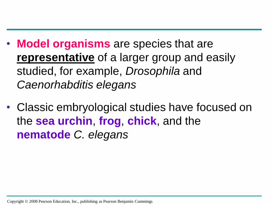
• Model organisms are species that are
representative of a larger group and easily
studied, for example, Drosophila and
Caenorhabditis elegans
• Classic embryological studies have focused on
the sea urchin, frog, chick, and the
nematode C. elegans
Copyright © 2008 Pearson Education, Inc., publishing as Pearson Benjamin Cummings
Concept 47.1: After fertilization, embryonic development proceeds through cleavage, gastrulation, and organogenesis
• Important events regulating development occur
during fertilization and the next three
stages that build the animal’s body
– Cleavage: cell division creates a hollow ball of
cells called a blastula
– Gastrulation: cells are rearranged into a three-
layered gastrula
– Organogenesis: the three layers interact and
move to give rise to organs
Copyright © 2008 Pearson Education, Inc., publishing as Pearson Benjamin Cummings
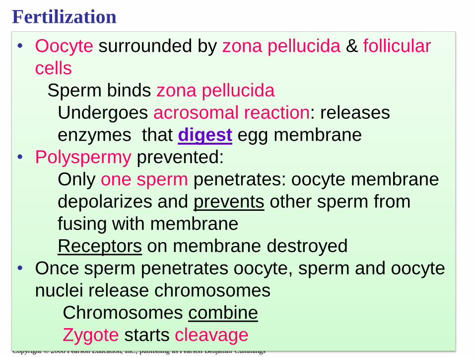
Fertilization
• Oocyte surrounded by zona pellucida & follicular
cells
Sperm binds zona pellucida
Undergoes acrosomal reaction: releases
enzymes that digest egg membrane
• Polyspermy prevented:
Only one sperm penetrates: oocyte membrane
depolarizes and prevents other sperm from
fusing with membrane
Receptors on membrane destroyed
• Once sperm penetrates oocyte, sperm and oocyte
nuclei release chromosomes
Chromosomes combine
Zygote starts cleavage
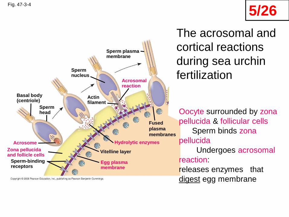
Fig. 47-3-4
Basal body(centriole)
Spermhead
Sperm-bindingreceptors
Acrosome
Zona pellucidaand follicle cells
Vitelline layer
Egg plasmamembrane
Hydrolytic enzymes
Acrosomalreaction
Actinfilament
Spermnucleus
Sperm plasmamembrane
Fused
plasma
membranes
Oocyte surrounded by zona
pellucida & follicular cells
Sperm binds zona
pellucida
Undergoes acrosomal
reaction:
releases enzymes that
digest egg membrane
The acrosomal and
cortical reactions
during sea urchin
fertilization
5/26
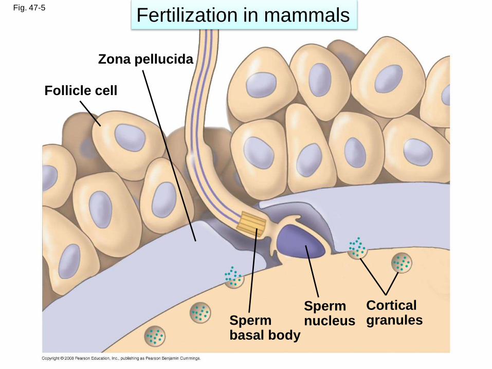
Fig. 47-5
Follicle cell
Zona pellucida
Cortical granules
SpermnucleusSperm
basal body
Fertilization in mammals
Copyright © 2008 Pearson Education, Inc., publishing as Pearson Benjamin Cummings
Placentation
• Placentation: formation of placenta from embryo
and uterus
Trophoblasts form chorion: site of nutrient and
gas exchange
• Extraembryonic membranes
Amnion: membranous sac containing embryo
Yolk sac:
Forms part of gut
Produces blood cells and vessels
Allantois: forms umbilical cord and bladder
Chorion
Copyright © 2008 Pearson Education, Inc., publishing as Pearson Benjamin Cummings
Cleavage
• Fertilization is followed by cleavage, a period
of rapid cell division without growth
• Cleavage partitions the cytoplasm of one large
cell into many smaller cells called blastomeres
• The blastula is a ball of cells with a fluid-filled
cavity called a blastocoel
• The inner cell mass forms embryonic disc, or
blastodisc
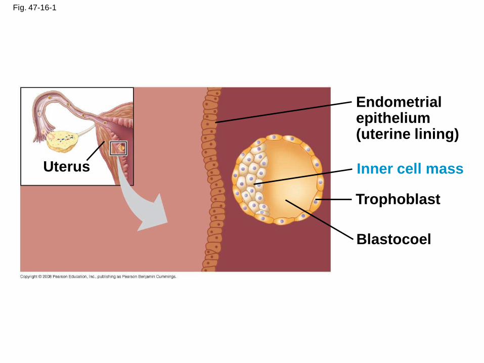
Fig. 47-16-1
Blastocoel
Trophoblast
Uterus
Endometrialepithelium(uterine lining)
Inner cell mass
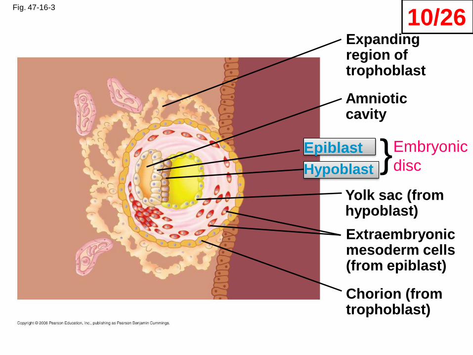
Fig. 47-16-3
Yolk sac (fromhypoblast)
Hypoblast
Expandingregion oftrophoblast
Amnioticcavity
Epiblast
Extraembryonicmesoderm cells(from epiblast)
Chorion (fromtrophoblast)
}Embryonic
disc
10/26
Copyright © 2008 Pearson Education, Inc., publishing as Pearson Benjamin Cummings
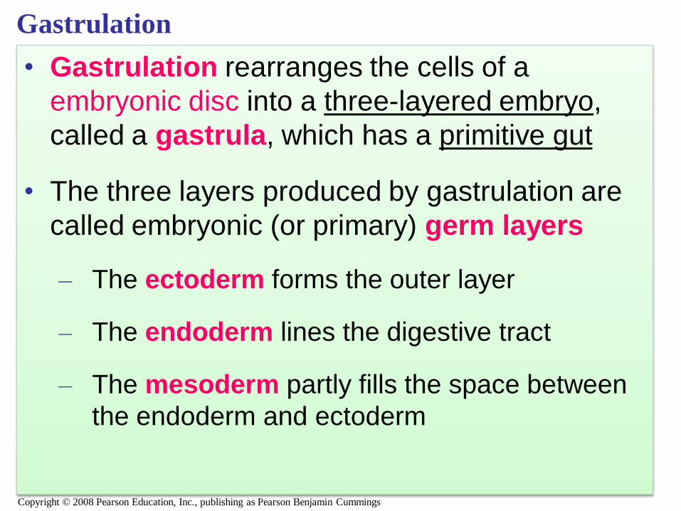
Gastrulation
• Gastrulation rearranges the cells of a
embryonic disc into a three-layered embryo,
called a gastrula, which has a primitive gut
• The three layers produced by gastrulation are
called embryonic (or primary) germ layers
– The ectoderm forms the outer layer
– The endoderm lines the digestive tract
– The mesoderm partly fills the space between
the endoderm and ectoderm
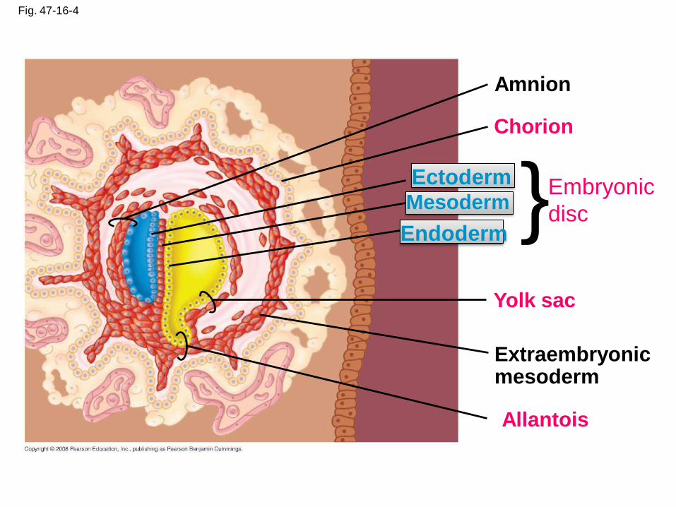
Fig. 47-16-4
Yolk sac
Mesoderm
Amnion
Chorion
Ectoderm
Extraembryonicmesoderm
Allantois
Endoderm}Embryonic
disc
Copyright © 2008 Pearson Education, Inc., publishing as Pearson Benjamin Cummings
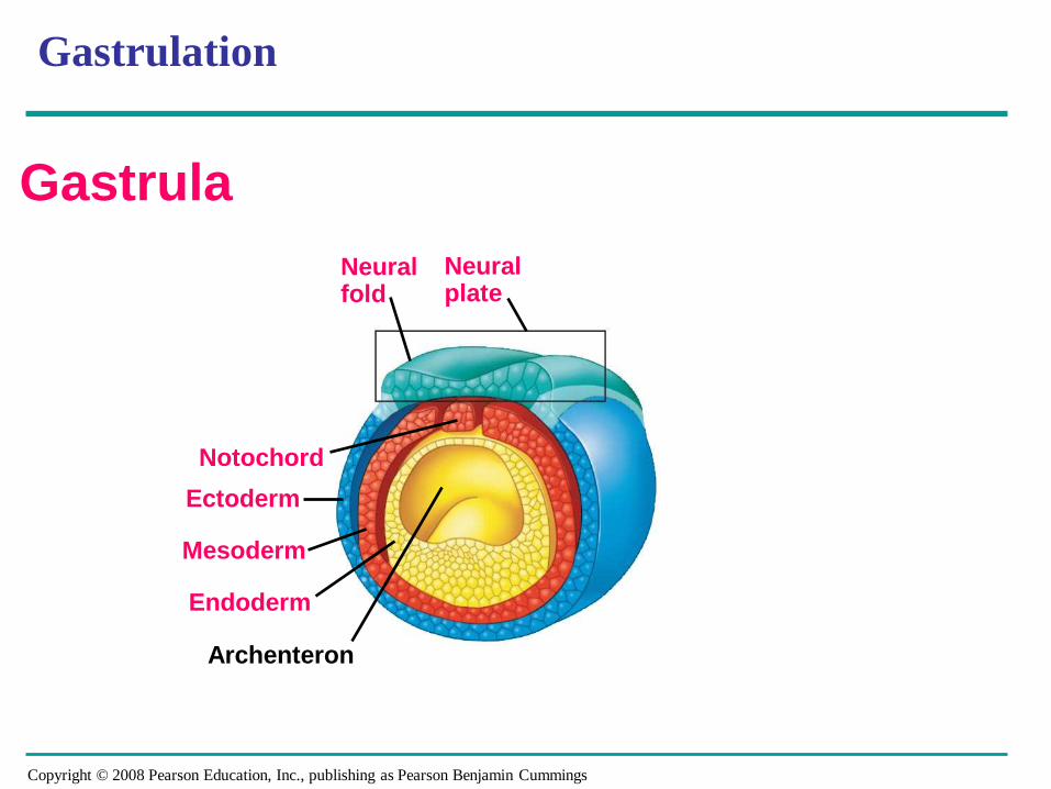
Gastrulation
Neuralfold
Neural plate
Mesoderm
Notochord
Archenteron
Ectoderm
Endoderm
Gastrula
Copyright © 2008 Pearson Education, Inc., publishing as Pearson Benjamin Cummings
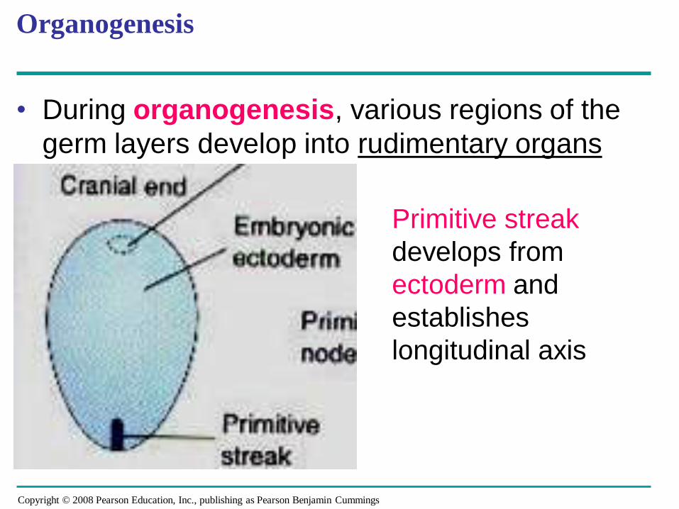
Organogenesis
• During organogenesis, various regions of the
germ layers develop into rudimentary organs
Primitive streak
develops from
ectoderm and
establishes
longitudinal axis
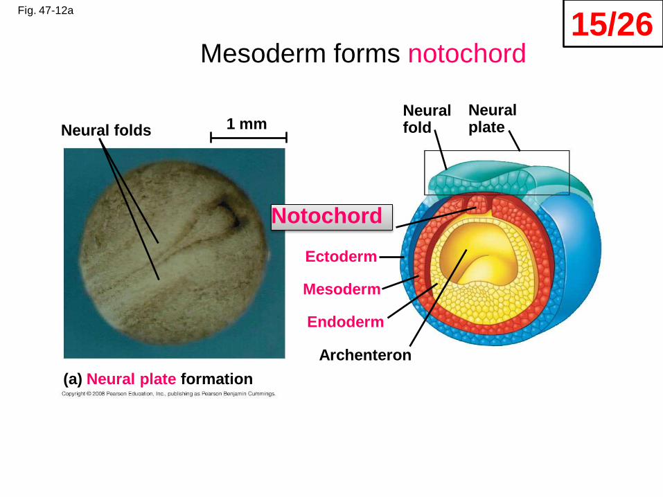
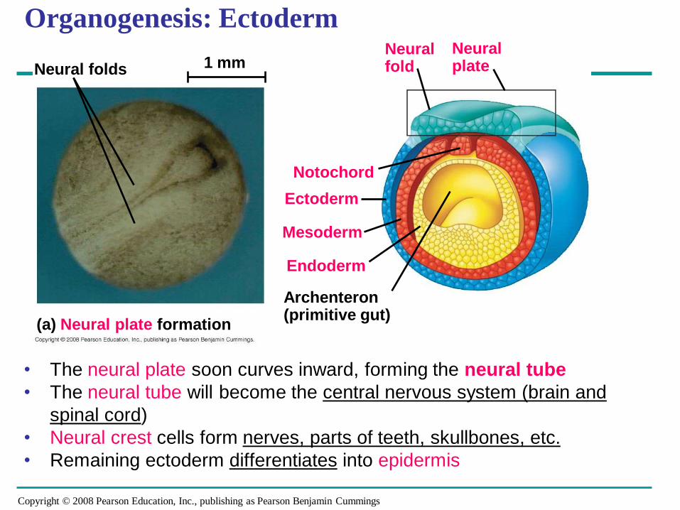
Fig. 47-12a
Neural folds
Neuralfold
Neural plate
Mesoderm
Notochord
Archenteron
Ectoderm
Endoderm
(a) Neural plate formation
1 mm
Mesoderm forms notochord15/26
Copyright © 2008 Pearson Education, Inc., publishing as Pearson Benjamin Cummings
• The neural plate soon curves inward, forming the neural tube
• The neural tube will become the central nervous system (brain and
spinal cord)
• Neural crest cells form nerves, parts of teeth, skullbones, etc.
• Remaining ectoderm differentiates into epidermis
Organogenesis: Ectoderm
Neural folds
Neuralfold
Neural plate
Mesoderm
Notochord
Archenteron(primitive gut)
Ectoderm
Endoderm
(a) Neural plate formation
1 mm
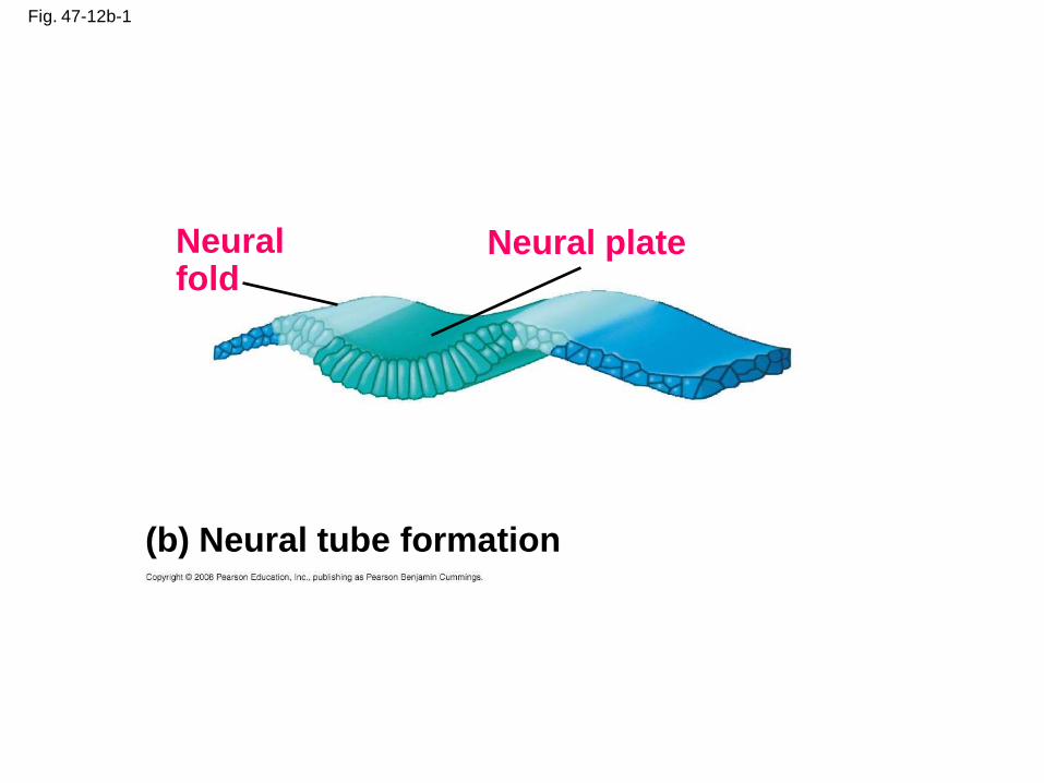
Fig. 47-12b-1
(b) Neural tube formation
Neuralfold
Neural plate
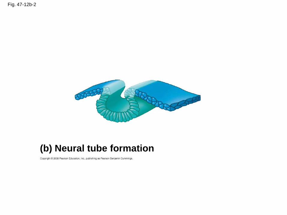
Fig. 47-12b-2
(b) Neural tube formation
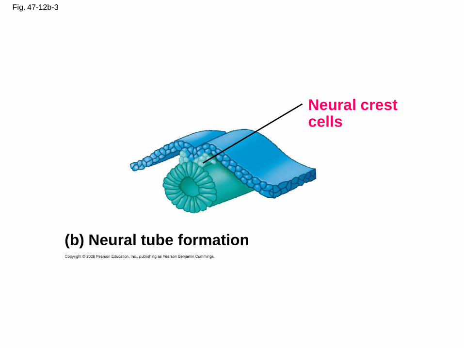
Fig. 47-12b-3
Neural crestcells
(b) Neural tube formation
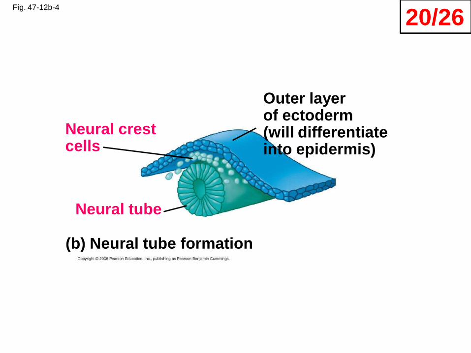
Fig. 47-12b-4
Neural tube
Neural crestcells
Outer layerof ectoderm (will differentiate into epidermis)
(b) Neural tube formation
20/26
Copyright © 2008 Pearson Education, Inc., publishing as Pearson Benjamin Cummings
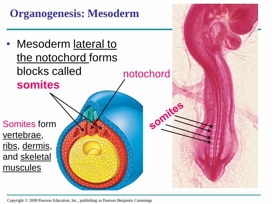
• Mesoderm lateral to
the notochord forms
blocks called
somites
Organogenesis: Mesoderm
notochord
Somites form
vertebrae,
ribs, dermis,
and skeletal
muscules
Copyright © 2008 Pearson Education, Inc., publishing as Pearson Benjamin Cummings
Organogenesis: Mesoderm
Mesoderm also form kidneys, gonads,
bones, heart, vessels, smooth muscles, and
connective tissue
Copyright © 2008 Pearson Education, Inc., publishing as Pearson Benjamin Cummings
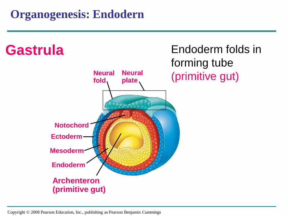
Organogenesis: Endodern
Neuralfold
Neural plate
Mesoderm
Notochord
Archenteron(primitive gut)
Ectoderm
Endoderm
Gastrula Endoderm folds in
forming tube
(primitive gut)

Copyright © 2008 Pearson Education, Inc., publishing as Pearson Benjamin Cummings
Organogenesis: Endoderm
Primitive gut is shown in yellow
Endoderm forms epithelial lining
of digestive tract
Organs of digestive tract develop from
primitive gut
Respiratory tract lining develops
from outpouching of foregut (1) and
forms lung buds
Endoderm gives develop to glands
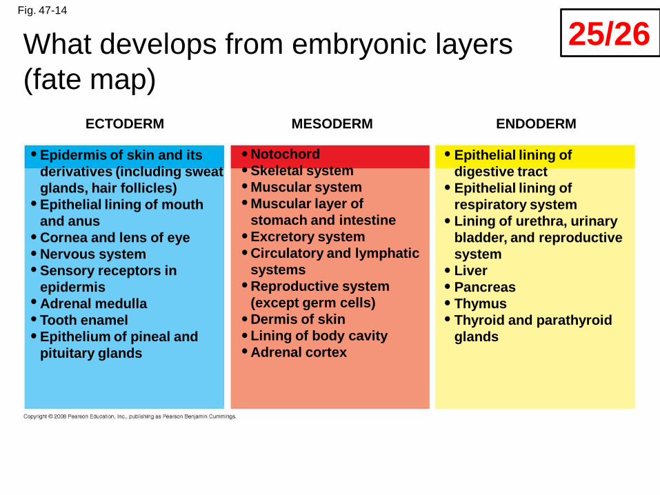
Fig. 47-14
ECTODERM MESODERM ENDODERM
Epidermis of skin and its
derivatives (including sweat
glands, hair follicles)
Epithelial lining of mouth
and anus
Cornea and lens of eye
Nervous system
Sensory receptors in
epidermis
Adrenal medulla
Tooth enamel
Epithelium of pineal and
pituitary glands
Notochord
Skeletal system
Muscular system
Muscular layer of
stomach and intestine
Excretory system
Circulatory and lymphatic
systems
Reproductive system
(except germ cells)
Dermis of skin
Lining of body cavity
Adrenal cortex
Epithelial lining of
digestive tract
Epithelial lining of
respiratory system
Lining of urethra, urinary
bladder, and reproductive
system
Liver
Pancreas
Thymus
Thyroid and parathyroid
glands
What develops from embryonic layers
(fate map)
25/26
Fig. 47-16-5
Yolk sac
Mesoderm
Amnion
Chorion
Ectoderm
Extraembryonicmesoderm
Trophoblast
Endoderm
Hypoblast
Expandingregion oftrophoblast
Epiblast
Maternalbloodvessel
Allantois
Trophoblast
Hypoblast
Endometrialepithelium(uterine lining)
Inner cell mass
Blastocoel
Uterus
Epiblast
Amnioticcavity
Expandingregion oftrophoblast
Yolk sac (fromhypoblast)
Chorion (fromtrophoblast)
Extraembryonicmesoderm cells(from epiblast)
Thank you for
your attention and
participation!
Copyright © 2008 Pearson Education, Inc., publishing as Pearson Benjamin Cummings
You should now be able to:
1. Describe the acrosomal reaction
2. Describe the formation of a blastula and
gastrulation
3. Describe the main processes in
organogenesis