Embed Size (px)
Citation preview

PAPER
Animal, but not human, faces engage the distributed facenetwork in adolescents with autism
Elisabeth M. Whyte,1 Marlene Behrmann,2 Nancy J. Minshew,3
Natalie V. Garcia1 and K. Suzanne Scherf1
1. Department of Psychology, Penn State University, USA2. Departments of Psychiatry and Neurology, University of Pittsburgh Medical School, USA3. Department of Psychology, Carnegie Mellon University, USA
Abstract
Multiple hypotheses have been offered to explain the impaired face-processing behavior and the accompanying underlyingdisruptions in neural circuitry among individuals with autism.We explored the specificity of atypical face-processing activation andpotential alterations to fusiform gyrus (FG) morphology as potential underlying mechanisms. Adolescents with high functioningautism(HFA)and age-matched typically developing (TD)adolescentswere scannedwith sMRI and fMRIas theyobserved humanand animal faces. In spite of exhibiting comparable face recognition behavior, the HFA adolescents evinced hypo-activationthroughout the face-processing system in response to unfamiliar human, but not animal, faces. Theyalso exhibitedgreateractivationin affective regions of the face-processing network in response to animal, but not human, faces. Importantly, this atypical pattern ofactivation in response to human faces was not related to atypical structural properties of the FG. This atypical neural response tohuman faces in autism may stem from abnormalities in the ability to represent the reward value of social (i.e. conspecific) stimuli.
Research highlights
• High functioning adolescents (HFA) with autism andage-matched typically developing (TD) adolescentswere scanned with sMRI and fMRI as they observedhuman and animal faces.
• TD adolescents exhibited comparable activation tohuman and animal faces throughout the distributedface-processing neural network.
• HFA adolescents exhibited hypo-activation only tohuman, but not animal, faces, compared to the TDadolescents.
• Morphology in the fusiform gyri was comparableacross the groups.
Introduction
The impaired development of face-processing behaviorand the accompanying disruptions to the underlying
neural circuitry are widely reported findings in theautism literature (e.g. Corbett, Carmean, Ravizza, Wen-delken, Henry et al., 2009; Dalton, Nacewicz, Johnstone,Schaefer, Gernsbacher et al., 2005; Grelotti, Klin, Gau-their, Skudlarski, Cohen et al., 2005; Humphreys, Has-son, Avidan, Minshew & Behrmann, 2008; Pierce,Muller, Ambrose, Allen & Courchesne, 2001; Scherf,Luna, Minshew & Behrmann, 2010; Schultz, Gauthier,Klin, Fulbright, Anderson et al., 2000). These atypical-ities are particularly apparent among children andadolescents. For example, although face recognitionbehavior typically improves through adolescence andearly adulthood among typically developing (TD) indi-viduals (O’Hearn, Schroer, Minshew & Luna, 2010), itfails to improve beyond childhood among individualswith high functioning autism (HFA) (Greimel, Schulte-R€uther, Kamp-Becker, Remschmidt, Herpertz-Dahl-mann et al., 2014; O’Hearn et al., 2010; O’Hearn,Tanaka, Lynn, Fedor, Minshew et al., 2014). Similarly,the size of the fusiform face area (FFA) increases as a
Address for correspondence: Elisabeth M. Whyte, Department of Psychology, Pennsylvania State University, 110 Moore Building, University Park, PA16802, USA; e-mail: [email protected]
© 2015 John Wiley & Sons Ltd
Developmental Science 19:2 (2016), pp 306–317 DOI: 10.1111/desc.12305
function of age from childhood to adolescence amongTD individuals (Scherf, Behrmann, Humphreys & Luna,2007; Golarai, Liberman, Yoon & Grill-Spector, 2010),yet it remains smaller and hypoactive in HFA adoles-cents compared to TD adolescents (Scherf et al., 2010).To date, there have been multiple hypotheses offered toexplain these deficits. Here, we evaluated two alternative,but not mutually exclusive, theories using fMRI.
One potential explanation for this atypical face-relatedactivation in autism is that it is derived from structuralalterations in the fusiform gyrus that likely emerge earlyin development (Dziobek, Bahnemann, Convit & Heek-eren, 2010; Raznahan, Toro, Daly, Robertson, Murphyet al., 2010; Trontel, Duffield, Bigler, Froehlich, Priggeet al., 2013; Toal, Bloemen, Deeley, Tunstall, Daly et al.,2009; Van Kooten, Palmen, Von Cappeln, Steinbusch,Korr et al., 2008). For example, in autism, alterations inleft fusiform gyrus (FG) volume in children and adoles-cents (Trontel et al., 2013) and cortical thickness inadults (Dziobek et al., 2010) are negatively correlatedwith face memory abilities. This theory predicts that thefundamental structural alterations likely impact the fullrange of stimuli that are processed by the FG, ratherthan being limited to human faces in particular.
An alternative hypothesis is that face-processingdifficulties stem from abnormalities in the ability torepresent the reward value of social (i.e. conspecific)stimuli (Dawson, Carver, Meltzoff, Panagiotides,McPartland et al., 2002; Dawson, Webb & McPartland,2005; Chevallier, Kohls, Troiani, Brodkin & Schultz,2012). In turn, this leads to atypical tuning of neuralregions that process social information about conspecif-ics, including the fusiform face area (FFA) and otherface- and body-processing regions over the course ofdevelopment (Schultz, 2005). In support of this hypoth-esis, there are hints in the literature that highly familiarstimuli, which may be particularly rewarding, do elicitmore normal levels of activation, specifically in theFFA, in autism. For example, Grelotti et al. (2005)found that a 12-year-old boy with autism showed strongFFA activation while observing Digimon cartoon char-acters, which were objects of expertise for him. Anotherstudy reported comparable responses in the FFA amongchildren and young adolescents with autism and TDindividuals when they viewed personally familiar faces(e.g. own mother; Pierce & Redcay, 2008). However,there are no conditions that reportedly lead to morenormal levels of activation in any of the other regions(beyond the FFA) of the distributed face-processingnetwork in autism (Bookheimer, Wang, Scott, Sigman,Dapretto et al., 2008; Dapretto, Davies, Pfeifer,Scott, Sigman et al., 2006; Hadjikhani, Joseph, Snyder &Tager-Flusberg, 2007; Perlman, Hudac, Pegors, Minshew
& Pelphrey, 2011; Pierce & Redcay, 2008). This complexset of findings leads to open questions about thefull range of face properties that may or may not elicitmore typical levels of neural activation in autism andthe extent to which modulation of neural activity isevident in multiple neural regions or is restricted to theFG/FFA.
In the current study, we explored the extent to whichatypical activation of the face-processing network isexplained by either of these two mechanisms, structuralalterations in FG morphology or human face-specifichypo-activation. To do so, we measured the morphologiccharacteristics (volume and cortical thickness) of thefusiform gyrus to determine whether face recognitionbehavior and/or neural responses to faces for HFAadolescents are related to aberrant morphology withinthese regions. We also measured neural responses to bothhuman and animal faces. Faces of cats and dogs are idealstimuli to evaluate the specificity of the atypicalresponses in the face-processing system in autism fortwo reasons. First, animal faces share similar perceptualfeatures with human faces, although they are notconspecifics; thus they likely engage similar visuoper-ceptual processing strategies as do human faces (seeDiamond & Carey, 1986). Second, a growing body ofevidence suggests that animal faces (particularly dogs)evoke similar activation to human faces in the FFA(Blonder, Smith, Davis, Kesler/West, Garrity et al., 2004;Tong, Nakayama, Moscovitch, Weinrib & Kanwisher,2000) and other regions involved in face processing(Anzellotti & Caramazza, 2014; Blonder et al., 2004;Stoeckel, Pailley, Gollub, Niemi & Evins, 2014; Yang,Bellgowan & Martin, 2012) in TD adults. Therefore,evaluating neural responses to animal and human facesin autism can help determine whether the neural circuitryinvolved in face processing is generally disrupted(regardless of the type of face) or is specifically affectedin the processing of human faces.
We scanned HFA adolescents and age-matched TDadolescents as they observed unfamiliar human faceswith a range of emotional expressions and eye gazedirection, animal faces, and common objects. Wehypothesized that if there is a generalized deficit in theunderlying neural system in autism, we would observehypo-activation to both human and animal faces in theFFA (and potentially in other face-processing regions)and that FFA hypo-activation might be related toaberrant structural properties of the FG. In contrast, ifthe deficit in autism is highly specific to human faces, wewould observe selective hypo-activation only in responseto human, but not animal, faces in the FFA and otherface-processing regions, and no obvious relationshipwith altered morphological brain characteristics.
© 2015 John Wiley & Sons Ltd
Autism human and animal face activation 307
Methods
Participants
Participants included 14 HFA adolescents (13 male, 1female), ages 13–18 years (M = 15, SD = 2) and 14 TDadolescents (13 male, 1 female), ages 13–18 years(M = 15, SD = 2). One additional HFA adolescent wasexcluded due to excessive head motion. The groups werematched on sex, handedness, age [t(26) = .23, p = .82],Full Scale IQ [t(26) = .53, p = .60], Verbal IQ [t(26) = .11,p = .5], and Performance IQ [t(26) = 1.37, p = .18] (seeTable 1). IQ was assessed in the HFA adolescents usingthe Weschler Abbreviated Scales of Intelligence (WASI;Wechsler, 1999) and in the TD adolescents using theKauffman Brief Intelligence Test-2 (KBIT-2; Kaufman& Kaufman, 2004).The diagnosis of autism was established using the
Autism Diagnostic Interview-Revised (ADI-R; Lord,Rutter & Le Couteur, 1994), the Autism DiagnosticObservation Schedule-G (ADOS; Lord, Rutter, DiLa-vore & Risi, 2001), and expert clinical diagnosis (Min-shew, 1996). The HFA adolescents were medicallyhealthy; had no identifiable genetic, metabolic, orinfectious etiology for their disorder; and were free oftraumatic brain injury, seizures, attention deficit disor-der, and depression. HFA participants were not asked towithhold medication prior to testing. TD participantswere medically healthy, free of regular medication usage,had no history of autism, neurological or psychiatricillness, acquired brain injury, learning disabilities, devel-opmental disabilities, school problems, or substanceabuse in themselves or their first-degree relatives.Adolescents were recruited from several participant
databases to be part of a longitudinal study investigating
the effects of visuoperceptual training. The data reportedhere are from the pre-training assessment. Writteninformed consent was obtained from the parents of theadolescents, along with written assent or consent fromthe adolescents, using procedures approved by theInternal Review Boards of Penn State University andCarnegie Mellon University.
Measures
Face recognition behavior
The upright and inverted versions of the Cambridge FaceMemory Test (CFMT) were used to measure unfamiliarface recognition outside the scanner (Duchaine &Nakay-ama, 2006). This task has beenused previously tomeasureface recognition abilities in TD and HFA adolescents(O’Hearn et al., 2010). One HFA and one TD adolescentdid not complete the inverted version of the task.Accuracy was collected as a measure of performance.
MRI acquisition
Prior to scanning, all participants were placed in a mockMR scanner for approximately 20 minutes and com-pleted practice versions of the tasks that were adminis-tered in the full scan. This procedure acclimatesparticipants to the scanner environment and minimizesmotion artifact and anxiety.Participants were scanned using a Siemens 3T Magne-
tomTriowhole bodyMRI scanner with a 12-channel headcoil. High-resolution T1-weighted anatomical images(3D-MPRAGE) were acquired with 176 sagittal slices(TR/TE/TI = 1700, 1.78, 850 ms; voxel size = 1 mm3,FOV = 256 9 256). Functional EPI images were acquiredin 34 slices (3 mm isotropic voxels; TR/TE = 2000, 25 ms;FOV = 210; matrix 70 9 70; Flip angle = 80o). Thefunctional images were aligned approximately 30o in therostral direction from the AC-PC line, which minimizesnoise from the eye orbits and nasal sinuses and maximizessignal in the medial temporal lobes (Whalen, Johnstone,Somerville, Nitschke, Polis et al., 2008). This protocolallowed for full coverage of the ventral visual pathway aswell as of the frontal and occipital lobes. For participantswith larger head size, the superior parietal lobe was notcompletely covered. High-resolution structural imagesand functional images were acquired in a single session.
fMRI human–animal face task
Participants observed color pictures from four categoriesof human faces (fearful, happy, and neutral expressionswith direct eye gaze, and neutral expressions with averted
Table 1 Demographic characteristics for the adolescentswith high functioning autism (HFA) and typically developing(TD) adolescents
HFA TD
# of participants 14 14Age (years) 15 (2) 15 (2)Full Scale IQa 112 (11) 110 (12)Verbal IQ 109 (12) 109 (13)Performance IQ 113 (13) 107 (10)Handedness 12R / 2L 12R / 2LGender 13M / 1F 13M / 1FADOS Total 13 (3)ADOS Comm 5 (1)ADOS Social 9 (2)
Note: aThe HFA adolescents completed the Weschler Abbreviated Scaleof Intelligence and the TD adolescents completed the Kauffman BriefIntelligence Test-2. SRS = Social Responsiveness Scale
© 2015 John Wiley & Sons Ltd
308 Elisabeth M. Whyte et al.
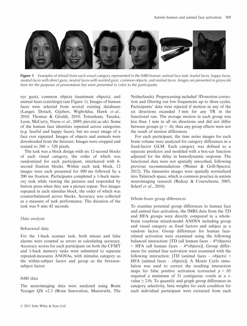
eye gaze), common objects (inanimate objects), andanimal faces (cats/dogs) (see Figure 1). Images of humanfaces were selected from several existing databases(Langer, Dotsch, Gijsbert, Wigboldus, Hawk et al.,2010; Thomaz & Giraldi, 2010; Tottenham, Tanaka,Leon, McCarry, Nurse et al., 2009; pics.stir.ac.uk). Someof the human face identities repeated across categories(e.g. fearful and happy faces), but no exact image of aface ever repeated. Images of objects and animals weredownloaded from the Internet. Images were cropped andresized to 280 9 320 pixels.
The task was a block design with six 12-second blocksof each visual category, the order of which wasrandomized for each participant, interleaved with 6-second fixation blocks. Within each task block, 12images were each presented for 800 ms followed by a200 ms fixation. Participants completed a 1-back mem-ory task while viewing the pictures and responded bybutton press when they saw a picture repeat. Two imagesrepeated in each stimulus block, the order of which wascounterbalanced across blocks. Accuracy was collectedas a measure of task performance. The duration of thetask was 9 min 42 seconds.
Data analysis
Behavioral data
For the 1-back scanner task, both misses and falsealarms were counted as errors in calculating accuracy.Accuracy scores for each participant on both the CFMTand 1-back memory tasks were submitted to separaterepeated-measures ANOVAs, with stimulus category asthe within-subject factor and group as the between-subject factor.
fMRI data
The neuroimaging data were analyzed using BrainVoyager QX v2.3 (Brain Innovation, Maastricht, The
Netherlands). Preprocessing included 3D-motion correc-tion and filtering out low frequencies up to three cycles.Participants’ data were rejected if motion in any of thesix directions exceeded 3 mm for any TR in thefunctional run. The average motion in each group wasless than 1 mm in all six directions and did not differbetween groups (p > .4); thus any group effects were notthe result of motion differences.
For each participant, the time series images for eachbrain volume were analyzed for category differences in afixed-factor GLM. Each category was defined as aseparate predictor and modeled with a box-car functionadjusted for the delay in hemodynamic response. Thefunctional data were not spatially smoothed, followingprevious recommendations (Weiner & Grill-Spector,2012). The timeseries images were spatially normalizedinto Talairach space, which is common practice in autismneuroimaging research (Redcay & Courschesne, 2005;Scherf et al., 2010).
Whole-brain group differences
To examine potential group differences in human faceand animal face activation, the fMRI data from the TDand HFA groups were directly compared in a whole-brain voxelwise mixed-model ANOVA including groupand visual category as fixed factors and subject as arandom factor. Group differences for human face-related activation were examined using the followingbalanced interaction: [TD (all human faces – 4*objects)> HFA (all human faces – 4*objects)]. Group differ-ences for animal face activation were examined with thefollowing interaction: [TD (animal faces – objects) >HFA (animal faces – objects)]. A Monte Carlo simu-lation was used to correct the resulting interactionmaps for false positive activation (corrected p < .05required a minimum of 31 contiguous voxels at a t-value ≥ 2.0). To quantify and graph group differences incategory selectivity, beta weights for each condition foreach individual participant were extracted from each
Figure 1 Examples of stimuli from each visual category represented in the fMRI human–animal face task: fearful faces, happy faces,neutral faces with direct gaze, neutral faces with averted gaze, common objects, and animal faces. Images are presented in greyscalehere for the purposes of presentation but were presented in color to the participants.
© 2015 John Wiley & Sons Ltd
Autism human and animal face activation 309
significant ROI and difference scores were plotted forhuman (average of human face categories – objects) andanimal (animal – objects) faces.
Individually defined fusiform face area
Given our a priori predictions about group differences inthe profile of activation for the FFA, individually definedFFA regions were demarcated separately for human andanimal faces in each hemisphere in each participant(Scherf et al., 2010; Weiner & Grill-Spector, 2012).Selectivity for human faces was defined in a balancedcontrast: [(happy + fearful + neutral-direct + neutral-averted human faces) � (4*objects)]. Animal face selec-tivity was defined as: [(animal faces) � (objects)]. Eachresulting individual contrast map was corrected for falsepositive activation using the False Discovery Rateprocedure (Genovese, Lazar & Nichols, 2002) with aq < .05. The right and left FFA ROIs were defined as themost anterior cluster of contiguous significant voxels inthe fusiform gyrus (FG) separately for each participant’shuman-selective and animal-selective activation, andwere quantified in terms of the number of voxels (e.g,ROI size). These ROIs were defined to ensure that theBOLD signal comparison between groups was derivedfrom the optimal (i.e. most selective) region on a case-by-case basis rather than superimposing a group-definedROI on each brain. This approach allowed us todifferentially optimize the BOLD signal for human andanimal faces separately. Therefore, any emerging groupdifferences in BOLD response resulting from theseindividual ROI analyses cannot be attributed to partialvolume effects or differences in ROI definition across thetwo groups.Within each individually defined ROI for each partic-
ipant, a separate ROI-based GLM was conducted thatmodeled stimulus condition as a fixed factor. Theresulting beta weights were extracted for each of the sixvisual categories. Beta weight difference scores werecomputed to examine the magnitude of face-relatedactivation in the FFA for human faces (average ofhuman face categories – objects) and animal faces(animal faces – objects). The size and the beta weightdifference scores were then submitted to separaterepeated-measures ANOVAs with the factors of facecategory (human, animal), hemisphere (right, left), andgroup (HFA, TD).
Independently defined fusiform face area
To confirm the results from the individually defined ROIanalysis, beta weights were also extracted from an
independently defined right FFA ROI. This ROI wasidentified in previous work evaluating human face-related activation in TD adolescents and adults. Wecentered a 4 mm sphere on the functionally definedTalairach coordinates for the right FFA (40, �41, �21)reported in a previous study (Scherf et al., 2007).
sMRI analyses
To evaluate whether structural differences mightaccount for observed functional differences, weextracted FG volume and cortical thickness for eachparticipant in each hemisphere, as well as whole-brainvolume. The structural data were analyzed using thestandard cortical reconstruction pipeline in FreeSurferv5.3.0 (surfer.nmr.mgh.harvard.edu), including stan-dardization to a common atlas (Fischl, Salat, Busa,Albert, Dieterich et al., 2002), which is a validatedapproach for use with children (Ghosh, Kakunoori,Augustinack, Nieto-Castanon, Kovelman et al., 2010).Once the cortical model was completed for eachparticipant, the cerebral cortex was parcellated intounits based on gyral and sulcal structure, creatingsurface-based maps of gyral curvature and sulcal depth(Fischl, Sereno & Dale, 1999). Calculations of corticaland subcortical volumes using this method are compa-rable to manual segmentation (e.g. Lehmann, Douiri,Kim, Modat, Chan et al., 2010). We normalized theFG volumes for each participant using their whole-brain volume (e.g. [region/whole brain volume]*1000).Whole brain volume was calculated as the SupraTen-torial volume, excluding ventricles, CSF, and choroidplexus. The structural measures were submitted toseparate ANOVAs, with the factors of group andhemisphere.
Structure–function correlations
To test the hypothesis that structural properties of theleft and right FG may predict atypical behavior orfunctional activation for HFA adolescents or TD ado-lescents, Pearson product correlations were conductedbetween the structural and functional measures obtainedfor these regions separately for each group. We examinedthe relations between structural properties of the FG(volume and cortical thickness) with FFA size (sepa-rately for human and animal faces) and activation (betaweight difference scores). These correlations were con-ducted separately within each hemisphere. For eachsignificant correlation, we evaluated robustness in sep-arate bootstrap analyses using 1000 iterations to obtain a95% confidence interval (CI).
© 2015 John Wiley & Sons Ltd
310 Elisabeth M. Whyte et al.
Results
Behavioral tasks
On the CFMT, both groups exhibited a face inversioneffect; there was a main effect of orientation, with higherperformance for upright than inverted faces, F(1, 24) =83.78, p < .001, g2 = .78. There was no main effect ofgroup nor an interaction between group and orientation(Supplemental Figure 1a). During the scanning task,recognition performance was comparable across the twogroups for all conditions and there were no main effectsor interactions (all p > .05) (Supplemental Figure 1b).Therefore, group differences in the BOLD responsecannot be attributed to differences in behavioral perfor-mance.
Whole-Brain Group Differences
Human face activation comparison
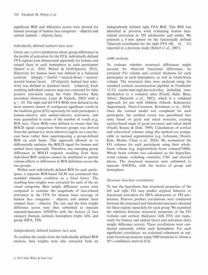
In spite of the comparable face recognition behavioracross the groups, the TD adolescents exhibited greaterhuman face activation than HFA adolescents in the rightFFA, right occipital face area (OFA), left amygdala, leftputamen, and the posterior cingulate cortex (PCC)(Figure 2). There were no regions where HFA adoles-cents showed greater activation than TD adolescents forhuman faces (Table 2). Figure 2 shows the activation forhuman and animal faces extracted from the right FFA,right OFA, left amygdala, and PCC. In each of theseregions there was a group difference for human faces (bydefinition). The key question concerns whether there
(a)
(b)
Figure 2 Group differences for HFA adolescents and TD adolescents in response to human faces compared to objects. The mapswere generated by computing a whole-brain analysis, corrected at p < .05. Adolescents with HFA exhibited less activation than TDadolescents for human faces compared to objects in the left putamen (not pictured), (a) right fusiform face area, left amygdala,posterior cingulate cortex, and (b) the right occipital face area. The mean (SEM) beta weight difference scores (human faces minusobjects) and (animal faces minus objects) were extracted from these regions. Adolescents with HFA show hypo-activation for humanfaces, but not animal faces, in these regions.
© 2015 John Wiley & Sons Ltd
Autism human and animal face activation 311
were group differences for animal faces. Interestingly,there were no significant differences between HFAadolescents and TD adolescents for the beta-weightsextracted for animal faces in any of these ROIs (p = ns).In addition, while TD adolescents did not show differ-ences between human and animal activation in theseROIs, the HFA adolescents showed significantly reducedactivation for human compared to animal faces (allp < .05), indicating that the hypo-activation was specificto human faces.
Animal face activation comparison
The whole-brain analysis contrasting group responses toanimal faces revealed no regions of significant differencebetween HFA and TD adolescents for animal face-selective activation.
Individually defined fusiform face area
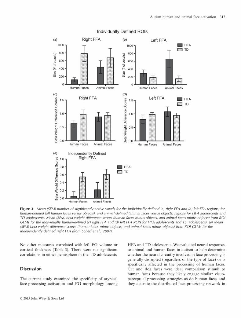
FFA size
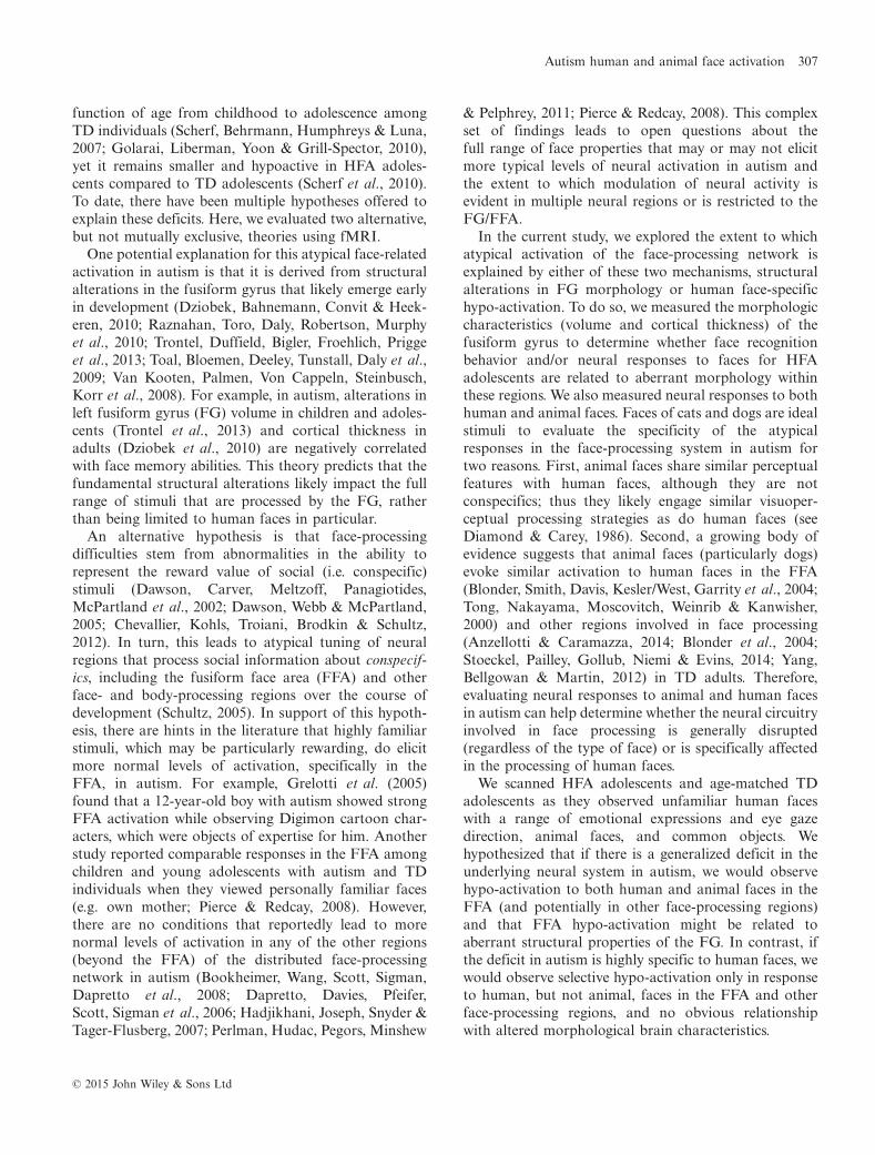
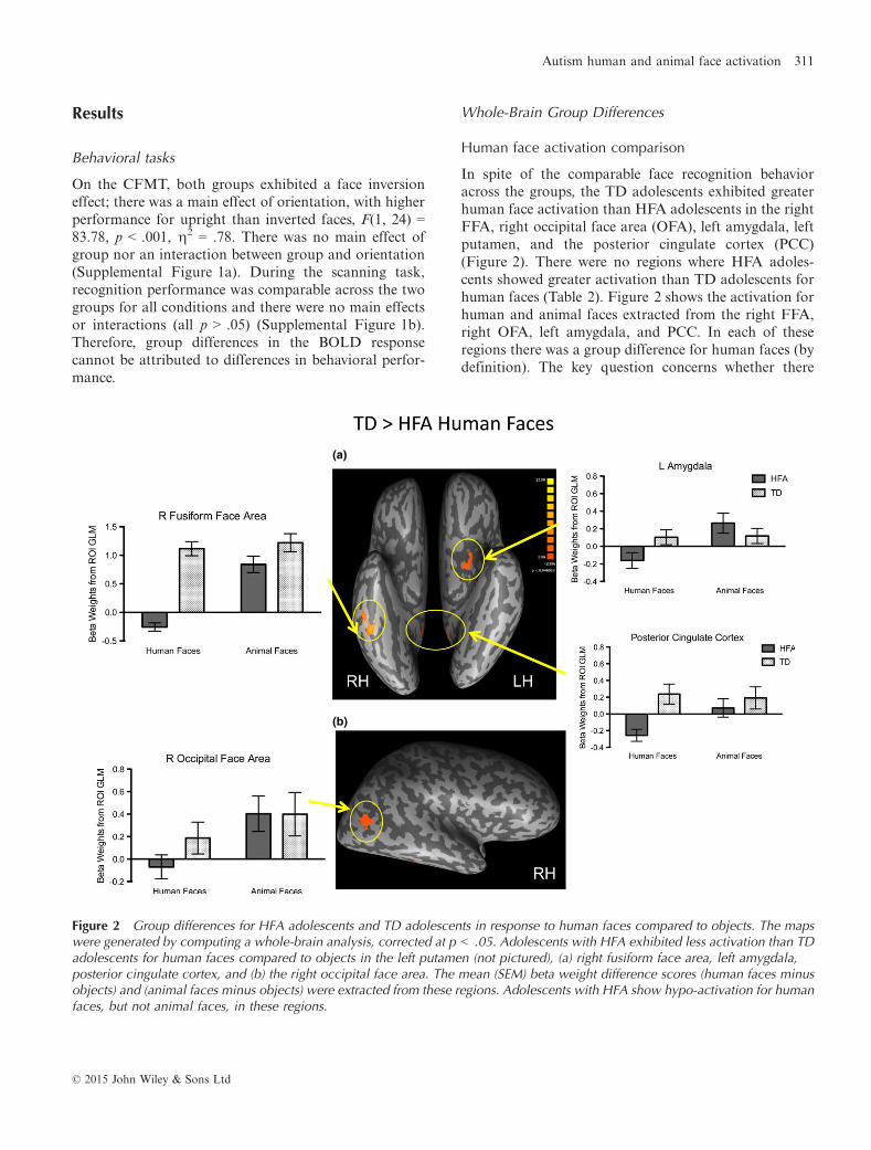
Figure 3a–b shows the size of the human- and animal-defined right and left FFA ROIs for the two groups.There were no main effects of group or of hemisphere.However, there were interactions between face category9 group, F(1, 26) = 4.75, p < .05, g2 = .15, and betweenhemisphere 9 group, F(1, 26) = 89.99, p < .01, g2 = .28.To investigate the face category 9 group interaction, weevaluated category effects within each group collapsedacross hemisphere. Across both hemispheres, the HFAadolescents had smaller human- than animal-definedFFA regions, t(13) = �2.17, p < .05. Among TDadolescents, the size of the human-defined and animal-defined FFA regions did not differ (p = ns). Toinvestigate the hemisphere by group interaction, weevaluated hemisphere effects within each group collapsedacross face category. HFA adolescents had larger leftthan right FFA regions, t(13) = �2.30, p < .05. Incontrast, TD adolescents, had larger right than left FFAregions, t(13) = 2.55, p < .05.
FFA magnitude
Figure 3c–d shows the beta weight difference scores inthe human-defined bilateral FFA regions, for bothhuman faces and animal faces. There was a main effectof face category, F(1, 25) = 6.21, p < .05, g2 = .19.Surprisingly, animal faces elicited higher magnituderesponses than did human faces in the human-definedFFA. However, this was qualified by a face category 9
group interaction, F(1, 25) = 11.06, p < .01, g2 = .31, andno main effect or interactions with hemisphere. Toinvestigate this interaction, we evaluated the effect offace category within each group. HFA adolescents hadlower activation for human than animal faces in theirhuman-defined FFA regions, t(12) = 3.37, p < .05. Incontrast, TD adolescents showed comparable activationfor human and animal faces in the FFA (p = ns).
Independently defined right fusiform face area
Within the independently defined right FFA, HFAadolescents showed hypo-activation for human facescompared to TD adolescents, t(26) = �3.00, p < .01, butnot for animal faces, replicating the pattern found for thewhole-brain analysis (Figure 3e).
Structural MRI
For all ROIs, there was no main effect of group, no maineffect of hemisphere, and no interaction between groupand hemisphere (p = ns) (Supplemental Figure 2a–c).
Structure–function correlations
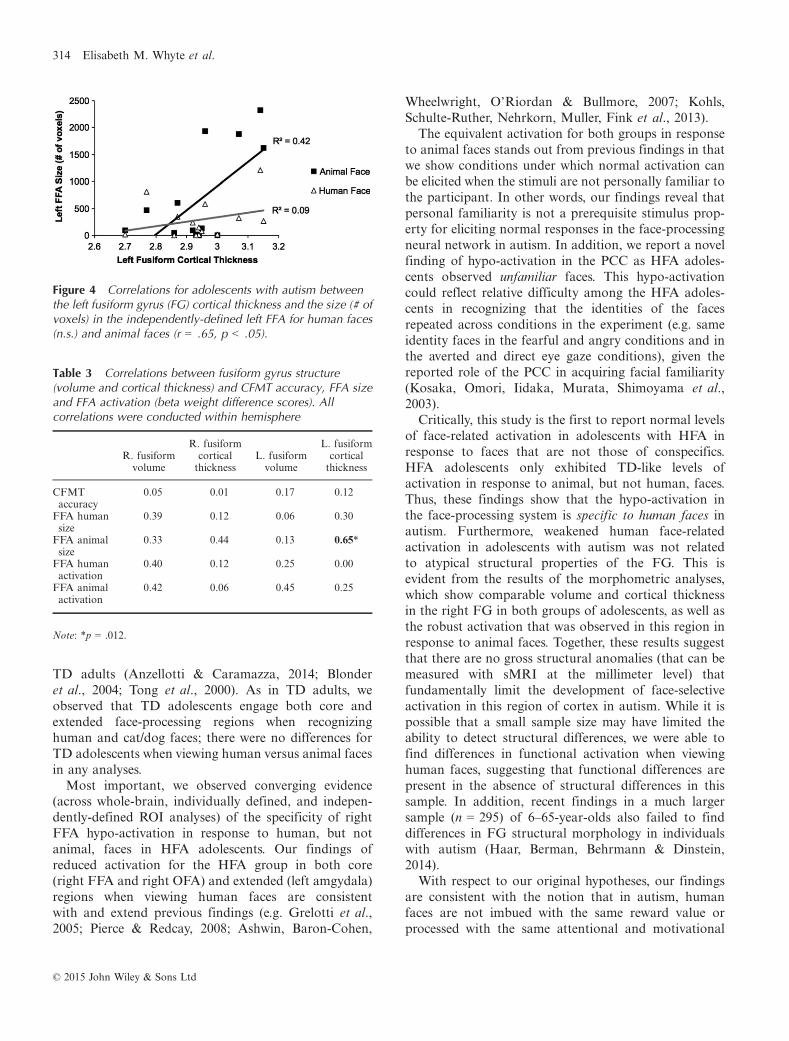
For HFA adolescents, in the right hemisphere, there wereno correlations between FG structure and either behav-ior or functional activation. In the left hemisphere, FGcortical thickness correlated with the size of the left FFAROI for animal faces, r(14) = .65, p = .012, (bootstrapanalysis: r = .63, 95% CIs of .34/.92, which is differentfrom 0 at p < .05), but not for human faces (Figure 4).
Table 2 Results from whole-brain comparisons between TD and HFA adolescents for human and animal face activation
Group Contrast Region Hemisphere Size (#voxels) Talairach coordinates X Y Z
TD > HFA Human > object FFA R 940 41 �41 �22OFA R 1642 45 �65 8Amygdala L 417 �20 3 �13Putamen L 870 �22 4 0PCC 2327 15 �51 �0
TD > HFA Animal > Object nsHFA > TD Human > Object nsHFA > TD Animal > Object ns
© 2015 John Wiley & Sons Ltd
312 Elisabeth M. Whyte et al.
No other measures correlated with left FG volume orcortical thickness (Table 3). There were no significantcorrelations in either hemisphere in the TD adolescents.
Discussion
The current study examined the specificity of atypicalface-processing activation and FG morphology among
HFA and TD adolescents. We evaluated neural responsesto animal and human faces in autism to help determinewhether the neural circuitry involved in face processing isgenerally disrupted (regardless of the type of face) or isspecifically affected in the processing of human faces.Cat and dog faces were ideal comparison stimuli tohuman faces because they likely engage similar visuo-perceptual processing strategies as do human faces andthey activate the distributed face-processing network in
(a) (b)
(c)
(e)
(d)
Figure 3 Mean (SEM) number of significantly active voxels for the individually defined (a) right FFA and (b) left FFA regions, forhuman-defined (all human faces versus objects), and animal-defined (animal faces versus objects) regions for HFA adolescents andTD adolescents. Mean (SEM) beta weight difference scores (human faces minus objects, and animal faces minus objects) from ROIGLMs for the individually human-defined (c) right FFA and (d) left FFA ROIs for HFA adolescents and TD adolescents. (e) Mean(SEM) beta weight difference scores (human faces minus objects, and animal faces minus objects) from ROI GLMs for theindependently defined right FFA (from Scherf et al., 2007).
© 2015 John Wiley & Sons Ltd
Autism human and animal face activation 313
TD adults (Anzellotti & Caramazza, 2014; Blonderet al., 2004; Tong et al., 2000). As in TD adults, weobserved that TD adolescents engage both core andextended face-processing regions when recognizinghuman and cat/dog faces; there were no differences forTD adolescents when viewing human versus animal facesin any analyses.Most important, we observed converging evidence
(across whole-brain, individually defined, and indepen-dently-defined ROI analyses) of the specificity of rightFFA hypo-activation in response to human, but notanimal, faces in HFA adolescents. Our findings ofreduced activation for the HFA group in both core(right FFA and right OFA) and extended (left amgydala)regions when viewing human faces are consistentwith and extend previous findings (e.g. Grelotti et al.,2005; Pierce & Redcay, 2008; Ashwin, Baron-Cohen,
Wheelwright, O’Riordan & Bullmore, 2007; Kohls,Schulte-Ruther, Nehrkorn, Muller, Fink et al., 2013).The equivalent activation for both groups in response
to animal faces stands out from previous findings in thatwe show conditions under which normal activation canbe elicited when the stimuli are not personally familiar tothe participant. In other words, our findings reveal thatpersonal familiarity is not a prerequisite stimulus prop-erty for eliciting normal responses in the face-processingneural network in autism. In addition, we report a novelfinding of hypo-activation in the PCC as HFA adoles-cents observed unfamiliar faces. This hypo-activationcould reflect relative difficulty among the HFA adoles-cents in recognizing that the identities of the facesrepeated across conditions in the experiment (e.g. sameidentity faces in the fearful and angry conditions and inthe averted and direct eye gaze conditions), given thereported role of the PCC in acquiring facial familiarity(Kosaka, Omori, Iidaka, Murata, Shimoyama et al.,2003).Critically, this study is the first to report normal levels
of face-related activation in adolescents with HFA inresponse to faces that are not those of conspecifics.HFA adolescents only exhibited TD-like levels ofactivation in response to animal, but not human, faces.Thus, these findings show that the hypo-activation inthe face-processing system is specific to human faces inautism. Furthermore, weakened human face-relatedactivation in adolescents with autism was not relatedto atypical structural properties of the FG. This isevident from the results of the morphometric analyses,which show comparable volume and cortical thicknessin the right FG in both groups of adolescents, as well asthe robust activation that was observed in this region inresponse to animal faces. Together, these results suggestthat there are no gross structural anomalies (that can bemeasured with sMRI at the millimeter level) thatfundamentally limit the development of face-selectiveactivation in this region of cortex in autism. While it ispossible that a small sample size may have limited theability to detect structural differences, we were able tofind differences in functional activation when viewinghuman faces, suggesting that functional differences arepresent in the absence of structural differences in thissample. In addition, recent findings in a much largersample (n = 295) of 6–65-year-olds also failed to finddifferences in FG structural morphology in individualswith autism (Haar, Berman, Behrmann & Dinstein,2014).With respect to our original hypotheses, our findings
are consistent with the notion that in autism, humanfaces are not imbued with the same reward value orprocessed with the same attentional and motivational
Table 3 Correlations between fusiform gyrus structure(volume and cortical thickness) and CFMT accuracy, FFA sizeand FFA activation (beta weight difference scores). Allcorrelations were conducted within hemisphere
R. fusiformvolume
R. fusiformcorticalthickness
L. fusiformvolume
L. fusiformcorticalthickness
CFMTaccuracy
0.05 0.01 0.17 0.12
FFA humansize
0.39 0.12 0.06 0.30
FFA animalsize
0.33 0.44 0.13 0.65*
FFA humanactivation
�0.40 �0.12 �0.25 0.00
FFA animalactivation
�0.42 �0.06 �0.45 0.25
Note: *p = .012.
Figure 4 Correlations for adolescents with autism betweenthe left fusiform gyrus (FG) cortical thickness and the size (# ofvoxels) in the independently-defined left FFA for human faces(n.s.) and animal faces (r = .65, p < .05).
© 2015 John Wiley & Sons Ltd
314 Elisabeth M. Whyte et al.
resources as they are in TD individuals (Chevallier et al.,2012; Schultz, 2005). Our findings of stronger responsesto animal faces in this same network may indicate thatindividuals with autism find animal faces more sociallyrewarding than human faces. There is behavioral evi-dence that is consistent with this notion. Individuals withautism often show strong motivational preferences andsocial behaviors directed towards common animals suchas cats and dogs (Carlisle, 2014; Celani, 2002; O’Haire,McKenzie, Beck & Slaughter, 2013; Prothmann, Ettrich& Prothmann, 2009). In support of this interpretation,we found greater activation in areas related to rewardand emotional arousal (amygdala, putamen) in responseto animal faces but not human faces among the HFAadolescents. Future work investigating the associationbetween perceived reward value of both human andanimal faces and category-selective neural activation isneeded to directly evaluate this interpretation. In addi-tion, future research using eye-tracking technology mayprovide specific information about whether and howvisually attention to human and animal faces is deployeddifferently and is or is not related to the differentialpatterns of activation in either the visuoperceptual and/or affective regions of the face-processing network.
There were no differences in human face-processingbehavior between the HFA and TD adolescents. In spiteof the comparable behavior, the adolescents with autismstill showed reduced activation for human faces, partic-ularly in the right hemisphere. Also, the two groupsexhibited different patterns of laterality in face activa-tion. The TD adolescents exhibited right hemispherelaterality for both human and animal faces, while theHFA adolescents exhibited left hemisphere laterality.Together, this pattern of results suggests that the HFAadolescents may be using different strategies from theTD adolescents to recognize both human and animalfaces. We suggest that these processing strategies mayrely disproportionately on the left hemisphere. Thishypothesis is informed by the results from the structure–function analyses. Among the HFA adolescents, therewas a relation between the cortical thickness of the leftFG and the size of the animal face defined functionalROI. All together, these results may reflect individualdifferences in the extent of cortical thinning (or rather,lack thereof) during development that have consequencesfor and/or are related to the emerging specialization forfaces in the autism brain. Consistent with this notion areprevious findings that children with autism do not beginto show a left visual hemifield advantage for processingboth human and dog faces that young TD children do(consistent with emerging right-lateralization for faceprocessing) (Guillon, Hadjikhani, Baduel, Kruck, Ar-naud et al., 2014). Longitudinal data will determine
whether this left lateralization and heightened selectivityfor animal faces is contributing to the development ofhypo-activation in the face-processing system duringhuman face processing, is a compensatory response, or iseven unrelated to this hypo-activation.
In conclusion, the current study found that adoles-cents with autism evince consistent hypo-activationthroughout the face-processing network specificallywhen viewing human, but not animal, faces. Groupdifferences in social motivation specific to human facesmay be central to this atypical neural response in autism.Future research should examine how motivationalsalience impacts activation of face-processing regionsfor both individuals with autism and typical develop-ment.
Acknowledgements
This work was supported by Pennsylvania Departmentof Health SAP grant 4100047862 (MB, KSS, NM),NICHD/NIDCD P01/U19 (MB, PI: NM), and a grantfrom the Simons Foundation to MB (PI: D. Heeger).This research was supported by the Social ScienceResearch Institute and the Center for Online Innovationin Learning at Penn State University. This research wasalso supported by Scifund Challenge contributors. Wewould like to thank the Director, Rick Gilmore, and staffat the Social, Life, and Engineering Center as well asGiorgia Picci and Susan Bowser for their assistance withdata collection for this project. We are also grateful toour study families for making this research possible.
References
Anzellotti, S., & Caramazza, A. (2014). Individuating theneural bases for the recognition of conspecifics with MVPA.NeuroImage, 89, 165–170.
Ashwin, C., Baron-Cohen, S., Wheelwright, S., O’Riordan, M.,& Bullmore, E.T. (2007). Differential activation of theamygdala and the ‘social brain’ during fearful face process-ing in asperger syndrome. Neuropsychologia, 45 (1), 2–14.
Blonder, L.X., Smith, C.D., Davis, C.E., Kesler/West, M.L., &Garrity, T.F. et al. (2004). Regional brain response to faces ofhumans and dogs. Cognitive Brain Research, 20, 384–394.
Bookheimer, S.Y., Wang, A.T., Scott, A., Sigman, M., Dapret-to, M. et al. (2008). Frontal contributions to face processingdifferences in autism: evidence from fMRI of inverted faceprocessing. Journal of the International NeuropsychologicalSociety, 14 (06), 922–932.
Carlisle, G.K. (2014). Pet ownership decisions for parents ofchildren with autism spectrum disorder. Journal of PediatricNursing, 29 (2), 114–123.
© 2015 John Wiley & Sons Ltd
Autism human and animal face activation 315
Celani,G. (2002).Humanbeings, animals and inanimate objects:what do people with autism like? Autism, 6 (1), 93–102.
Chevallier, C., Kohls, G., Troriani, V., Brodkin, E.S., & Schultz,R.T. (2012). The social motivation theory of autism. Trendsin Cognitive Sciences, 16 (4), 231–239.
Corbett, B., Carmean, V., Ravizza, S., Wendelken, C., Henry,M. et al. (2009). A functional and structural study ofemotion and face processing in children with autism.Psychiatry Research: Neuroimaging, 173 (3), 196–205.
Dalton, K.M., Nacewicz, B.M., Johnstone, T., Schaefer, H.S.,Gernsbacher, M.A. et al. (2005). Gaze fixation and theneural circuitry of face processing in autism. Nature Neuro-science, 8 (4), 519–526.
Dapretto, M., Davies, M.S., Pfeifer, J.H., Scott, A.A., Sigman,M. et al. (2006). Understanding emotions in others: mirrorneuron dysfunction in children with autism spectrum disor-ders. Nature Neuroscience, 9 (1), 28–30.
Dawson, G., Carver, L., Meltzoff, A.N., Panagiotides, H.,McPartland, J. et al. (2002). Neural correlates of face andobject recognition in young children with autism spectrumdisorder, developmental delay, and typical development.Child Development, 73 (3), 700–717.
Dawson, G., Webb, S.J., & McPartland, J. (2005). Understand-ing the nature of face processing impairment in autism:insights from behavioral and electrophysiological studies.Developmental Neuropsychology, 27 (3), 403–424.
Diamond, R., & Carey, S. (1986). Why faces are and are notspecial: an effect of expertise. Journal of ExperimentalPsychology, 115 (2), 107–117.
Duchaine, B., & Nakayama, K. (2006). The Cambridge facememory test: results for neurologically intact individuals andan investigation of its validity using inverted face stimuli andprosopagnosic participants. Neuropsychologia, 44 (4), 576–585.
Dziobek, I., Bahnemann, M., Convit, A., & Heekeren, H.R.(2010). The role of the fusiform-amygdala system in thepathophysiology of autism. Archives of General Psychiatry, 67(4), 397–405. doi:10.1001/archgenpsychiatry.2010.31
Fischl, B., Salat, D.H., Busa, E., Albert, M., Dieterich, M.et al. (2002). Whole brain segmentation: automated labelingof neuroanatomical structures in the human brain. Neuron,33 (3), 341–355.
Fischl, B., Sereno, M.I., & Dale, A.M. (1999). Cortical surface-based analysis. II: Inflation, flattening, and a surface-basedcoordinate system. NeuroImage, 9 (2), 195–207.
Genovese, C.R., Lazar, N.A., & Nichols, T. (2002). Threshold-ing of statistical maps in functional neuroimaging using thefalse discovery rate. NeuroImage, 15 (4), 870–878.
Ghosh, S.S., Kakunoori, S., Augustinack, J., Nieto-Castanon,A., Kovelman, I. et al. (2010). Evaluating the validity ofvolume-based and surface-based brain image registration fordevelopmental cognitive neuroscience studies in children 4 to11 years of age. NeuroImage, 53 (1), 85–93.
Golarai, G., Liberman, A., Yoon, J.M., & Grill-Spector, K.(2010). Differential development of the ventral visual cortexextends through adolescence. Frontiers in Human Neurosci-ence, 3, 80.
Greimel, E., Schulte-R€uther, M., Kamp-Becker, I., Remsch-midt, H., Herpertz-Dahlmann, B. et al. (2014). Impairmentin face processing in autism spectrum disorder: a develop-mental perspective. Journal of Neural Transmission, 121 (9),1171–1181.
Grelotti, D.J., Klin, A.J., Gautheir, I., Skudlarski, P., Cohen,D.J. et al. (2005). fMRI activation of the fusiform gyrus andamygdala to cartoon characters but not to faces in a boy withautism. Neuropsychologia, 43 (4), 373–385.
Guillon, Q., Hadjikhani, N., Baduel, S., Kruck, J., Arnaud, M.et al. (2014). Both dog and human faces are exploredabnormally by young children with autism spectrum disor-ders. NeuroReport, 25 (15), 1237–1241.
Haar, S., Berman, S., Behrmann, M., & Dinstein, I. (2014).Anatomical abnormalities in autism? Cerebral Cortex.Advance online publication. doi:10.1093/cercor/bhu242
Hadjikhani, N., Joseph, R.M., Snyder, J., & Tager-Flusberg, H.(2007). Abnormal activation of the social brain during faceperception in autism.Human Brain Mapping, 28 (5), 441–449.
Humphreys, K., Hasson, U., Avidan, G., Minshew, N., &Behrmann, M. (2008). Cortical patterns of category-selectiveactivation for faces, places and objects in adults with autism.Autism Research, 1 (1), 52–63.
Kaufman, A.S., & Kaufman, N.L. (2004). Kaufman BriefIntelligence Test (2nd edn.). Circle Pines, MN: AGS Pub-lishing.
Kohls, G., Schulte-Ruther, M., Nehrkorn, B., Muller, K., Fink,G.R. et al. (2013). Reward system dysfunction in autismspectrum disorders. Social Cognitive and Affective Neurosci-ence, 8 (5), 565–572.
Kosaka, H., Omori, M., Iidaka, T., Murata, T., Shimoyama, T.et al. (2003). Neural substrates participating in acquisition offacial familiarity: an fMRI study. NeuroImage, 20, 1734–1742.
Langer, O., Dotsch, R., Gijsbert, B., Wigboldus, D.H.J., Hawk,S.T. et al. (2010). Presentation and validation of the RadboudFaces Database. Cognition and Emotion, 24 (8), 1377–1388.
Lehmann, M., Douiri, A., Kim, L.G., Modat, M., Chan, D.et al. (2010). Atrophy patterns in Alzheimer’s disease andsemantic dementia: a comparison of FreeSurfer and manualvolumetric measurements. NeuroImage, 49 (3), 2264–2274.
Lord, C., Rutter, M., DiLavore, P.C., & Risi, S. (2001). AutismDiagnostic Observation Schedule (ADOS). Lost Angeles,CA: Western Psychological Services.
Lord, C., Rutter, M., & Le Couteur, A. (1994). Autismdiagnostic interview-revised: a revised version of a diagnosticinterview for caregivers of individuals with possible pervasivedevelopmental disorders. Journal of Autism and Developmen-tal Disorders, 24 (5), 659–685.
Minshew, N. (1996). Autism. In B.O. Berg (Ed.), Principles ofchild neurology (pp. 1713–1729). New York: McGraw-Hill.
O’Haire, M.E., McKenzie, S.J., Beck, A.M., & Slaughter, V.(2013). Social behaviors increase in children with autism inthe presence of animals compared to toys. Plos ONE, 8 (2),e57010.
O’Hearn, K., Schroer, E., Minshew, N., & Luna, B. (2010).Lack of developmental improvement on a face memory task
© 2015 John Wiley & Sons Ltd
316 Elisabeth M. Whyte et al.
during adolescence in autism. Neuropsychologia, 48 (13),3955–3960.
O’Hearn, K., Tanaka, J., Lynn, A., Fedor, J., Minshew, N. et al.(2014). Developmental plateau in visual object processingfrom adolescence to adulthood in autism. Brain and Cogni-tion, 90, 124–134. doi:10.1016/j.bandc.2014.06.004
Perlman, S.B., Hudac, C.M., Pegors, T., Minshew, N.J., &Pelphrey, K.A. (2011). Experimental manipulation of face-evoked activity in the fusiform gyrus of individuals withautism. Social Neuroscience, 6 (1), 22–30.
Pierce, K., Muller, R.A., Ambrose, J., Allen, G., & Courchesne,E. (2001). Face processing occurs outside the fusiform ‘facearea’ in autism: evidence from functional MRI. Brain, 124,2059–2073.
Pierce, K., & Redcay, E. (2008). Fusiform function in childrenwith autism spectrum disorder is a matter of ‘who’.Biological Psychiatry, 64 (7), 552–560.
Prothmann, A., Ettrich, E., & Prothmann, S. (2009). Preferencefor, and responsiveness to, people, dogs, and objects inchildren with autism. Anthrozoos: A Multidisciplinary Jour-nal of the Interactions of People and Animals, 22 (2), 161–171.
Raznahan, A., Toro, R., Daly, E., Robertson, D., Murphy, C.et al. (2010). Cortical anatomy in autism spectrum disorder:an in vivo MRI study on the effect of age. Cerebral Cortex, 20(6), 1332–1340.
Redcay, E., & Courchesne, E. (2005). When is the brainenlarged in autism? A meta-analysis of all brain size reports.Biological Psychiatry, 58 (1), 1–9.
Scherf, K., Behrmann, M., Humphreys, K., & Luna, B. (2007).Visual category-selectivity for faces, places and objectsemerges along different developmental trajectories. Develop-mental Science, 10 (4), F15–F31.
Scherf, K., Luna, B., Minshew, N., & Behrmann, M. (2010).Location, location, location: alterations in the functionaltopography of face- but not object- or place-related cortex inadolescentswith autism.Frontiers inHumanNeuroscience, 4, 26.
Schultz, R.T. (2005). Developmental deficits in social percep-tion in autism: the role of the amygdala and fusiform facearea. International Journal of Developmental Neuroscience,23, 125–141.
Schultz, R.T., Gauthier, I., Klin, A., Fulbright, R.K., Ander-son, A.W. et al. (2000). Abnormal ventral temporal corticalactivity during face discrimination among individuals withautism and Asperger syndrome. Archives in General Psychi-atry, 57 (4), 331–340.
Stoeckel, L.E., Palley, L.S., Gollub, R.L., Niemi, S.M., &Evins, A.E. (2014). Patterns of brain activation whenmothers view their own child and dog: an fMRI study. PloSONE, 9 (10), e107205.
Thomaz, C.E., & Giraldi, G.A. (2010). A new rankingmethod for principal components analysis and its applica-
tion to face image analysis. Image and Vision Computing, 28(6), 902–913.
Toal, F., Bloemen, O.J., Deeley, Q., Tunstall, N., Daly, E.M.et al. (2009). Psychosis and autism: magnetic resonanceimaging study of brain anatomy. British Journal of Psychi-atry: The Journal of Mental Science, 194 (5), 418–425.
Tong, F., Nakayama, K., Moscovitch, M., Weinrib, O., &Kanwisher, N. (2000). Response properties of the humanfusiform face area. Cognitive Neuropsychology, 17 (1), 257–280.
Tottenham, N., Tanaka, J.W., Leon, A.C., McCarry, T., Nurse,M. et al. (2009). The Nimstim set of facial expressions:judgments from untrained research participants. PsychiatryResearch, 168 (3), 242–249.
Trontel, H.G., Duffield, T.C., Bigler, E.D., Froehlich, A.,Prigge, M.B. et al. (2013). Fusiform correlates of facialmemory in autism. Behavioral Sciences, 3 (3), 348–371.
Van Kooten, I., Palmen, S., Von Cappeln, P., Steinbusch, H., &Korr, H. et al. (2008). Neurons in the fusiform gyrus arefewer and smaller in autism. Brain: A Journal of Neurology,131 (4), 987–999.
Wechsler, D. (1999). Wechsler Abbreviated Scale of Intelligence(WASI). San Antonio, TX: The Psychological Corporation.
Weiner, K.S., & Grill-Spector, K. (2012). The improbablesimplicity of the fusiform face area. Trends in CognitiveSciences, 16 (5), 251–254.
Whalen, P.J., Johnstone, T., Somerville, L.H., Nitschke, J.B.,Polis, S. et al. (2008). A functional magnetic resonanceimaging predictor of treatment response to venlafaxine ingeneralized anxiety disorder. Biological Psychiatry, 63 (9),858–863.
Yang, J., Bellgowan, P.S., & Martin, A. (2012). Threat, domain-specificity and the human amygdala. Neuropsychologia, 50(11), 2566–2572.
Received: 16 September 2014Accepted: 17 February 2015
Supporting Information
Additional Supporting Information may be found in the onlineversion of this article:Supplemental Figure 1. Mean (SEM) behavioral accuracy on
the A) Cambridge Face Memory Test (CFMT) and B) 1-backmemory task performed during scanning by HFA adolescentsand TD adolescents.Supplemental Figure 2. The mean (SEM) structural data for
A) fusiform volume corrected by whole brain volume, B)fusiform cortical thickness, and C) whole brain volume forHFA adolescents and TD adolescents.
© 2015 John Wiley & Sons Ltd
Autism human and animal face activation 317


![Matching 3D Faces with Partial Data - BMVA · Matching 3D Faces with Partial Data ... [12] an ICP based 3D face recognition approach is presented, where the face is first detected](https://img.pdfslide.us/doc/110x75/5e2a1c17a6b0d11c993ac500/matching-3d-faces-with-partial-data-matching-3d-faces-with-partial-data-12.jpg)