Embed Size (px)
Citation preview

REVIEW PAPER
Angiotensin II AT1 Receptor Blockers Ameliorate InflammatoryStress: A Beneficial Effect for the Treatment of Brain Disorders
Juan M. Saavedra
Received: 13 July 2011 / Accepted: 26 August 2011 / Published online: 22 September 2011
� Springer Science+Business Media, LLC (outside the USA) 2011
Abstract Excessive allostatic load as a consequence of
deregulated brain inflammation participates in the devel-
opment and progression of multiple brain diseases,
including but not limited to mood and neurodegenerative
disorders. Inhibition of the peripheral and brain Renin–
Angiotensin System by systemic administration of Angio-
tensin II AT1 receptor blockers (ARBs) ameliorates
inflammatory stress associated with hypertension, cold-
restraint, and bacterial endotoxin administration. The
mechanisms involved include: (a) decreased inflammatory
factor production in peripheral organs and their release to
the circulation; (b) reduced progression of peripherally
induced inflammatory cascades in the cerebral vasculature
and brain parenchyma; and (c) direct anti-inflammatory
effects in cerebrovascular endothelial cells, microglia, and
neurons. In addition, ARBs reduce bacterial endotoxin-
induced anxiety and depression. Further pre-clinical
experiments reveal that ARBs reduce brain inflammation,
protect cognition in rodent models of Alzheimer’s disease,
and diminish brain inflammation associated with genetic
hypertension, ischemia, and stroke. The anti-inflammatory
effects of ARBs have also been reported in circulating
human monocytes. Clinical studies demonstrate that ARBs
improve mood, significantly reduce cognitive decline after
stroke, and ameliorate the progression of Alzheimer’s
disease. ARBs are well-tolerated and extensively used to
treat cardiovascular and metabolic disorders such as
hypertension and diabetes, where inflammation is an inte-
gral pathogenic mechanism. We propose that including
ARBs in a novel integrated approach for the treatment of
brain disorders such as depression and Alzheimer’s disease
may be of immediate translational relevance.
Keywords Brain Renin–Angiotensin system �Angiotensin II AT1 receptor blockers � Stress �Brain inflammation � Depression � Anxiety �Mood disorders � Neurodegenerative disorders �Alzheimer’s disease � Stroke � Cognition
Introduction
Mood and neurodegenerative disorders are devastating
diseases of high prevalence and poorly understood etiol-
ogy, without adequate treatment. These diseases are the
consequence of failure to maintain homeostasis, a condi-
tion associated with multiple constellations of factors on a
background of genetic vulnerability (Fig. 1).
A major influence in the development and progression
of many brain disorders is the failure of the mechanisms to
regulate and control the necessary, adaptive inflammatory
responses in the brain (Dantzer et al. 2008; Rivest 2010).
When not adequately restricted, brain inflammation may
lead to altered behavior and neuronal damage, depression,
progressive loss of cognition, and reduced neurological
performance (Pascoe et al. 2011; Marchesi 2011; Anisman
2009; Leonard 2007; Tansey and Goldberg 2010). Unfor-
tunately, at present there are no effective and safe treat-
ments to control deregulated inflammatory processes in the
brain (Editorial 2007; Nimmo and Vink 2009). For these
reasons the search for novel, safe, and effective central
anti-inflammatory drugs is of major interest.
J. M. Saavedra (&)
Section on Pharmacology, Division of Intramural Research
Programs, National Institute of Mental Health, National
Institutes of Health, 10 Center Drive, Bldg. 10, Room 2D-57,
Bethesda, MD 20892, USA
e-mail: [email protected]
123
Cell Mol Neurobiol (2012) 32:667–681
DOI 10.1007/s10571-011-9754-6
The present review summarizes our observations
demonstrating that inhibition of the peripheral and brain
Renin–Angiotensin System (RAS) by systemic adminis-
tration of Angiotensin II AT1 receptor blockers (ARBs)
ameliorates brain inflammatory stress. ARBs are safe
compounds that while commonly used for the treatment of
cardiovascular and metabolic disorders where inflamma-
tion is a major pathogenic factor (Savoia and Schiffrin
2007; Barra et al. 2009), may have other uses that have not
been adequately tested. For example, there is accumulating
evidence that ARBs are not only neuroprotective in stroke
and diabetes, but also ameliorate age-related cognitive loss,
anxiety, and depression (Saavedra et al. 2011). Because of
their safety and demonstrated central anti-inflammatory
effects, we propose to utilize ARBs as a novel additional
component of an integrative treatment of brain disorders
such as depression and Alzheimer’s disease, a concept of
potential immediate translational value.
What follows is a description of our research findings
leading to this proposal, and a brief description of sup-
portive pre-clinical and clinical evidence obtained by other
laboratories.
For both our pre-clinical experiments using rodents and
for our studies on human circulating monocytes, we used
the ARB candesartan at doses and concentrations in the
same order of magnitude than those used in clinical set-
tings, supporting the clinical relevance of our findings (Lee
et al. 1995; Nishimura et al. 2000a, b; Weinberg et al.
2004; Benicky et al. 2011).
The Brain Angiotensin II System
Angiotensin II was discovered as a circulating pro-hyper-
tensive peptide of renal origin, the active principle of the
RAS. Circulating Angiotensin II, through activation of its
physiological AT1 receptors, was characterized as a major
regulator of vascular tone and fluid metabolism (Skrbic and
Igic 2009). Inhibition of peripheral Angiotensin II AT1
receptors may be achieved with the use of ARBs, non-
peptidic, orally active, and well-tolerated compounds
(Timmermans et al. 1993). ARBs are currently a main-
stream treatment for cardiovascular and metabolic diseases,
including essential hypertension and diabetes (McFarlane
2009). The beneficial effects of ARBs are not only limited
to reduction of vasoconstriction, but also include a signif-
icant decrease in vascular and end-organ inflammation, the
result of excessive AT1 receptor activation and a major
participant in the pathogenesis of hypertension and diabe-
tes (Savoia and Schiffrin 2007; Marchesi et al. 2008)
(Fig. 2).
Initially, the effects of Angiotensin II in the brain were
thought to be restricted to regulation of thirst and sodium
appetite (Skrbic and Igic 2009). Later, multiple local RAS
systems were described in all tissues studied, including the
brain (Bader 2010; Saavedra 1992). Subsequent studies
demonstrated that brain Angiotensin II is a pleiotropic
neuroregulator, and that brain AT1 receptors are involved
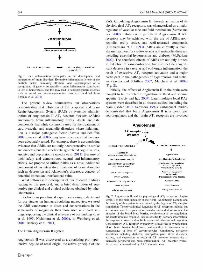
Fig. 1 Brain inflammation participates in the development and
progression of brain disorders. Excessive inflammation is one of the
multiple factors increasing allostatic load. Superimposed on a
background of genetic vulnerability, brain inflammation contributes
to loss of homeostasis, and this may lead to neuropsychiatric diseases
such as mood and neurodegenerative disorders (modified from
Benicky et al. 2011)
Fig. 2 Angiotensin II and its physiological AT1 receptors. Angio-
tensin II is the main mediator of the Renin–Angiotensin System, and
the activity of this system is determined by the degree of AT1 receptor
stimulation. The physiological functions of AT1 receptors include, but
are not restricted to, regulation of vascular tone and blood volume, the
integrity of the blood brain barrier, cerebrovascular autoregulation,
the innate immune response, insulin sensitivity, sensory information,
the response to stress and multiple aspects of behavior and cognition.
Consequently, AT1 receptor overactivity is involved in hypertension,
blood brain barrier breakdown, vulnerability to ischemia as a
consequence of loss of cerebrovascular compliance, metabolic
alterations including diabetes, neuropathic pain, stress disorders,
anxiety, and depression. A result of AT1 receptor overactivity is
increased peripheral and brain inflammation. AT1 receptor overac-
tivity may be normalized by ARB administration
668 Cell Mol Neurobiol (2012) 32:667–681
123
in multiple functions including, but not limited to the con-
trol of the cerebral circulation, hormonal, and autonomic
regulation and in particular the regulation of the response to
stress and behavior (Tsutsumi and Saavedra 1991; Saavedra
1992; Saavedra 2005; Saavedra et al. 2011) (Fig. 2).
In the brain and the periphery, Angiotensin II has been
proposed to stimulate, in addition to AT1 receptors, a
second receptor type, the AT2 receptor. AT2 receptor
stimulation by Angiotensin II has been proposed to play a
protective role and to balance AT1 receptor stimulation.
However, the emerging consensus is that the AT2 receptor
expression in the human brain is limited, the receptor can
be activated without Angiotensin II participation, published
results are controversial, and there is no clear mechanistic
model for its signal transduction (Porrello et al. 2009; De
Gasparo and Siragy 1999; Allen et al. 1999; Saavedra
2005; Rompe et al. 2010). For these reasons the proposed
role of AT2 receptors in the neuroprotective effects of
ARBs will not be discussed here.
The Consequences of Enhanced Brain Angiotensin II
AT1 Receptor Activity
Evidence accumulated in many laboratories including ours
demonstrated an association of enhanced brain Angiotensin
II activity with increased allostatic load leading to vul-
nerability to stress, anxiety, depression, brain ischemia
with associated cerebrovascular remodeling and loss of
compliance, and inflammation (Castren and Saavedra
1988; Xang et al. 1993; Edwards et al. 1999; Nishimura
et al. 2000a; Saavedra and Benicky 2007; Zhang et al.
2010; Saavedra et al. 2011) (Fig. 2). Some of the initial
information was obtained in a rodent model of essential,
genetic hypertension, the Spontaneously Hypertensive Rat
(SHR). In this model, enhanced Angiotensin II AT1
receptor expression is indicative of increased activation
and it is associated with cerebrovascular stiffness and
inflammation (Nishimura et al. 2000a).
The Results of AT1 Receptor Blockade
Since it was demonstrated that ARBs reverse peripheral
vascular inflammation and excessive vascular remodeling
in hypertension, atherosclerosis, and diabetes (Savoia and
Schiffrin 2007), we hypothesized that the beneficial effects
of ARBs may extend to the brain circulation in hyperten-
sive animals. To test our hypothesis we asked:
Are ARBs able to reverse the cerebrovascular alterations
characteristic of genetic hypertension? First, we demon-
strated that systemically administered ARBs crossed the
blood brain barrier and blocked AT1 receptors in the brain
parenchyma (Nishimura et al. 2000b). We subsequently
found that systemic administration of the ARB candesartan
protected cerebrovascular flow, reversing cerebrovascular
remodeling and inflammation in SHR, effects independent
of its blood pressure-lowering effects (Ito et al. 2002;
Yamakawa et al. 2003; Ando et al. 2004; Zhou et al. 2005).
The end result of ARB therapy was to reduce stroke damage
(Fig. 3).
Parallel experiments in our laboratory revealed that
systemic candesartan administration completely prevented
the hormonal and sympathoadrenal response to isolation
stress in normotensive rats (Armando et al. 2001, 2007) and
the stimulation of central sympathetic activity during cold-
restraint stress in SHR (Bregonzio et al. 2008). Although
anti-stress properties of ARBs were of a major interest, we
recognized that elimination of the stress response to psy-
chogenic stimuli may not necessarily be a beneficial effect.
To establish whether or not ARBs manifested anti-stress
properties of therapeutic benefit, we asked:
May systemic administration of ARBs prevent a stress-
induced disorder? To answer this question we used a
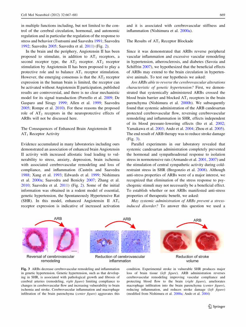
Fig. 3 ARBs decrease cerebrovascular remodeling and inflammation
in genetic hypertension. Genetic hypertension, such as that develop-
ing in SHR, is associated with pathological growth and fibrosis of
cerebral arteries (remodeling, right figure) limiting compliance to
changes in cerebrovascular flow and increasing vulnerability to brain
ischemia and stroke. Cerebrovascular inflammation and macrophage
infiltration of the brain parenchyma (center figure) aggravates this
condition. Experimental stroke in vulnerable SHR produces major
loss of brain tissue (left figure). ARB administration reverses
cerebrovascular remodeling improving vascular compliance and
protecting blood flow to the brain (right figure), ameliorates
macrophage infiltration into the brain parenchyma (center figure),
reducing inflammation, and reduces stroke damage (left figure)
(modified from Nishimura et al. 2000a; Ando et al. 2004)
Cell Mol Neurobiol (2012) 32:667–681 669
123
rodent model of cold-restraint gastric ulceration. We found
that systemic candesartan effectively abolished gastric
ulceration in stress-vulnerable SHR (Bregonzio et al.
2003). A mechanistic analysis revealed that in addition to
prevention of circulatory vasoconstriction by reduction of
the stress-induced sympathoadrenal activation, candesartan
significantly prevented the inflammatory response to stress
in the gastric mucosa (Bregonzio et al. 2003) (Fig. 4). It
was of interest that, while ARBs prevented the HPA axis
response to the psychogenic stress of isolation, they did not
influence the glucocorticoid production and release during
cold-restraint stress (Bregonzio et al. 2003). We interpreted
this observation, initially surprising, as further evidence of
selective, beneficial anti-stress effects of ARBs, since
production and release of glucocorticoids, major anti-
inflammatory hormones, is essential for the protection of
the gastric mucosa during stress (Bregonzio et al. 2003).
Thus, two independent lines of evidence strongly sug-
gested major beneficial anti-inflammatory effects of ARBs,
ameliorating inflammation in the cerebral vasculature
during hypertension and in the gastric mucosa during
stress. For these reasons we hypothesized that ARBs might
possess general anti-inflammatory properties, not only in
the periphery but also in the brain, and that amelioration of
pathological inflammation might be beyond their direct
cardiovascular effects.
AT1 Receptor Blockade in Normotensive Rats
To rule out the participation of blood pressure changes on
the anti-inflammatory effects of ARBs, we asked: Does
ARB administration ameliorate brain inflammation unre-
lated to alterations in blood pressure? To test our
hypothesis, we selected a well-characterized model of
inflammation, the activation of the innate immune response
in the periphery and the brain by systemic administration of
the bacterial endotoxin lipopolysaccharide (LPS) (Dantzer
et al. 1998; Rivest 2010). Brain inflammatory processes are
necessary for the maintenance of homeostasis. The brain
innate immune response, the initial reaction to inflamma-
tory challenges, is an essential mechanism to restore
homeostasis in response to stress, infection, or neuronal
injury (Dantzer et al. 1998; Rivest 2010). Exaggerated,
disturbed inflammatory responses, however, lead to chronic
inflammation and neuronal damage (Dantzer et al. 1998;
Rivest 2010). It was therefore of interest to establish whe-
ther systemic ARB administration controlled, but not
eliminated, the innate immune response to LPS.
To avoid confounding cardiovascular effects we
administered LPS to normotensive rats at a subseptic dose
not influencing blood pressure (Sanchez-Lemus et al.
2008). These concentrations are similar to those found in
humans affected by metabolic disorders (Schwartz et al.
2010), supporting the clinical relevance of our findings.
Systemic LPS stimulates peripheral target cells, namely
vascular endothelial cells and circulating and tissue mac-
rophages located in multiple organs (Guha and Mackman
2001; Quan and Banks 2007; Rivest 2010). In turn, these
cells produce and release to the circulation large amounts
of pro-inflammatory cytokines directly affecting the brain
(Rivest 2010). Circulating pro-inflammatory cytokines and
LPS target cerebrovascular endothelial cells, stimulating
inflammatory cascades that promote further inflammation
and microglia activation in the brain parenchyma (Quan
and Banks 2007). If the inflammatory response is not bal-
anced, it leads to neuronal injury (Guha and Mackman
2001; Quan and Banks 2007; Rivest 2010). In addition,
systemic LPS strongly activates the HPA axis, a charac-
teristic inflammatory stress with enhanced production and
release of pro-inflammatory aldosterone from the adrenal
gland (Sanchez-Lemus et al. 2008). The HPA axis reaction
to LPS includes enhanced production and release of anti-
inflammatory corticosterone, responsible for the feedback
control and regulation of the hormonal and systemic
inflammatory responses (Sanchez-Lemus et al. 2008). The
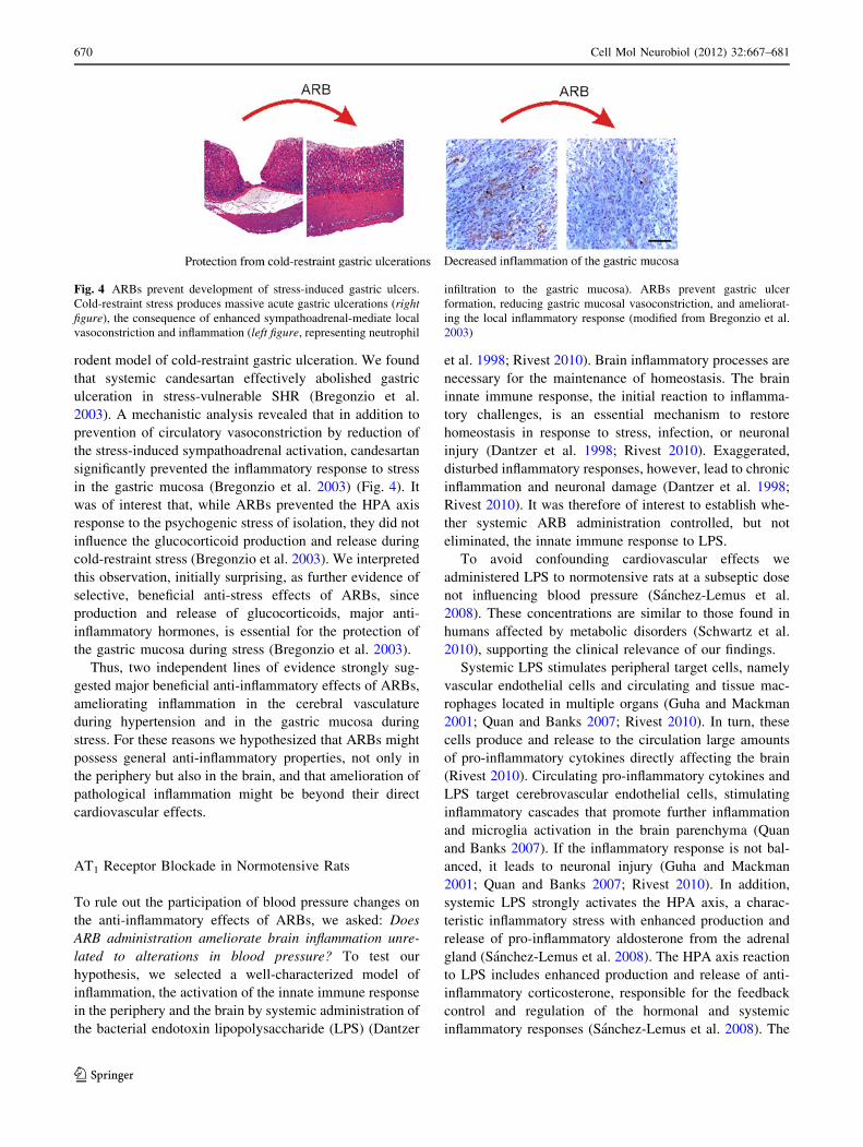
Fig. 4 ARBs prevent development of stress-induced gastric ulcers.
Cold-restraint stress produces massive acute gastric ulcerations (rightfigure), the consequence of enhanced sympathoadrenal-mediate local
vasoconstriction and inflammation (left figure, representing neutrophil
infiltration to the gastric mucosa). ARBs prevent gastric ulcer
formation, reducing gastric mucosal vasoconstriction, and ameliorat-
ing the local inflammatory response (modified from Bregonzio et al.
2003)
670 Cell Mol Neurobiol (2012) 32:667–681
123
LPS-induced peripheral and brain inflammation is associ-
ated with an initial behavioral reaction or ‘‘sickness syn-
drome’’ with fever, diminished locomotion, decreased
social interactions, and anorexia leading to body weight
loss and depression (Dantzer et al. 1998).
Effects of ARBs on the Peripheral and Brain Innate
Immune Response
Consequently, we tested the effects of ARB administration
on the LPS-induced innate immune response, and we asked
the following questions: Does ARB administration influ-
ence circulating biomarkers of inflammation? In normo-
tensive rats, a subseptic LPS dose not affecting systemic
blood pressure produced a major increase in inflammatory
factors in the general circulation (Benicky et al. 2011). The
ARB candesartan decreased or eliminated LPS induction of
circulating inflammatory factors without affecting LPS
induction of anti-inflammatory factors. This significantly
decreased the LPS-induced pro-inflammatory profile in the
general circulation (Benicky et al. 2011; Sanchez-Lemus
et al. 2008, 2009a, b) (Fig. 5).
Does ARB administration influence peripheral inflam-
matory responses in target organs, indirectly affecting the
brain? Circulating markers of inflammation have their origin
in target cells for LPS, namely endothelial cells in the vas-
culature, macrophages in peripheral organs and components
of the HPA axis. We studied the pituitary and adrenal glands,
major components of the HPA axis, and the macrophage-rich
spleen, a major target for systemic LPS (Sanchez-Lemus
et al. 2008, 2009a, b). LPS-induced profound inflammatory
responses in all tissues studied. In the adrenal gland, can-
desartan prevented LPS-induced upregulation of pro-
inflammatory aldosterone production without affecting that
of anti-inflammatory glucocorticoids (Sanchez-Lemus et al.
2008). In the adrenal gland, pituitary gland, and spleen,
candesartan significantly reduced the LPS-induced upregu-
lation of all inflammatory pathways studied, including
cytokine production, COX-2 and iNOS transcription,
ROS formation and NF-jB activation (Sanchez-Lemus
et al. 2008, 2009a, b) (Fig. 6). This indicates that the
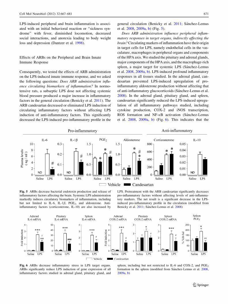
Fig. 5 ARBs decrease bacterial endotoxin production and release of
inflammatory factors affecting the brain. Systemic LPS administration
markedly induces circulatory biomarkers of inflammation, including
but not limited to IL-6, IL-1b, PGE2, and aldosterone. Anti-
inflammatory factors (corticosterone, IL-10) are also increased by
LPS. Pretreatment with the ARB candesartan significantly decreases
pro-inflammatory factors without affecting levels of anti-inflamma-
tory markers. The net result is a significant decrease in the LPS-
induced pro-inflammatory profile in the circulation (modified from
Benicky et al. 2011; Sanchez-Lemus et al. 2008)
Fig. 6 ARBs decrease inflammatory stress in LPS target organs.
ARBs significantly reduce LPS induction of gene expression of all
inflammatory factors studied in adrenal gland, pituitary gland, and
spleen, including but not restricted to IL-6 and COX-2, and PGE2
formation in the spleen (modified from Sanchez-Lemus et al. 2008,
2009a, b)
Cell Mol Neurobiol (2012) 32:667–681 671
123

anti-inflammatory effect of ARBs was widespread through-
out the organism, and amelioration of peripheral inflamma-
tion was not the consequence of hemodynamic effects.
We had demonstrated that systemic ARB administration
reduces inflammation in peripheral tissues and diminishes
production of circulating inflammatory factors affecting the
brain. The reduction in peripheral inflammation may
decrease inflammatory stress to the brain (Dantzer et al.
2008; Rivest 2010). To answer this question, we asked:
Does systemic ARB administration influence brain
inflammation? LPS increased AT1 receptor mRNA in the
PVN, suggesting a participation of AT1 receptor activation
in inflammatory stress (Sanchez-Lemus et al. 2009b).
Systemic candesartan administration blocking not only
peripheral but also brain AT1 receptors significantly
decreased LPS-induced upregulation of inflammatory
cytokines, their receptors, adhesion molecules, iNOS and
COX-2 production, c-fos and NF-jB induction, and
microglia activation (Benicky et al. 2009; Benicky et al.
2011) (Figs. 7, 8). The effects of LPS were widespread,
and were documented not only in the PVN and SFO,
characteristic brain targets for circulating LPS and pro-
inflammatory cytokines, but also in the prefrontal cortex,
amygdala and hippocampus (Benicky et al. 2011) (Figs. 7,
8). In all cases, candesartan significantly limited LPS
inflammatory effects (Benicky et al. 2011). In parallel with
the peripheral anti-inflammatory effects and decreased
circulating inflammatory factors, systemic administration
of candesartan reduced brain inflammation.
Since systemically administered candesartan blocks both
peripheral and brain AT1 receptors (Nishimura et al.
2000b), and all sartans studied, when administered periph-
erally, are able to cross the blood–brain barrier and enter the
brain (Li et al. 1993; Polidori et al. 1998; Wang et al. 2003)
the question remained as to the relative role of peripheral
and brain AT1 receptor blockade on the anti-inflammatory
effects of ARBs. To address this point, we asked:
Is there a central component to the anti-inflammatory
effects of systemically administered ARBs in the brain? We
believe this question may be answered in the affirmative,
since we have earlier demonstrated that brain AT1 receptors
were involved in the effects of systemically administered
ARBs; orally administered candesartan administration
abrogated the drinking response and the increase in blood
pressure produced by intracerebral administration of
Angiotensin II (Seltzer et al. 2004). In addition, ARBs
administered into the cerebral ventricles in preclinical ani-
mal studies prevent the effects of peripherally administered
Angiotensin II in the brain (Kang et al. 2009). However,
since direct infusion of ARBs into the brain is not used in
clinical settings, these experiments do not directly clarify
whether or not ARBs, as administered in the clinic, have
central effects in addition to peripheral actions.
To definitely answer the question of a participation of
brain AT1 receptors in the anti-inflammatory effects of
systemically administered ARBs, we asked:
Do ARBs reduce inflammation directly in brain cells?
We studied the direct effects of candesartan on LPS
inflammation in cultured brain cells. We selected rat pri-
mary cerebellar granule cells because brain AT1 receptors
have a predominant expression in neurons (Tsutsumi and
Saavedra 1991), microglia because of their fundamental
role in central inflammatory responses (Hanisch 2002), and
cerebral microvascular endothelial cells as targets of LPS,
circulating pro-inflammatory factors and Angiotensin II
(Benicky et al. 2011). Candesartan reduced LPS-induced
production of inflammatory cytokines in all cultures,
TNF-a release from cerebellar granule cells, and IL-1brelease from microglia (Benicky et al. 2011) (Fig. 9). In
addition, candesartan prevented NF-jB activation, IL-1b,
IL-6, and TNF-a mRNA expression in cerebellar granule
cells, iNOS upregulation in cerebral microvascular endo-
thelial cells, and IL-1b mRNA expression in cultured cortical
microglia (Benicky et al. 2011). We concluded that ARBs
are capable of reducing inflammation directly on cerebro-
vascular endothelial cells, neurons, and microglia through
multiple mechanisms. For this reason, and because orally
administered ARBs prevent the effects of centrally admin-
istered Angiotensin II (Seltzer et al. 2004), we hypothesize
that part of the anti-inflammatory effects of ARBs may be the
consequence of direct actions in the brain (Figs. 10, 11).
ARB Effects on Behavior
Severe brain inflammation is accompanied by profound
mood and behavioral alterations, including enhanced anxiety
and sickness behavior, representing depressive symptom-
atology (Dantzer et al. 1998; Dantzer et al. 2008). We have
previously demonstrated that systemically administered
ARBs reduced anxiety (Saavedra et al. 2006), an effect
associated with a normalization of stress-induced alterations
in the cortical corticotrophin-releasing factor 1 receptors, a
pro-anxiety regulatory system (Keck and Holsboer 2001;
Bravo et al. 2011), and the anti-anxiety benzodiazepine-1
receptors, part of the inhibitory GABAA complex (Domschke
and Zwanzger 2008). Again, a separated line of evidence
suggested that ARBs may ameliorate anxiety and depression
associated with brain inflammation (Saavedra et al. 2006).
To test our hypothesis, we asked:
Does ARB administration influence inflammation-
induced sickness behavior and anxiety? We found that
candesartan prevented the LPS-induced sickness behavior,
the anorexia leading to weight loss, which are core
symptoms of depression, and reduced anxiety (Benicky
et al. 2011) (Fig. 12). We conclude that ARBs may exert
anti-depressant and anti-anxiety properties.
672 Cell Mol Neurobiol (2012) 32:667–681
123
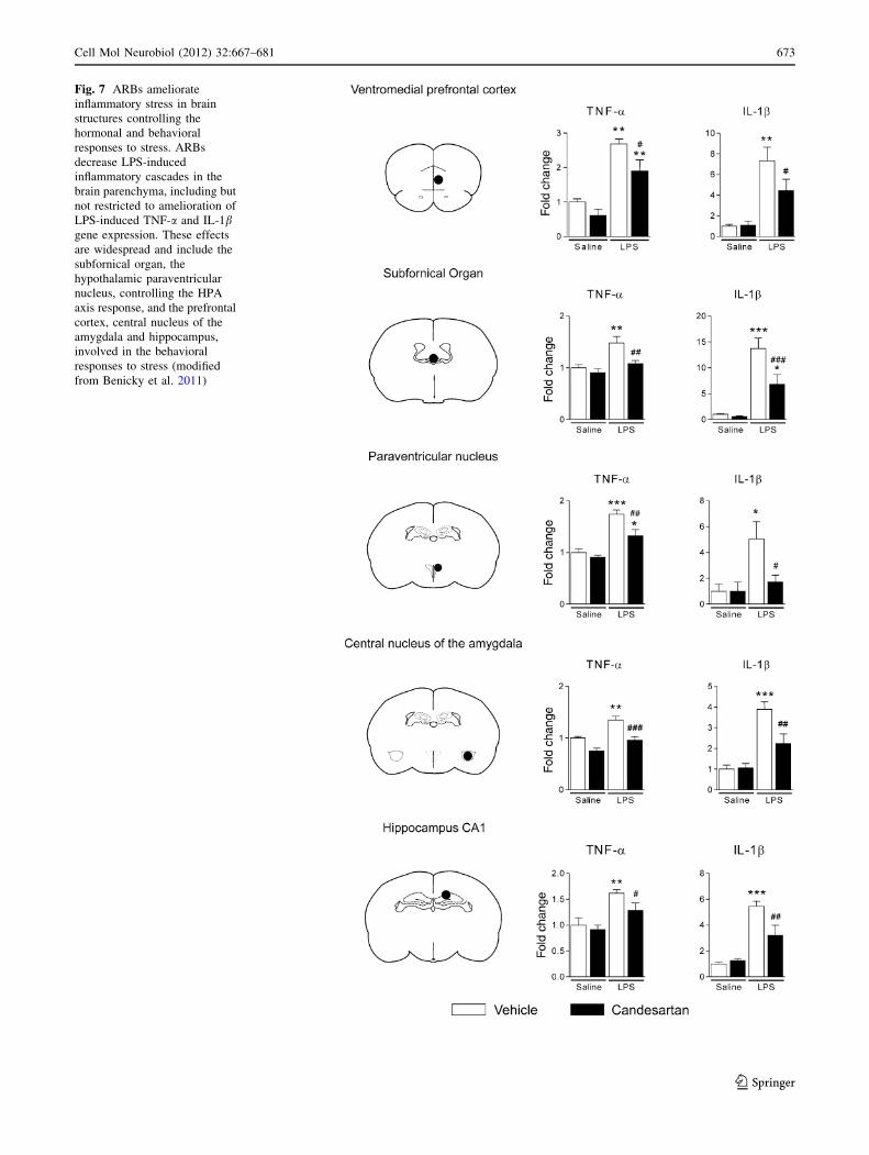
Fig. 7 ARBs ameliorate
inflammatory stress in brain
structures controlling the
hormonal and behavioral
responses to stress. ARBs
decrease LPS-induced
inflammatory cascades in the
brain parenchyma, including but
not restricted to amelioration of
LPS-induced TNF-a and IL-1bgene expression. These effects
are widespread and include the
subfornical organ, the
hypothalamic paraventricular
nucleus, controlling the HPA
axis response, and the prefrontal
cortex, central nucleus of the
amygdala and hippocampus,
involved in the behavioral
responses to stress (modified
from Benicky et al. 2011)
Cell Mol Neurobiol (2012) 32:667–681 673
123
Results obtained in pre-clinical experiments are not
always substantiated in the clinic. For this reason it was
important to consider whether ARBs ameliorated inflam-
matory stress in normal human cells. To answer this
question, we asked:
Do ARBs directly ameliorate inflammation in human
cells? We studied human circulating monocytes obtained
from healthy volunteers. We used LPS at a concentration
substantially below the levels encountered during sepsis
(Larrayoz et al. 2009), and similar to the LPS
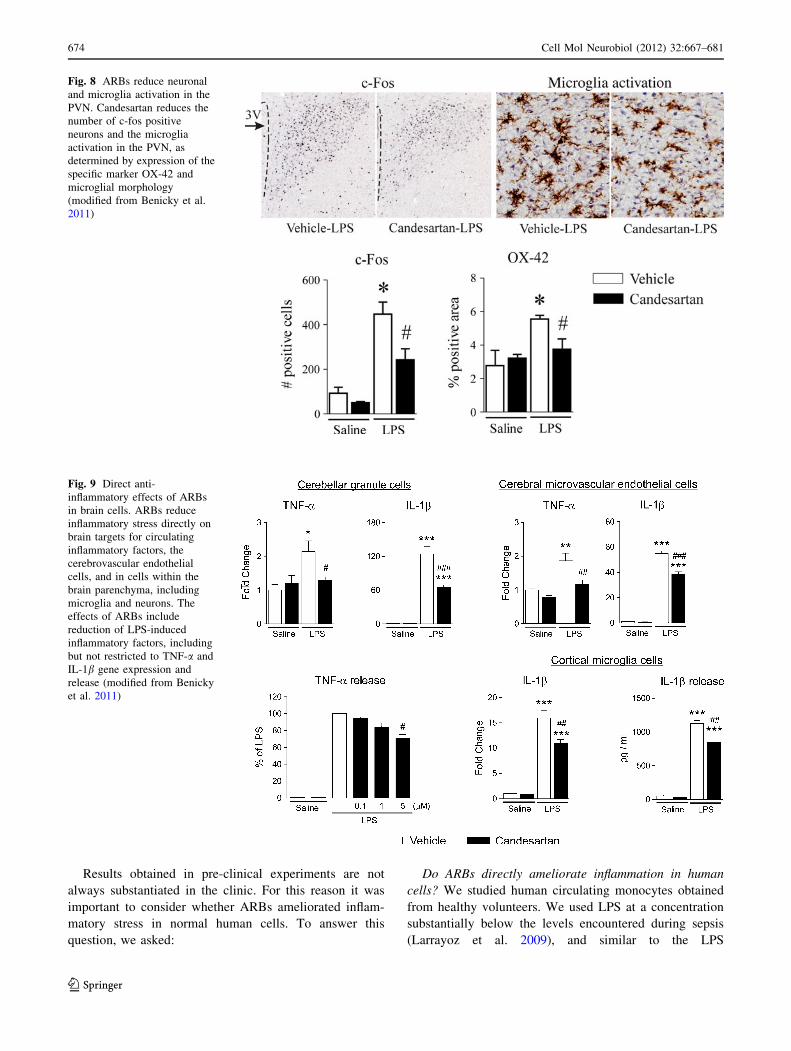
Fig. 8 ARBs reduce neuronal
and microglia activation in the
PVN. Candesartan reduces the
number of c-fos positive
neurons and the microglia
activation in the PVN, as
determined by expression of the
specific marker OX-42 and
microglial morphology
(modified from Benicky et al.
2011)
Fig. 9 Direct anti-
inflammatory effects of ARBs
in brain cells. ARBs reduce
inflammatory stress directly on
brain targets for circulating
inflammatory factors, the
cerebrovascular endothelial
cells, and in cells within the
brain parenchyma, including
microglia and neurons. The
effects of ARBs include
reduction of LPS-induced
inflammatory factors, including
but not restricted to TNF-a and
IL-1b gene expression and
release (modified from Benicky
et al. 2011)
674 Cell Mol Neurobiol (2012) 32:667–681
123
concentrations found in obesity, insulin resistance, diabe-
tes, and those resulting from a high fat and carbohydrate
meal (Schwartz et al. 2010). In human monocytes, cande-
sartan significantly reduced LPS-induced inflammation,
including a reduction of inflammatory cytokine production
and release to the medium, decreased ROS formation and
diminishing NF-jB activation, without affecting secretion
of the anti-inflammatory cytokine IL-10 (Larrayoz et al.
2009) (Fig. 13). We conclude that ARBs directly amelio-
rate inflammatory stress in human circulating monocytes.
Additional Supportive Pre-clinical Evidence
Findings from a single laboratory may not be sufficient to
consider further investment on potential clinical applica-
tions. A consideration of the literature revealed substantial
and highly relevant contributions from other laboratories.
Early developmental stress produces life-long increases in
basal HPA axis and RAS activity (Edwards et al. 1999).
Conversely, anti-anxiety effects of ARBs, of potency
similar to that of benzodiazepines, and antidepressant
effects in mice and rats have been reported by other groups
(Kaiser et al. 1992; Gard et al. 2001; Shekhar et al. 2006).
Other pre-clinical studies on stroke models confirmed our
initial observations of the neuroprotective, anti-inflamma-
tory effects of ARBs (Ozacmak et al. 2007; Hallevi et al.
2007; Jung et al. 2007). These studies also substantiated
our hypothesis of beneficial effects of ARBs unrelated to
hypertension (Sironi et al. 2004), because they demon-
strated that ARBs protect from experimental stroke in
normotensive animals (Lou et al. 2004). Other studies
revealed that ARBs protect from neuronal injury during
retinal inflammation, whole brain irradiation, hypoxia
and experimental injury of dopamine neurons (Robbins
et al. 2009; Kurihara et al. 2006; Mertens et al. 2010;
Grammatopoulos et al. 2007; Nagai et al. 2007; Conner
et al. 2010; Saavedra et al. 2011). ARB administration was
reported to be neuroprotective in experimental models of
autoimmune diseases, such as experimental autoimmune
encephalomyelitis, an animal model of multiple sclerosis,
where ARBs decrease macrophage infiltration and reduce
paralysis (Platten et al. 2009; Lanz et al. 2010). Other
research revealed that ARBs decreased the cognitive
decline produced by central administration of amyloid-beta
(Tsukuda et al. 2009).
Supportive Evidence from Clinical Studies
Clinical reports have been for the most part focused on the
role of ARBs on the prevention and treatment of stroke.
Multicenter studies demonstrated that ARBs are of superior
benefit when compared to other anti-hypertensive therapies
for the treatment of stroke and the preservation of cognition
(Papademetriou et al. 2004; Zanchetti and Elmfeldt 2006;
Julius et al. 2006; Chrysant 2006; Devereux and Dahlof
2007; Saxby et al. 2008; Lu et al. 2009). Although nor-
malization of blood pressure is the cornerstone for the
treatment and prevention of cardiovascular disease, these
findings strongly suggest that factors in addition to simple
blood pressure normalization account for the superior
beneficial effects of ARBs.
Previous uncontrolled studies suggesting that decreased
activation of the brain RAS improved the quality of life and
exerted beneficial effects on mood (Braszko et al. 2003;
Weber 2005) are increasingly confirmed by further clinical
observations (Fogari and Zoppi 2004). ARBs have been
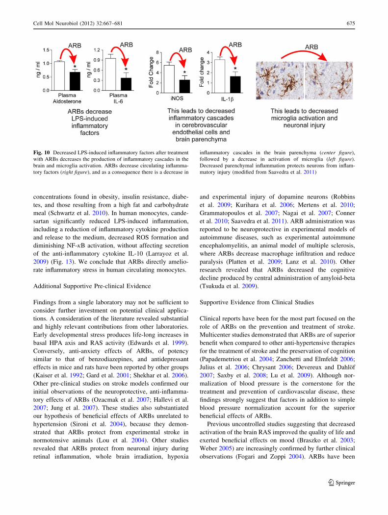
Fig. 10 Decreased LPS-induced inflammatory factors after treatment
with ARBs decreases the production of inflammatory cascades in the
brain and microglia activation. ARBs decrease circulating inflamma-
tory factors (right figure), and as a consequence there is a decrease in
inflammatory cascades in the brain parenchyma (center figure),
followed by a decrease in activation of microglia (left figure).
Decreased parenchymal inflammation protects neurons from inflam-
matory injury (modified from Saavedra et al. 2011)
Cell Mol Neurobiol (2012) 32:667–681 675
123
reported to improve mood and the regulation of the HPA
axis in diabetic patients (Pavlatou et al. 2008). Depressed
patients medicated with ARBs to treat co-morbid hyper-
tension improve their response to and require lower doses
of antidepressants to achieve therapeutic efficacy (Nasr
et al. 2011), and a similar finding was earlier reported in
patients treated with inhibitors of Angiotensin II formation
the Angiotensin Converting Enzyme inhibitors (ACEI)
(Braszko et al. 2003; Hertzman et al. 2005).
A recent cohort prospective analysis revealed that ARB
administration protects cognition and significantly decrea-
ses the progression of Alzheimer’s disease (Li et al. 2010).
This indicated that ARBs have not only potent anti-
inflammatory but also neuroprotective properties, in
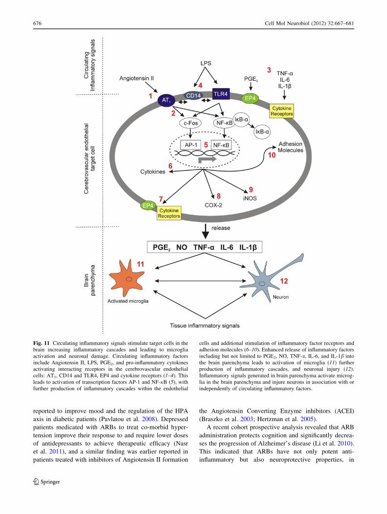
Fig. 11 Circulating inflammatory signals stimulate target cells in the
brain increasing inflammatory cascades and leading to microglia
activation and neuronal damage. Circulating inflammatory factors
include Angiotensin II, LPS, PGE2, and pro-inflammatory cytokines
activating interacting receptors in the cerebrovascular endothelial
cells: AT1, CD14 and TLR4, EP4 and cytokine receptors (1–4). This
leads to activation of transcription factors AP-1 and NF-jB (5), with
further production of inflammatory cascades within the endothelial
cells and additional stimulation of inflammatory factor receptors and
adhesion molecules (6–10). Enhanced release of inflammatory factors
including but not limited to PGE2, NO, TNF-a, IL-6, and IL-1b into
the brain parenchyma leads to activation of microglia (11) further
production of inflammatory cascades, and neuronal injury (12).
Inflammatory signals generated in brain parenchyma activate microg-
lia in the brain parenchyma and injure neurons in association with or
independently of circulating inflammatory factors.
676 Cell Mol Neurobiol (2012) 32:667–681
123
agreement with the multicenter clinical studies on stroke
(Papademetriou et al. 2004; Zanchetti and Elmfeldt 2006;
Julius et al. 2006; Saxby et al. 2008; Lu et al. 2009;
Matsumoto et al. 2010). The neuroprotective effects of
ARBs are increasingly recognized in the cardiovascular
field (Anderson 2010). If the initial data analysis is con-
firmed by well-controlled studies, the beneficial effects of
ARBs on the long-term protection of cognitive function
will represent a major innovative finding of immediate
translational value.
Cardiovascular, metabolic, neurodegenerative, and mood
disorders are substantially influenced by age. This is not sur-
prising because allostatic load is strongly dependent on time.
In pre-clinical studies, it is well-recognized that long-term
ARB or ACEI administration prolongs life (Linz et al. 2000;
Baiardi et al. 2004), a result commonly considered as the
consequence of decreased allostatic load to the cardiovascular
system. These findings have been recently confirmed by the
observation that life-long depletion (Benigni et al. 2009) of
AT1 receptors also significantly increases the life span.
We are left with the intriguing question of whether or
not long-term ARB administration will extend the life span
and improve the quality of life in human populations.
Conclusions
1. Systemic ARB administration ameliorates inflam-
matory stress in the brain.
2. These effects can be demonstrated in genetically
hypertensive rats by the reversal of cerebrovascular
inflammation, after stroke, and following systemic
administration of LPS to normotensive animals.
3. The anti-inflammatory effects of ARBs can be
demonstrated in peripheral organs, the circulation
and the brain, and are independent of their hemody-
namic effects.
4. In the brain, anti-inflammatory effects of ARBs are
widespread, occurring not only in the hypothalamic
centers regulating the HPA axis response to stress,
but also in regions modulating the behavioral
responses to stress, such as the prefrontal cortex
and the hippocampus.
5. ARBs reduce the unwanted behavioral consequences
of brain inflammation, protecting from anxiety and
sickness behavior, core symptoms associated with
depression.
6. ARBs reduce stress-induced allostatic load. While
these compounds prevent the HPA axis and sympa-
thetic over activation characteristic of emotional
stress, they protect from stress-induced disorders
such as gastric ulcerations while conserving benefi-
cial aspects of the HPA axis response (secretion of
anti-inflammatory glucocorticoids) and decreasing its
negative consequences (secretion of pro-inflamma-
tory aldosterone).
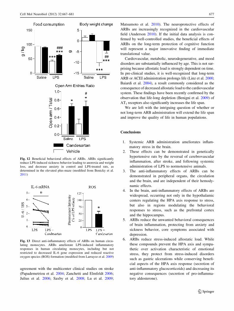
Fig. 12 Beneficial behavioral effects of ARBs. ARBs significantly
reduce LPS-induced sickness behavior leading to anorexia and weight
loss, and decrease anxiety in control and LPS-treated rats, as
determined in the elevated plus-maze (modified from Benicky et al.
2011)
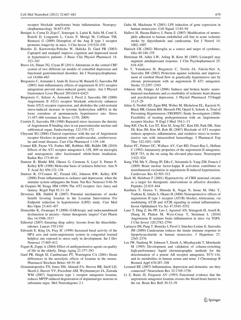
Fig. 13 Direct anti-inflammatory effects of ARBs on human circu-
lating monocytes. ARBs ameliorate LPS-induced inflammatory
responses in human circulating monocytes, including but not
restricted to decreased IL-6 gene expression and reduced reactive
oxygen species (ROS) formation (modified from Larrayoz et al. 2009)
Cell Mol Neurobiol (2012) 32:667–681 677
123

7. Associated mechanisms are responsible for the anti-
inflammatory effects of ARBs. They include reduc-
tion of pro-inflammatory factors in the circulation
leading to the production of inflammatory cascades in
the brain parenchyma. They also exert direct anti-
inflammatory effects on brain cells target for circu-
lating inflammatory factors, such as the cerebrovas-
cular endothelial cells, and in cells located within the
brain parenchyma, including microglia and neurons.
8. Amelioration of inflammatory stress in the brain is a
factor involved in the neuroprotective effects of
ARBs in a number of conditions (stroke, irradiation,
hypoxia) leading to neuronal injury.
9. The neuroprotective effects of ARB explain the
increasing evidence linking the use of these com-
pounds with the protection of cognition in stroke and
Alzheimer’s disease.
10. ARB-induced neuroprotection may be the combina-
tion not only of their anti-inflammatory effects but of
a number of additional mechanisms, including pro-
tection of the cerebrovascular flow, regulation of the
HPA axis response to stress and direct neuroprotec-
tive effects in the brain parenchyma.
Relevance
Excessive, poorly controlled inflammation is recognized as
a major stress factor increasing allostatic load in peripheral
organs, contributing to the loss of homeostasis and cardio-
vascular and metabolic disease. Regulatory mechanisms
and disease factors share multiple points of contact between
the periphery and the brain, and allostatic load in the
periphery and the brain influence each other. This explains
the significant co-morbidity between mood and degenera-
tive diseases of the brain, cardiovascular, and metabolic
disorders (Johnson and Grippo 2006; Szczepanska-Sado-
wska et al. 2010). Because of their pleiotropic anti-inflam-
matory and beneficial metabolic effects and their excellent
safety record, ARBs are increasingly utilized as first line of
treatment in hypertension, metabolic syndrome, and dia-
betes. The willingness to consider alternative and additional
research avenues has lead to the utilization of novel thera-
pies for the treatment of hypertension and diabetes, result-
ing in incremental and sustained improvements in the
treatment of these conditions. In contrast, there have been
no major advances in the therapy of mood and neurode-
generative disorders during the last decades, and several
major pharmaceutical companies have recently made the
decision to substantially reduce or eliminate their CNS drug
discovery operations (Miller 2010). The treatment of major
brain disorders remains incomplete and unsatisfactory.
Several conclusions may be drawn from the above.
Research in the mechanisms and treatment of brain disor-
ders should be open to the consideration of novel alterna-
tives. Increasing consideration should be made not only of
the fundamental differences between brain and peripheral
disorders, but also on their points of contact leading to their
recognized co-morbidity (Szczepanska-Sadowska et al.
2010). Considering the above, the findings demonstrating
that ARBs ameliorate inflammatory stress in the brain,
reduce allostatic load and are neuroprotective remain,
while novel, not entirely surprising. However, the use of
ARBs in mood and neurodegenerative disorders has not
been adequately tested. We propose to consider the ARBs
as part of an integrative novel approach for the treatment of
brain disorders.
Acknowledgments This study was supported by the Division of
Intramural Research Programs, National Institute of Mental Health,
National Institutes of Health, Department of Health and Human
Services, USA.
References
Allen AM, MacGregor DP, McKinley MJ, Mendelsohn FA (1999)
Angiotensin II receptors in the human brain. Regul Pept 79:1–7
Anderson C (2010) More indirect evidence of potential neuroprotec-
tive benefits of Angiotensin receptor blockers. J Hypertens
28:429
Ando H, Zhou J, Macova M, Imboden H, Saavedra JM (2004)
Angiotensin II AT1 receptor blockade reverses pathological
hypertrophy and inflammation in brain microvessels of sponta-
neously hypertensive rats. Stroke 35:1726–1731
Anisman H (2009) Cascading effects of stressors and inflammatory
immune system activation: implications for major depressive
disorder. J Psychiatry Neurosci 34:4–20
Armando I, Carranza A, Nishimura Y, Hoe KL, Barontini M, Terron JA,
Falcon-Neri A, Ito T, Juorio AV, Saavedra JM (2001) Peripheral
administration of an angiotensin II AT(1) receptor antagonist
decreases the hypothalamic-pituitary-adrenal response to isolation
stress. Endocrinology 142:3880–3889
Armando I, Volpi S, Aguilera G, Saavedra JM (2007) Angiotensin II
AT1 receptor blockade prevents the hypothalamic corticotropin-
releasing factor response to isolation stress. Brain Res 1142:
92–99
Bader M (2010) Tissue renin-angiotensin-aldosterone systems: targets
for pharmacological therapy. Ann Rev Pharm Tox 50:439–465
Baiardi G, Bregonzio C, Jezova M, Armando I, Saavedra JM (2004)
Angiotensin II AT1 receptor blockade prolongs the lifespan of
spontaneously hypertensive rats and reduces stress-induced
release of catecholamines, glucocorticoids, and vasopressin.
Ann N Y Acad Sci 1018:131–136
Barra S, Vitagliano A, Cuomo V, Vitagliano G, Gaeta G (2009)
Vascular and metabolic effects of angiotensin II receptor
blockers. Exp Opin Pharmacother 10:173–189
Benicky J, Sanchez-Lemus E, Pavel J, Saavedra JM (2009) Anti-
inflammatory effects of angiotensin receptor blockers in the
brain and the periphery. Cell Mol Neurobiol 29:781–792
Benicky J, Sanchez-Lemus E, Honda M, Pang T, Orecna M, Wang J,
Leng Y, Chuang DM, Saavedra JM (2011) Angiotensin II AT(1)
678 Cell Mol Neurobiol (2012) 32:667–681
123
receptor blockade ameliorates brain inflammation. Neuropsy-
chopharmacology 36:857–870
Benigni A, Corna D, Zoja C, Sonzogni A, Latini R, Salio M, Conti S,
Rottoli D, Longaretti L, Cassis P, Morigi M, Coffman TM,
Remuzzi G (2009) Disruption of the Ang II type 1 receptor
promotes longevity in mice. J Clin Invest 119:524–530
Braszko JJ, Karwowska-Polecka W, Halicka D, Gard PR (2003)
Captopril and enalapril improve cognition and depressed mood
in hypertensive patients. J Basic Clin Physiol Pharmacol 14:
323–343
Bravo JA, Dinan TG, Cryan JF (2011) Alterations in the central CRF
system of two different rat models of comorbid depression and
functional gastrointestinal disorders. Int J Neuropsychopharma-
col 14:666–683
Bregonzio C, Armando I, Ando H, Jezova M, Baiardi G, Saavedra JM
(2003) Anti-inflammatory effects of angiotensin II AT1 receptor
antagonism prevent stress-induced gastric injury. Am J Physiol
Gastrointest Liver Physiol 285:G414–G423
Bregonzio C, Seltzer A, Armando I, Pavel J, Saavedra JM (2008)
Angiotensin II AT(1) receptor blockade selectively enhances
brain AT(2) receptor expression, and abolishes the cold-restraint
stress-induced increase in tyrosine hydroxylase mRNA in the
locus coeruleus of spontaneously hypertensive rats. Stress
11:457–466 (erratum in Stress 12:95, 2009)
Castren E, Saavedra JM (1988) Repeated stress increases the density
of Angiotensin II binding sites in rat paraventricular nucleus and
subfornical organ. Endocrinology 122:370–372
Chrysant SG (2006) Clinical experience with the use of Angiotensin
receptor blockers in patients with cardiovascular, cerebrovascu-
lar and renal diseases. Curr Clin Pharmacol 1:139–146
Conner KR, Payne VS, Forbes ME, Robbins ME, Riddle DR (2010)
Effects of the AT1 receptor antagonist L-158, 809 on microglia
and neurogenesis after fractionated whole-brain irradiation.
Radiat Res 173:49–61
Dantzer R, Bluthe RM, Gheusi G, Cremona S, Laye S, Parnet P,
Kelley KW (1998) Molecular basis of sickness behavior. Ann N
Y Acad Sci 856:132–138
Dantzer R, O’Connor JC, Freund GG, Johnson RW, Kelley KW
(2008) From inflammation to sickness and depression: when the
immune system subjugates the brain. Nat Rev Neurosci 9:46–56
de Gasparo M, Siragy HM (1999) The AT2 receptor: fact, fancy and
fantasy. Regul Pept 81:11–24
Devereux RB, Dahlof B (2007) Potential mechanisms of stroke
benefit favoring losartan in the Losartan Intervention For
Endpoint reduction in hypertension (LIFE) study. Curr Med
Res Opin 23:443–457
Domschke K, Zwanzger P (2008) GABAergic and endocannabinoid
dysfunction in anxiety—future therapeutic targets? Curr Pharm
Des 14:3508–3517
Editorial (2007) Ensuring drug safety: lessons from the thiazolidin-
ediones. Lancet 370:1101
Edwards E, King JA, Fray JC (1999) Increased basal activity of the
HPA axis and renin-angiotensin system in congenital learned
helpless rats exposed to stress early in development. Int J Dev
Neurosci 17:805–812
Fogari R, Zoppi A (2004) Effect of antihypertensive agents on quality
of life in the elderly. Drugs Aging 21:377–393
Gard PR, Haigh SJ, Cambursano PT, Warrington CA (2001) Strain
differences in the anxiolytic effects of losartan in the mouse.
Pharmacol Biochem Behav 69:35–40
Grammatopoulos TN, Jones SM, Ahmadi FA, Hoover BR, Snell LD,
Skoch J, Jhaveri VV, Poczobutt AM, Weyhenmeyer JA, Zawada
WM (2007) Angiotensin type 1 receptor antagonist losartan,
reduces MPTP-induced degeneration of dopaminergic neurons in
substantia nigra. Mol Neurodegener 2:1
Guha M, Mackman N (2001) LPS induction of gene expression in
human monocytes. Cell Signal 13:85–94
Hallevi H, Hazan-Halevy I, Paran E (2007) Modification of neutro-
phils adhesion to human endothelial cell line in acute ischemic
stroke by dipyridamole and candesartan. Eur J Neurol 14:
1002–1007
Hanisch UK (2002) Microglia as a source and target of cytokines.
Glia 40:140–155
Hertzman M, Adler LW, Arling B, Kern M (2005) Lisinopril may
augment antidepressant response. J Clin Psychopharmacol 25:
618–620
Ito T, Yamakawa H, Bregonzio C, Terron JA, Falcon-Neri A,
Saavedra JM (2002) Protection against ischemia and improve-
ment of cerebral blood flow in genetically hypertensive rats by
chronic pretreatment with an angiotensin II AT1 antagonist.
Stroke 33:2297–2303
Johnson AK, Grippo AJ (2006) Sadness and broken hearts: neuro-
humoral mechanisms and co-morbidity of ischemic heart disease
and psychological depression. J Physiol Pharmacol 57(Suppl
11):5–29
Julius S, Nesbitt SD, Egan BM, Weber M, Michelson EL, Kaciroti N,
Black HR, Grimm RH, Messerli FH, Oparil S, Schork A, Trial of
Preventing Hypertension (TROPHY) Study Investigators (2006)
Feasibility of treating prehypertension with an Angiotensin-
receptor blocker. N Engl J Med 354:1–13
Jung KH, Chu K, Lee ST, Kim SJ, Song EC, Kim EH, Park DK, Sinn
DI, Kim JM, Kim M, Roh JK (2007) Blockade of AT1 receptor
reduces apoptosis, inflammation, and oxidative stress in normo-
tensive rats with intracerebral hemorrhage. J Pharmacol Exp
Ther 322:1051–1058
Kaiser FC, Palmer GC, Wallace AV, Carr RD, Fraser-Rae L, Hallam
C (1992) Antianxiety properties of the angiotensin II antagonist,
DUP 753, in the rat using the elevated plus-maze. Neuroreport
3:922–924
Kang YM, Ma Y, Zheng JP, Elks C, Sriramula S, Yang ZM, Francis J
(2009) Brain nuclear factor-kappa B activation contributes to
neurohumoral excitation in angiotensin II-induced hypertension.
Cardiovasc Res 82:503–512
Keck M, Holsboer F (2001) Hyperactivity of CRH neuronal circuits
as a target for therapeutic interventions in affective disorders.
Peptides 22:835–844
Kurihara T, Ozawa Y, Shinoda K, Nagai N, Inoue M, Oike Y,
Tsubota K, Ishida S, Okano H (2006) Neuroprotective effects of
angiotensin II type 1 receptor (AT1R) blocker, telmisartan, via
modulating AT1R and AT2R signaling in retinal inflammation.
Invest Ophthalmol Vis Sci 47:5545–5552
Lanz T, Ding Z, Ho PP, Luo J, Agrawal AN, Srinagesh H, Axtell R,
Zhang H, Platten M, Wyss-Coray T, Steinman L (2010)
Angiotensin II sustains brain inflammation in mice via TGFb.
J Clin Invest 120:2782–2794
Larrayoz IM, Pang T, Benicky J, Pavel J, Sanchez-Lemus E, Saavedra
JM (2009) Candesartan reduces the innate immune response to
lipopolysaccharide in human monocytes. J Hypertens 27:
2365–2376
Lee JW, Naidong W, Johnson T, Dzerk A, Miyabayashi T, Motohashi
M (1995) Development and validation of column-switching
high-performance liquid chromatographic methods for the
determination of a potent AII receptor antagonist, TCV-116,
and its metabolites in human serum and urine. J Chromatogr B
Biomed Appl 670:287–298
Leonard BE (2007) Inflammation, depression and dementia: are they
connected? Neurochem Res 32:1749–1756
Li Z, Bains JS, Ferguson AV (1993) Functional evidence that the
angiotensin antagonist losartan crosses the blood-brain barrier in
the rat. Brain Res Bull 30:33–39
Cell Mol Neurobiol (2012) 32:667–681 679
123
Li NC, Lee A, Whitmer RA, Kivipelto M, Lawler E, Kazis LE,
Wolozin B (2010) Use of Angiotensin receptor blockers and risk
of dementia in a predominantly male population: prospective
cohort analysis. BMJ 340:b5465
Linz W, Heitsch H, Scholkens BA, Wiemer G (2000) Long-term
angiotensin II type 1 receptor blockade with fonsartan doubles
lifespan of hypertensive rats. Hypertension 35:908–913
Lou M, Blume A, Zhao Y, Gohlke P, Deuschl G, Herdegen T,
Culman J (2004) Sustained blockade of brain AT1 receptors
before and after focal cerebral ischemia alleviates neurologic
deficits and reduces neuronal injury, apoptosis, and inflammatory
responses in the rat. J Cereb Blood Flow Metab 24:536–547
Lu GC, Cheng JW, Zhu KM, Ma XJ, Shen FM, Su DF (2009) A
systematic review of Angiotensin receptor blockers in preventing
stroke. Stroke 40:3876–3878
Marchesi VT (2011) Alzheimer’s dementia begins as a disease of
small blood vessels, damaged by oxidative-induced inflamma-
tion and dysregulated amyloid metabolism: implications for
early detection and therapy. FASEB J 25:5–13
Marchesi C, Paradis P, Schiffrin EL (2008) Role of the renin-
angiotensin system in vascular inflammation. Trends Pharmacol
Sci 29:367–374
Matsumoto S, Shimodozono M, Miyata R, Kawahira K (2010) The
Angiotensin II type 1 receptor antagonist olmesartan preserves
cerebral blood flow and cerebrovascular reserve capacity, and
accelerates rehabilitative outcomes in hypertensive patients with
a history of stroke. Int J Neurosci 120:372–380
McFarlane SI (2009) Role of angiotensin receptor blockers in
diabetes: implications of recent clinical trials. Expert Rev
CardiovascTher 7:1363–1371
Mertens B, Vanderheyden P, Michotte Y, Sarre S (2010) The role of
the central renin-angiotensin system in Parkinson’s disease.
J Renin Angiotensin Aldosterone Syst 11:49–56
Miller G (2010) Is pharma running out of brainy ideas? Science
329:502–504
Nagai N, Izumi-Nagai K, Oike Y, Koto T, Satofuka S, Ozawa Y,
Yamashiro K, Inoue M, Tsubota K, Umezawa K, Ishida S (2007)
Suppression of diabetes-induced retinal inflammation by block-
ing the Angiotensin II type 1 receptor or its downstream nuclear
factor-kB pathway. Invest Ophthalmol Vis Sci 48:4342–4350
Nasr SJ, Crayton JW, Agarwal B, Wendt B, Kora R (2011) Lower
frequency of antidepressant use in patients on renin-angiotensin-
aldosterone system modifying medications. Cell Mol Neurobiol
31:615–618
Nimmo AJ, Vink R (2009) Recent patents in CNS drug discovery: the
management of inflammation in the central nervous system.
Recent Pat CNS Drug Discov 4:86–95
Nishimura Y, Ito T, Saavedra JM (2000a) Angiotensin II AT(1)
blockade normalizes cerebrovascular autoregulation and reduces
cerebral ischemia in spontaneously hypertensive rats. Stroke
31:2478–2486
Nishimura Y, Ito T, Hoe K, Saavedra JM (2000b) Chronic
peripheral administration of the angiotensin II AT(1) receptor
antagonist candesartan blocks brain AT(1) receptors. Brain Res
871:29–38
Ozacmak VH, Sayan H, Cetin A, Akyildiz-Igdem A (2007) AT1
receptor blocker candesartan-induced attenuation of brain injury
of rats subjected to chronic cerebral hypoperfusion. Neurochem
Res 32:1314–1321
Papademetriou V, Farsang C, Elmfeldt D, Hofman A, Lithell H,
Olofsson B, Skoog I, Trenkwalder P, Zanchetti A (2004) Study
on Cognition and Prognosis in the Elderly study group. Stroke
prevention with the angiotensin II type 1-receptor blocker
candesartan in elderly patients with isolated systolic hyperten-
sion: the Study on Cognition and Prognosis in the Elderly
(SCOPE). J Am Coll Cardiol 44:1175–1180
Pascoe MC, Crewther SG, Carey LM, Crewther DP (2011) Inflam-
mation and depression: why poststroke depression may be the
norm and not the exception. Int J Stroke 6:128–135
Pavlatou MG, Mastorakos G, Lekakis I, Liatis S, Vamvakou G,
Zoumakis E, Papassotiriou I, Rabavilas AD, Katsilambros N,
Chrousos GP (2008) Chronic administration of an angiotensin II
receptor antagonist resets the hypothalamic-pituitary-adrenal
(HPA) axis and improves the affect of patients with diabetes
mellitus type 2: preliminary results. Stress 11:62–72
Platten M, Youssef S, Hur EM, Ho PP, Han MH, Lanz TV, Phillips
LK, Goldstein MJ, Bhat R, Raine CS, Sobel RA, Steinman L
(2009) Blocking Angiotensin-converting enzyme induces potent
regulatory T cells and modulates TH1-andTH17-mediated
autoimmunity. Proc Natl Acad Sci USA 106:14948–14953
Polidori C, Ciccocioppo R, Nisato D, Cazaubon C, Massi M (1998)
Evaluation of the ability of irbesartan to cross the blood-brain
barrier following acute intragastric treatment. Eur J Pharmacol
352:15–21
Porrello ER, Delbridge LM, Thomas WG (2009) The angiotensin II
type 2 (AT2) receptor: an enigmatic seven transmembrane
receptor. Front Biosci 14:958–972
Quan N, Banks WA (2007) Brain-immune communication pathways.
Brain Behav Immun 21:727–735
Rivest S (2010) Interactions between the immune and neuroendocrine
systems. Prog Brain Res 181:43–53
Robbins ME, Payne V, Tommasi E, Diz DI, Hsu FC, Brown WR,
Wheeler KT, Olson J, Zhao W (2009) The AT1 receptor
antagonist, L-158, 809, prevents or ameliorates fractionated
whole-brain irradiation-induced cognitive impairment. Int.
J Radiat Oncol Biol Phys 73:499–505
Rompe F, Unger T, Steckelings UM (2010) The angiotensin AT2
receptor in inflammation. Drug News Perspect 23:104–111
Saavedra JM (1992) Brain and pituitary Angiotensin. Endocrin Rev
13:329–380
Saavedra JM (2005) Brain angiotensin II: new developments,
unanswered questions and therapeutic opportunities. Cell Mol
Neurobiol 25:485–512
Saavedra JM, Benicky J (2007) Brain and peripheral angiotensin II
play a major role in stress. Stress 10:185–193
Saavedra JM, Armando I, Bregonzio C, Juorio A, Macova M, Pavel J,
Sanchez-Lemus E (2006) A centrally acting, anxiolytic angio-
tensin II AT1 receptor antagonist prevents the isolation stress-
induced decrease in cortical CRF1 receptor and benzodiazepine
binding. Neuropsychopharmacology. 31:1123–1134
Saavedra JM, Sanchez-Lemus E, Benicky J (2011) Blockade of brain
angiotensin II AT1 receptors ameliorates stress, anxiety, brain
inflammation and ischemia: therapeutic implications. Psycho-
neuroendocrinology 36:1–18
Sanchez-Lemus E, Murakami Y, Larrayoz-Roldan IM, Moughamian
AJ, Pavel J, Nishioku T, Saavedra JM (2008) Angiotensin II AT1
receptor blockade decreases lipopolysaccharide-induced inflam-
mation in the rat adrenal gland. Endocrinology 149:5177–5188
Sanchez-Lemus E, Benicky J, Pavel J, Larrayoz IM, Zhou J, Baliova
M, Nishioku T, Saavedra JM (2009a) Angiotensin II AT1
blockade reduces the lipopolysaccharide-induced innate immune
response in rat spleen. Am J Physiol Regul Integr Comp Physiol
296:R1376–R1384
Sanchez-Lemus E, Benicky J, Pavel J, Saavedra JM (2009b) In vivo
Angiotensin II AT1 receptor blockade selectively inhibits LPS-
induced innate immune response and ACTH release in rat
pituitary gland. Brain Behav Immun 23:945–957
Savoia C, Schiffrin EL (2007) Vascular inflammation in hypertension
and diabetes: molecular mechanisms and therapeutic interven-
tions. Clin Sci (Lond) 112:375–384
Saxby BK, Harrington F, Wesnes KA, McKeith IG, Ford GA (2008)
Candesartan and cognitive decline in older patients with
680 Cell Mol Neurobiol (2012) 32:667–681
123
hypertension: a substudy of the SCOPE trial. Neurology 70:
1858–1866
Schwartz EA, Zhang WY, Karnik SK, Borwege S, Anand VR, Laine
PS, Su Y, Reaven PD (2010) Nutrient modification of the innate
immune response: a novel mechanism by which saturated fatty
acids greatly amplify monocyte inflammation. Arterioscler
Thromb Vasc Biol 30:802–808
Seltzer A, Bregonzio C, Armando I, Baiardi G, Saavedra JM (2004)
Oral administration of an AT1 receptor antagonist prevents the
central effects of angiotensin II in spontaneously hypertensive
rats. Brain Res 1028:9–18
Shekhar A, Johnson PL, Sajdyk TJ, Fitz SD, Keim SR, Kelley PE,
Gehlert DR, DiMicco JA (2006) Angiotensin-II is a putative
neurotransmitter in lactate-induced panic-like responses in rats
with disruption of GABAergic inhibition in the dorsomedial
hypothalamus. J Neurosci 26:9205–9215
Sironi L, Gelosa P, Guerrini U, Banfi C, Crippa V, Brioschi M,
Gianazza E, Nobili E, Gianella A, de Gasparo M, Tremoli E
(2004) Anti-inflammatory effects of AT1 receptor blockade
provide end-organ protection in stroke-prone rats independently
from blood pressure fall. J Pharmacol Exp Ther 311:989–995
Skrbic R, Igic R (2009) Seven decades of angiotensin (1939–2009).
Peptides 30:1945–1950
Szczepanska-Sadowska E, Cudnoch-Jedrzejewska A, Ufnal M, Zera
T (2010) Brain and cardiovascular diseases: common neurogenic
background of cardiovascular, metabolic and inflammatory
diseases. J Physiol Pharmacol 61:509–521
Tansey MG, Goldberg MS (2010) Neuroinflammation in Parkinson’s
disease: its role in neuronal death and implications for
therapeutic intervention. Neurobiol Dis 37:510–518
Timmermans PB, Wong PC, Chiu AT, Herblin WF, Benfield P,
Carini DJ, Lee RJ, Wexler RR, Saye JA, Smith RD (1993)
Angiotensin II receptors and angiotensin II receptor antagonists.
Pharmacol Rev 45:205–251
Tsukuda K, Mogi M, Iwanami J, Min LJ, Sakata A, Jing F, Iwai M,
Horiuchi M (2009) Cognitive deficit in amyloid-beta-injected
mice was improved by pretreatment with a low dose of
telmisartan partly because of peroxisome proliferator-activated
receptor-gamma activation. Hypertension 54:782–787
Tsutsumi K, Saavedra JM (1991) Characterization and development
of angiotensin II receptor subtypes (AT1 and AT2) in rat brain.
Am J Physiol 261:R209–R216
Wang JM, Tan J, Leenen FH (2003) Central nervous system blockade
by peripheral administration of AT1 receptor blockers. J Cardio-
vasc Pharmacol 41:593–599
Weber MA (2005) Angiotensin-II receptor blockers for hypertension
and heart failure: quality of life and outcomes. Manag Care
Interface 18:47–54
Weinberg AJ, Zappe DH, Ashton M, Weinberg MS (2004) Safety and
tolerability of high-dose angiotensin receptor blocker therapy in
patients with chronic kidney disease: a pilot study. Am J Nephrol
24:340–345
Xang G, Xi ZX, Wan Y, Wang H, Bi G (1993) Changes in circulating
and tissue Angiotensin II during acute and chronic stress. Biol
Signals 2:166–172
Yamakawa H, Jezova M, Ando H, Saavedra JM (2003) Normalization
of endothelial and inducible nitric oxide synthase expression in
brain microvessels of spontaneously hypertensive rats by
angiotensin II AT1 receptor inhibition. J Cereb Blood Flow
Metab 23:371–380
Zanchetti A, Elmfeldt D (2006) Findings and implications of the
Study on Cognition and Prognosis in the Elderly (SCOPE)—a
review. Blood Press 15:71–79
Zhang M, Mao Y, Ramirez SH, Tuma RF, Chabrashvili T (2010)
Angiotensin II induced cerebral microvascular inflammation and
increased blood-brain barrier permeability via oxidative stress.
Neuroscience 171:852–858
Zhou J, Ando H, Macova M, Dou J, Saavedra JM (2005) Angiotensin
II AT1 receptor blockade abolishes brain microvascular inflam-
mation and heat shock protein responses in hypertensive rats.
J Cereb Blood Flow Metab 25:878–886
Cell Mol Neurobiol (2012) 32:667–681 681
123