Embed Size (px)
Citation preview
Angiosomes andWound Care in theDiabetic Foot
Mark W. Clemens, MDa, Christopher E. Attinger, MDb,*
KEYWORDS
� Angiosomes � Wound care � Diabetic foot � Vascular anatomy
Successful limb salvage is dependent on detailed knowledge of the vascular anatomyof the foot and ankle. The foot and ankle are composed of 6 distinct angiosomes;three-dimensional blocks of tissue fed by source arteries with functional vascularinterconnections between muscle, fascia and skin. Because the foot and ankle arean end organ, their main arteries have numerous direct arterial-arterial connectionsthat allow alternative routes of blood flow to develop if the direct route is disruptedor compromised. Understanding the boundaries of the angiosome and the vascularconnections among its source arteries provides the basis for logically rather thanempirically designed incisions for tissue exposure or to plan reconstructions or ampu-tations that ultimately preserve blood flow for a surgical wound to heal.
Ian Taylor1 first defined the angiosome principle by expanding on the work of previousanatomists2–9 to further define the vascular anatomy of muscle and skin. He defined anangiosome as a three-dimensional anatomic unit of tissue fed by a source artery. Taylorand Minabe10 defined at least 40 angiosomes in the body that were interconnected byeither reduced caliber choke vessels or by similar caliber anastomotic arteries.11,12
These choke vessels can be important safety conduits that allow one angiosome toeventually provide blood flow to an adjacent angiosome if the source artery of the latteris damaged. A unified network can be created so that one source artery can provideblood flow to multiple angiosomes beyond its immediate border. Occluding or interrupt-ing one source artery surgically manipulates the system so that blood flows through theneighboring choke vessels. This is an anatomic explanation for the delay phenom-enon.13,14 Although choke vessels provide an indirect connection among angiosomes,there are also direct arterial-arterial connections that allow blood flow to immediatelybypass local obstructions in the vascular tree. The 6 angiosomes of the foot and ankleoriginate from the 3 main source arteries: the posterior tibial artery supplies the medial
Drs Clemens and Attinger have no financial disclosures.a Department of Plastic Surgery, Georgetown University Medical Center, 3800 Reservoir RoadNorthwest, Washington, DC 20007, USAb Division of Wound Healing, Department of Plastic Surgery, Georgetown University MedicalCenter, 1st Floor Bles Building, 3800 Reservoir Road Northwest, Washington, DC 20007, USA* Corresponding author.E-mail address: [email protected]
Foot Ankle Clin N Am 15 (2010) 439–464doi:10.1016/j.fcl.2010.04.003 foot.theclinics.com1083-7515/10/$ – see front matter ª 2010 Elsevier Inc. All rights reserved.
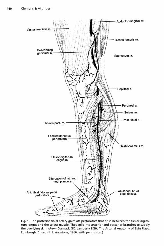
Fig. 1. The posterior tibial artery gives off perforators that arise between the flexor digito-rum longus and the soleus muscle. They split into anterior and posterior branches to supplythe overlying skin. (From Cormack GC, Lamberty BGH. The Arterial Anatomy of Skin Flaps.Edinburgh: Churchill Livingstone, 1986; with permission.)
Clemens & Attinger440
Angiosomes and Wound Care in the Diabetic Foot 441
ankle and the plantar foot, the anterior tibial artery supplies the dorsum of the foot, andthe peroneal artery supplies the anterolateral ankle and the lateral rear foot. These largeangiosomes of the foot can be further broken into angiosomes of the major branches ofthe above arteries. The 3 main branches of the posterior tibial artery each supply distinctportions of the plantar foot: the calcaneal branch (heel), the medial plantar artery(instep), and the lateral plantar artery (lateral midfoot and forefoot). The 2 branches ofthe peroneal artery supply the anterolateral portion of the ankle and rear foot, the ante-rior perforating branch (lateral anterior upper ankle), and the calcaneal branch (lateraland plantar heel). The anterior tibial artery supplies the anterior ankle and then becomesthe dorsalis pedis artery, which supplies the dorsum of the foot. Detailed descriptions ofthe vascular anatomy15 and angiosomes of the lower leg, foot, and ankle have beenthoroughly illustrated elsewhere.16–18
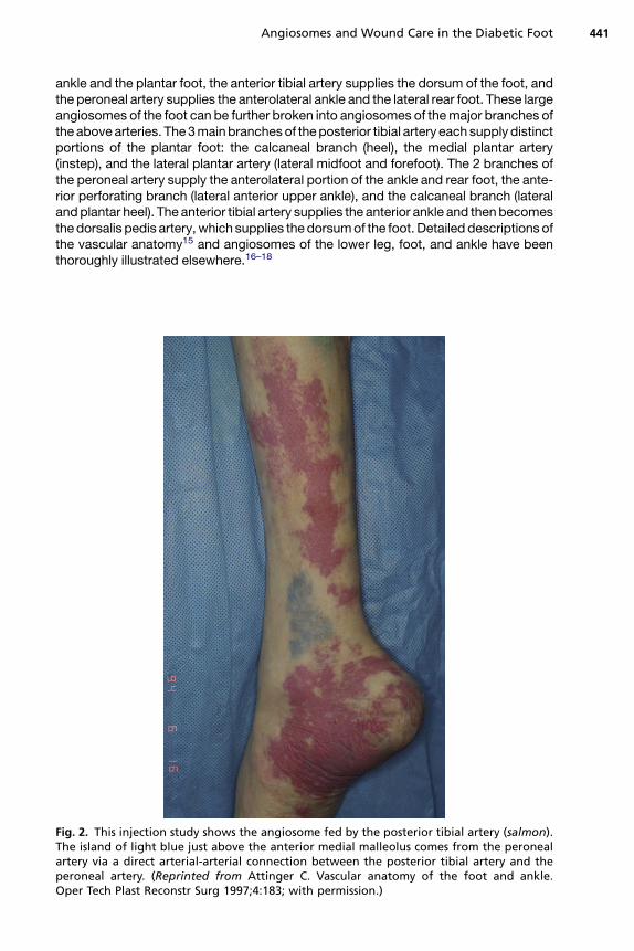
Fig. 2. This injection study shows the angiosome fed by the posterior tibial artery (salmon).The island of light blue just above the anterior medial malleolus comes from the peronealartery via a direct arterial-arterial connection between the posterior tibial artery and theperoneal artery. (Reprinted from Attinger C. Vascular anatomy of the foot and ankle.Oper Tech Plast Reconstr Surg 1997;4:183; with permission.)
Clemens & Attinger442
THE 3 POSTERIOR TIBIAL ARTERY ANGIOSOMES FED BY THE CALCANEAL, MEDIALPLANTAR, AND LATERAL PLANTAR ARTERIES
In the leg, the posterior tibial artery supplies the medial lower leg, starting from theanterior medial border of the tibia and extending posteriorly to the midline of thecalf over the central raphe of the Achilles tendon (Figs. 1 and 2). There are smallerperforator arteries along the course of the posterior tibial artery that perforate throughthe flexor digitorum longus and/or soleus to supply the overlying skin. In addition,there are smaller serial branches to the deep flexor muscles, the medial half of thesoleus muscle, and the Achilles tendon.16,18
In the foot, this artery gives off the posterior medial malleolar branch at the medialmalleolus. The posterior medial malleolar branch joins the anterior medial malleolarbranch from the anterior tibial artery, giving rise to an important interconnectionbetween the posterior tibial artery and the anterior tibial artery. This system suppliesthe medial malleolar area. At the same level, the medial calcaneal artery branchesoff the posterior tibial artery inferiorly and arborizes into multiple branches that travelin a coronal direction to supply the heel. The angiosome boundary of the medial calca-neal artery includes the medial and plantar heel, with its most distal boundary beingthe glabrous junction of the lateral posterior and plantar heel (Figs. 3 and 4).19
The posterior tibial artery then enters the calcaneal canal underneath the flexor reti-naculum and bifurcates into the medial and lateral plantar arteries at the level of thetransverse septum, between the abductor hallucis longus and the flexor digitorum bre-vis muscles. The angiosome boundaries of the medial plantar artery encompass theinstep (Fig. 5). Its boundaries are as follows: posteriorly, the distal-medial edge of
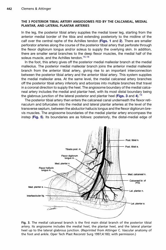
Fig. 3. The medial calcaneal branch is the first main distal branch of the posterior tibialartery. Its angiosome includes the medial heel, the plantar heel, and the lateral plantarheel up to the lateral glabrous junction. (Reprinted from Attinger C. Vascular anatomy ofthe foot and ankle. Oper Tech Plast Reconstr Surg 1997;4:183; with permission.)
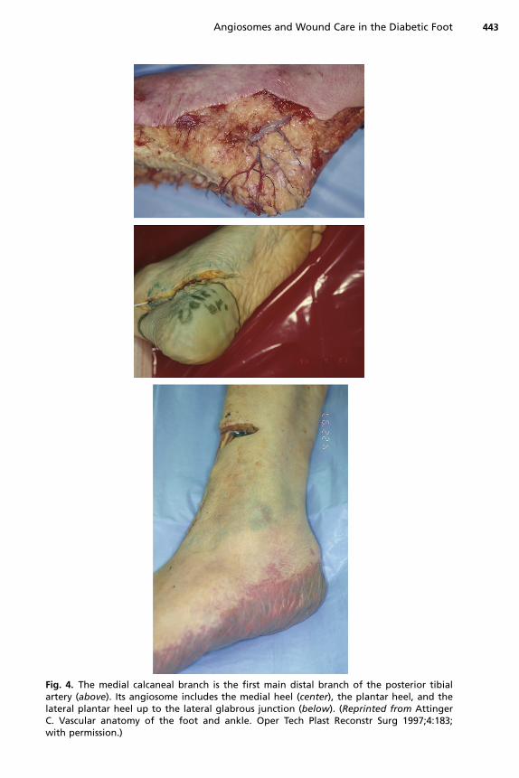
Fig. 4. The medial calcaneal branch is the first main distal branch of the posterior tibialartery (above). Its angiosome includes the medial heel (center), the plantar heel, and thelateral plantar heel up to the lateral glabrous junction (below). (Reprinted from AttingerC. Vascular anatomy of the foot and ankle. Oper Tech Plast Reconstr Surg 1997;4:183;with permission.)
Angiosomes and Wound Care in the Diabetic Foot 443
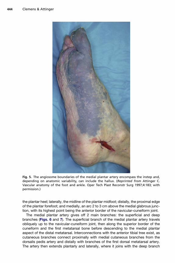
Fig. 5. The angiosome boundaries of the medial plantar artery encompass the instep and,depending on anatomic variability, can include the hallux. (Reprinted from Attinger C.Vascular anatomy of the foot and ankle. Oper Tech Plast Reconstr Surg 1997;4:183; withpermission.)
Clemens & Attinger444
the plantar heel; laterally, the midline of the plantar midfoot; distally, the proximal edgeof the plantar forefoot; and medially, an arc 2 to 3 cm above the medial glabrous junc-tion, with its highest point being the anterior border of the navicular-cuneiform joint.
The medial plantar artery gives off 2 main branches: the superficial and deepbranches (Figs. 6 and 7). The superficial branch of the medial plantar artery travelsobliquely up to the navicular-cuneiform joint, then along the superior border of thecuneiform and the first metatarsal bone before descending to the medial plantaraspect of the distal metatarsal. Interconnections with the anterior tibial tree exist, ascutaneous branches connect proximally with medial cutaneous branches from thedorsalis pedis artery and distally with branches of the first dorsal metatarsal artery.The artery then extends plantarly and laterally, where it joins with the deep branch
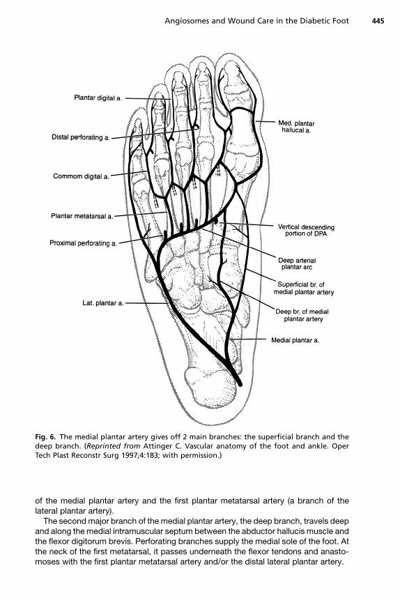
Fig. 6. The medial plantar artery gives off 2 main branches: the superficial branch and thedeep branch. (Reprinted from Attinger C. Vascular anatomy of the foot and ankle. OperTech Plast Reconstr Surg 1997;4:183; with permission.)
Angiosomes and Wound Care in the Diabetic Foot 445
of the medial plantar artery and the first plantar metatarsal artery (a branch of thelateral plantar artery).
The second major branch of the medial plantar artery, the deep branch, travels deepand along the medial intramuscular septum between the abductor hallucis muscle andthe flexor digitorum brevis. Perforating branches supply the medial sole of the foot. Atthe neck of the first metatarsal, it passes underneath the flexor tendons and anasto-moses with the first plantar metatarsal artery and/or the distal lateral plantar artery.
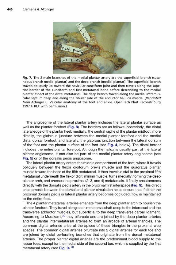
Fig. 7. The 2 main branches of the medial plantar artery are the superficial branch (cuta-neous branch medial plantar) and the deep branch (medial plantar). The superficial branchtravels obliquely up toward the navicular-cuneiform joint and then travels along the supe-rior border of the cuneiform and first metatarsal bone before descending to the medialplantar aspect of the distal metatarsal. The deep branch travels along the medial intramus-cular septum deep and along the fibular side of the abductor hallucis muscle. (Reprintedfrom Attinger C. Vascular anatomy of the foot and ankle. Oper Tech Plast Reconstr Surg1997;4:183; with permission.)
Clemens & Attinger446
The angiosome of the lateral plantar artery includes the lateral plantar surface aswell as the plantar forefoot (Fig. 8). The borders are as follows: posteriorly, the distallateral edge of the plantar heel; medially, the central raphe of the plantar midfoot; moredistally, the glabrous juncture between the medial plantar forefoot and the medialdistal dorsal forefoot; and laterally, the glabrous junction between the lateral dorsumof the foot and the plantar surface of the foot (see Fig. 4, below). The distal borderincludes the entire plantar forefoot. Although the hallux is usually part of the lateralplantar angiosome, it can also be part of the medial plantar artery angiosome (seeFig. 5) or of the dorsalis pedis angiosome.
The lateral plantar artery enters the middle compartment of the foot, where it travelsobliquely between the flexor digitorum brevis muscle and the quadratus plantarmuscle toward the base of the fifth metatarsal. It then travels distal to the proximal fifthmetatarsal underneath the flexor digiti minimi muscle, turns medially, forming the deepplantar arch, and crosses the proximal (2, 3, and 4) metatarsals. It finally anastomosesdirectly with the dorsalis pedis artery in the proximal first interspace (Fig. 9). This directanastomosis between the dorsal and plantar circulation helps ensure that if either theproximal dorsalis pedis or lateral plantar artery becomes occluded, flow is maintainedto the entire foot.
The 4 plantar metatarsal arteries emanate from the deep plantar arch to nourish theplantar forefoot. They travel along each metatarsal shaft deep to the interossei and thetransverse adductor muscles, but superficial to the deep transverse carpal ligament.According to Murakami,20 they bifurcate and are joined by the deep plantar arteriesand the plantar intermetatarsal arteries to form an arcade of arterial triangles. Thecommon digital arteries arise at the apices of these triangles in the proximal webspaces. The common digital arteries bifurcate into 2 digital arteries for each toe andare joined by distal perforating branches that originate from the dorsal metatarsalarteries. The proper plantar digital arteries are the predominant blood supply to thelesser toes, except for the medial side of the second toe, which is supplied by the firstmetatarsal artery (see Fig. 9).20
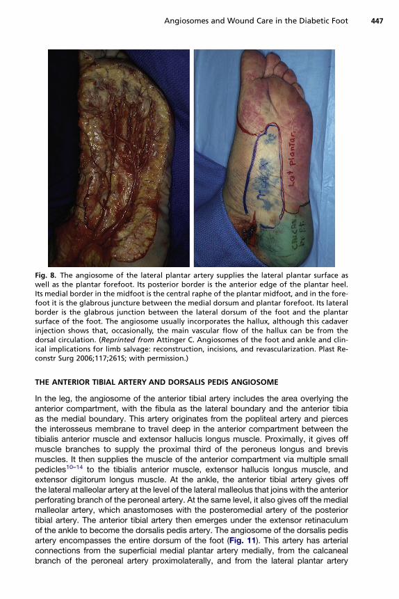
Fig. 8. The angiosome of the lateral plantar artery supplies the lateral plantar surface aswell as the plantar forefoot. Its posterior border is the anterior edge of the plantar heel.Its medial border in the midfoot is the central raphe of the plantar midfoot, and in the fore-foot it is the glabrous juncture between the medial dorsum and plantar forefoot. Its lateralborder is the glabrous junction between the lateral dorsum of the foot and the plantarsurface of the foot. The angiosome usually incorporates the hallux, although this cadaverinjection shows that, occasionally, the main vascular flow of the hallux can be from thedorsal circulation. (Reprinted from Attinger C. Angiosomes of the foot and ankle and clin-ical implications for limb salvage: reconstruction, incisions, and revascularization. Plast Re-constr Surg 2006;117;261S; with permission.)
Angiosomes and Wound Care in the Diabetic Foot 447
THE ANTERIOR TIBIAL ARTERY AND DORSALIS PEDIS ANGIOSOME
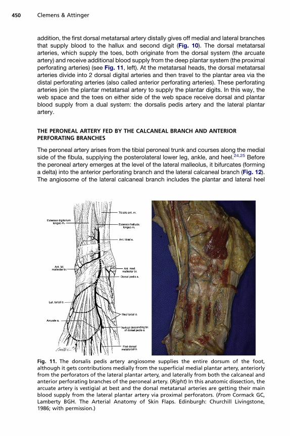
In the leg, the angiosome of the anterior tibial artery includes the area overlying theanterior compartment, with the fibula as the lateral boundary and the anterior tibiaas the medial boundary. This artery originates from the popliteal artery and piercesthe interosseus membrane to travel deep in the anterior compartment between thetibialis anterior muscle and extensor hallucis longus muscle. Proximally, it gives offmuscle branches to supply the proximal third of the peroneus longus and brevismuscles. It then supplies the muscle of the anterior compartment via multiple smallpedicles10–14 to the tibialis anterior muscle, extensor hallucis longus muscle, andextensor digitorum longus muscle. At the ankle, the anterior tibial artery gives offthe lateral malleolar artery at the level of the lateral malleolus that joins with the anteriorperforating branch of the peroneal artery. At the same level, it also gives off the medialmalleolar artery, which anastomoses with the posteromedial artery of the posteriortibial artery. The anterior tibial artery then emerges under the extensor retinaculumof the ankle to become the dorsalis pedis artery. The angiosome of the dorsalis pedisartery encompasses the entire dorsum of the foot (Fig. 11). This artery has arterialconnections from the superficial medial plantar artery medially, from the calcanealbranch of the peroneal artery proximolaterally, and from the lateral plantar artery
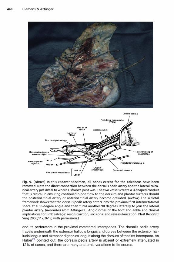
Fig. 9. (Above) In this cadaver specimen, all bones except for the calcaneus have beenremoved. Note the direct connection between the dorsalis pedis artery and the lateral calca-neal artery just distal to where Lisfranc’s joint was. The two vessels create a U-shaped conduitthat is critical in ensuring continued blood flow to the dorsum and plantar surfaces shouldthe posterior tibial artery or anterior tibial artery become occluded. (Below) The skeletalframework shows that the dorsalis pedis artery enters into the proximal first intrametatarsalspace at a 90-degree angle and then turns another 90 degrees laterally to join the lateralplantar artery. (Reprinted from Attinger C. Angiosomes of the foot and ankle and clinicalimplications for limb salvage: reconstruction, incisions, and revascularization. Plast ReconstrSurg 2006;117;261S; with permission.)
Clemens & Attinger448
and its perforators in the proximal metatarsal interspaces. The dorsalis pedis arterytravels underneath the extensor hallucis longus and curves between the extensor hal-lucis longus and extensor digitorum longus along the dorsum of the first interspace. AsHuber21 pointed out, the dorsalis pedis artery is absent or extremely attenuated in12% of cases, and there are many anatomic variations to its course.
Angiosomes and Wound Care in the Diabetic Foot 449
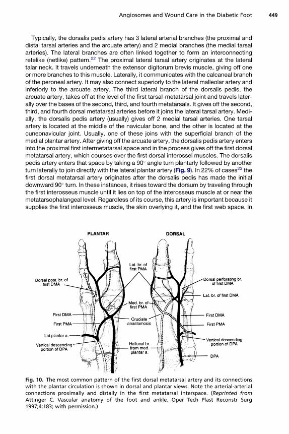
Typically, the dorsalis pedis artery has 3 lateral arterial branches (the proximal anddistal tarsal arteries and the arcuate artery) and 2 medial branches (the medial tarsalarteries). The lateral branches are often linked together to form an interconnectingretelike (netlike) pattern.22 The proximal lateral tarsal artery originates at the lateraltalar neck. It travels underneath the extensor digitorum brevis muscle, giving off oneor more branches to this muscle. Laterally, it communicates with the calcaneal branchof the peroneal artery. It may also connect superiorly to the lateral malleolar artery andinferiorly to the arcuate artery. The third lateral branch of the dorsalis pedis, thearcuate artery, takes off at the level of the first tarsal-metatarsal joint and travels later-ally over the bases of the second, third, and fourth metatarsals. It gives off the second,third, and fourth dorsal metatarsal arteries before it joins the lateral tarsal artery. Medi-ally, the dorsalis pedis artery (usually) gives off 2 medial tarsal arteries. One tarsalartery is located at the middle of the navicular bone, and the other is located at thecuneonavicular joint. Usually, one of these joins with the superficial branch of themedial plantar artery. After giving off the arcuate artery, the dorsalis pedis artery entersinto the proximal first intermetatarsal space and in the process gives off the first dorsalmetatarsal artery, which courses over the first dorsal interossei muscles. The dorsalispedis artery enters that space by taking a 90� angle turn plantarly followed by anotherturn laterally to join directly with the lateral plantar artery (Fig. 9). In 22% of cases23 thefirst dorsal metatarsal artery originates after the dorsalis pedis has made the initialdownward 90� turn. In these instances, it rises toward the dorsum by traveling throughthe first interosseus muscle until it lies on top of the interosseus muscle at or near themetatarsophalangeal level. Regardless of its course, this artery is important because itsupplies the first interosseus muscle, the skin overlying it, and the first web space. In
Fig. 10. The most common pattern of the first dorsal metatarsal artery and its connectionswith the plantar circulation is shown in dorsal and plantar views. Note the arterial-arterialconnections proximally and distally in the first metatarsal interspace. (Reprinted fromAttinger C. Vascular anatomy of the foot and ankle. Oper Tech Plast Reconstr Surg1997;4:183; with permission.)
Clemens & Attinger450
addition, the first dorsal metatarsal artery distally gives off medial and lateral branchesthat supply blood to the hallux and second digit (Fig. 10). The dorsal metatarsalarteries, which supply the toes, both originate from the dorsal system (the arcuateartery) and receive additional blood supply from the deep plantar system (the proximalperforating arteries) (see Fig. 11, left). At the metatarsal heads, the dorsal metatarsalarteries divide into 2 dorsal digital arteries and then travel to the plantar area via thedistal perforating arteries (also called anterior perforating arteries). These perforatingarteries join the plantar metatarsal artery to supply the plantar digits. In this way, theweb space and the toes on either side of the web space receive dorsal and plantarblood supply from a dual system: the dorsalis pedis artery and the lateral plantarartery.
THE PERONEAL ARTERY FED BY THE CALCANEAL BRANCH AND ANTERIORPERFORATING BRANCHES
The peroneal artery arises from the tibial peroneal trunk and courses along the medialside of the fibula, supplying the posterolateral lower leg, ankle, and heel.24,25 Beforethe peroneal artery emerges at the level of the lateral malleolus, it bifurcates (forminga delta) into the anterior perforating branch and the lateral calcaneal branch (Fig. 12).The angiosome of the lateral calcaneal branch includes the plantar and lateral heel
Fig. 11. The dorsalis pedis artery angiosome supplies the entire dorsum of the foot,although it gets contributions medially from the superficial medial plantar artery, anteriorlyfrom the perforators of the lateral plantar artery, and laterally from both the calcaneal andanterior perforating branches of the peroneal artery. (Right) In this anatomic dissection, thearcuate artery is vestigial at best and the dorsal metatarsal arteries are getting their mainblood supply from the lateral plantar artery via proximal perforators. (From Cormack GC,Lamberty BGH. The Arterial Anatomy of Skin Flaps. Edinburgh: Churchill Livingstone,1986; with permission.)
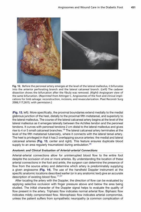
Fig. 12. Before the peroneal artery emerges at the level of the lateral malleolus, it bifurcatesinto the anterior perforating branch and the lateral calcaneal branch. (Left) The cadaverdissection shows the bifurcation after the fibula was removed. (Right) Angiogram view ofthe same bifurcation. (Reprinted from Attinger C. Angiosomes of the foot and clinical impli-cations for limb salvage: reconstruction, incisions, and revascularization. Plast Reconstr Surg2006;117;261S; with permission.)
Angiosomes and Wound Care in the Diabetic Foot 451
(Fig. 13, left). More specifically, the proximal boundaries extend medially to the medialglabrous junction of the heel, distally to the proximal fifth metatarsal, and superiorly tothe lateral malleolus. The course of the lateral calcaneal artery begins at the level of thelateral malleolus as it emerges laterally between the Achilles tendon and the peronealtendons. It curves with peroneal tendons 2 cm distal to the lateral malleolus and givesrise to 4 or 5 small calcaneal branches.19 The lateral calcaneal artery terminates at thelevel of the fifth metatarsal tuberosity, where it connects with the lateral tarsal artery.The heel is privileged in that it has 2 overlapping source arteries: the medial and lateralcalcaneal arteries (Fig. 13, center and right). This feature ensures duplicate bloodsupply to an area regularly traumatized during ambulation.26
Anatomic and Clinical Evaluation of Arterial-arterial Connections
Arterial-arterial connections allow for uninterrupted blood flow to the entire footdespite the occlusion of one or more arteries. By understanding the location of thesearterial connections in the foot and ankle, the surgeon can determine the presence offlow from the source artery and determine which artery is predominately supplyinga given angiosome (Fig. 14). The use of the handheld Doppler instrument at thespecific anatomic locations described earlier (or in any anatomic text) give an accuratedescription of existing blood flow.17,27
After locating the artery with the Doppler, the direction of flow can be evaluated byapplying selective occlusion with finger pressure above and below the area beingstudied. The initial character of the Doppler signal helps to evaluate the quality offlow present in the artery. Triphasic flow indicates normal arterial flow. Biphasic flowindicates mildly compromised flow. Monophasic flow indicates arterial compromise,unless the patient suffers from sympathetic neuropathy (a common complication of
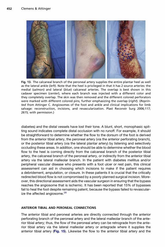
Fig. 13. The calcaneal branch of the peroneal artery supplies the entire plantar heel as wellas the lateral ankle (left). Note that the heel is privileged in that it has 2 source arteries: themedial (salmon) and lateral (blue) calcaneal arteries. The overlap is best shown in thiscadaver specimen (center), where each branch was injected with a different color andthey completely overlap. The skin was then removed and the different colored perforatorswere marked with different colored pins, further emphasizing the overlap (right). (Reprin-ted from Attinger C. Angiosomes of the foot and ankle and clinical implications for limbsalvage: reconstruction, incisions, and revascularization. Plast Reconstr Surg 2006;117;261S; with permission.)
Clemens & Attinger452
diabetes) and the distal vessels have lost their tone. A blunt, short, monophasic spit-ting sound indicates complete distal occlusion with no runoff. For example, it shouldbe straightforward to determine whether the flow to the dorsum of the foot is derivedfrom the anterior tibial artery, the peroneal artery (via the anterior perforating branch),or the posterior tibial artery (via the lateral plantar artery) by listening and selectivelyoccluding these areas. In addition, one should be able to determine whether the bloodflow to the heel is coming directly from the calcaneal branch of the posterior tibialartery, the calcaneal branch of the peroneal artery, or indirectly from the anterior tibialartery via the lateral malleolar branch. In the patient with diabetes mellitus and/orperipheral vascular disease who presents with a foot ulcer or rest pain, this clinicalassessment can aid in choosing which incisions to make if the patient requiresa debridement, amputation, or closure. In these patients it is crucial that the criticallyredirected blood flow is not compromised by a poorly planned surgical incision. More-over, this directional assessment aids the vascular surgeon in ensuring that the bypassreaches the angiosome that is ischemic. It has been reported that 15% of bypassesfail to heal the foot despite remaining patent, because the bypass failed to revascular-ize the affected angiosome.28
ANTERIOR TIBIAL AND PERONEAL CONNECTIONS
The anterior tibial and peroneal arteries are directly connected through the anteriorperforating branch of the peroneal artery and the lateral malleolar branch of the ante-rior tibial artery; thus, the flow of the peroneal artery can be retrograde from the ante-rior tibial artery via the lateral malleolar artery or antegrade where it supplies theanterior tibial artery (Fig. 15). Likewise the flow to the anterior tibial artery and the

Fig. 14. The anterior perforating branch of the peroneal artery is located in the lateral softarea just above the ankle joint between the tibia and the fibula (left). Then, the anteriortibial artery is occluded at the takeoff of the lateral malleolar branch (right). If the Dopplersounds continue, then there is antegrade flow along the anterior perforating branch of theperoneal artery. (Reprinted from Attinger C. Angiosomes of the foot and ankle and clinicalimplications for limb salvage: reconstruction, incisions, and revascularization. Plast ReconstrSurg 2006;117:261S; with permission.)
Angiosomes and Wound Care in the Diabetic Foot 453
dorsalis pedis can be antegrade from the proximal anterior tibial artery or retrogradefrom the anterior perforating branch of the peroneal artery via the lateral malleolarartery.
PERONEAL AND POSTERIOR TIBIAL CONNECTIONS
The peroneal artery communicates distally with the posterior tibial artery via 1 to 3transverse communicating branches that are located within the fat pad deep to theAchilles tendon (Fig. 15). These branches are located 5 to 7 cm above the ankle joint,at the ankle joint, and just above the insertion of the Achilles tendon. Because of theseconnections, it is impossible by Doppler imaging to know whether the flow along thedistal posterior tibial artery originates directly from the proximal posterior tibial arteryor indirectly from the distal peroneal artery via the above perforators. Likewise, onecannot tell whether the flow along the peroneal artery originates from the peronealartery or from the posterior tibial artery via those same perforators.18
ANTERIOR TIBIAL AND POSTERIOR TIBIAL CONNECTIONS
The anterior tibial artery and posterior tibial artery are also directly connected distal tothe Lisfranc joint, where the dorsalis pedis artery enters into the proximal first
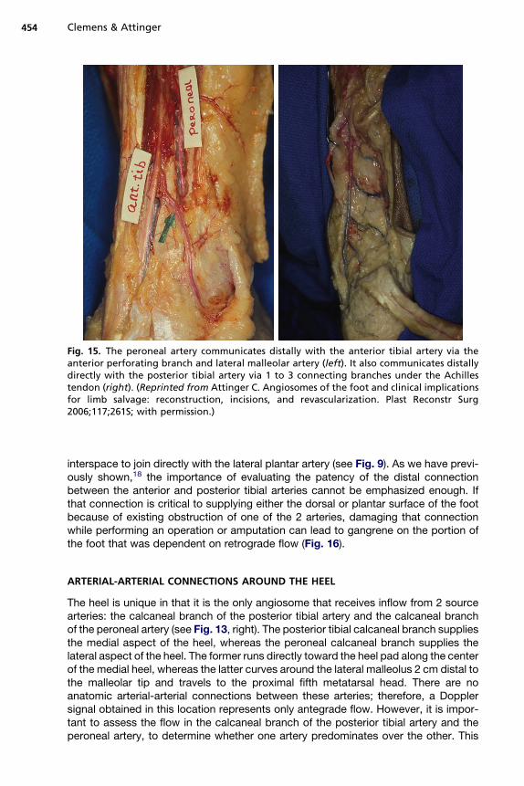
Fig. 15. The peroneal artery communicates distally with the anterior tibial artery via theanterior perforating branch and lateral malleolar artery (left). It also communicates distallydirectly with the posterior tibial artery via 1 to 3 connecting branches under the Achillestendon (right). (Reprinted from Attinger C. Angiosomes of the foot and clinical implicationsfor limb salvage: reconstruction, incisions, and revascularization. Plast Reconstr Surg2006;117;261S; with permission.)
Clemens & Attinger454
interspace to join directly with the lateral plantar artery (see Fig. 9). As we have previ-ously shown,18 the importance of evaluating the patency of the distal connectionbetween the anterior and posterior tibial arteries cannot be emphasized enough. Ifthat connection is critical to supplying either the dorsal or plantar surface of the footbecause of existing obstruction of one of the 2 arteries, damaging that connectionwhile performing an operation or amputation can lead to gangrene on the portion ofthe foot that was dependent on retrograde flow (Fig. 16).
ARTERIAL-ARTERIAL CONNECTIONS AROUND THE HEEL
The heel is unique in that it is the only angiosome that receives inflow from 2 sourcearteries: the calcaneal branch of the posterior tibial artery and the calcaneal branchof the peroneal artery (see Fig. 13, right). The posterior tibial calcaneal branch suppliesthe medial aspect of the heel, whereas the peroneal calcaneal branch supplies thelateral aspect of the heel. The former runs directly toward the heel pad along the centerof the medial heel, whereas the latter curves around the lateral malleolus 2 cm distal tothe malleolar tip and travels to the proximal fifth metatarsal head. There are noanatomic arterial-arterial connections between these arteries; therefore, a Dopplersignal obtained in this location represents only antegrade flow. However, it is impor-tant to assess the flow in the calcaneal branch of the posterior tibial artery and theperoneal artery, to determine whether one artery predominates over the other. This
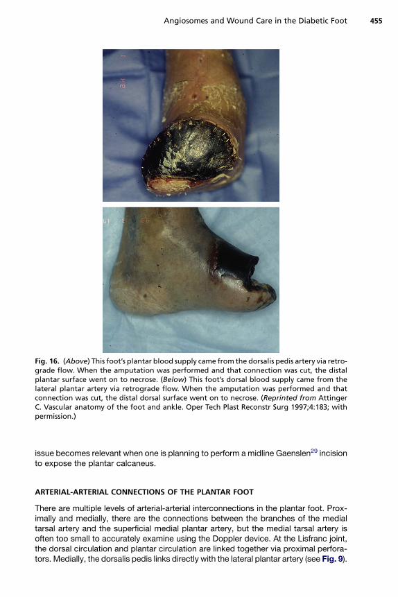
Fig. 16. (Above) This foot’s plantar blood supply came from the dorsalis pedis artery via retro-grade flow. When the amputation was performed and that connection was cut, the distalplantar surface went on to necrose. (Below) This foot’s dorsal blood supply came from thelateral plantar artery via retrograde flow. When the amputation was performed and thatconnection was cut, the distal dorsal surface went on to necrose. (Reprinted from AttingerC. Vascular anatomy of the foot and ankle. Oper Tech Plast Reconstr Surg 1997;4:183; withpermission.)
Angiosomes and Wound Care in the Diabetic Foot 455
issue becomes relevant when one is planning to perform a midline Gaenslen29 incisionto expose the plantar calcaneus.
ARTERIAL-ARTERIAL CONNECTIONS OF THE PLANTAR FOOT
There are multiple levels of arterial-arterial interconnections in the plantar foot. Prox-imally and medially, there are the connections between the branches of the medialtarsal artery and the superficial medial plantar artery, but the medial tarsal artery isoften too small to accurately examine using the Doppler device. At the Lisfranc joint,the dorsal circulation and plantar circulation are linked together via proximal perfora-tors. Medially, the dorsalis pedis links directly with the lateral plantar artery (see Fig. 9).
Clemens & Attinger456
More laterally, the dorsal and plantar metatarsal arteries are linked at their takeoff bythe proximal perforating branches. At the web spaces, distal perforating arteries againlink the dorsal and plantar metatarsal arteries. The final arterial-arterial interconnectionis a fine subdermal arteriolar plexus linking the dorsalis pedis with the lateral plantarartery in a circumferential wraparound pattern about the plantar foot.
DORSALIS PEDIS, LATERAL PLANTAR ARTERIES, AND CRUCIATE ANASTOMOSIS
In the plantar foot, the principle connection to evaluate is that between the dorsalispedis and lateral plantar arteries. First, use the Doppler device to examine the lateralplantar artery proximal to the base of the proximal first interspace. Then, occlude thedorsalis pedis at the tarsal-metatarsal joint. If the signal disappears, then flow in thelateral plantar artery depends on the dorsalis pedis arterial flow. However, if the soundremains, it means that there is antegrade flow from the posterior tibialis artery to thelateral plantar artery.
A second source of arterial-arterial anastomosis occurs proximal to the first meta-tarsal head at the cruciate anastomosis, where the superficial and deep medial plantararteries join (see Fig. 10). The distal lateral plantar artery also joins the cruciate anas-tomosis, linking the medial plantar artery with the lateral plantar artery. The bloodsupply to the first toe depends on which arteries anastomose and which providethe major blood supply to the cruciate anastomosis: the medial plantar artery, lateralplantar artery, or first dorsal metatarsal artery.
The final arterial-arterial interconnection was first described by Hidalgo and Shaw,30
who showed a fine subdermal arteriolar plexus linking the dorsalis pedis with thelateral plantar artery in a circumferential wraparound pattern about the plantar foot.They span the angiosome boundaries of the dorsalis pedis artery, medial plantarartery, and lateral plantar artery.
CONNECTIONS ON THE DORSUM OF THE FOOT
As discussed earlier, the dorsal and plantar arterial systems are closely linked atmultiple levels. The most proximal is located in the medial foot, where the medial tarsalartery communicates with the superficial (medial branch) of the medial plantar artery. Itis usually too difficult to use Doppler imaging on this small connection. Laterally, thereis the rete that connects the proximal lateral tarsal artery, the distal tarsal and arcuatearteries, and the lateral malleolar artery and the anterior perforating branch of theperoneal superiorly. In addition, the calcaneal branch of the peroneal artery connectswith the lateral tarsal artery. Because of this complex network of connections, it is diffi-cult to determine the source of retrograde flow over the major tarsal artery when it isoccluded proximally. If there is retrograde flow along the proximal lateral tarsal artery,it signifies an intact network of connections that can include the anterior perforatingbranch of the lateral plantar artery, the lateral malleolar artery, the calcaneal branchof the peroneal artery, the distal tarsal artery arcuate artery, and the arcuate artery.The arterial connection described in detail earlier occurs just distal to the Lisfranc joint,where the dorsalis pedis artery joins the lateral plantar artery in the proximal first inter-space (see Fig. 9). At the proximal metatarsal interspaces and at distal web spaces,the proximal and distal perforating arteries, respectively, link the dorsal and plantarmetatarsal arteries (see Fig. 11, left). The direction(s) of flow along the dorsal metatar-sals can be easily determined. This close linkage ensures that collateral flow compen-sates for occlusions to either the dorsalis pedis or posterior tibial artery.
Angiosomes and Wound Care in the Diabetic Foot 457
Using the Principles of Angiosomes to Make Safe Incisions in Normaland Vascularly Compromised Patients
As we have previously reported in more detail,18 there are 4 important factors to beconsidered and balanced when choosing where to place an incision. The incisionmust provide adequate exposure for the planned procedure. In addition, there mustbe adequate blood supply on either side of the incision to optimize healing. Third,the incision should spare the sensory and motor nerves. Fourth, the incision shouldnot be placed perpendicular to a joint, because of the risk of causing scar contractureand resultant joint immobility. Although adequate exposure, nerve location, and scarcontracture are important factors, we focus primarily on the vascular ramifications oftypical incisions in the foot and ankle.
We have described earlier in detail the importance of assessing the blood flow toeach angiosome. As we stated, the presence of a palpable pulse or a Doppler-detectable triphasic sound over the source artery to a given angiosome indicatesadequate blood flow to that angiosome. If there is good blood flow from the sourceartery feeding each angiosome, the safest incisions to make are along the borderbetween 2 adjacent angiosomes, because each side of the incision has maximal bloodflow. Therefore, incisions along the central raphe over the Achilles tendon, along theglabrous junction separating the sole of the foot from the dorsum of the foot, or alongthe midline of the sole of the foot are safe incisions.
One cannot reach all areas of the foot through these incisions, and blood flow toeach angiosome is not always satisfactory; thus, well-deliberated compromisesneed to be made. When the signal of a source artery to one of 2 adjacent angiosomesis absent, the affected ischemic angiosome depends on the surrounding angiosomesfor blood flow via the choke vessels. Because the choke vessels can require 4 to 10days to become patent after a given angiosome becomes ischemic, incisions placedtoo soon after an arterial occlusion for collateral circulation to develop run the risk ofpoor healing, necrosis, or gangrene.13,14 However, in patients with chronic arterioscle-rosis, the occlusion is gradual and the choke vessels are usually patent by the time thesource vessel closes.
In the vascularly compromised patient, collateral flow may keep the ischemic angio-some vascularized, and incisions must be planned so that this collateral flow is notdisturbed. In the more extreme ischemic cases, in which there are no palpable pulsesand the Doppler sounds are monophasic, then possible surgical revascularizationshould be entertained before proceeding. When one of the pulses is not present, itis best to place the incision away from the patent source artery, as we have previouslyreported.18 This is the safest location, because there is minimal risk of damaging thepatent source artery or the crucial choke vessels. We briefly discuss the mostfrequently used incisions and refer the reader to the article by Attinger andcolleagues18 for a more detailed discussion.
INCISIONS AT THE ACHILLES TENDON
Incisions over the Achilles tendon are the safest if they are made along the midline thatdivides the peroneal angiosome from the posterior tibial angiosome. Incisions oneither side of the Achilles tendon to expose the distal tibia or fibula are also safe,provided that the posterior tibial artery and the peroneal artery are patent. The richinterconnecting vascular plexus around the Achilles tendon keeps the skin abovethe Achilles tendon viable. A medial to lateral S-shaped incision minimizes injury tothe sural nerve and lesser saphenous vein. If an incision is made along the glabrousjunction of the posterior heel, the medial portion of the incision should not extend to
Clemens & Attinger458
the medial edge of the Achilles tendon, to avoid damaging the medial calcaneal neuro-vascular structures. It is safer to make the incision laterally along the glabrous junction,which represents the distal angiosome boundary of the calcaneal branch of the poste-rior tibial artery.
INCISIONS AT THE LATERAL CALCANEUS
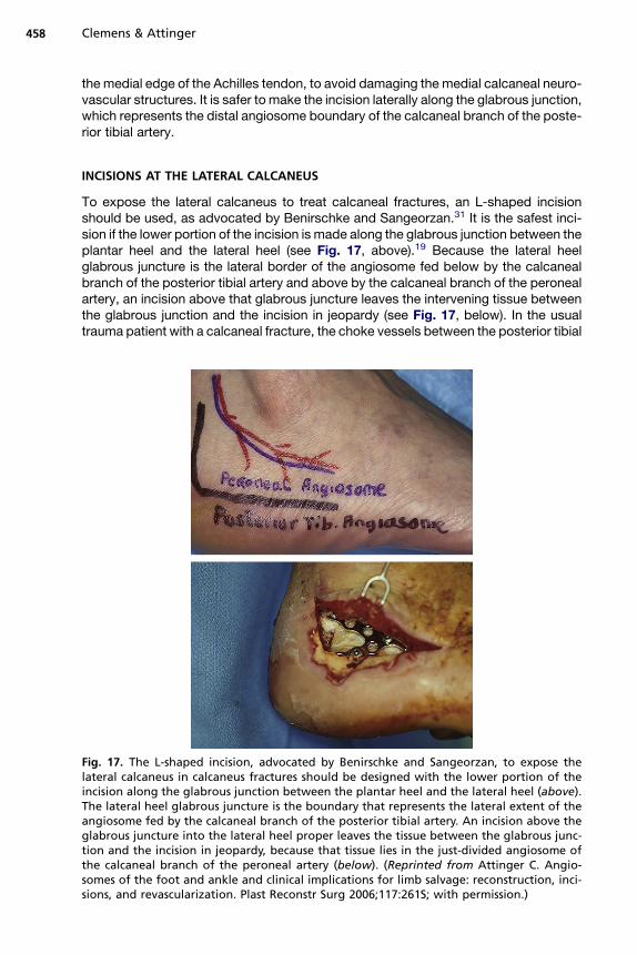
To expose the lateral calcaneus to treat calcaneal fractures, an L-shaped incisionshould be used, as advocated by Benirschke and Sangeorzan.31 It is the safest inci-sion if the lower portion of the incision is made along the glabrous junction between theplantar heel and the lateral heel (see Fig. 17, above).19 Because the lateral heelglabrous juncture is the lateral border of the angiosome fed below by the calcanealbranch of the posterior tibial artery and above by the calcaneal branch of the peronealartery, an incision above that glabrous juncture leaves the intervening tissue betweenthe glabrous junction and the incision in jeopardy (see Fig. 17, below). In the usualtrauma patient with a calcaneal fracture, the choke vessels between the posterior tibial
Fig. 17. The L-shaped incision, advocated by Benirschke and Sangeorzan, to expose thelateral calcaneus in calcaneus fractures should be designed with the lower portion of theincision along the glabrous junction between the plantar heel and the lateral heel (above).The lateral heel glabrous juncture is the boundary that represents the lateral extent of theangiosome fed by the calcaneal branch of the posterior tibial artery. An incision above theglabrous juncture into the lateral heel proper leaves the tissue between the glabrous junc-tion and the incision in jeopardy, because that tissue lies in the just-divided angiosome ofthe calcaneal branch of the peroneal artery (below). (Reprinted from Attinger C. Angio-somes of the foot and ankle and clinical implications for limb salvage: reconstruction, inci-sions, and revascularization. Plast Reconstr Surg 2006;117:261S; with permission.)
Angiosomes and Wound Care in the Diabetic Foot 459
calcaneal and peroneal calcaneal angiosomes have not had the time to open up andallow the calcaneal branch of the posterior tibial artery to feed the tissue beyond itsboundary. As we stated earlier, it usually takes 4 to 10 days for the choke vesselsto become patent, and it may take even longer in the setting of overlying soft-tissuedamage and inflammation.13
INCISIONS OVER THE PLANTAR HEEL
In general, incisions over the plantar heel are reserved for hindfoot limb salvage in thepresence of osteomyelitis. Safe incisions over the plantar heel from a vascularperspective can be coronal or sagittal in orientation, if the posterior tibial and peronealarteries are patent. Whether the resultant scar is acceptable is another question.31
Recall that the blood flow to the heel lies primarily in a coronal direction from the calca-neal branch of the posterior tibial artery (medial) and the peroneal artery (lateral). Thecoronal incision does not disturb the coronal flow or the sensory nerves that travel inthe same direction.
If the incision is in the sagittal direction, then the flow comes to each side of the inci-sion from the respective calcaneal arteries. However, the sensory nerves will bedamaged, which is less problematic if the patient is neuropathic. In that instance,a Gaenslen29 incision down the central heel pad is the ideal choice to expose thecalcaneus for calcanectomy. Taking care to adequately evert the edges when closingthe incision avoids an inverted and chronically calloused scar.
INCISIONS AT THE PLANTAR MEDIAL MIDFOOT
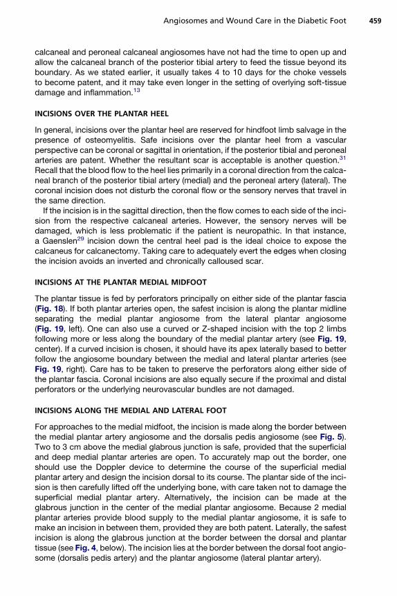
The plantar tissue is fed by perforators principally on either side of the plantar fascia(Fig. 18). If both plantar arteries open, the safest incision is along the plantar midlineseparating the medial plantar angiosome from the lateral plantar angiosome(Fig. 19, left). One can also use a curved or Z-shaped incision with the top 2 limbsfollowing more or less along the boundary of the medial plantar artery (see Fig. 19,center). If a curved incision is chosen, it should have its apex laterally based to betterfollow the angiosome boundary between the medial and lateral plantar arteries (seeFig. 19, right). Care has to be taken to preserve the perforators along either side ofthe plantar fascia. Coronal incisions are also equally secure if the proximal and distalperforators or the underlying neurovascular bundles are not damaged.
INCISIONS ALONG THE MEDIAL AND LATERAL FOOT
For approaches to the medial midfoot, the incision is made along the border betweenthe medial plantar artery angiosome and the dorsalis pedis angiosome (see Fig. 5).Two to 3 cm above the medial glabrous junction is safe, provided that the superficialand deep medial plantar arteries are open. To accurately map out the border, oneshould use the Doppler device to determine the course of the superficial medialplantar artery and design the incision dorsal to its course. The plantar side of the inci-sion is then carefully lifted off the underlying bone, with care taken not to damage thesuperficial medial plantar artery. Alternatively, the incision can be made at theglabrous junction in the center of the medial plantar angiosome. Because 2 medialplantar arteries provide blood supply to the medial plantar angiosome, it is safe tomake an incision in between them, provided they are both patent. Laterally, the safestincision is along the glabrous junction at the border between the dorsal and plantartissue (see Fig. 4, below). The incision lies at the border between the dorsal foot angio-some (dorsalis pedis artery) and the plantar angiosome (lateral plantar artery).
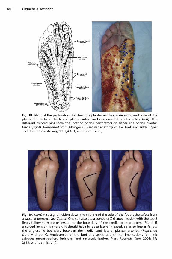
Fig. 18. Most of the perforators that feed the plantar midfoot arise along each side of theplantar fascia from the lateral plantar artery and deep medial plantar artery (left). Thedifferent colored pins show the location of the perforators on either side of the plantarfascia (right). (Reprinted from Attinger C. Vascular anatomy of the foot and ankle. OperTech Plast Reconstr Surg 1997;4:183; with permission.)
Fig. 19. (Left) A straight incision down the midline of the sole of the foot is the safest froma vascular perspective. (Center) One can also use a curved or Z-shaped incision with the top 2limbs following more or less along the boundary of the medial plantar artery. (Right) Ifa curved incision is chosen, it should have its apex laterally based, so as to better followthe angiosome boundary between the medial and lateral plantar arteries. (Reprintedfrom Attinger C. Angiosomes of the foot and ankle and clinical implications for limbsalvage: reconstruction, incisions, and revascularization. Plast Reconstr Surg 2006;117;261S; with permission.)
Clemens & Attinger460
Angiosomes and Wound Care in the Diabetic Foot 461
INCISIONS ON THE DORSUM OF THE FOOT
When considering dorsal foot incisions, recall that the dorsal circulation proximal tothe Lisfranc joint travels in a coronal direction, and distal to the Lisfranc joint it travelsin a sagittal direction (see Fig. 11, left). The lateral proximal dorsum of the foot iscomposed of a rete of coronally interconnected arteries of the lateral malleolar, tarsal(proximal and distal), and arcuate arteries. This rete is linked superiorly to the anteriorperforating branch and laterally to the calcaneal branch of the peroneal arteries.Medial to the dorsalis pedis artery is the medial tarsal artery, which may be directlylinked to the superficial medial plantar artery. We placing the incision parallel to thedirection of the arterial supply. Coronal incisions in the lateral proximal dorsal footare parallel to the coronally directed arteries (proximal tarsal, distal tarsal, arcuatearteries, and their perforators). In addition, the dorsalis pedis artery should be identi-fied and spared, unless it is clear that the antegrade and retrograde flow is strong. Forapproaches to the medial proximal dorsal foot, the safest incision is along the borderbetween the medial plantar artery angiosome and the dorsalis pedis angiosome. Forincisions of the distal forefoot, it is important not to place an incision through the meta-tarsal arteries, unless they both have antegrade flow (from the arcuate artery and prox-imal perforators) and retrograde flow (from the distal perforators). Recall that themetatarsal arteries arise from the arcuate artery, travel along the interosseus space,and are connected to plantar circulation proximally and distally by perforators. If themetatarsal artery flow is bidirectional, then coronally directed incisions are safe.However, if the flow is unidirectional, the incisions should be in the sagittal direction,over the metatarsal bones themselves, in order not to disturb the dorsal metatarsalarteries. Multiple parallel sagittal incisions over the distal dorsal forefoot can be per-formed as long as the dorsal metatarsal arteries are preserved. Only 3 incisions arenecessary to gain access to all metatarsals, and the incisions should be short, withlittle undermining of the skin overlying the interosseus muscles.
INCISIONS FOR AMPUTATIONS
In general, performing forefoot and midfoot amputations in patients who have intactcirculation with the dorsal and plantar antegrade blood flow has minimal risk. All inci-sions should be designed at the angiosome boundaries to maximize blood flow at theedges of the amputation. Medial and lateral incisions should be at the glabrous junc-ture between the dorsal and plantar circulation, whereas dorsal and plantar incisionsshould be to bone, without undermining to preserve the metatarsal arteries in the flaps.When there is compromised flow and a forefoot or midfoot amputation is planned, it isimportant that the remaining blood flow and arterial-arterial connections are mappedcompletely. If the dorsal circulation depends on the plantar circulation or vice versa,then connections between the 2 regions cannot be disturbed (see Fig. 17). That is,the connection between the dorsalis pedis and lateral plantar arteries at the proximalfirst interspace must be maintained. To preserve that connection when performinga short transmetatarsal or Lisfranc amputation, the lateral 4 metatarsals are removedlaterally whereas the first metatarsal is removed medially.
Using the Angiosome Principle in Planning the Optimal Revascularization
Despite the current advances in revascularization techniques, vascular bypasssurgery fails to heal approximately 15% of ischemic lower extremity wounds witha patent bypass.28–43 Gooden and colleagues44 found that up to 25% of patientswith heel ulcers ultimately succumbed to a proximal leg amputation despite a palpablepedal pulse. The failures may be due in part to inadequate postoperative wound
Clemens & Attinger462
care,45 but the major part of the problem is caused by the inadequate revasculariza-tion of the local ischemic area, because the vascular connections between the revas-cularized vessel and the source vessel nourishing the ischemic area are absent oroccluded.
Thus, successful revascularization for ischemic wounds is more complex thansimply restoring circulation to a specific artery. The failure of limb salvage ina percentage of successful bypasses suggests that more effective revascularizationmay occur if the bypassed vessel directly feeds the source artery of the angiosomecontaining the ulceration. That is, revascularization of the major artery directlysupplying the ischemic and ulcerated angiosome should be more successful thanrevascularizing one of the other 2 major arteries and hoping that existing arterial-arterial connections for the blood flow reach the ischemic ulcerated angiosome.46
We retrospectively examined the results of direct versus indirect consecutive revas-cularization of 52 limbs. There was a 9.1% failure rate when wounds were directlyrevascularized versus a 38.1% failure rate in the wounds indirectly bypassed (P 5.03). Those who failed to heal went on to a major leg amputation. The amputationrate, therefore, in the indirectly bypassed group was 4 times that of the directlybypassed group. This study supports the suggestion that direct revascularization ofthe affected angiosome leads to higher limb salvage rates.
If the vascular surgeon has more than one vessel to bypass to, or has the choice ofendovascularly opening more than one vessel, they should preferentially open thevessel that directly feeds the affected angiosome. For heel wounds, the peroneal orposterior tibial artery should be preferentially revascularized. For plantar foot wounds,the posterior tibial artery should be preferentially revascularized. For lateral anklewounds, the peroneal artery should be preferentially revascularized. For dorsal footwounds, the anterior tibial artery should be preferentially revascularized. If the vascularsurgeon cannot revascularize the source artery to the affected angiosome, they canthen predict a failure rate of the revascularization to be 15% or higher unless thesurgeon can show that the arterial-arterial connections between the artery to be revas-cularized and the source artery of the affected angiosome are open.
SUMMARY
Three major arteries supply the foot and ankle and create vascular redundancythrough multiple arterial-arterial connections. A Doppler device may be used to mapout the patient’s vascular tree, including the direction of flow. Knowledge of the 6angiosomes of the foot and ankle combined with an adequate Doppler examinationcan optimize the success of any planned treatment or procedure. This informationis essential for successful limb salvage in diabetic patients and patients with peripheralvascular disease and ultimately helps a surgeon to decide whether a reconstruction ispossible or an amputation is indicated.
REFERENCES
1. Taylor GI, Palmer JH. The vascular territories (angiosomes) of the body: experi-mental studies and clinical applications. Br J Plast Surg 1990;43:1.
2. Morain WD, Ristic J. Manchot: the cutaneous arteries of the human body. NewYork: Springer Verlag; 1983.
3. Salmon M. In: Taylor GI, Tempest MN, editors. Arteries of the skin. Edinburgh(UK): Churchill Livingston; 1988.
4. McGregor IA, Morgan G. Axial and random pattern flaps. Br J Plast Surg 1973;26:202.
Angiosomes and Wound Care in the Diabetic Foot 463
5. Daniel RK, Cunningham DM, Taylor GI. The deltopectoral flap: an anatomical andhemodynamic approach. Plast Reconstr Surg 1975;55:275.
6. Mathes SJ, Nahai F. Clinical atlas of muscle and musculocutaneous flaps. StLouis (MO): Mosby; 1979.
7. Ger R. Operative treatment of the advanced stasis ulcer using muscle transposi-tion. Am J Surg 1970;120:376.
8. Orticochea M. The musculocutaneous flap method: an immediate and heroicsubstitute for the method of delay. Br J Plast Surg 1972;25:106.
9. McCraw JB, Dibell DG. Experimental definition of independent myocutaneousvascular territories. Plast Reconstr Surg 1977;60:341.
10. Taylor GI, Minabe T. The angiosomes of the mammals and other vertebrates.Plast Reconstr Surg 1992;89:181.
11. Calligari PR, Taylor GI, Caddy CM, et al. An anatomic review of the delayphenomenon: I. Experimental studies. Plast Reconstr Surg 1992;89:397.
12. Taylor GI, Corlett RJ, Caddy CM, et al. An anatomic review of the delay phenom-enon: II. Clinical applications. Plast Reconstr Surg 1992;89:408.
13. MorrisSF,TaylorGI. The timesequenceof thedelayphenomenon: when is asurgicaldelay effective? An experimental study. Plast Reconstr Surg 1995;95:526.
14. Dhar SC, Taylor GI. The delay phenomenon: the story unfolds. Plast ReconstrSurg 1999;104:2079.
15. Sarrafian SK. Anatomy of the foot and ankle. Philadelphia: Lippincott; 1993. pp.294–355.
16. Taylor GI, Pan WR. Angiosomes of the leg: anatomic study and clinical implica-tions. Plast Reconstr Surg 1998;102:599.
17. Attinger CE, Evans KK, Bulan E, et al. Angiosomes of the foot and ankle clinicalimplications for limb salvage: reconstruction, incisions, and revascularization.Plast Reconstr Surg 2006;117:261S–93S.
18. Attinger CE, Cooper P, Blume P, et al. The safest surgical incision and amputa-tions applying the angiosomes principle and using the Doppler to assess thearterial-arterial connections of the foot and ankle. Foot Ankle Clin North Am2001;6:745.
19. Attinger C, Cooper P. Soft tissue reconstruction for calcaneal fractures or osteo-myelitis. Orthop Clin North Am 2001;32:135.
20. Murakami T. On the position and course of the deep plantar arteries, with specialreference to the so called plantar metatarsal arteries. Okajimas Folia Anat Jpn1971;48:295.
21. Huber JF. The arterial network supplying the dorsum of the foot. Anat Rec 1941;80:373.
22. Adachi B. Das arteriensystem der Japaner. Kyoto (Japan): Maruzen; 1928. pp.246–48.
23. May JW, Chait LA, Cohen BE. Free neurovascular flap from the first web of thefoot in hand reconstruction. J Hand Surg Am 1977;5:387.
24. Shusterman MA, Reece GP, Milller MJ. The osteocutaneous free fibula flap: is theskin paddle reliable? Plast Reconstr Surg 1992;90:787.
25. Jones NF, Monstrey MD, Gambier BA. Reliability of the fibular osteocutaneousflap for mandibular reconstruction: anatomical and surgical confirmation. PlastReconstr Surg 1996;97:707.
26. Masqualet AC, Beveridge J, Romana C. The lateral supramalleolar flap. Plast Re-constr Surg 1988;81:74.
27. Taylor GI, Doyle M, McCarten G. The Doppler probe for planning flaps: anatom-ical study and clinical applications. Br J Plast Surg 1990;43:1.
Clemens & Attinger464
28. Berceli SA, Chan AK, Pomposelli FB, et al. Efficacy of dorsal pedal artery bypassin limb salvage for ischemic heel ulcers. J Vasc Surg 1999;30:499.
29. Gaenslen FJ. Split heel approach in osteomyelitis of the os calcis. J Bone JointSurg 1931;13:759.
30. Hidalgo DA, Shaw WW. Anatomic basis of plantar flap design. Plast ReconstrSurg 1986;78:267.
31. Benirschke SK, Sangeorzan BJ. Extensive intra-articular fractures of the foot:surgical management of calcaneal fractures. Clin Orthop 1993;291:128.
32. Jahss MH. Surgical principles and the plantigrade foot. In: Jahss MH, editor.Disorder of the foot and ankle: medical and surgical management. 2nd edition.Philadelphia: Saunders; 1991. p. 236–79.
33. Treiman GS, Oderich GS, Ashrafi A, et al. Management of ischemic heel ulcera-tion and gangrene: an evaluation of factors associated with successful healing.J Vasc Surg 2000;31:1110.
34. Carsten CG III, Taylor SM, Langan EM III, et al. Factors associated with limb lossdespite a patent infrainguinal bypass graft. Am Surg 1998;64:33.
35. Edwards JM, Taylor LM, Porter JM. Limb salvage in end-stage renal disease(ESRD), comparison of modern results in patients with and without ESRD. ArchSurg 1998;123:1164.
36. Chang BB, Paty PK, Shah DM, et al. Results of infrainguinal bypass for limbsalvage in patients with end-stage renal disease. Surgery 1990;108:742.
37. Andros G, Harris RW, Dulawa LB, et al. The need for arteriography in diabeticpatients with gangrene and palpable foot pulses. Arch Surg 1984;119:1260.
38. Johnson BL, Glickman MH, Bandyk DF, et al. Failure of foot salvage in patientswith end-stage renal disease after surgical revascularization. J Vasc Surg1995;22:280.
39. Elliot BM, Robison JG, Brothers TE, et al. Limitations of peroneal artery bypassgrafting for limb salvage. J Vasc Surg 1993;18:881.
40. Bergamini TM, George SM, Massey H, et al. Pedal or peroneal bypass: which isbetter when both are patent? J Vasc Surg 1994;20:347.
41. Seeger JM, Pretus HA, Carlton L, et al. Potential predictors of outcome in patientswith tissue loss who undergo infrainguinal vein bypass grafting. J Vasc Surg1999;30:427.
42. Darling RC III, Chang BB, Paty PS, et al. Choice of peroneal or dorsalis pedisartery bypass for limb salvage. Am J Surg 1995;170:109.
43. Abou-Zamzam AM, Moneta GL, Lee R, et al. Peroneal bypass is equivalent to in-framalleolar bypass for ischemic pedal gangrene. Arch Surg 1996;131:894.
44. Gooden MA, Gentile AT, Mills JL, et al. Free tissue transfer to extend the limits oflimb salvage for lower extremity tissue loss. Am J Surg 1997;174:644.
45. Attinger CE, Ducic I, Neville RF, et al. The relative roles of aggressive wound careversus revascularization in salvage of the threatened lower extremity in the renalfailure diabetic patient. Plast Reconstr Surg 2002;109:1281.
46. Neville RF, Attinger CE, Bulan EJ, et al. Revascularization of a specific angiosomefor limb salvage: Does the target artery matter? Ann of Vasc Surg 2009;23(3):367–73.