Embed Size (px)
Citation preview
1
Angiopoietin-1 mediates the pro-angiogenic activity of the Bone Morphogenic Protein
antagonist Drm
Stefania Mitola, Emanuela Moroni, Cosetta Ravelli, German Andres, Mirella Belleri, and Marco
Presta
Unit of General Pathology and Immunology, Department of Biomedical Sciences and
Biotechnology, University of Brescia, Italy.
Correspondence to: Marco Presta General Pathology, Department of Biomedical Sciences and Biotechnology, University of Brescia, Viale Europa 11 25123 Brescia, Italy Email: [email protected].
Blood First Edition Paper, prepublished online May 27, 2008; DOI 10.1182/blood-2007-09-111450
Copyright © 2008 American Society of Hematology
For personal use only.on February 1, 2018. by guest www.bloodjournal.orgFrom
2
ABSTRACT
Recent observations have shown that Drm, a member the Dan family of bone morphogenic protein
(BMP) antagonists, induces endothelial cell (EC) sprouting in vitro and angiogenesis in vivo by
interacting with signaling EC receptors in a BMP-independent manner. Here, recombinant Drm
(rDrm) upregulates angiopoientin-1 (Ang-1) expression in EC without affecting Ang-2 and Tie-2
receptor expression. Ang-1 upregulation is mediated by the activation of the transcription factor
NF-kB. Specific inhibition of Ang-1 activity by anti-Ang-1 antibodies, soluble Tie-2 receptor, or
Ang-1 siRNA transfection significantly reduced the rDrm-mediated sprouting of EC in three-
dimensional fibrin and type I collagen gels. Also, Ang-1 antagonists inhibited the angiogenic
activity exerted by rDrm in the chick embryo chorioallantoic membrane. Taken together, the data
indicate that the proangiogenic activity of Drm is mediated by the activation of an Ang-1-dependent
autocrine loop of stimulation in EC.
For personal use only.on February 1, 2018. by guest www.bloodjournal.orgFrom
3
INTRODUCTION
The angiopoietin/Tie receptor system, that consists of four angiopoietin (Ang) ligands and the Tie1
and Tie-2 receptors, plays a nonredundant role in embryonic vascularization and tumor
angiogenesis1,2. An autocrine Ang-1/Tie-2 axis modulates blood vessel plasticity and contributes to
vascular maintenance. Accordingly, Ang-1 enhances survival, migration, and network formation of
endothelial cells (EC) in vitro3 and induces neovascularization in vivo4,5.
Drm, a member the Dan family of bone morphogenic protein (BMP) antagonists6,7, interacts with
signaling EC receptors in a BMP-independent manner8. The productive interaction stimulates EC
sprouting and migration in vitro and induces angiogenesis in vivo8. Drm is overexpressed in human
cancers by tumor and stromal cells, thus suggesting that Drm may contribute to tumor
neovascularization8-10.
Here, we investigated the role of the Ang/Tie-2 system in mediating the angiogenic activity
of Drm. The results demonstrate that Drm upregulates Ang-1 expression in EC via
activation of the transcription factor NF-kB. This leads to an autocrine loop of stimulation
that mediates Drm-induced EC sprouting in vitro and angiogenesis in vivo.
MATERIALS AND METHODS
Cell cultures
Approval for these studies was obtained from the Ethical Committee of the School of Medicine of
the University of Brescia. Subcutaneous murine microvascular EC (SIE cells) and vascular
endothelial growth factor (VEGF) receptor-2-transduced murine aortic EC (VEGFR2-MAE cells)
were cultured in DMEM supplemented with 10% calf serum (Gibco Life Technologies). Human
umbilical vein EC (HUVE cells) were grown as described11. ECs were transfected (70-80%
efficiency) with siRNAs using the SiRNA Transfection Reagent (Santa Cruz Biotechnology)
according to manufacturer’s instructions.
Western blotting
Western blotting was performed according to standard procedures on the conditioned media (0.5
ml), total cell lysates (20 µg), nuclear extracts (5 µg), and cytoplasmic fractions (10 µg) from
serum-starved EC cultures stimulated with 50 ng/mL of recombinant Drm (rDrm, R&D Systems).
For personal use only.on February 1, 2018. by guest www.bloodjournal.orgFrom
4
Electrophoretic mobility shift assay (EMSA)
Nuclear extracts (2 µg) were incubated with biotin-labeled NF-kB double-stranded DNA
oligonucleotide probe (5’-AGTTGAGGGGACTTTCCCAGGC) according to LightShift
Chemiluminescent EMSA Kit instructions (Pierce, Rockford, IL) and analyzed on a native 6%
polyacrylamide gel.
EC sprouting
Fibrin gel and type I collagen gel invasion assays were performed on VEGFR2-MAE and HUVEC
cell aggregates, respectively12,13. Also, HUVEC cell aggregates were immunostained with anti-Ang-
1 antibodies (Santa Cruz) and analyzed under a confocal laser microscope.
Artery ring assay
One mm-human umbilical artery rings were embedded in fibrin gel14 and cultured in human EC
medium SFM (Gibco) with different stimuli. After 3 days, EC sprouts were counted under an
inverted microscope at 10x magnification.
Gene expression analysis
cDNA was generated from total RNA using the MMLV-RT kit (Invitrogen). RT–polymerase chain
reaction was performed using murine specific primers for Ang-1 (NM_009640: 5’-
TCATGCTAACAGGAGGTTGGT-3’; 5’-ATGGTGGTGGAACGTAAGGA-3’); Ang-2
(NM_007426: 5’-TCGGGAGCCCTCTGGGAGAGTACT-3’; 5’-AGCGAATGCGCCTCGTTGC-
3’); β-actin (NM_009073: 5’-CGTAAAGACCTCTATGCCAACA-3’;
5’-CCACCGATCCACACAGAGTA-3’).
Chick embryo chorioallantoic membrane (CAM) assay
Alginate beads (5 µL) containing vehicle, rDrm (100 ng/embryo), parental or Drm-overexpressing
COS cells8 (10,000 cells/embryo) were placed on the CAM of fertilized White Leghorn chicken
eggs at day 11 of incubation15. After 72 hours, new blood vessels converging towards the implant
were counted under a stereomicroscope at 5x magnification16.
RESULTS AND DISCUSSION
Steady-state levels of Ang-1, Ang-2, Tie-2, and VEGF-A transcripts were evaluated in rDrm-treated
SIE cells by RT-PCR analysis. Within 2 hours, rDrm causes the upregulation of Ang-1 expression
For personal use only.on February 1, 2018. by guest www.bloodjournal.orgFrom
5
without affecting the expression of Ang-2 (Figure 1A) or of Tie-2 and VEGF-A (data not shown).
Accordingly, Western blot analysis of rDrm-treated EC extracts demonstrates an increase in the
levels of cell-associated Ang-1 protein at 18 hours after stimulation (Figure 1B). The effect was
dose-dependent, maximal stimulation occurring at 50 ng/mL rDrm (data not shown). Secreted Ang-
1 induces Tie-2 phosphorylation in SIEC cells (Figure 1F) that was prevented by neutralizing anti-
Ang-1 antibodies (Supplementary Figure 1), pointing to an Ang-1/Tie-2 autocrine loop of
stimulation in Drm-activated ECs (see below).
Proangiogenic cytokines and growth factors cause the activation of the transcription factor NF-kB17.
Immunofluorescence and Western blot analysis demonstrate the rapid nuclear translocation of the
NF-kB RelA/p65 subunit in rDrm-treated ECs paralleled by the degradation of the NF-kB inhibitor
IKBα (Figure 1C,D). EMSA of the nuclear extracts with a NF-kB double-stranded DNA
oligonucleotide probe revealed an increase in the formation of protein-DNA complexes 20 minutes
after rDrm stimulation (Figure 1E). Complex formation was prevented by an excess of unlabeled
NF-kB probe but not of a mutant NF-kB probe and the presence of RelA/p65 in the complex(es)
was confirmed by EMSA supershifting (Supplementary Figure 2).
NF-kB activation may cause Ang-1 induction18. Accordingly, inhibition of NF-kB activity by the
NF-kB nuclear translocation inhibitor SN50 or by the IkBα phosphorylation inhibitor BAY 11-
7085 abrogates rDrm-mediated Ang-1 upregulation and release in SIE cells, thus preventing Tie-2
phosphorylation (Figure 1F). Accordingly, RelA/p65 down-modulation by siRNA transfection
inhibits Ang-1 upregulation (Supplementary Figure 3). These data demonstrate that Ang-1
induction by rDrm in EC is mediated, at least in part, by NF-kB activation.
Drm stimulates EC sprouting and invasive activity8. Ang-1 siRNA transfection, that suppresses
Ang-1 upregulation in rDrm-treated cells, prevents rDrm-mediated sprouting of murine EC
aggregates in fibrin gel (Figure 2A) without affecting cell survival and serum-dependent
proliferation (data not shown). No inhibition was instead exerted by Ang-1 siRNA on human Ang-
1-transfected ECs both in the absence or in the presence of rDrm treatment (Supplementary Figure
4). Similar to Ang-1 siRNA transfection, neutralizing anti-Ang-1 antibodies affected rDrm-
mediated EC sprouting (Figure 2A). Also, a soluble Tie-2 receptor (sTie-2) inhibited rDrm-
mediated invasion of type I collagen matrix by HUVE cell spheroids without affecting Ang-1
upregulation, as shown by Ang-1 immunostaining (Figure 2B). Moreover, anti-Ang-1 antibodies
and sTie-2 abrogate the capacity of rDrm to induce EC sprouting from fibrin-embedded human
umbilical artery rings (Figure 2C and Supplementary Figure 5A). Accordingly, anti-Ang-1
antibodies prevent EC sprouting in a murine aorta ring assay (data not shown). Finally, the two
Ang-1 antagonists significantly inhibit the angiogenic response triggered in vivo by rDrm or by
For personal use only.on February 1, 2018. by guest www.bloodjournal.orgFrom
6
Drm-overexpressing COS cells8 on the chick embryo CAM (Figure 2D and Supplementary Figure
5B).
Vascular homeostasis is controlled by a balanced Tie-2 signaling2. Ang-1 is constitutively
expressed at low levels in adult vasculature2. However, proangiogenic stimuli, including nitric
oxide, hypoxia, TNFα, and VEGF, cause Ang-1 upregulation18-20. Also, Ang-1 triggers
neovascularization in vivo4,5 and modulates EC sprouting and vascular network formation in vitro3.
Here, we demonstrate the capacity of the proangiogenic factor Drm to increase Ang-1 production in
murine and human ECs. Ang-1 upregulation plays a nonredundant role in the angiogenic process
triggered by Drm. Indeed, inhibition of the Ang-1/Tie-2 autocrine loop of stimulation by Ang-1
antagonists or by siRNA transfection hampers Drm-mediated EC sprouting in vitro and
angiogenesis in vivo. Interestingly, Ang-1 antagonists cause also a ~50% inhibition of the
angiogenic response triggered by VEGF-A or Fibroblast Growth Factor-2 in the CAM assay (S.
Mitola, unpublished observations, January 2008), indicating that activation of the Ang-1/Tie-2
system may represent a common step in the angiogenic process.
Drm-induced Ang-1 upregulation in EC is mediated, at least in part, by activation of NF-kB. NF-kB
triggers a proinflammatory/proangiogenic transcription program in endothelium and regulates the
production of various angiogenic factors17. Accordingly, various NF-kB-modulated chemokines
(i.e. CCL2, CCL7, CXCL1, and CXCL2) and cell adhesion molecules (i.e. ICAM1 and VCAM1)
are upregulated in rDrm-treated SIE cells in addition to Ang-1 (G. Andres, unpublished
observations, September 2007). Interestingly, Ang-1 exerts anti-inflammatory effects2. NF-kB-
mediated Ang-1 upregulation by Drm may represent a negative feedback mechanism of control of
the inflammatory response during angiogenesis.
ACKNOWLEDGEMENTS
The authors wish to thank F. Bussolino and L. Napione (IRCC Institute, Candiolo, Turin) for
having provided the pcDNA3-human Ang-1 expression plasmid.
This work was supported by grants from Istituto Superiore di Sanità (Oncotechnological Program),
Ministero dell’Istruzione, Università e Ricerca (Centro di Eccellenza per l’Innovazione Diagnostica
e Terapeutica, Cofin projects), Associazione Italiana per la Ricerca sul Cancro, Fondazione
Berlucchi, and NOBEL Project Cariplo to MP.
For personal use only.on February 1, 2018. by guest www.bloodjournal.orgFrom

7
AUTHOR CONTRIBUTIONS
S.M., E.M., G.A. and M.P. designed research; S.M., E.M., C.R., G.A., and M.B. performed
research; S.M., E.M., and M.P analyzed data; and S.M., E.M., and M.P. wrote the paper.
Conflict of Interest Disclosure: The authors declare no competing financial interests.
For personal use only.on February 1, 2018. by guest www.bloodjournal.orgFrom
8
REFERENCES
1. Carmeliet P, Jain RK. Angiogenesis in cancer and other diseases. Nature. 2000;407:249-
257
2. Fiedler U, Augustin HG. Angiopoietins: a link between angiogenesis and inflammation.
Trends Immunol. 2006;27:552-558
3. Cascone I, Audero E, Giraudo E, Napione L, Maniero F, Philips MR, Collard JG, Serini
G, Bussolino F. Tie-2-dependent activation of RhoA and Rac1 participates in endothelial cell
motility triggered by angiopoietin-1. Blood. 2003;102:2482-2490
4. Cho CH, Sung HK, Kim KT, Cheon HG, Oh GT, Hong HJ, Yoo OJ, Koh GY. COMP-
angiopoietin-1 promotes wound healing through enhanced angiogenesis, lymphangiogenesis, and
blood flow in a diabetic mouse model. Proc Natl Acad Sci U S A. 2006;103:4946-4951
5. Suri C, McClain J, Thurston G, McDonald DM, Zhou H, Oldmixon EH, Sato TN,
Yancopoulos GD. Increased vascularization in mice overexpressing angiopoietin-1. Science.
1998;282:468-471
6. Pearce JJ, Penny G, Rossant J. A mouse cerberus/Dan-related gene family. Dev Biol.
1999;209:98-110
7. Balemans W, Van Hul W. Extracellular regulation of BMP signaling in vertebrates: a
cocktail of modulators. Dev Biol. 2002;250:231-250
8. Stabile H, Mitola S, Moroni E, Belleri M, Nicoli S, Coltrini D, Peri F, Pessi A, Orsatti L,
Talamo F, Castronovo V, Waltregny D, Cotelli F, Ribatti D, Presta M. Bone morphogenic protein
antagonist Drm/gremlin is a novel proangiogenic factor. Blood. 2007;109:1834-1840
9. Namkoong H, Shin SM, Kim HK, Ha SA, Cho GW, Hur SY, Kim TE, Kim JW. The
bone morphogenetic protein antagonist gremlin 1 is overexpressed in human cancers and interacts
with YWHAH protein. BMC Cancer. 2006;6:74
10. Sneddon JB, Zhen HH, Montgomery K, van de Rijn M, Tward AD, West R, Gladstone
H, Chang HY, Morganroth GS, Oro AE, Brown PO. Bone morphogenetic protein antagonist
gremlin 1 is widely expressed by cancer-associated stromal cells and can promote tumor cell
proliferation. Proc Natl Acad Sci U S A. 2006;103:14842-14847
11. Poliani PL, Mitola S, Ravanini M, Ferrari-Toninelli G, D'Ippolito C, Notarangelo LD,
Bercich L, Wagener C, Memo M, Presta M, Facchetti F. CEACAM1/VEGF cross-talk during
neuroblastic tumour differentiation. J Pathol. 2007;211:541-549
For personal use only.on February 1, 2018. by guest www.bloodjournal.orgFrom

9
12. Belleri M, Ribatti D, Nicoli S, Cotelli F, Forti L, Vannini V, Stivala LA, Presta M.
Antiangiogenic and vascular targeting activity of the microtubule-destabilizing trans-resveratrol
derivative 3,5,4'-trimethoxystilbene. Mol Pharmacol. 2005
13. Audero E, Cascone I, Maniero F, Napione L, Arese M, Lanfrancone L, Bussolino F.
Adaptor ShcA protein binds tyrosine kinase Tie2 receptor and regulates migration and sprouting but
not survival of endothelial cells. J Biol Chem. 2004;279:13224-13233
14. Belleri M, Ribatti D, Nicoli S, Cotelli F, Forti L, Vannini V, Stivala LA, Presta M.
Antiangiogenic and vascular-targeting activity of the microtubule-destabilizing trans-resveratrol
derivative 3,5,4'-trimethoxystilbene. Mol Pharmacol. 2005;67:1451-1459
15. Knoll A, Schmidt S, Chapman M, Wiley D, Bulgrin J, Blank J, Kirchner L. A
comparison of two controlled-release delivery systems for the delivery of amiloride to control
angiogenesis. Microvasc Res. 1999;58:1-9
16. Ribatti D, Nico B, Vacca A, Presta M. The gelatin sponge-chorioallantoic membrane
assay. Nat Protoc. 2006;1:85-91
17. De Martin R, Hoeth M, Hofer-Warbinek R, Schmid JA. The transcription factor NF-
kappa B and the regulation of vascular cell function. Arterioscler Thromb Vasc Biol. 2000;20:E83-
88
18. Scott BB, Zaratin PF, Gilmartin AG, Hansbury MJ, Colombo A, Belpasso C, Winkler
JD, Jackson JR. TNF-alpha modulates angiopoietin-1 expression in rheumatoid synovial fibroblasts
via the NF-kappa B signalling pathway. Biochem Biophys Res Commun. 2005;328:409-414
19. Park YS, Kim NH, Jo I. Hypoxia and vascular endothelial growth factor acutely up-
regulate angiopoietin-1 and Tie2 mRNA in bovine retinal pericytes. Microvasc Res. 2003;65:125-
131
20. Zacharek A, Chen J, Zhang C, Cui X, Roberts C, Jiang H, Teng H, Chopp M. Nitric
oxide regulates Angiopoietin1/Tie2 expression after stroke. Neurosci Lett. 2006;404:28-32
For personal use only.on February 1, 2018. by guest www.bloodjournal.orgFrom
10
FIGURE LEGENDS
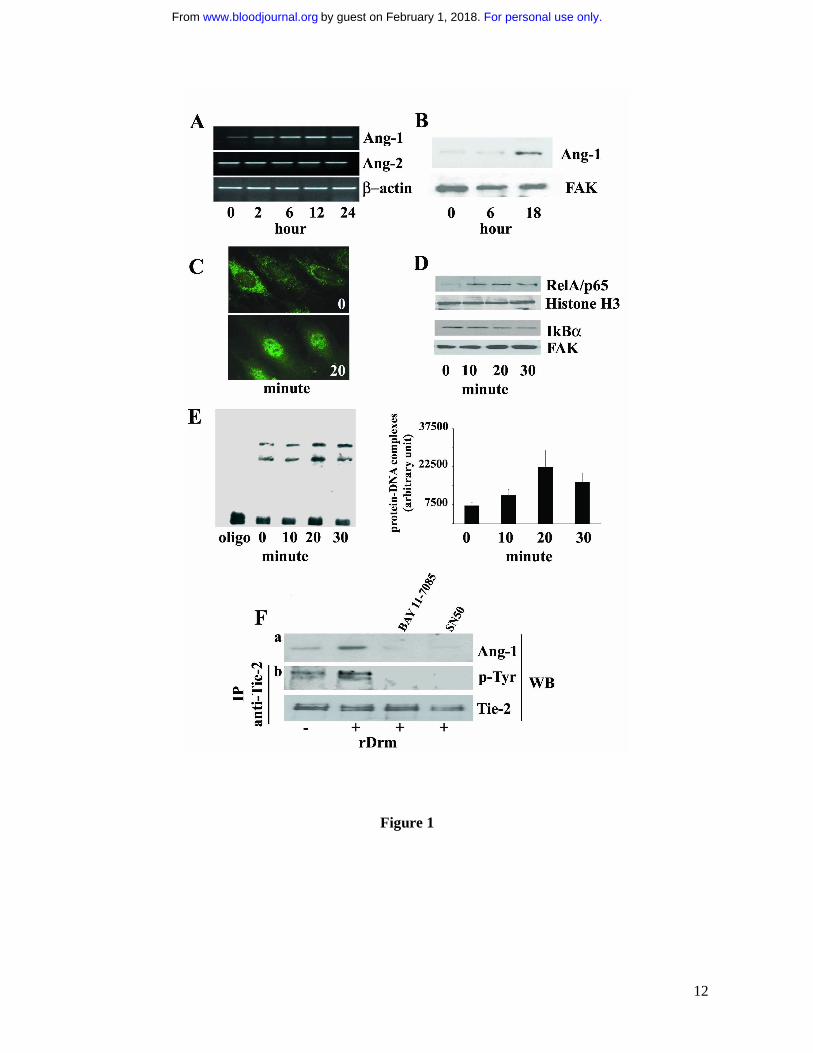
Figure 1. rDrm induces Ang-1 expression in EC in a NF-kB-dependent manner. A, Serum-
starved SIE cells were stimulated for 2, 6, 12, and 24 hours with 50 ng/mL rDrm. At the end of
incubation, total RNA was extracted and Ang-1 and Ang-2 transcripts were analyzed by
semiquantitative RT-PCR. β-actin was used as a control. B, Cell lysates were prepared from SIE
cells stimulated for 0, 6, and 18 hours with 50 ng/mL rDrm. Then, 20 µg-aliquots were probed by
Western blotting with anti-Ang-1 antibodies (Santa Cruz). Uniform loading of the gels was
confirmed by incubation of the membranes with anti-focal adhesion kinase (FAK) antibodies. C,
Serum-starved EC were incubated for 20 minutes with 50 ng/mL rDrm and immunostained with an
anti-RelA/p65 antibody (Santa Cruz) followed by anti-rabbit FITC (Molecular Probes). Control
cells show a diffuse cytoplasmic RelA/p65 immunoreactivity (upper panel) that localizes into the
nucleus after rDrm stimulation (lower panel). Analysis was performed using a Zeiss Axiovert 200M
epifluorescence microscope equipped with a Plan-Apochromat 63x/1.4 NA oil objective. D, SIE
cells were stimulated for 0-30 minutes with 50 ng/mL rDrm. Then, nuclear (5 µg) and cytoplasmic
(10 µg) extracts were probed by Western blotting with anti-RelA/p65 and anti-IkBα (Santa Cruz)
antibodies, respectively. Uniform loading of the gels was confirmed by incubation of the
membranes with anti-histone H3 (Santa Cruz) and anti-FAK antibodies, respectively. E, Nuclear
extracts (2 µg) of SIE cells treated for 0-30 minutes with 50 ng/mL rDrm were incubated with a
biotin-labeled NF-kB double-stranded DNA oligonucleotide probe. Left panel: the protein-DNA
complexes were analyzed by EMSA onto a native 6% polyacrylamide gel; the first lane shows the
migration of the labeled probe in the absence of nuclear extract. Right panel: densitometric analysis
of the protein-DNA complexes shown in the left panel; data are the mean ± SEM of three
independent experiments. F, SIE cells were treated for 1 hour with NF-kB inhibitors SN50 (45
pg/mL, US Biological) or BAY 11-7085 (5 µM, Sigma) before rDrm administration. After 18 hours
cell extracts were probed by Western blotting with anti-Ang-1 antibodies (a). Also, conditioned
media from the corresponding cell cultures were added for 10 minutes to naïve serum-starved SIE
cells. Then, immunoprecipitation with anti-Tie-2 antibodies was performed on the cell extracts
followed by Western blotting with anti-phosphotyrosine antibodies (Santa Cruz) (b). Uniform
loading of the gel was confirmed by incubation of the membrane with anti-Tie-2 antibodies. Similar
results were obtained in two independent experiments.
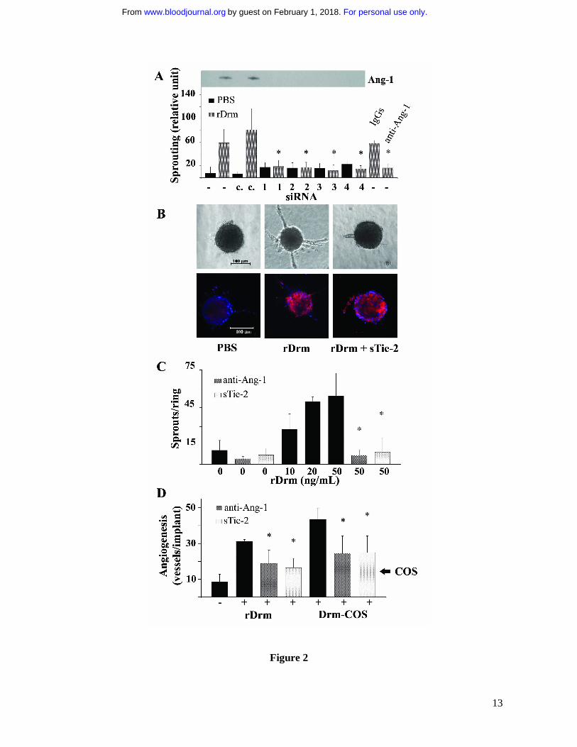
Figure 2. Ang-1 modulates the proangiogenic activity of rDrm. A, Cell aggregates from naïve
VEGFR2-MAE cells (-) or from VEGFR2-MAE cells transfected with fluorescein-conjugated
For personal use only.on February 1, 2018. by guest www.bloodjournal.orgFrom
11
control siRNA (c.) or four different Ang-1 siRNA [1-3, three distinct single oligonucleotides
(Qiagen); 4, one pool of three different oligonucleotides (Santa Cruz)] were embedded in fibrin gel.
Then, 50 ng/mL rDrm were added on the top of the gel in medium containing 10 µg/mL aprotinin.
Also, some rDrm-treated aggregates were incubated in the presence of 1.0 µg/mL of neutralizing
anti-Ang-1 antibodies or irrelevant IgGs. Formation of radially growing cell sprouts was observed
during the next 48 hours8. Sprouts were photographed at 40x magnification with an IX51 inverted
microscope equipped with a 4x/0.10 numerical aperture objective and a Camedia C-4040 digital
camera (Olympus, Melville, NY). Sprouting was quantified by computerized analysis of the
digitalized images8. Data are expressed as mean ± SEM (n = 10); *, statistically different from
rDrm-treated naïve cells (p<0.01, Student’s t test). Conditioned media from the corresponding
transfectants were analyzed for Ang-1 protein content by Western blotting (inset). B, HUVE cell
spheroids prepared in medium supplemented with carboxymethylcellulose were embedded in type I
collagen gel and treated with vehicle or 30 ng/mL rDrm in the absence or in the presence of 100
ng/mL sTie-2 (RELIA Tech GmbH, Woburn, MA). After 24 hours, cell sprouts were photographed
at 200x magnification using an Axiovert 200M microscope equipped with a 20x objective (LD A
PLAN 20X/0,30PH1, Zeiss). Aggregates were fixed in 4% paraformaldehyde, permeabilized in
TBS-0.2% Triton X100, and co-stained with anti-Ang-1 antibodies/anti-rabbit Alexa594 (Molecular
Probes) (in red) and counterstained with 4',6-diamidino-2-phenylindole (Sigma) (in blue). Images
were acquired using a LSM510Meta confocal microscope equipped with objective LD A PLAN
32X/0,40PH1 and processed using the LSM software (Zeiss, Jena, Germany). C, One mm-human
umbilical artery rings were embedded in fibrin gel and incubated with the indicated concentrations
of rDrm in the absence or in the presence of 1.0 µg/mL of neutralizing anti-Ang-1 antibodies or 100
ng/mL sTie-2. After 3 days, EC sprouts were counted under an IX51 inverted microscope at 10x
magnification. *, statistically different from rDrm-treated rings (p< 0.01, Student’s t test). D,
Alginate beads containing vehicle, 100 ng of rDrm, parental (�) or Drm-overexpressing-COS cells
(10,000 cells/embryo) were implanted on the top of chick embryo CAMs at day 11 of development.
When indicated, pellets contained also 1.0 µg of neutralizing anti-Ang-1 antibodies or 100 ng of
sTie-2. After 72 hours, newly formed blood vessels converging towards the implant were counted in
ovo at 5x magnification using a STEMI SR stereomicroscope equipped with an objective f equal to
100 mm with adapter ring 475070 (Zeiss). Data are expressed as mean ± SEM (n = 6-8); *,
statistically different from the corresponding stimulus in the absence of any inhibitor (p< 0.01,
Student’s t test).
For personal use only.on February 1, 2018. by guest www.bloodjournal.orgFrom
12
Figure 1
For personal use only.on February 1, 2018. by guest www.bloodjournal.orgFrom
13
Figure 2
For personal use only.on February 1, 2018. by guest www.bloodjournal.orgFrom
doi:10.1182/blood-2007-09-111450Prepublished online May 27, 2008;
Stefania Mitola, Emanuela Moroni, Cosetta Ravelli, German Andres, Mirella Belleri and Marco Presta morphogenic protein antagonist DrmAngiopoietin-1 mediates the pro-angiogenic activity of the bone
http://www.bloodjournal.org/site/misc/rights.xhtml#repub_requestsInformation about reproducing this article in parts or in its entirety may be found online at:
http://www.bloodjournal.org/site/misc/rights.xhtml#reprintsInformation about ordering reprints may be found online at:
http://www.bloodjournal.org/site/subscriptions/index.xhtmlInformation about subscriptions and ASH membership may be found online at:
digital object identifier (DOIs) and date of initial publication. indexed by PubMed from initial publication. Citations to Advance online articles must include final publication). Advance online articles are citable and establish publication priority; they areappeared in the paper journal (edited, typeset versions may be posted when available prior to Advance online articles have been peer reviewed and accepted for publication but have not yet
Copyright 2011 by The American Society of Hematology; all rights reserved.Hematology, 2021 L St, NW, Suite 900, Washington DC 20036.Blood (print ISSN 0006-4971, online ISSN 1528-0020), is published weekly by the American Society of
For personal use only.on February 1, 2018. by guest www.bloodjournal.orgFrom