Embed Size (px)
Citation preview
Med Oral Patol Oral Cir Bucal. 2011 Jul 1;16 (4):e482-7. Angioleiomyoma of the lips
e482
Journal section: Oral Medicine and PathologyPublication Types: Case Report
Angioleiomyoma affecting the lips: Report of 3 cases and review of the literature
Luiz-Alcino Gueiros 1, Mário-José Romañach 1, Ana-Maria Pires-Soubhia 2, Fábio-Ramôa Pires 3, Oslei Paes-de-Almeida 1, Pablo-Agustin Vargas 1
1 Department of Oral Diagnosis – Laboratory of Oral Pathology. Piracicaba Dental School – University of Campinas2 Department of Pathology and Clinical Propedeutics – Laboratory of Oral Pathology. Araçatuba Dental School – Júlio Mesquista State University3 Stomatology, Estácio de Sá University
Correspondence:Department of Oral Diagnosis Oral Pathology Section Piracicaba Dental School State University of CampinasP.O. Box 52 - 13414-903, Piracicaba, SP, Brazil [email protected]
Received: 27/02/2010Accepted: 25/03/2010
AbstractAngioleiomyoma is an uncommon benign soft tissue tumor usually found in the lower extremities and rarely observed in oral tissues. It is microscopically characterized as a proliferation of smooth muscle cells intermin-gled with abundant vascular channels. Oral angioleiomyomas affect mostly the lips, palate, buccal mucosa and tongue, and appears as a submucosal painless nodule. Upper lip is seldom affected and only few cases have been reported. We report three additional cases of angioleiomyoma affecting the lips of elderly patients. All lesions were asymptomatic and presented as submucosal nodules of approximately 1cm. Microscopic analysis on H&E sections revealed similar pattern in all cases, showing well-circumscribed and encapsulated tumors characterized by proliferation of smooth muscle cells and large amount of wide vascular spaces of varying sizes. Most tumor cells were immunoreactive for α-smooth muscle actin, desmin and HHF-35. CD34 was also positive on the en-dothelial cells. All patients were surgically treated and no recurrence was observed so far. The oral pathologists and clinicians should consider this entity when assessing nodular lesions on upper lip.
Key words: Lip neoplasms, vascular neoplasms, angioleiomyoma.
Gueiros LA, Romañach MJ, Pires-Soubhia AM, Pires FR, Paes-de-Almeida O, Vargas PA. Angioleiomyoma affecting the lips: Report of 3 cases and review of the literature. Med Oral Patol Oral Cir Bucal. 2011 Jul 1;16 (4):e482-7. http://www.medicinaoral.com/medoralfree01/v16i4/medoralv16i4p482.pdf
Article Number: 16889 http://www.medicinaoral.com/© Medicina Oral S. L. C.I.F. B 96689336 - pISSN 1698-4447 - eISSN: 1698-6946eMail: [email protected] Indexed in:
Science Citation Index ExpandedJournal Citation ReportsIndex Medicus, MEDLINE, PubMedScopus, Embase and Emcare Indice Médico Español
doi:10.4317/medoral.16.e482http://dx.doi.org/doi:10.4317/medoral.16.e482
Introduction Leiomyomas are benign smooth muscle tumors that preferably affect the genitals, skin and gastrointestinal tract, being rarely seen in the oral cavity and commonly classified into three microscopic patterns: solid leio-myoma, angioleiomyoma (ALM) and epithelioid leio-myoma (1). ALM is a relatively common neoplasm ac-
counting for about 5% of all benign soft tissue tumours and almost three fourths of all oral leiomyomas (1,2). Approximately 150 cases of oral ALM have been re-ported up to now, being frequently identified as a bluish submucosal painless nodule affecting the lips, palate, buccal mucosa, and tongue, presenting important clini-cal differential diagnosis (1).
Med Oral Patol Oral Cir Bucal. 2011 Jul 1;16 (4):e482-7. Angioleiomyoma of the lips
e483
Microscopically, ALM consists in a well-circumscribed lesion presenting vascular spaces with varied size and thick muscle walls, containing concentrically arranged smooth muscle cells within the muscular walls and interspersed with collagen fibers between vessels (3). The smooth muscle origin can be confirmed through a strong cytoplasmic staining pattern of all tumor cells by antibodies directed against smooth muscle actin (3). According to the dominant microscopic pattern some authors additionally classify oral ALM as solid/capil-lary, venous, and cavernous. The latter is characterized by the majority of large vascular spaces and it is the most uncommon subtype (3). The aims of this paper are to report three cases of ALM affecting the lips and to discuss its clinicopathological and immunohistochemi-cal features.
Case Reports Case 1A 54 year-old male was referred for evaluation com-plaining of an asymptomatic lower lip nodule with 6 months of evolution (Fig. 1). He was HIV positive without any other remarkable disease. On physical ex-amination it could be noted a tiny painless submucosal nodule on the right portion of the lower lip, fibrous on palpation and measuring 1 x 1 cm. Under the clinical differential diagnosis of deep-seated mucocele, heman-gioma, fibrous hyperplasia and pleomorphic adenoma, the patient was submitted to an excisional biopsy af-ter local anesthesia. The specimen was processed and histopathological examination revealed a well-circum-scribed smooth muscle proliferation (Fig. 2A1). The le-sion presented a uniform pattern composed mostly of spindle-shaped cells and blood vessels (Fig. 2A2 and 2A3). A diagnosis of angioleiomyoma was rendered and the patient presented no further complications follow-ing two years of follow up. Case 2 A 66 year-old male presented with an asymptomatic well-defined fibrous nodule in the central portion of the upper lip lasting 19 years. Medical history was non-contributory and an excisional biopsy was performed. Histopathological examination revealed a multinodular mass full of large vascular spaces. Stroma was com-posed of spindled cells and small blood vessels (Fig. 2B1 and 2B3). A final diagnosis of angioleiomyoma was rendered and the patient undertook periodic evaluations and failed to show recurrence after 1 year following sur-gery.Case 3A 53 year-old male presented with a slightly bluish painless fibrous nodule with a smooth surface, mea-suring 0.8 x 0.5 x 0.5 cm in the upper lip, lasting 5 months. Medical history was non-contributory. Clinical diagnosis included fibrous hyperplasia, hemangioma,
pleomorphic adenoma and canalicular adenoma, and the lesion was submitted to an excisional biopsy. Gross pathological examination demonstrated a white nodule measuring 0.6 x 0.5 x 0.4 cm and after sectioning dark brownish areas could be seen in the inner portion (Fig. 3A). Histological examination revealed a multilobulated well-circumscribed lesion (Fig. 3B and 3C). The tumor presented bands of smooth muscle cells surrounding multiple vascular spaces of varying sizes. The spindle-shaped cells showed elongated blunt-ended nuclei and eosinophilic cytoplasm forming smooth muscle bundles showing interlacing pattern with collagen fibers between dilated and slit-like vascular spaces (Fig. 3D). Final di-agnosis was angioleiomyoma and no local recurrence has been detected after 6 months of follow up.
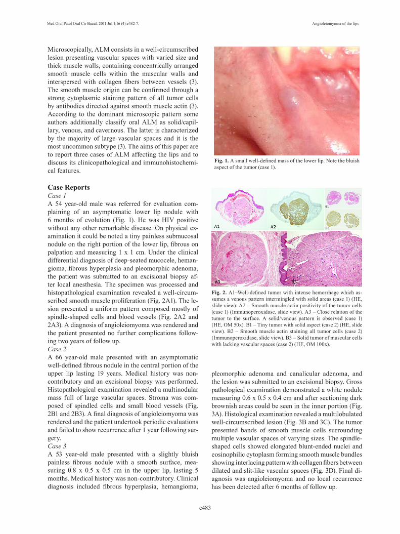
Fig. 1. A small well-defined mass of the lower lip. Note the bluish aspect of the tumor (case 1).
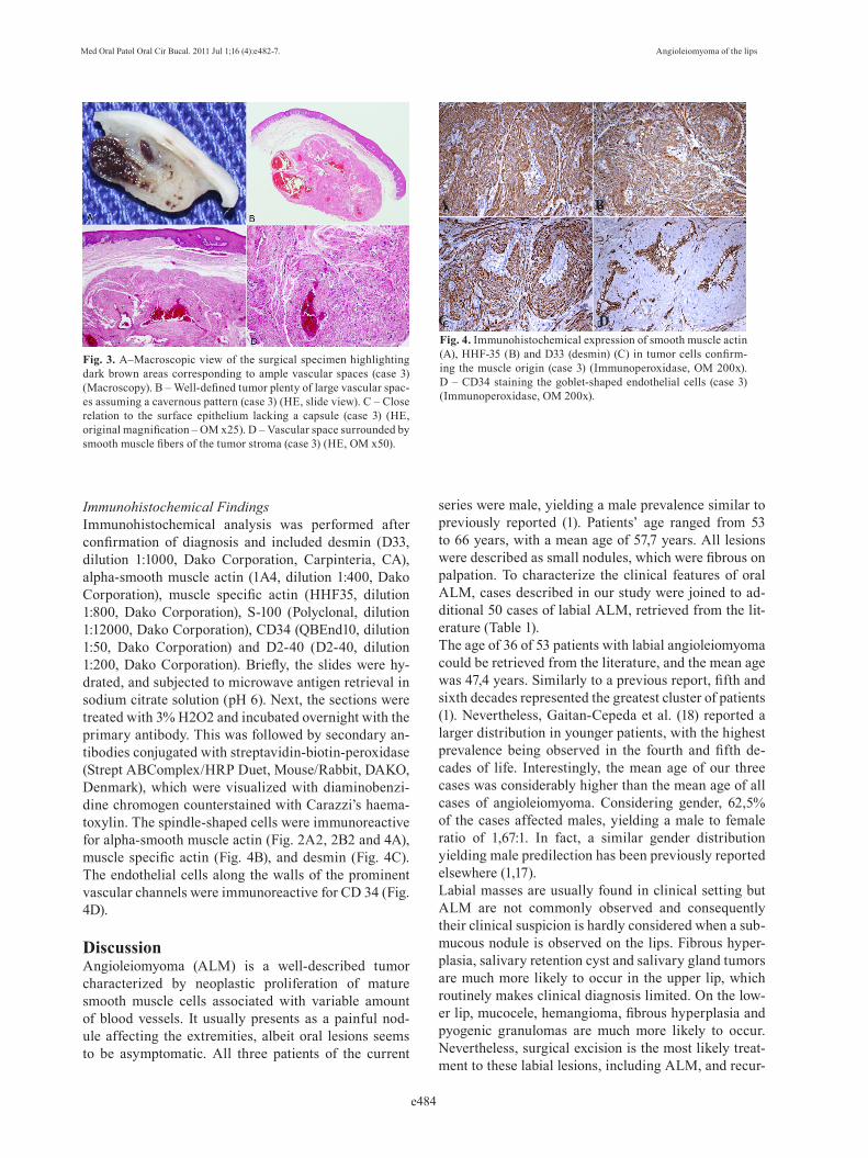
Fig. 2. A1–Well-defined tumor with intense hemorrhage which as-sumes a venous pattern intermingled with solid areas (case 1) (HE, slide view). A2 – Smooth muscle actin positivity of the tumor cells (case 1) (Immunoperoxidase, slide view). A3 – Close relation of the tumor to the surface. A solid/venous pattern is observed (case 1) (HE, OM 50x). B1 – Tiny tumor with solid aspect (case 2) (HE, slide view). B2 – Smooth muscle actin staining all tumor cells (case 2) (Immunoperoxidase, slide view). B3 – Solid tumor of muscular cells with lacking vascular spaces (case 2) (HE, OM 100x).
Med Oral Patol Oral Cir Bucal. 2011 Jul 1;16 (4):e482-7. Angioleiomyoma of the lips
e484
Immunohistochemical FindingsImmunohistochemical analysis was performed after confirmation of diagnosis and included desmin (D33, dilution 1:1000, Dako Corporation, Carpinteria, CA), alpha-smooth muscle actin (1A4, dilution 1:400, Dako Corporation), muscle specific actin (HHF35, dilution 1:800, Dako Corporation), S-100 (Polyclonal, dilution 1:12000, Dako Corporation), CD34 (QBEnd10, dilution 1:50, Dako Corporation) and D2-40 (D2-40, dilution 1:200, Dako Corporation). Briefly, the slides were hy-drated, and subjected to microwave antigen retrieval in sodium citrate solution (pH 6). Next, the sections were treated with 3% H2O2 and incubated overnight with the primary antibody. This was followed by secondary an-tibodies conjugated with streptavidin-biotin-peroxidase (Strept ABComplex/HRP Duet, Mouse/Rabbit, DAKO, Denmark), which were visualized with diaminobenzi-dine chromogen counterstained with Carazzi’s haema-toxylin. The spindle-shaped cells were immunoreactive for alpha-smooth muscle actin (Fig. 2A2, 2B2 and 4A), muscle specific actin (Fig. 4B), and desmin (Fig. 4C). The endothelial cells along the walls of the prominent vascular channels were immunoreactive for CD 34 (Fig. 4D).
DiscussionAngioleiomyoma (ALM) is a well-described tumor characterized by neoplastic proliferation of mature smooth muscle cells associated with variable amount of blood vessels. It usually presents as a painful nod-ule affecting the extremities, albeit oral lesions seems to be asymptomatic. All three patients of the current
Fig. 3. A–Macroscopic view of the surgical specimen highlighting dark brown areas corresponding to ample vascular spaces (case 3) (Macroscopy). B – Well-defined tumor plenty of large vascular spac-es assuming a cavernous pattern (case 3) (HE, slide view). C – Close relation to the surface epithelium lacking a capsule (case 3) (HE, original magnification – OM x25). D – Vascular space surrounded by smooth muscle fibers of the tumor stroma (case 3) (HE, OM x50).
Fig. 4. Immunohistochemical expression of smooth muscle actin (A), HHF-35 (B) and D33 (desmin) (C) in tumor cells confirm-ing the muscle origin (case 3) (Immunoperoxidase, OM 200x). D – CD34 staining the goblet-shaped endothelial cells (case 3) (Immunoperoxidase, OM 200x).
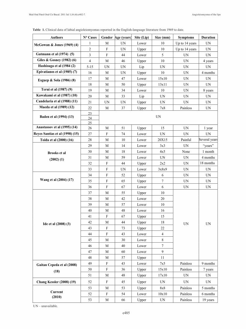
series were male, yielding a male prevalence similar to previously reported (1). Patients’ age ranged from 53 to 66 years, with a mean age of 57,7 years. All lesions were described as small nodules, which were fibrous on palpation. To characterize the clinical features of oral ALM, cases described in our study were joined to ad-ditional 50 cases of labial ALM, retrieved from the lit-erature (Table 1). The age of 36 of 53 patients with labial angioleiomyoma could be retrieved from the literature, and the mean age was 47,4 years. Similarly to a previous report, fifth and sixth decades represented the greatest cluster of patients (1). Nevertheless, Gaitan-Cepeda et al. (18) reported a larger distribution in younger patients, with the highest prevalence being observed in the fourth and fifth de-cades of life. Interestingly, the mean age of our three cases was considerably higher than the mean age of all cases of angioleiomyoma. Considering gender, 62,5% of the cases affected males, yielding a male to female ratio of 1,67:1. In fact, a similar gender distribution yielding male predilection has been previously reported elsewhere (1,17). Labial masses are usually found in clinical setting but ALM are not commonly observed and consequently their clinical suspicion is hardly considered when a sub-mucous nodule is observed on the lips. Fibrous hyper-plasia, salivary retention cyst and salivary gland tumors are much more likely to occur in the upper lip, which routinely makes clinical diagnosis limited. On the low-er lip, mucocele, hemangioma, fibrous hyperplasia and pyogenic granulomas are much more likely to occur. Nevertheless, surgical excision is the most likely treat-ment to these labial lesions, including ALM, and recur-
A B
C D
Med Oral Patol Oral Cir Bucal. 2011 Jul 1;16 (4):e482-7. Angioleiomyoma of the lips
e485
Table 1. Clinical data of labial angioleiomyomas reported in the English-language literature from 1969 to date.
UN – unavailable.
Authors N° Cases Gender Age (years) Site (Lip) Size (mm) Symptoms Duration
McGowan & Jones (1969) (4) 1 M UN Lower 10 Up to 14 years UN 2 F UN Upper 10 Up to 14 years UN
Gutmann et al (1974) (5) 3 F 44 Lower 5 UN UN Giles & Gosney (1982) (6) 4 M 46 Upper 10 UN 4 years Hashisuga et al (1984 (2) 5-15 UN UN Lip UN UN UN
Epivatianos et al (1985) (7) 16 M UN Upper 10 UN 4 months
Esguep & Sola (1986) (8) 17 M 47 Lower 15x10 UN UN 18 M 50 Upper 13x11 UN UN
Terui et al (1987) (9) 19 M 34 Lower 10 UN 8 years Kawakami et al (1987) (10) 20 M 33 Lip UN UN UN Candelaria et al (1988) (11) 21 UN UN Upper UN UN UN
Maeda et al (1989) (12) 22 M 37 Upper 7x8 Painless UN
Baden et al (1994) (13) 23
UN 2425
Anastassov et al (1995) (14) 26 M 51 Upper 15 UN 1 year Reyes Santías et al (1998) (15) 27 F 74 Lower UN UN UN
Toida et al (2000) (16) 28 M 10 Lower 20X15 Painful Several years
Brooks et al
(2002) (1)
29 M 14 Lower 3x3 UN “years” 30 M 18 Lower 4x5 None 1 month 31 M 59 Lower UN UN 4 months 32 F 44 Upper 2x2 UN 18 months
33 F UN Lower 3x8x9 UN UN
Wang et al (2004) (17) 34 F 52 Upper 6 UN UN 35 F 65 Upper 7 UN UN 36 F 67 Lower 6 UN UN
Ide et al (2008) (3)
37 M 55 Upper 10
UN UN
38 M 42 Lower 20 39 M 57 Lower 10 40 M 48 Lower 16 41 F 67 Upper 15 42 M 44 Upper 18 43 F 73 Upper 22 44 F 43 Lower 4 45 M 30 Lower 8 46 M 40 Lower 7 47 M 60 Lower 9 48 M 57 Upper 11
Gaitan Cepeda et al (2008)
(18)
49 F 43 Lower 7x5 Painless 9 months 50 F 36 Upper 15x10 Painless 7 years 51 M 48 Upper 17x10 UN UN
Chang Kessler (2008) (19) 52 F 45 Upper UN UN UN
Current (2010)
53 M 53 Upper 8x8 Painless 5 months 52 F 54 Lower 10x10 Painless 6 months 53 M 66 Upper UN Painless 19 years
Med Oral Patol Oral Cir Bucal. 2011 Jul 1;16 (4):e482-7. Angioleiomyoma of the lips
e486
References with links to Crossref - DOI
rences are not expected. Bleeding is seldom observed when removing labial ALM despite the prominent vas-cular component, possibly because the lesion is usually well-circumscribed and well-demarcated (1). Macroscopic aspects of ALM can be highly sugges-tive. Evaluation of case 3 revealed a whitish mass of fibroelastic consistency during gross examination and after sectioning dark brownish areas could be observed intermingled with pale fibrous structures. The presence of dark-brown areas in the cut surface is expected in such cases, representing large vascular spaces charac-teristic of the cavernous pattern (2,4). ALM can be microscopically subclassified into three distinct patterns: solid, venous or cavernous (2,20). The solid-type is composed of compacted bundles of smooth muscle cells and thin-walled vessels; the venous type shows thick walls of the vascular channels and blend-ing of the vascular musculature with the intervascular muscle bundles. The cavernous-type is characterized by large vascular channels with thin muscular walls, which are difficult to differentiate from intervascular muscle bundles. There can be also a mixture of patterns, lead-ing to biphasic lesions. Matsuyama et al. reviewed 122 ALM and found only 23 mixed cases and only two of these were venous-cavernous type (20). To the best of our knowledge, case 3 is the first case of mixed oral ALM displaying a venous-cavernous pattern. Also, a great amount of morphological variations have already been described. Areas of recent hemorrhage and deposits of hemossiderin have been described in solid-type tumors. Dense hyalinization of collagen, foci of calcification and areas with mature fat cells may be also part of the tumor stroma (2,20). None of these fea-tures were seen in our cases, but some foci of goblet-shaped endothelial cells could be observed in the con-tracted blood vessels, similar to the picture described by Guttmann et al. (5). This feature is rarely described and its endothelial origin can be confirmed through CD34 positive staining (Fig. 4D). The presence of nerve fibers in the capsule have already been described, and cases 1 and 3 showed S-100 immunostaining in the periphery of the tumor (2). Interestingly, case 2 presented with S100 positive cells intermingled with tumor stroma, what can possibly represent a cross reaction because of the poly-clonality of S100 antibody. Despite of the presence of S100 positive nerve fibers on the periphery of cases 1 and 3, no symptoms were reported. The immunohistochemical pattern of ALM is well rec-ognized and may in turn be distinguished from other perivascular myoid tumors easier. Strong and diffuse immunopositivity for SMA is observed in this subset of lesions, which include ALM, myopericytoma (MPC), myofibroma and glomus tumor (3,18). Other smooth muscle markers, such as HHF-35, calponin and h-calde-smon, also show similar immunostaining pattern in
ALM and MPC. Nevertheless, desmin is more specific for ALM, and 75% of the solid-type, 51% of the venous-type and 18% of the cavernous type tumors are positive for this marker. Interestingly, the MPC-like portions in the ALM are desmin-negative and thus allow adequate distinction in difficult cases (17). All three cases were 100% positive for alpha-smooth muscle actin, HHF-35 and desmin (Figs. 4A, 4B and 4C).Although the classic presentation lacks diagnostic dif-ficulties, some tumors (specially the venous-type) pres-ent perivascular concentric arrangement of myoid cells similar to those observed in myopericytoma which may turn the diagnosis somewhat complex (17). Distinction between classic histopathologic presentation of ALM and MPC does not present a diagnostic challenge. Any-how, approximately 21% of all ALM may present some MPC-like areas with perivascular concentric smooth muscle cells proliferation, which are desmin-negative. On the other hand, a great proportion (50%) of MPC may also present ALM-like areas with variable amount of eosinophilic spindle cells and venous or cavern-ous pattern (17). It seems important to highlight that perivascular concentric growth of myoid cells cannot be considered the only distinctive aspect of MPC. In summary, ALM may be considered as a perivascular myoid tumor with some distinctive aspects. As they can be considered rare in the lips, their clinical suspicion is not common. Surgical excision is the adequate treat-ment and no recurrence is expected. Desmin immunos-taining seems to be helpful when morphological aspects are not conclusive.
References1. Brooks JK, Nikitakis NG, Goodman NJ, Levy BA. Clinicopatho-logic characterization of oral angioleiomyomas. Oral Surg Oral Med Oral Pathol Oral Radiol Endod. 2002;94:221-7. 2. Hachisuga T, Hashimoto H, Enjoji M. Angioleiomyoma. A clinico-pathologic reappraisal of 562 cases. Cancer. 1984;54:126-30. 3. Ide F, Mishima K, Yamada H, Saito I, Horie N, Shimoyama T, et al. Perivascular myoid tumors of the oral region: a clinicopathologic re-evaluation of 35 cases. J Oral Pathol Med. 2008;37:43-9. 4. McGowan DA, Jones JH. Angioma (vascular leiomyoma) of the oral cavity. Oral Surg Oral Med Oral Pathol. 1969;27:649-52. 5. Gutmann J, Cifuentes C, Balzarini MA, Sobarzo V, Vicuña R. Angiomyoma of the oral cavity. Oral Surg Oral Med Oral Pathol. 1974;38:269-73. 6. Giles AD, Gosney MB. Oral angiomyoma: a case report. Br J Oral Surg. 1982;20:142-6. 7. Epivatianos A, Trigonidis G, Papanayotou P. Vascular leiomyoma of the oral cavity. J Oral Maxillofac Surg. 1985;43:377-82. 8. Esguep A, Solar M. Oral vascular leiomyoma--report of 5 cases and review of the literature. J Oral Med. 1986;41:126-9,133. 9. Terui T, Takahashi M, Tagami H. Angioleiomyoma of the lip. Int J Dermatol. 1987;26:119-20. 10. Kawakami T, Hasegawa H, Chino T. A transmission electron mi-croscopic study of two cases of oral smooth muscle neoplasm. J Oral Maxillofac Surg. 1987;45:551-5. 11. Candelaria LM, Warnock GR, Pankey G. Nonpainful, smooth-surfaced, bluish nodule on the upper lip. J Am Dent Assoc. 1988;117:487-8. 12. Maeda Y, Hirota J, Osaki T, Hayashi K, Sonobe H, Otsuki Y. An-
Med Oral Patol Oral Cir Bucal. 2011 Jul 1;16 (4):e482-7. Angioleiomyoma of the lips
e487
giomyoma of the upper lip: report of a case with electron microscopic and immunohistochemical observation. Br J Oral Maxillofac Surg. 1989;27:236-42. 13. Baden E, Doyle JL, Lederman DA. Leiomyoma of the oral cav-ity: a light microscopic and immunohistochemical study with review of the literature from 1884 to 1992. Eur J Cancer B Oral Oncol. 1994;30B:1-7. 14. Anastassov GE, Van Damme PA. Angioleiomyoma of the upper lip: report of a case. Int J Oral Maxillofac Surg. 1995;24:301-2. 15. Reyes Santías RM, Suárez Peñaranda JM, Martín Martín C, Ab-dulkader I, Forteza Vila J. [Smooth muscle tumor of the lower lip. A report of a case of rapid growing and increased mitotic activity]. Acta Otorrinolaringol Esp. 1998;49:495-7. 16. Toida M, Koizumi H, Shimokawa K. Painful angiomyoma of the oral cavity: report of a case and review of the literature. J Oral Maxil-lofac Surg. 2000;58:450-3. 17. Wang CP, Chang YL, Sheen TS. Vascular leiomyoma of the head and neck. Laryngoscope. 2004;114:661-5. 18. Gaitan Cepeda LA, Quezada Rivera D, Tenorio Rocha F, Leyva Huerta ER, Mendez Sánchez ER. Vascular leiomyoma of the oral cav-ity. Clinical, histopathological and immunohistochemical characteris-tics. Presentation of five cases and review of the literature. Med Oral Patol Oral Cir Bucal. 2008;13:E483-8. 19. Chang JY, Kessler HP. Masson trichrome stain helps differentiate myofibroma from smooth muscle lesions in the head and neck region. J Formos Med Assoc. 2008 ;107:767-73. 20. Matsuyama A, Hisaoka M, Hashimoto H. Angioleiomyoma: a clinicopathologic and immunohistochemical reappraisal with spe-cial reference to the correlation with myopericytoma. Hum Pathol. 2007;38:645-51.