Embed Size (px)
Citation preview
Aneurysmal Bone Cysts of Soft TissueRepresent True Neoplasms
A Report of Two Cases
By Matthias F. Pietschmann, MD, Andre M. Oliveira, MD, Margaret M. Chou, PhD, Stefan Ihrler, MD,Manuel Niederhagen, MD, Andrea Baur-Melnyk, MD, and Hans Roland Durr, MD
Investigation performed at Ludwig-Maximilians University, Munich, Germany, and the Department of Anatomicand Clinical Pathology, Mayo Clinic, Rochester, Minnesota
Aneurysmal bone cyst was first described by Jaffe andLichtenstein in 19421. It is considered a benign, locallyaggressive lesion with a potential for local recurrence,
and it typically appears in the metaphysis of the long bones andin the vertebral column2-4. Mostly, children and young adults areaffected. No sex predilection has been observed. Radiographi-cally, aneurysmal bone cyst is seen as a lytic lesion, usuallyeccentrically located and expansile but with well-defined mar-gins. Histologically, there are blood-filled cysts separated byfibrous septa, with fibroblasts as well as osteoclast-type giant
cells and reactive woven bone5. Historically, aneurysmal bonecyst was believed to occur exclusively in bone6. In 1972, Salmand Sissons noted soft-tissue lesions resembling aneurysmalbone cysts, and this was probably the first description of thisentity7. For many years, aneurysmal bone cyst was thought to bea lesion, reactive in nature, caused by a circulatory abnormalityleading to an increased venous pressure and resulting in dilationof the vascular network2,8,9. In recent years, strong evidence hassupported the neoplastic nature of aneurysmal bone cyst10-13.In 1999, Panoutsakopoulos et al.10 demonstrated chromosomal
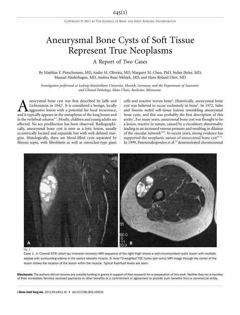
Fig. 1
Case 1. A: Coronal STIR (short tau inversion recovery) MRI sequence of the right thigh shows a well-circumscribed cystic lesion with multiple
septae with surrounding edema in the vastus lateralis muscle. B: Axial T2-weighted TSE (turbo spin echo) MRI image through the center of the
lesion shows the location of the lesion within the muscle. Typical fluid-fluid levels are seen.
Disclosure: The authors did not receive any outside funding or grants in support of their research for or preparation of this work. Neither they nor a memberof their immediate families received payments or other benefits or a commitment or agreement to provide such benefits from a commercial entity.
e45(1)
COPYRIGHT � 2011 BY THE JOURNAL OF BONE AND JOINT SURGERY, INCORPORATED
J Bone Joint Surg Am. 2011;93:e45(1-8) d doi:10.2106/JBJS.J.00534
translocation t(16;17)(q22;p13) as a recurrent cytogenetic ab-normality in primary aneurysmal bone cyst, which was con-firmed by other groups11-13. We report two cases of soft-tissueaneurysmal bone cyst with USP6 locus rearrangement onchromosome 17p13. The patients were informed that dataconcerning their cases would be submitted for publication, andthey consented.
Methods
Tissue specimens from two cases of primary soft-tissue aneurysmal bonecyst were collected in 2007, fixed in 5% buffered formalin, and processed in
standard fashion after decalcification, and micrometer sections were preparedwith hematoxylin and eosin staining. The histological findings were reviewedby two bone and soft-tissue pathologists, and imaging studies were reviewed byan expert radiologist; the diagnosis of aneurysmal bone cyst was made with useof established diagnostic criteria
14. Molecular cytogenetic analysis with fluo-
rescence in situ hybridization (FISH) studies was performed on the paraffin-embedded tissue.
Fluorescence in Situ Hybridization (FISH)15
Bacterial artificial chromosome (BAC) clones flanking the USP6 locus onchromosome 17p13 were obtained from the Children’s Hospital Oakland Re-search Institute (Oakland, California). DNA isolation was performed accordingto Qiagen plasmid Maxi Kit specifications (Qiagen, Valencia, California). DNA
was labeled with use of a Nick Translation Kit from Abbott Molecular (Vysis,Downers Grove, Illinois). Interphase molecular cytogenetic studies were per-formed on 4-mm paraffin-embedded thin sections that were deparaffinized inxylene (twice for fifteen minutes), dehydrated twice in 100% ethyl alcohol forfive minutes, and treated with 10 mmol/L citric acid for ten minutes in a humidmicrowave. Tissue sections were then transferred to 37�C 2· standard salinecitrate for five minutes, and protein was digested with Digest All-3 (Zymed, SanFrancisco, California). After brief washing in 1· phosphate-buffered salinesolution, the slides were sequentially dehydrated in alcohol (70%, 85%, and100%) and air-dried at room temperature. Tissue sections were denatured at80�C for five minutes, and BAC probe hybridization was carried out overnightin a humidified chamber at 37�C. Tissue sections were then washed in 0.1%NP40/2· standard saline citrate at 76�C for four minutes and subsequentlywashed in 0.1% NP40/2· standard saline citrate at room temperature for oneminute. Slides were then mounted in Vectashield mounting medium (VectorLaboratories, Burlingame, California) with 1.5 mg/mL of 49,6-diamidino-2-phenylindole. Tumor samples were scored by two independent investigatorsand considered positive if >5% of 200 cells analyzed showed splitting apart ofthe flanking fluorescence in situ hybridization probes.
Case Reports
CASE 1. A twenty-six-year-old woman had a two-monthhistory of pain in the right thigh. Although she worked
in a gym, no specific traumatic event was identified. On ex-amination, a painful lump, 7 · 5 cm, was identified in the
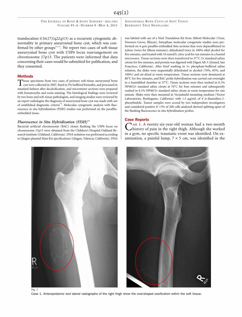
Fig. 2
Case 1. Anteroposterior and lateral radiographs of the right thigh show the oval-shaped ossification within the soft tissue.
e45(2)
TH E J O U R N A L O F B O N E & JO I N T SU R G E RY d J B J S . O R G
VO LU M E 93-A d NU M B E R 9 d M AY 4, 2011AN E U RY S M A L B O N E CY S T S O F SO F T TI S S U E
RE P R E S E N T TR U E NE O P L A S M S
anterolateral aspect of the right thigh. There was no sign ofinflammation, and the findings on examination were otherwiseunremarkable. Radiographs showed a mass with a thin periph-
eral shell of ossification not connected to bone. Magnetic reso-nance imaging (MRI) showed a mass with multiloculated cysticspaces and fluid-fluid levels in the superficial aspect of the vastus
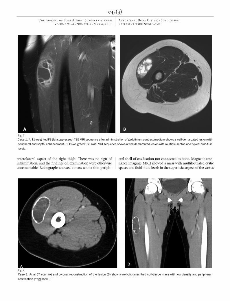
Fig. 3
Case 1. A: T1-weighted FS (fat-suppressed) TSE MRI sequence after administration of gadolinium contrast medium shows a well-demarcated lesion with
peripheral and septal enhancement. B: T2-weighted TSE axial MRI sequence shows a well-demarcated lesion with multiple septae and typical fluid-fluid
levels.
Fig. 4
Case 1. Axial CT scan (A) and coronal reconstruction of the lesion (B) show a well-circumscribed soft-tissue mass with low density and peripheral
ossification (‘‘eggshell’’).
e45(3)
TH E J O U R N A L O F B O N E & JO I N T SU R G E RY d J B J S . O R G
VO LU M E 93-A d NU M B E R 9 d M AY 4, 2011AN E U RY S M A L B O N E CY S T S O F SO F T TI S S U E
RE P R E S E N T TR U E NE O P L A S M S

lateralis muscle (Fig. 1). As there was no sign of malignantdisease, it was decided to follow the patient.
Four months later, the patient reported a change in thepain and consistency of the lesion. Radiographs and computedtomography (CT) revealed increased ossification (Figs. 2, 3, and4), and follow-up MRI showed an increase in the size of thelesion with more prominent septae and fluid levels and dimin-ished edema. Marginal excision of the lesion was performed.
After recovery from the surgery, the patient remainedfree of pain and recurrence, with unrestricted physical activity,as noted at the time of the latest follow-up, at thirty-six months.
Histological findings: Gross examination of the resectionspecimen showed a well-circumscribed 8 · 6 · 3-cm mass coveredby muscle fibers. Sectioning revealed multiple cystic spaces bor-dered by an eggshell of bone at the perimeter of the lesion. Foci of
multinucleated osteoclast giant cells were identified histologically.Atypical cells were not evident, and an infiltrative pattern was notseen (Fig. 5).
CASE 2. A thirty-eight-year-old man presented with a lump in thesoft tissue of the distal part of the left upper arm, which he hadhad for one month. There was no history of trauma, and onexamination there was a walnut-sized painful tense mass proximalto the lateral humeral condyle, which was firmly attached to thesurrounding soft tissues. No other abnormalities were identified.
Radiographs showed a soft-tissue lesion with discreteossifications proximal to the lateral humeral condyle. MRIshowed a soft-tissue mass located in the brachioradialis musclethat was highly suspicious for sarcoma (Fig. 6). MRI withcontrast medium showed uptake mainly in the periphery of the
Fig. 5
Case 1. Histological features of the aneurysmal bone cyst of soft tissue (hematoxylin and eosin). A: Anastomosing branching of fibrous septa with
bone formation of broad cystic and blood-filled spaces (·25). B: Collapsed cystic spaces with cellular fibrous septa without atypia and osteoid
formation (·100). C: Eggshell layer of mature bone (single arrow) at the peripheral border of the lesion and osteoclastic giant cells in fibrous, less
cellular septa associated with osteoid and woven bone trabeculae (double arrows) (·100). D: Higher magnification of fibrous septa with fibroblastic
stroma component, scattered lymphocytes, and immature bone formation with osteoblastic rimming (single arrow) and osteoclastic resorption of
bone (double arrows) (·200).
e45(4)
TH E J O U R N A L O F B O N E & JO I N T SU R G E RY d J B J S . O R G
VO LU M E 93-A d NU M B E R 9 d M AY 4, 2011AN E U RY S M A L B O N E CY S T S O F SO F T TI S S U E
RE P R E S E N T TR U E NE O P L A S M S
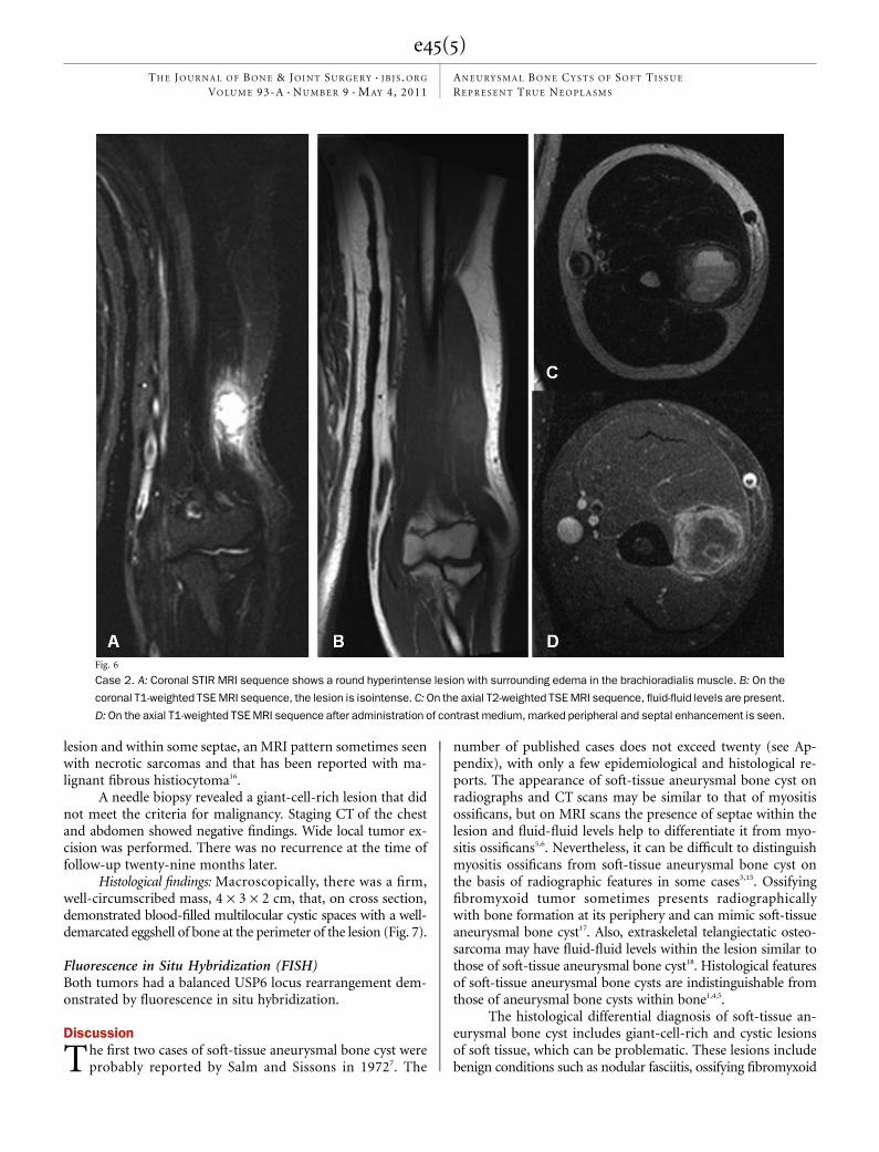
lesion and within some septae, an MRI pattern sometimes seenwith necrotic sarcomas and that has been reported with ma-lignant fibrous histiocytoma16.
A needle biopsy revealed a giant-cell-rich lesion that didnot meet the criteria for malignancy. Staging CT of the chestand abdomen showed negative findings. Wide local tumor ex-cision was performed. There was no recurrence at the time offollow-up twenty-nine months later.
Histological findings: Macroscopically, there was a firm,well-circumscribed mass, 4 · 3 · 2 cm, that, on cross section,demonstrated blood-filled multilocular cystic spaces with a well-demarcated eggshell of bone at the perimeter of the lesion (Fig. 7).
Fluorescence in Situ Hybridization (FISH)Both tumors had a balanced USP6 locus rearrangement dem-onstrated by fluorescence in situ hybridization.
Discussion
The first two cases of soft-tissue aneurysmal bone cyst wereprobably reported by Salm and Sissons in 19727. The
number of published cases does not exceed twenty (see Ap-pendix), with only a few epidemiological and histological re-ports. The appearance of soft-tissue aneurysmal bone cyst onradiographs and CT scans may be similar to that of myositisossificans, but on MRI scans the presence of septae within thelesion and fluid-fluid levels help to differentiate it from myo-sitis ossificans5,6. Nevertheless, it can be difficult to distinguishmyositis ossificans from soft-tissue aneurysmal bone cyst onthe basis of radiographic features in some cases5,15. Ossifyingfibromyxoid tumor sometimes presents radiographicallywith bone formation at its periphery and can mimic soft-tissueaneurysmal bone cyst17. Also, extraskeletal telangiectatic osteo-sarcoma may have fluid-fluid levels within the lesion similar tothose of soft-tissue aneurysmal bone cyst18. Histological featuresof soft-tissue aneurysmal bone cysts are indistinguishable fromthose of aneurysmal bone cysts within bone1,4,5.
The histological differential diagnosis of soft-tissue an-eurysmal bone cyst includes giant-cell-rich and cystic lesionsof soft tissue, which can be problematic. These lesions includebenign conditions such as nodular fasciitis, ossifying fibromyxoid
Fig. 6
Case 2. A: Coronal STIR MRI sequence shows a round hyperintense lesion with surrounding edema in the brachioradialis muscle. B: On the
coronal T1-weighted TSE MRI sequence, the lesion is isointense. C: On the axial T2-weighted TSE MRI sequence, fluid-fluid levels are present.
D: On the axial T1-weighted TSE MRI sequence after administration of contrast medium, marked peripheral and septal enhancement is seen.
e45(5)
TH E J O U R N A L O F B O N E & JO I N T SU R G E RY d J B J S . O R G
VO LU M E 93-A d NU M B E R 9 d M AY 4, 2011AN E U RY S M A L B O N E CY S T S O F SO F T TI S S U E
RE P R E S E N T TR U E NE O P L A S M S
tumor, and giant-cell tumor of the tendon sheath as well aspotentially malignant or malignant lesions such as giant-celltumor of soft tissue and the telangiectatic subtype of extra-skeletal osteosarcoma19-21.
Aneurysmal bone cysts have been shown to have recur-rent rearrangements of the USP6 gene on chromosome17p1311,22,23. USP6—also known as TRE2 or TRE17—was firstidentified as a potential oncogene on the basis of its trans-forming properties when NIH-3T3 cells were transfected withEwing sarcoma DNA24,25. It encodes a ubiquitin-specific pro-tease (USP) and a TBC domain that mediates binding to theArf6 GTPase26. USP6 has effects on cell adhesion and actinremodeling27. Oliveira et al. reported, in 2004, that the productof this chromosomal translocation creates a fusion gene inwhich the osteoblast cadherin 11 gene (CDH11) promoterregion on 16q22 is juxtaposed to the entire ubiquitin-specific
protease USP6 (Tre2) coding sequence on 17p1328. The fusiongene CDH11-USP6 and that USP6 rearrangement are specificfor primary aneurysmal bone cyst and not found in the so-called secondary aneurysmal bone cyst, which is commonlyassociated with giant cell tumor, chondroblastoma, osteoblas-toma, and fibrous dysplasia28.
Rearrangements of USP6 have been found in approxi-mately 70% of aneurysmal bone cysts (70% sensitivity) buthave not been found in other tumors (100% specificity)15,28.
Petrik et al. described an aneurysmal bone cyst-like re-action in the left carotid artery bifurcation in an otherwisehealthy seven-year-old29. Since that time, there have been fewerthan twenty case reports of soft-tissue aneurysmal bone cysts inthe literature5-7,15,19,28-33. The histological features of soft-tissueaneurysmal bone cyst are identical to those of intraosseousaneurysmal bone cyst except for its extraosseous location19.
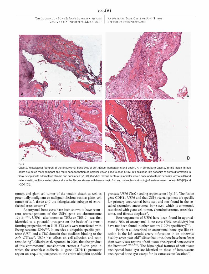
Fig. 7
Case 2. Histological features of the aneurysmal bone cyst of soft tissue (hematoxylin and eosin). A: In contrast to Case 1, in this lesion fibrous
septa are much more compact and more bone formation of lamellar woven bone is seen (·25). B: Focal lace-like deposits of osteoid formation in
fibrous septa with edematous stroma and capillaries (·100). C and D: Fibrous septa with lamellar woven bone and osteoid deposits (arrow in C) and
osteoclastic, multinucleated giant cells in the fibrous stroma with hemorrhagic foci and osteoblastic rimming of mature woven bone (·100 [C] and
·200 [D]).
e45(6)
TH E J O U R N A L O F B O N E & JO I N T SU R G E RY d J B J S . O R G
VO LU M E 93-A d NU M B E R 9 d M AY 4, 2011AN E U RY S M A L B O N E CY S T S O F SO F T TI S S U E
RE P R E S E N T TR U E NE O P L A S M S
Histological features of aneurysmal bone cyst overlapwith those of other osseous lesions such as myositis ossificans,cherubism, and brown tumor14,15. In a 2008 study, Sukov et al.looked for USP6 rearrangements in soft-tissue aneurysmalbone cyst, myositis ossificans, cherubism, and brown tumorand found no such rearrangements in cherubism or browntumor15. However, molecular cytogenetic studies revealedUSP6 rearrangement in two of twelve specimens previouslyclassified as myositis ossificans on the basis of their radio-graphic appearance15. One of the two patients presented ini-tially with classic radiographic features of myositis ossificans,but the radiographic appearance changed to that of an aneu-rysmal bone cyst over time. It is also of interest that no incitingtrauma could be identified for this patient. These data reportedby Sukov et al. were verified by the analysis of our patients, bothof whom were found to have USP6 rearrangements and nohistory of trauma. Nielsen et al. reported five cases of soft-tissueaneurysmal bone cyst, with no patient having a known historyof trauma5.
Nielsen et al. reported only one recurrence in their fivepatients with soft-tissue aneurysmal bone cyst, in whom anintralesional resection had been performed5. The other fourpatients had been free of recurrence for sixteen months to tenyears, findings in concordance with those in other reports of along disease-free survival after resection of soft-tissue aneurysmalbone cyst6,7.
Given that soft-tissue aneurysmal bone cyst may beconfused with other, similarly appearing lesions on radio-graphs, we believe that soft-tissue aneurysmal bone cyst mightbe more frequent than one would assume on the basis of the
published literature. Therefore, fluorescence in situ hybridi-zation analysis of USP6 rearrangement could be a very helpfultool for differentiating soft-tissue aneurysmal bone cyst fromother soft-tissue tumors, especially myositis ossificans.
AppendixA table reviewing cases of soft-tissue aneurysmal bone cystsin the literature is available with the online version of this
article on our web site at jbjs.org. n
Matthias F. Pietschmann, MDStefan Ihrler, MDManuel Niederhagen, MDAndrea Baur-Melnyk, MDHans Roland Durr, MDOrthopaedic Oncology, Department of Orthopaedics (M.F.P. and H.R.D.),Department of Pathology (S.I. and M.N.), and Department of Radiology(A.B.-M.), Ludwig-Maximilians University, Campus Grosshadern,Marchioninistrasse 15, 81377 Munich, Germany.E-mail address for H.R. Durr: [email protected]
Andre M. Oliveira, MDDepartment of Anatomic and Clinical Pathology, Mayo Clinic,200 First Street S.W., Rochester, MN 55905
Margaret M. Chou, PhDDepartment of Pathology and Laboratory Medicine, Children’s Hospital ofPhiladelphia and University of Pennsylvania,3615 Civic Center Boulevard, ARC 816E, Philadelphia, PA 19105
References
1. Jaffe HL, Lichtenstein L. Solitary unicameral bone cyst: with emphasis on theroentgen picture, the pathologic appearance and the pathogenesis. Arch Surg.1942;44:1004-25.2. Martinez V, Sissons HA. Aneurysmal bone cyst. A review of 123 cases includingprimary lesions and those secondary to other bone pathology. Cancer. 1988;61:2291-304.3. Tillman BP, Dahlin DC, Lipscomb PR, Stewart JR. Aneurysmal bone cyst: ananalysis of ninety-five cases. Mayo Clin Proc. 1968;43:478-95.4. Vergel De Dios AM, Bond JR, Shives TC, McLeod RA, Unni KK. Aneurysmal bonecyst. A clinicopathologic study of 238 cases. Cancer. 1992;69:2921-31.5. Nielsen GP, Fletcher CD, Smith MA, Rybak L, Rosenberg AE. Soft tissue aneu-rysmal bone cyst: a clinicopathologic study of five cases. Am J Surg Pathol. 2002;26:64-9.6. Rodrıguez-Peralto JL, Lopez-Barea F, Sanchez-Herrera S, Atienza M. Primary an-eurysmal cyst of soft tissues (extraosseous aneurysmal cyst). Am J Surg Pathol.1994;18:632-6.7. Salm R, Sissons HA. Giant-cell tumours of soft tissues. J Pathol. 1972;107:27-39.8. Kransdorf MJ, Sweet DE. Aneurysmal bone cyst: concept, controversy, clinicalpresentation, and imaging. AJR Am J Roentgenol. 1995;164:573-80.9. Clough JR, Price CH. Aneurysmal bone cyst: pathogenesis and long term resultsof treatment. Clin Orthop Relat Res. 1973;97:52-63.10. Panoutsakopoulos G, Pandis N, Kyriazoglou I, Gustafson P, Mertens F, MandahlN. Recurrent t(16;17)(q22;p13) in aneurysmal bone cysts. Genes ChromosomesCancer. 1999;26:265-6.11. Oliveira AM, Hsi BL, Weremowicz S, Rosenberg AE, Dal Cin P, Joseph N, BridgeJA, Perez-Atayde AR, Fletcher JA. USP6 (Tre2) fusion oncogenes in aneurysmal bonecyst. Cancer Res. 2004;64:1920-3.12. Althof PA, Ohmori K, Zhou M, Bailey JM, Bridge RS, Nelson M, Neff JR, Bridge JA.Cytogenetic and molecular cytogenetic findings in 43 aneurysmal bone cysts: ab-errations of 17p mapped to 17p13.2 by fluorescence in situ hybridization. ModPathol. 2004;17:518-25.
13. Baruffi MR, Neto JB, Barbieri CH, Casartelli C. Aneurysmal bone cyst withchromosomal changes involving 7q and 16p. Cancer Genet Cytogenet. 2001;129:177-80.14. Rosenberg AE, Nielsen GP, Fletcher JA. Aneurysmal bone cyst. In: Fletcher CDM,Unni KK, Mertens F, editors. World Health Organization classification of tumours:pathology and genetics of tumours of soft tissue and bone. Lyon: IARC Press; 2002.p 338-9.15. Sukov WR, Franco MF, Erickson-Johnson M, Chou MM, Unni KK, Wenger DE,Wang X, Oliveira AM. Frequency of USP6 rearrangements in myositis ossificans,brown tumor, and cherubism: molecular cytogenetic evidence that a subset of"myositis ossificans-like lesions" are the early phases in the formation of soft-tissueaneurysmal bone cyst. Skeletal Radiol. 2008;37:321-7.16. Alyas F, Lee J, Ahmed M, Connell D, Saifuddin A. Prevalence and diagnosticsignificance of fluid-fluid levels in soft-tissue neoplasms. Clin Radiol. 2007;62:769-75.17. Enzinger FM, Weiss SW, Liang CY. Ossifying fibromyxoid tumor of soft parts.A clinicopathological analysis of 59 cases. Am J Surg Pathol. 1989;13:817-27.18. Dubec JJ, Munk PL, O’Connell JX, Lee MJ, Janzen D, Connell D, Masri B, LoganPM. Soft tissue osteosarcoma with telangiectatic features: MR imaging findings intwo cases. Skeletal Radiol. 1997;26:732-6.19. Shannon P, Bedard Y, Bell R, Kandel R. Aneurysmal cyst of soft tissue: report ofa case with serial magnetic resonance imaging and biopsy. Hum Pathol. 1997;28:255-7.20. Weiss SW, Goldblum JR, Enzinger FM. Enzinger and Weiss’s soft tissue tumors.5th ed. Philadelphia: Mosby Elsevier; 2008. p 398-401.21. Miettinen M. Ossifying fibromyxoid tumor of soft parts. Additional observationsof a distinctive soft tissue tumor. Am J Clin Pathol. 1991;95:142-9.22. Panoutsakopoulos G, Pandis N, Kyriazoglou I, Gustafson P, Mertens F, MandahlN. Recurrent t(16;17)(q22;p13) in aneurysmal bone cysts. Genes ChromosomesCancer. 1999;26:265-6.
e45(7)
TH E J O U R N A L O F B O N E & JO I N T SU R G E RY d J B J S . O R G
VO LU M E 93-A d NU M B E R 9 d M AY 4, 2011AN E U RY S M A L B O N E CY S T S O F SO F T TI S S U E
RE P R E S E N T TR U E NE O P L A S M S
23. Winnepenninckx V, Debiec-Rychter M, Jorissen M, Bogaerts S, Sciot R. Aneurys-mal bone cyst of the nose with 17p13 involvement. Virchows Arch. 2001;439:636-9.
24. Nakamura T, Hillova J, Mariage-Samson R, Hill M. Molecular cloning of a noveloncogene generated by DNA recombination during transfection. Oncogene Res.1988;2:357-70.
25. Nakamura T, Hillova J, Mariage-Samson R, Onno M, Huebner K, Cannizzaro LA,Boghosian-Sell L, Croce CM, Hill M. A novel transcriptional unit of the tre oncogenewidely expressed in human cancer cells. Oncogene. 1992;7:733-41.
26. Ye Y, Pringle LM, Lau AW, Riquelme DN, Wang H, Jiang T, Lev D, Welman A,Blobel GA, Oliveira AM, Chou MM. TRE17/USP6 oncogene translocated in aneu-rysmal bone cyst induces matrix metalloproteinase production via activation ofNF-kappaB. Oncogene. 2010;29:3619-29.
27. Masuda-Robens JM, Kutney SN, Qi H, Chou MM. The TRE17 oncogene encodesa component of a novel effector pathway for Rho GTPases Cdc42 and Rac1 andstimulates actin remodeling. Mol Cell Biol. 2003;23:2151-61.
28. Oliveira AM, Perez-Atayde AR, Inwards CY, Medeiros F, Derr V, Hsi BL, GebhardtMC, Rosenberg AE, Fletcher JA. USP6 and CDH11 oncogenes identify the neoplasticcell in primary aneurysmal bone cysts and are absent in so-called secondary aneu-rysmal bone cysts. Am J Pathol. 2004;165:1773-80.
29. Petrik PK, Findlay JM, Sherlock RA. Aneurysmal cyst, bone type, primary in anartery. Am J Surg Pathol. 1993;17:1062-6.
30. Ajilogba KA, Kaur H, Duncan R, McFarlane JH, Watt AJ. Extraosseous aneurys-mal bone cyst in a 12-year-old girl. Pediatr Radiol. 2005;35:1240-2.
31. Wang XL, Gielen JL, Salgado R, Delrue F, De Schepper AM. Soft tissue aneu-rysmal bone cyst. Skeletal Radiol. 2004;33:477-80.
32. Lopez-Barea F, Rodrıguez-Peralto JL, Burgos-Lizaldez E, Alvarez-Linera J,Sanchez-Herrera S. Primary aneurysmal cyst of soft tissue. Report of a case withultrastructural and MRI studies. Virchows Arch. 1996;428:125-9.
33. Amir G, Mogle P, Sucher E. Case report 729. Myositis ossificans and aneu-rysmal bone cyst. Skeletal Radiol. 1992;21:257-9.
e45(8)
TH E J O U R N A L O F B O N E & JO I N T SU R G E RY d J B J S . O R G
VO LU M E 93-A d NU M B E R 9 d M AY 4, 2011AN E U RY S M A L B O N E CY S T S O F SO F T TI S S U E
RE P R E S E N T TR U E NE O P L A S M S