Embed Size (px)
Citation preview
American Journal of Medical Genetics 64:488492 (1996)
Aneuploidy Detection in Human Sperm Nuclei Using PRINS Technique
Anne Girardet, Lionel Coignet, Brigitte Andrko, Genevieve Lefort, Jean Paul Charlieu, and Franck Pellestor CNRS UPR 9008, Montpellier (A.G., B.A., L.C., F.P.), and Cytogenetics Laboratory, St . Charles Hospital, Montpellier, France (G.L.); MRC Human Genetics Unit, Western General Hospital, Crewe Road, Edinburgh, UK (J.P.C.)
Rapid and specific identification of chromo- somes can be attained in situ using the PRimed IN Situ (PRINS) labelling tech- nique. We have adapted this technique to mature human sperm in combination with a protocol for simultaneous decondensation and denaturation of sperm nuclei. This strategy allowed us to obtain double label- ling of human spermatozoa in a <2-hr reac- tion. In the present study, we report the es- timates of disomy for chromosomes 3, 7, 10, 11, and 17 on 64,642 spermatozoa from 2 nor- mal males. The incidences of disomy ranged from 0.28434%. There were no significant interindividual or interchromosomal differ- ences in disomy rates. @ 1996 Wiley-Liss, Inc.
KEY WORDS: PRINS, human sperm, chro- mosomes, aneuploidy
INTRODUCTION Assessment of the aneuploidy rate in human ga-
metes remains an important topic of interest and re- search, because nondisjunctions make a major contri- bution to the chromosomal abnormalities found in man.
In the last decade, direct information on the chromo- somal constitution of human sperm has been obtained because of the introduction of the in vitro human sperm- hamster eggs fertilization system [Rudak et al., 1978; Martin et al., 19831. This methodology has allowed the karyotyping of human sperm complements and has been applied in investigating various points concern- ing the occurrence and cause of chromosomal abnor- malities in male meiosis [Martin and Rademaker, 1987; Pellestor et al., 19891. However, this experimental method is labor-intensive and of little profit in terms of sperm karyotypes obtained. Recently, several laborato-
Received for publication August 2, 1995; revision received November 20,1995.
Address reprint requests to Dr. Franck Pellestor, CNRS UPR 9008, CRBM, Route de Mende, B.P. 5051, F-34033 Montpellier, France.
0 1996 Wiley-Liss, Inc.
ries have adapted interphase fluorescence in situ hy- bridization (FISH) technique on sperm in order to di- rectly assess the incidence of disomy in human gametes [reviewed in Williams et al., 1993; Bischoff et al., 19941. Chromosomal detection is usually performed with a- satellite repeat probes. The use of these satellite probes has some limitations; in particular, several chromo- somes (5119,13121, and 14/22) present high homology in their a-satellite DNA sequences, resulting in crosshy- bridization in FISH reaction [Waye and Willard, 1987; Lebo et al., 19921.
To overcome this problem, we have adapted the PRimed IN Situ (PRINS) labelling technique to human sperm. This technique is more efficient than the FISH procedure for discriminating among a-satellite DNA sequences [Koch et al., 1991; Pellestor et al., 19941. We have generated primers specific for several human chromosomes and have established a dual-color PRINS protocol which distinguishes diploid and dis- omic sperm nuclei. In the present study, we report the results of aneuploidy detection for chromosomes 3, 7, 10, 11, and 17.
MATERIALS AND METHODS Sperm Preparation
Sperm samples were obtained from 2 normal, healthy males, of proven fertility, ages 34 and 36 years, respec- tively. After liquifying at room temperature, freshly ejaculated samples were washed twice in phosphate buffered saline (PBS) by centrifugation (8 min a t 2,000 rpm) and fixed for 1 hr in fresh fixative (3:l methanol: glacial acetic acid) a t -20°C. Sperm suspensions were then dropped onto clean microscopic slides and air- dried. Slides were aged 1-8 days a t room temperature before use for PRINS reaction. Immediately before the reaction, the slides were denatured in 3 M NaOH at room temperature for 3-11 min, depending on their age.
Double PRINS Reaction Specific oligonucleotide primers for a-satellite DNA
of chromosomes 3, 7, 10, 11, and 17 were defined by comparing the a-satellite sequences of each chromo- some to the consensus a-satellite sequence of human chromosomes reported by Choo et al. [19911. The primer sequences were determined in the area with the
PRINS on Sperm Nuclei 489
orescein antibody (20 pg/ml) (Boehringer Mannheim). Signals were simultaneously detected by incubating the slides 30 rnin with both Texas red-avidin and antidigox- igenin-fluorescein. The preparations were counterstained with propidium iodide (0.02 pg/ml) and 4',6-diamidine- 2-phenylindole dihydrochloride (DAPI) (0.5 pg/ml) in the antifade solution Vectashield (Vector).
Scoring Criteria, Microscopic Analysis, and Statistical Calculations
Slides were analyzed by two independent observ- ers, using a Leitz microscope equipped with a triple band-pass filter (DAP1:fluoresceinisothiocyanate (FITC): Texas red). Sperm nuclei were scored as haploid when they displayed two distinct signals in different colors (green and red). Nuclei were considered disomic when they displayed two similar signals, in size and inten- sity, separated by at least one domain diameter. The same criteria were applied for scoring diploid nuclei containing four distinct fluorescent spots (two green and two red).
Comparisons between mean frequencies of disomy were made using analysis of variance and standard devi- ation (SD). P < 0.05 was considered significant. x2 tests were performed to check for homogeneity in the rates of disomy for different chromosomes. Similar statistical analysis was carried out to compare diploidy frequencies.
RESULTS The combination of 3 M NaOH treatment and the
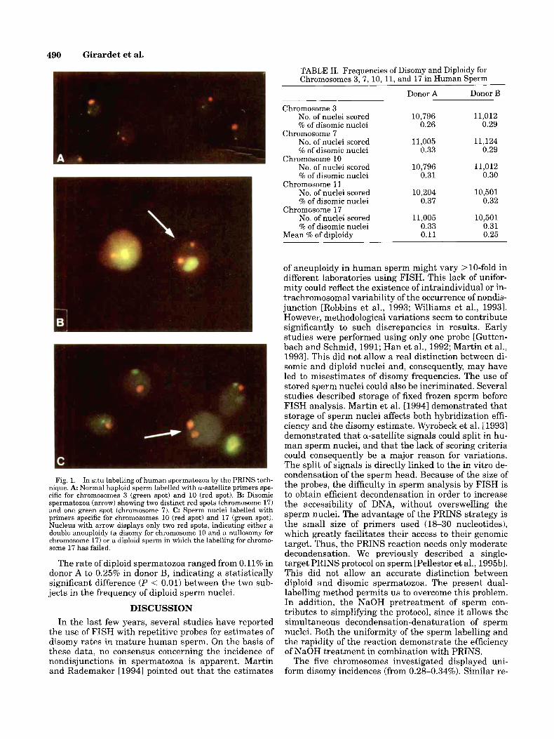
PRINS method allowed us to obtain an efficient la- belling of sperm nuclei (mean, 98.5%) in a reaction of <2 hr. The time of optimal NaOH treatment depended on the age of the sperm preparation slides. The longer the slides were aged, the longer they needed 3 M NaOH treatment (1 day old, 3 min; 2 days old, 5 min; 4 days old, 7 min; 6 days old, 9 min; 8 days old, 11 rnin). Chro- mosome-specific primers were used in various combi- nations. In all cases, fluorescent signals were distinct and easily scorable. Examples of labelling are shown in Figure 1. Occasional size variations were observed (Fig. 1A) and were the direct consequence of variations in a-satellite DNA size.
The data obtained for the different primers are pre- sented in Table 11. A minimum of 10,000 sperm nuclei per chromosome primer were analyzed by two indepen- dent observers, leading to a total number of 64,642 scored spermatozoa. The mean frequency of disomy was 0.28% for chromosome 3, 0.31% for chromosome 7, 0.305% for chromosome 10,0.345% for chromosome 11, and 0.32% for chromosome 17. There was no statistical difference (P > 0.5) in the frequencies of disomy among these five chromosomes.
most nucleotide divergences. The specificity of these primers was previously defined and tested on both metaphases and interphase nuclei [Pellestor et al., 1994,1995al. The sequences and the optimal technical conditions are given in Table I.
For each primer, a reaction mixture was prepared in a final volume of 50 pl, containing the oligonucleotide (50-200 pmol), 0.1 mM each of dATP, dCTP, and dGTP, 0.002 mM dTTP, 0.02 mM of biotin-16-dUTP or digoxi- genin-11-dUTP (Boehringer Mannheim, Meylan, France), 50 mM KC1, 10 mM Tris-HC1, pH 8.3, 1.5 mM MgC12, 0.01% bovine serum albumin (BSA), and 2 units of Taq DNA polymerase (Boehringer Mannheim). The reaction was performed on the programmable temperature cy- cler Techne PHC-3 (Techne Corporation, Cambridge, UK), fit with a flat plate block. Prepared slides and coverslip were put on the plate block. Each reaction program con- sisted of two steps: 1) 12 min at annealing temperature, wherein slides were heated alone for 5 min to get them to the annealing temperature, after which the reaction mix was deposited on warm slides, spread with a cover- slip, and heated 7 min; 2) 30 min at 72°C in order to al- low nucleotide chain elongation. At the beginning of this second step, the temperature was automatically raised to 72°C. The first PRINS reaction was arrested by im- mersing the slides in a stop solution (500 mM NaCU50 mM EDTA, pH 8) at 72°C for 3 min. Slides were then transferred from the stop solution to 1 x NT buffer (50 mM Tris-HC1, pH 7.2,lO mM MgS04, 100 pM DTT, and 150 yglml BSA) and washed twice at room temperature before being treated with a dideoxynucleotides mix (109 pM each of ddATP, ddCTP, ddGTP, and ddTTP, 4 p110 X NT buffer, and 2 U of Klenow enzyme) for 10 rnin at 37°C in order to block the free 3'-ends of the elongation fragments generated by the first PRINS reaction. This intermediate step prevented mixing of labelling. Slides were then passed in stop solution and washed twice in 1 x NT buffer at room temperature. The solution surplus was removed, and the slides were again placed on the plate block of the thermocycler. The second preheated PRINS reaction mix was then applied to the slides, which were heated 7 min at the annealing temperature of the second primer. The temperature was then auto- matically raised to 72"C, and slides were incubated 30 min. The reaction was stopped by immersing the slides in stop solution, and slides were transferred in 4 x SSC- 0.5% Tween-20 at room temperature.
Detection Biotinylated fragments were visualized using Texas
red-avidin-DCS (5 pg/ml) (Vector Labs, Burlingame, CA), whereas detection of digoxigenin incorporated into synthesized products was done with antidigoxigenin-flu-
TABLE I. Characteristics of Oligonucleotide Primers Used in the Present Study
Chromosome Annealing Optimal Name Locus location Sequences temperature ("C) concentration (pM)
3c a-satellite 3 5' TGAGTTGAACACACACGTAC 3' 66 150 7c a-satellite 7 5' AGCGATTTGAGGACAATTGC 3' 56 100 1Oc a-satellite 10 5' ACTGGAACGCACAGATGACAAAGC 3' 63 200 l l c a-satellite 11 5' GAGGGGTTTCAGAGCTGCTC 3' 65 200 17c a-satellite 17 5' AATTTCAGCTGACTAAACA 3' 51 200
490 Girardet et al.
TABLE 11. Frequencies of Disomy and Diploidy for Chromosomes 3, 7, 10, 11, and 17 in Human Sperm
Donor A Donor B
Chromosome 3 No. of nuclei scored % of disomic nuclei
No. of nuclei scored % of disomic nuclei
No. of nuclei scored % of disomic nuclei
No. of nuclei scored % of disomic nuclei
No. of nuclei scored % of disomic nuclei
Chromosome 7
Chromosome 10
Chromosome 11
Chromosome 17
Mean % of diploidy
10,796 0.26
11,005 0.33
10,796 0.31
10,204 0.37
11,005 0.33 0.11
11,012 0.29
11,124 0.29
11,012 0.30
10,501 0.32
10,501 0.31 0.25
Fig. 1. In situ labelling of human spermatozoa by the PRINS tech- nique. A Normal haploid sperm labelled with a-satellite primers spe- cific for chromosomes 3 (green spot) and 10 (red spot). B: Disomic spermatozoa (arrow) showing two distinct red spots (chromosome 17) and one green spot (chromosome 7). C: Sperm nuclei labelled with primers specific for chromosomes 10 (red spot) and 17 (green spot). Nucleus with arrow displays only two red spots, indicating either a double aneuploidy (a disomy for chromosome 10 and a nullosomy for chromosome 17) or a diploid sperm in which the labelling for chromo- some 17 has failed.
The rate of diploid spermatozoa ranged from 0.11% in donor A to 0.25% in donor B, indicating a statistically significant difference (P < 0.01) between the two sub- jects in the frequency of diploid sperm nuclei.
DISCUSSION In the last few years, several studies have reported
the use of FISH with repetitive probes for estimates of disomy rates in mature human sperm. On the basis of these data, no consensus concerning the incidence of nondisjunctions in spermatozoa is apparent. Martin and Rademaker [1994] pointed out that the estimates
of aneuploidy in human sperm might vary >lO-fold in different laboratories using FISH. This lack of unifor- mity could reflect the existence of intraindividual or in- trachromosomal variability of the occurrence of nondis- junction [Robbins et al., 1993; Williams et al., 19931. However, methodological variations seem to contribute significantly to such discrepancies in results. Early studies were performed using only one probe [Gutten- bach and Schmid, 1991; Han et al., 1992; Martin et al., 19931. This did not allow a real distinction between di- somic and diploid nuclei and, consequently, may have led to misestimates of disomy frequencies. The use of stored sperm nuclei could also be incriminated. Several studies described storage of fixed frozen sperm before FISH analysis. Martin et al. [ 19941 demonstrated that storage of sperm nuclei affects both hybridization effi- ciency and the disomy estimate. Wyrobeck et al. 119931 demonstrated that a-satellite signals could split in hu- man sperm nuclei, and that the lack of scoring criteria could consequently be a major reason for variations. The split of signals is directly linked to the in vitro de- condensation of the sperm head. Because of the size of the probes, the difficulty in sperm analysis by FISH is to obtain efficient decondensation in order to increase the accessibility of DNA, without overswelling the sperm nuclei. The advantage of the PRINS strategy is the small size of primers used (18-30 nucleotides), which greatly facilitates their access to their genomic target. Thus, the PRINS reaction needs only moderate decondensation. We previously described a single- target PRINS protocol on sperm [Pellestor et al., 1995133. This did not allow an accurate distinction between diploid and disomic spermatozoa. The present dual- labelling method permits us to overcome this problem. In addition, the NaOH pretreatment of sperm con- tributes to simplifying the protocol, since i t allows the simultaneous decondensation-denaturation of sperm nuclei. Both the uniformity of the sperm labelling and the rapidity of the reaction demonstrate the efficiency of NaOH treatment in combination with PRINS.
The five chromosomes investigated displayed uni- form disomy incidences (from 0.28-0.34%). Similar re-
PRINS on Sperm Nuclei 491
TABLE 111. Summary of Disomy Estimates (%) Reported for Chromosomes 3,7, 10,11, and 17
Chromosomes
References 3 7 10 11 17 Jackson-Cook and Haller [1991] 0.10 Han et al. [1992] 0.33 Bischoff et al. [1994] 0.34 0.04 0.19 0.09 Guttenbach et al. [19941 0.31 0.31 0.32 0.34 0.31 Lu et al. [1994] 0.40 Present study 0.28 0.31 0.30 0.34 0.32
sults were reported by Guttenbach et al. [19941 and, partially, by Bischoff et al. [19941 for the same chromo- somes. Variations are only significant for chromosomes 7 and 17, with estimates ranging from 0.04-0.40% for chromosome 7, and from 0.09-0.33% for chromosome 17 (Table 111). Due to the above reasons, it is difficult to determine if these variations reflect real variability in the occurrence of nondisjunction, or if they result from the methodological variations among different studies. Globally, reported rates were higher than those ob- tained by sperm karyotyping [Martin and Rademaker, 19901 or in some FISH studies for different chromo- somes [Williams et al., 1993; Spriggs et al., 19951. Pos- sible explanations for these discrepancies could involve the small number of sperm complements analyzed per donor using the human sperm-hamster eggs fertiliza- tion system, or the fluctuations of hybridization effi- ciency of probe coktails according to the quality of sperm and methodological factors. Also, the possibility of interindividual variations of aneuploidy frequency cannot be excluded.
Uniform disomy rates, as reported in both the present study and some previous reports [Guttenbach et al., 1994; Miharu et al., 1994; Lu et al., 19941, support the assumption of an equal occurrence of nondisjunction among autosomal chromosomes in male gametes. This finding agrees with the data drawn from the cytogenetic analysis of human spermatozoa [Martin and Rade- maker, 1990; Pellestor, 19911. However, because of the existence of contradictory results in some FISH studies, this hypothesis needs to be confirmed for all chromo- somes with a large sample of sperm from several indi- viduals. In addition, comparative studies need to be per- formed with PRINS and FISH protocols in order to determine the efficiency of the various decondensation methods described, and to define a standard protocol.
ACKNOWLEDGMENTS This work was supported by a grant from the Groupe-
ment de Recherches et dEtudes sur les GBnomes (G.R.E.G.).
REFERENCES Bischoff FZ, Nguyen DD, Burt KJ, Shaffer LG (1994): Estimates of
aneuploidy using multicolor fluorescence in situ hybridization on human sperm. Cytogenet Cell Genet 66:237-243.
Choo KH, Vissel B, Nagy A, Earle E, Kalitsis P (19911: A survey of the genomic distribution of alpha satellite DNAon all the human chro- mosomes, and derivation of a new consensus sequence. Nucleic Acids Res 19:1179-1182.
Guttenbach M, Schmid M (1991): Non-isotopic detection of chromo- some 1 in human meiosis and demonstration of disomic sperm nu- clei. Hum Genet 87:261-265.
Guttenbach M, Schakowski R, Schmid M (1994): Incidence of chromo- some 3,7,10,11,17 and X disomy in mature human sperm nuclei as determined by nonradioactive in situ hybridization. Hum Genet
Han TL, Webb GC, Flaherty SP, Correll A, Matthews CD, Ford JH (1992): Detection of chromosome 17- and X-bearing human sper- matozoa using fluorescence in situ hybridization. Mol Reprod Dev 33:189-194.
Jackson-Cook C, Haller VL (1991): Estimation of aneuploidy levels in human spermatozoa using chromosome complements and chromo- some specific probes. Am J Hum Genet [Suppll49:83.
Koch JE, Hindkjaer J , Mogensen J, Kolvraa S, Bolund L (1991): An improved method for chromosome-specific labeling of satellite DNA in situ by using denatured double-stranded DNA probes as primers in a primed in situ labelling (PRINS) procedure. Genet Anal Tech Appl8:171-178.
Lebo RV, Flandermeyer RR, Diukman R, Lynch ED, Lepercq JA, Gol- bus MS (1992): Prenatal diagnosis with repetitive in situ hy- bridization probes. Am J Med Genet 43:848-854.
Lu PY, Hammitt DG, Zinsmeister AR, Dewald GW (1994): Dual color fluorescence in situ hybridization to investigate aneuploidy in sperm from 33 normal males and a man with a t(2;4;8)(q23;q27;~21). Fertil Steril 62:394-399.
Martin RH, Rademaker AW (1987): The effect of age on the frequency of sperm chromosomal abnormalities in normal men. Am J Hum Genet 41:484-492.
Martin RH, Rademaker AW (1990): The frequency of aneuploidy among individual chromosomes in 6821 human sperm chromo- some complements. Cytogenet Cell Genet 53:103-107.
Martin RH, Rademaker A (1994): Reliability of aneuploidy estimates in human sperm: Results of fluorescence in situ hybridization studies using two different scoring criteria. Am J Hum Genet [Suppll 55:110.
Martin RH, Balkan W, Burns K, Rademaker AW (1983): The chromo- somal constitution of 1000 human spermatozoa. Hum Genet
Martin RH, KO E, Chan K (1993): Detection of aneuploidy in human interphase spermatozoa by fluorescence in situ hybridization (FISH). Cytogenet Cell Genet 64:23-26.
Martin RH, Chan K, KO E, Rademaker A (1994): Detection of aneu- ploidy in human sperm by fluorescence in situ hybridization (FISH): Different frequencies in fresh and stored sperm nuclei. Cy- togenet Cell Genet 65:95-96.
Miharu N, Best RG, Young R (1994): Numerical chromosome abnor- malities in spermatozoa of fertile and infertile men detected by fluorescence in situ hybridization. Hum Genet 93502-506.
Pellestor F (1991): Differential distribution of aneuploidies in human gametes according to their sex. Hum Reprod 6:1252-1258.
Pellestor F, Sele B, Jalbert H, Jalbert P (1989): Direct segregation analysis of reciprocal translocations: A study of 283 sperm kary- otypes from four carriers. Am J Hum Genet 44464-473.
Pellestor F, Girardet A, Andreo B, Charlieu JP (1994): A polymor- phic alpha satellite sequence specific for human chromosome 13
93 :7-12.
63~305-309.
492 Girardet et al.
detected by oligonucleotide PRimed IN Situ labelling (PRINS). Hum Genet 94:346-348.
Pellestor F, Girardet A, Lefort G, Andreo B, Charlieu J P (1995a): Selection of chromosome specific primers and their use in simple and double PRINS techniques for rapid in situ identification of human chromosomes. Cytogenet Cell Genet 70:138-142.
Pellestor F, Girardet A, Lefort G, Andreo B, Charlieu J P (1995b): PRINS as a method for rapid chromosomal labeling on human spermatozoa. Mol Reprod Dev 40:333-337.
Robbins WA, Seagraves R, Pinkel D, Wyrobek AJ (1993): Detection of aneuploid human sperm by fluorescence in situ hybridization: Ev- idence for a donor difference in frequency of sperm disomic for chromosomes 1 and Y. Am J Hum Genet 52799-807.
Rudak E, Jacobs PA, Yanagimachi L (1978): Direct analysis of the ch re mosome constitution of human spermatozoa. Nature 274:911-913.
Spriggs EL, Rademaker AW, Martin RH (1995): Aneuploidy in human sperm: Results of two- and three-color fluorescence in situ hy- bridization using centromeric probes for chromosomes 1, 12, 15, 18, X and Y. Cytogenet Cell Genet 71:47-53.
Waye JS, Willard HF (1987): Nucleotide sequence heterogeneity of al- pha satellite repetitive DNA A survey of alphoid sequences from different human chromosomes. Nucleic Acids Res 15:7549-7562.
Williams BJ, Ballenger CA, Malter HE, Bishop F, Tucker M, Zwing- man TA, Hassold TJ (1993): Non-disjunction in human sperm: Results of fluorescence in situ hybridization studies using two and three probes. Hum Mol Genet 21929-1936.
Wyrobek AJ, Robbins WA, Tang C, Kobayashi A, Baulch J, Weier HU, Pinkel D (1993): Hierarchical organization of human sperm chro- matin is a critical factor in the detection of chromosomal aneuploi- dies by fluorescence in situ hybridization. Am J Hum Genet 53:130.
![Chromosomal instability, aneuploidy, and gene mutations in ...3. Aneuploidy andAPC mutations A role of APC in the origin of CIN and aneuploidy in an in vitro model was suggested [8,18]](https://img.pdfslide.us/doc/110x75/61010a198f416a48f0302824/chromosomal-instability-aneuploidy-and-gene-mutations-in-3-aneuploidy-andapc.jpg)