Embed Size (px)
Citation preview

Wallace: Control of Blood Pressure and Vascular Tone 1
Anesthetic Pharmacology: Physiologic Principles and Clinical Practice: Control of Blood Pressure and Vascular Tone: Arthur Wallace, M.D., Ph.D. Professor of Anesthesiology and Perioperative Care University of California, San Francisco Address correspondence to: Arthur Wallace, M.D., Ph.D. VAMC Anesthesia (129) 4150 Clement St. San Francisco, CA 94121 Phone: 415-750-2069 Fax: 415-750-6653 Cell: 415-215-7979 Email: [email protected]; [email protected]; [email protected];
Primer: Minimum essential information that an anesthesiology trainee should retain
from this chapter.
1. Factors involved in the control of the blood pressure and vascular tone.
a. Vascular mechanics
b. Autonomic function
c. Hemodynamics
d. Ventricular function
e. Efficient Ventricular Function and the Vasculature
f. Heart Rate
2. Pharmacology
3. How do you solve hemodynamic problems?

Wallace: Control of Blood Pressure and Vascular Tone 2
The pharmacologic control of blood pressure and vascular tone can be addressed
using a number of approaches. The choice of an inotrope or vasoactive compound should
be based on an understanding of the cardiovascular system, the cause of the
hemodynamic perturbation being treated, and the pharmacology of the agent. The
appropriate management of hemodynamic perturbations must be based on a guess at the
etiology of the hemodynamic perturbation, the pathophysiology of the patient, the
pharmacology of the pharmacologic agents, and how these factors interact. This chapter
will discuss the physiologic and pharmacologic factors that go into the choice of
inotropes and vasoactive compounds. The response to a pharmacologic agent is a
complex combination of the direct pharmacologic actions, the hemodynamic response,
and the autonomic response. There are a set of basic anatomic and physiologic functions
that must be understood in order to master hemodynamics and to allow the diagnosis and
optimal treatment of hemodynamic problems. This chapter will briefly review
hemodynamics, autonomic control, cardiac physiology including ventricular function
and ventricular energetics, and then the pharmacology of inotropes, vasocontrictors,
vasodilators, and their use to control blood pressure and vascular tone.
The cardiovascular system, because of the Frank-Starling mechanism, is inherently
stable. The distribution of blood between the pulmonary and systemic circulations is
maintained without any outside inputs.1 The preload dependence of cardiac output, which
is defined by the Frank Starling mechanism, makes the relative distribution of pulmonic
and systemic blood volumes inherently stable and is controlled by ventricular mechanics.
Unfortunately, hemodynamic perturbations, either from changes in blood volume,
positional changes of the patient, or vasodilation of vascular beds as metabolic

Wallace: Control of Blood Pressure and Vascular Tone 3
requirements change, are not handled well solely by the Frank-Starling mechanism of the
heart. As vascular beds vasodilate in response to changes in metabolic load, or venous
beds dilate, or blood volume changes in response to hemorrhage, cardiac output and
blood pressure change without the control of the autonomic nervous system. The
autonomic nervous system controls vascular tone, which effects both systemic vascular
resistance and venous capacity,2 the relative distribution of blood flow to organs, and the
inotropic and chronotropic state of the heart. The combination of an inherently stable
cardiovascular system combined with a control system based on the autonomic nervous
system, makes a very stable hemodynamic system. Patients are able to change their
position in gravitational fields (an example would be standing up), exercise, and have
acute changes in blood volume with relatively minor changes in blood pressure and
cardiac output, because of the combination of the inherently stable cardiovascular system
and regulation by the autonomic nervous system.1,3,4
Anesthesia and many medications administered to patients inhibit the autonomic
nervous system and reduce hemodynamic stability. The entire point of anesthesia is to
!"#$%"&'("&)*#+,-&!"-.*/-"&'*&-$!01%23&-'14$315&6$..!"--1/0&2$'*/*41%&!"-.*/-"&1-&2&
fundamental component of anesthesia. Anesthetics inhibit the heart rate and blood
pressure response to pain.5 When patients undergo general anesthesia, the systemic
vasculature vasodilates, decreasing systemic vascular resistance, venous capacity vessels
dilate, reducing central venous pressure and venous return, filling pressures drop, blood
pressure drops, and breathing frequently stops. Vasodilation in response to anesthesia, is
sufficient to drop the core temperature by vasodilating the skin causing a redistribution
and mixing of peripheral and central blood volumes.6 General anesthesia inhibits the
Wallace: Control of Blood Pressure and Vascular Tone 4
autonomic nervous system reducing or eliminating the autonomic control of blood
pressure and heart rate.7 Heart rates, in response to synthetic opioid (fentanyl, sufentanil,
remifentanil, etc) stimulation of vagal reflexes, may become profoundly bradycardic (HR
7&89,-&'*&:9,-;58 Sufentanil can increase vagal effects to the point of asystole.9 The
carotid baroreceptor reflexes are inhibited by anesthesia.10 One of the major goals of
anesthesia is to reduce autonomic response to painful stimuli. Unfortunately one result of
anesthesia inhibiting the autonomic nervous system is a less stable hemodynamic system
in response to vasodilation, changes in blood volume, changes in patient position,
%(2/0"-&1/&'"4."!2'$!"<&(+.*=12<&*!&.21/5&>("&2/"-'("-1*3*01-'&)"%*4"-&'("&?2$'*/*41%&
/"!@*$-&-+-'"4A&B*!&'("&.2'1"/'&$/#"!&0"/"!23 anesthesia. The anesthesiologist is
responsible for taking over the function of the autonomic nervous system and for
maintaining blood pressure, cardiac output, blood volume, temperature, bladder function,
respiration, and other autonomic functions.
Autonomic Nervous System:
Closed loop control based on measurement of blood pressure by the baroreceptors,
and regulation by the sympathetic and parasympathetic nervous systems, maintains blood
pressure in response to changes in blood volume, metabolic demand, and vasodilation.
Anesthesia inhibits the autonomic nervous system reducing the efficacy of this control
system making the system less stable. The autonomic nervous system is divided into
sympathetic and parasympathetic systems (see Fig XX in Chapter 23). Parasympathetic
efferents (outputs) arise from the dorsal motor nucleus of X and form into the vagus
nerve.11 The most common vagal effect on hemodynamics, is slowing of the heart in
response to vagal stimulation. One of the more common hemodynamic perturbations

Wallace: Control of Blood Pressure and Vascular Tone 5
caused by the parasympathetic nervous system is fainting from vasovagal effects. This
reflex can be quite profound during ophthalmic surgery with the occulocardiac reflex
causing brief asystole. The most common autonomic effects of the sympathetic nervous
system in anesthetic care, are either vasodilation in response to inhibition of sympathetic
tone with induction of general anesthesia, or tachycardia and hypertension with
sympathetic stimulation in response to pain. The hypertensive and tachycardiac response
of the sympathetic nervous system to surgical stimulation is used to monitor depth of
anesthesia. Autonomic control of the cardiovascular system relies on multiple pressure
sensors and output control systems. Sympathetic afferents arise from the carotid sinus
baroreceptors and form into the carotid sinus nerve, then form the glossopharyngeal
nerve and synapse in the nucleus tractus solitarius (NTS) in the medulla oblongata.12
There are also afferent sympathetic nerves (inputs) from the aortic arch, right atrium, left
atrium, and pulmonary artery and venous baroreceptors.13-16 Projections then go from the
NTS to the dorsal motor nucleus of X17 and the intermediolateral column.18 Sympathetic
efferents from the intermediolateral column synapse in the sympathetic chain ganglion
prior to sending efferents to the blood vessels to control systemic vascular resistance and
venous capacity. Sympathetic efferents also project to the heart to control inotropic state.
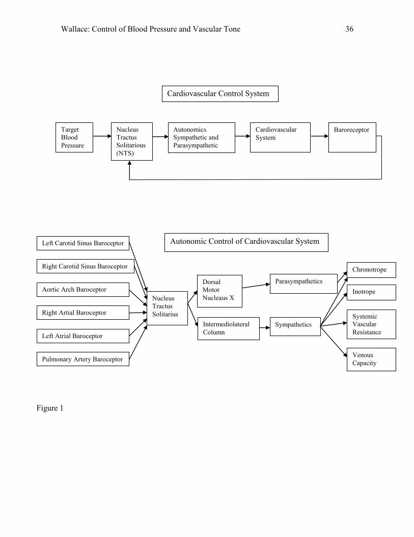
Figure 1 shows a system diagram for autonomic control of hemodynamics. The NTS
compares the pressure measured by the baroreceptors to a set point, and sends signals to
the dorsal motor nucleus of X for parasympathetic outputs,17 and the intermediolateral
column18 which controls sympathetic outputs, which combine to control the
cardiovascular system. Intermediolateral column nuclei are predominantly in the thoracic
cord (T1-T12) with cardioaccelerator fibers from T1-T5. The output of the

Wallace: Control of Blood Pressure and Vascular Tone 6
cardiovascular system (blood pressure) is then detected by the baroreceptors and fed back
to the NTS.12 Closed loop control based on measurement of blood pressure by the
baroreceptors and regulation by the sympathetic and parasympathetic nervous systems,
maintains blood pressure given changes in blood volume, metabolic demand, and
vasodilation. Figure 1 bottom panel shows the complete input and output system. There
are multiple interactions between the control systems with carotid reflexes inhibiting
many cardiopulmonary reflexes. Figure 2 shows an example of the response curves for
the interaction of left and right carotid baroreceptors (RCSP). The use of anesthetics,
including local anesthetics placed in the thoracic epidural space, can profoundly inhibit
this system.
H emodynamics:
Hemodynamic instability is common after the induction of anesthesia and can be
used to demonstrate the key elements in correcting hemodynamic problems. The first step
is to identify the etiology of the problem. Is the etiology vascular volume, systemic
vascular resistance, chronotropy (heart rate), inotropy (ventricular systolic function),
lusiotropy (ventricular diastolic function), or some extrinsic problem such as tamponade
(a lusiotropic problem), or tension pneumothorax (a problem with venous return)? The
diagnosis can usually be obtained by a combination of brief history and statistical
likelihood among common potential causes. A young patient who just had the induction
of anesthesia most likely has a decrease in systemic vascular resistance and venodilation
lowering preload. The most common therapy would be an infusion of volume. A
previously healthy patient with trauma most likely has a low blood volume. An elderly
patient with pre-existing coronary artery disease, who has just had the induction of

Wallace: Control of Blood Pressure and Vascular Tone 7
anesthesia, may have a low preoperative blood volume and vasodilation. The therapy
would be volume and possibly a vasoconstrictor. If the hypotension persists despite first
attempts at therapy, other etiologies such as ventricular dysfunction and/or myocardial
ischemia need to be addressed. History and statistical likelihood are the first approach. If
the first approach at therapy does not work, additional information should be obtained.
Therapy should be guided to correct the problem. Vasodilation, blood volume, or cardiac
problems should be investigated separately.
Vasodilation causes two problems which should be viewed separately. Arteriolar
vasodilation lowers systemic vascular resistance and afterload. Venous dilation dilates
systemic veins, increasing their unstressed vascular volume, decreasing central venous
pressure, and decreasing preload, which lowers cardiac output. Both changes in systemic
vascular resistance caused by arteriolar dilation, and changes in unstressed vascular
volume caused by @"/*$-„'1*/<&4$-'&)"&2##!"--"#&C("/&?@2-*#132'1*/A&*%%$!-5&
Systemic vascular resistance and blood volume must be increased to compensate for
?@2-*#132'1*/A5&
The next important point to note is that pharmacologic agents have three basic
effects. They have the direct effect noted in the package insert on the autonomic nervous
or vascular systems. They may also have direct effects on blood vessels and the heart.
Finally, the hemodynamic changes may cause reflex effects making the net response even
more complex. The combined effect is complex because is it rare that each component
can be isolated. For example, inhaled anesthetics inhibit autonomic tone,5,19 have direct
vasodilatory effects,20 inhibit ventricular function,21 dilate coronary arteries,22,23 and may
cause sympathetic stimulation through irritant receptors (desflurane24,25), all of which

Wallace: Control of Blood Pressure and Vascular Tone 8
may lead to reflex compensation in response to decreases in blood pressure. The net
result of this combination of autonomic, vasodilatory, and cardiac effects is both dose and
time dependent and complex to predict in magnitude. A patient with low blood volume
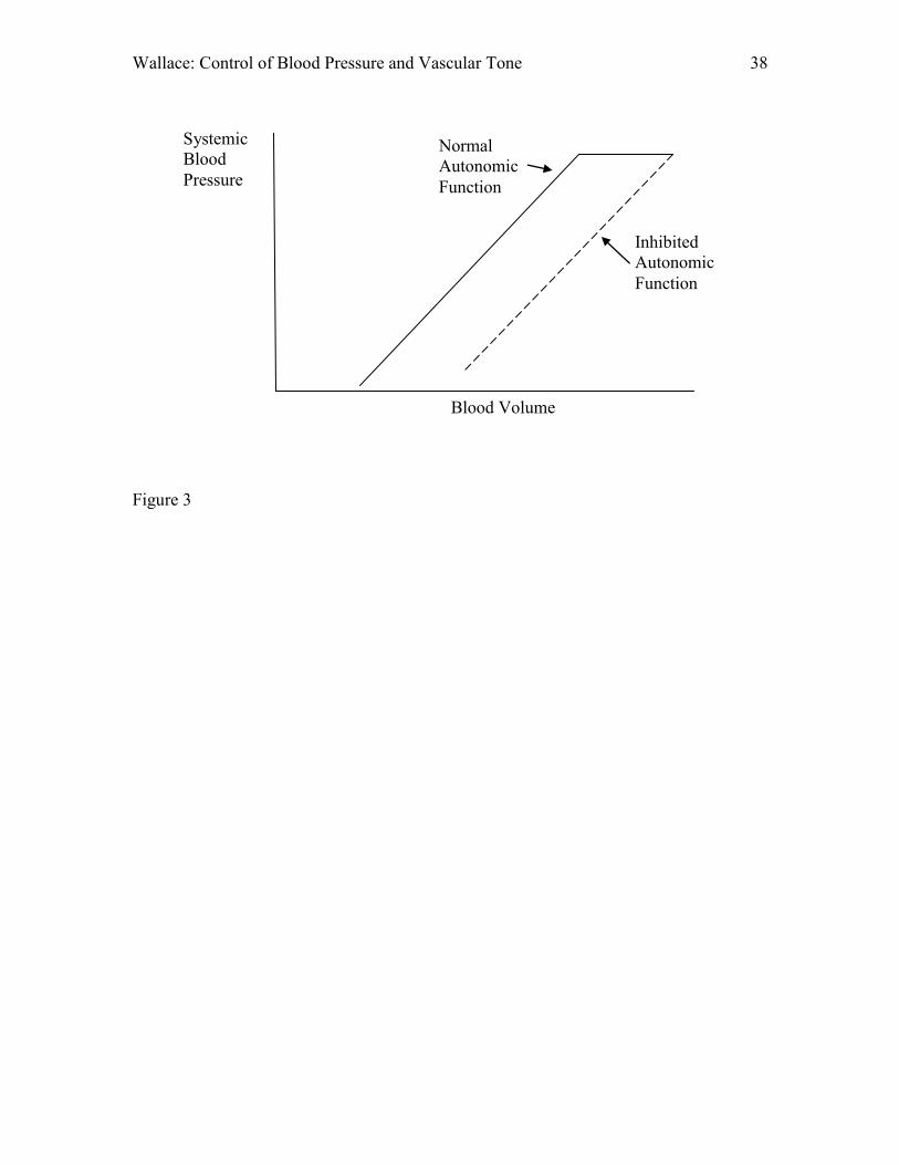
may not tolerate the vasodilation following induction of anesthesia. Figure 3 illustrates a
fundamentally important concept. Patients with an intact autonomic nervous system can
maintain normal blood pressure if blood volume is decreased by 10%.3,4 At a certain
point, further decreases in blood volume result in a decrease in blood pressure. Patients
with an inhibited autonomic nervous system (such as those under general anesthesia)
decrease their blood pressure with all decreases in blood volume. If a patient is already on
the decreasing slope of this relationship, then induction of general anesthesia (with a shift
from the solid to the dashed line) may result in dramatic and possibly lethal decreases in
blood pressure.
Vascular System:
The most common hemodynamic problems are the result of vascular problems,
either blood volume, venous capacity, or systemic vascular resistance. It is very difficult,
if not impossible, to have sufficient cardiac function to compensate for a vasculature that
is not functioning properly. Profound vasodilation either from anaphylaxis or septic
shock can progress to cardiac failure. Rapidly correcting the vascular problem is essential
to prevent the progression to cardiac failure. Profound volume depletion will rapidly lead
to hypotension, which will result in poor coronary perfusion, myocardial ischemia,
myocardial infarction, ventricular stunning, arrythmias, and ultimately cardiac failure.
The most common hemodyamic problems are the result of vascular problems, either
blood volume or resistance. An example will make the point clearer. Suppose systemic
Wallace: Control of Blood Pressure and Vascular Tone 9
vascular resistance which is normally 900D1200 dynes·sec/cm5 (90D120 MPa·s/m3) is
reduced during shock to 400 dynes·sec/cm5. What would the cardiac output need to be to
have a reasonable blood pressure?
80*)(C O
CVPMAPSVR
Where SVR is systemic vascular resistance, MAP is mean arterial pressure, CVP is
central venous pressure, and CO is cardiac output. The 80 converts the units to
dynes·sec/cm5. Rearranging, the cardiac output would be given by
80*)(SVR
CVPMAPC O
Substituting in SVR = 400 dynes·sec/cm5 and some typical values (MAP = 60 mmHg,
CVP = 10 mmHg), gives a cardiac output = 10 liters/minute! Unless the heart is able to
produce a cardiac output of 10 liters/minute, the blood pressure will be quite low. The
effect of systemic vascular resistance or afterload on cardiac output requirements is a
fundamentally important point. For the heart to work efficiently, the systemic vascular
resistance must be reasonable and the blood volume adequate. The first steps in solving
hemodynamic problems is to assess blood volume for adequacy and systemic vascular
resistance for reasonableness.
Ventricular Function:
The next important point in control of blood pressure and vascular tone is
ventricular function. Decisions on hemodynamic management must consider the effects
of the pharmacologic agent not only on blood pressure and cardiac output but also on
ventricular function, ventricular energetics, oxygen consumption, and efficiency. A brief
review of ventricular mechanics and energetics is essential to understand these effects.

Wallace: Control of Blood Pressure and Vascular Tone 10
There are many different models that have been used to explain ventricular mechanics.
The most successful has been the Sagawa model of pressure-volume analysis.26-28
Pressure-volume analysis plots simultaneously measured pressure and volume data on a
single graph. Sagawa, Suga, and Shoukas applied the techniques of pressure-volume
analysis used in thermodynamics of engines to the heart.26 A simple linear relationship
was identified between end-systolic pressure and volume. The end-systolic pressure-
volume relationship is an afterload independent measure of systolic ventricular function26
that completely describes the systolic properties of the ventricle.28 The end-diastolic
pressure-volume relationship completely describes the passive diastolic properties of the
ventricle. Together the end-systolic and end-diastolic pressure-volume relationships
provide a complete description of the mechanical properties of the ventricle. The
ventricle operates between the end-systolic and end-diastolic pressure-volume
relationships (Figure 4)
Ventricular Function and Energetics:
Pressure-volume analysis also describes myocardial energetics, oxygen
consumption, and efficiency.29 The area of the pressure-volume relationship describes
the work performed by the ventricle.30 The stroke work performed by the ventricle is the
integral of pressure with respect to volume integrated from the end-diastolic volume to
the end-systolic volume.31,32
ESV
E DVdvvPStrokeWork )(
The pressure-volume plot of an ejecting beat is shown in Figure 4, where ESV is
end-systolic volume and EDV is end-diastolic volume, P(v) is the pressure at a given
volume and dv is the change in volume. The grey area is the stroke work for a single

Wallace: Control of Blood Pressure and Vascular Tone 11
ejecting beat. End-diastolic volume is the lower right corner of the grey area. Tracing
around the plot in a counter-clockwise direction starting at end diastole, there is initial
isovolumic contraction. When left ventricular pressure exceeds aortic pressure, the aortic
valve opens and left ventricular ejection begins. When the pressure in the left ventricle
drops below aortic pressure, end-systole occurs, followed by isovolumic relaxation. Once
left ventricular pressure drops below left atrial pressure, the mitral valve opens and
ventricular filling begins. Stroke volume is the difference between end-diastolic volume
and end-systolic volume. The integral of pressure with respect to volume between end-
diastole and end-systole represents the work of ejection (gray area in Figure 4).
ESV
E DVdvvPStrokeWork )(
Another concept is that of potential energy (Figure 5). Consider an isovolumic
non-ejecting beat. If the ventricle is forced to contract without ejecting, it still consumes
energy on each beat. In a non-ejecting beat, there is only potential energy of the
pressurized ventricle on each beat. No external work is performed because there is no
ventricular ejection. The potential energy of the ventricle is given by the area of the black
triangle (Figure 5). In a non ejecting beat no external work is done but energy is
consumed (Figure 5 top panel). All energy in a non ejecting beat is potential energy of
the pressurized ventricle and is given by the area to the left of the isovolumic relaxation
line (Figure 5, top panel). In an ejecting beat, there is both potential energy of the
pressurized blood at end systole (black triangle) and the external work of the ejected
blood (gray square) (Figure 5, bottom Panel).
Ventricular Function and Energetics and Oxygen Consumption:
Wallace: Control of Blood Pressure and Vascular Tone 12
There is a very close relationship between ventricular energetics described by the
pressure-volume analysis and oxygen consumption.33 Total oxygen consumption per beat
can be described by the area between the end-systolic pressure-volume relationship and
the end-diastolic pressure-volume relationship for each beat.34 The total area in figure 6
top panel, which is the sum of the potential energy and the external work, gives the total
energy for each beat and the total energy is proportional to oxygen consumption for each
beat. The relationship between the pressure-volume area and oxygen consumption per
beat can be used to analyze ventricular performance. Prior to doing that, 3"',-&#"!1@"&*/"&
more term. The efficiency of ventricular function can be calculated by the ratio of
external work divided by total energy consumed per beat.32 Total work is equal to the
total energy consumed per beat and is proportional to the total oxygen consumed per
beat. The total energy consumed per beat is equal to the sum of the external work and the
potential energy per beat. The efficiency can therefore be derived from the oxygen
consumed to pump blood divided by the total oxygen consumed per beat.32
rkExternalWonergyPotentialETotalWork
TotalWorkrkExternalWoEfficiency
nergyPotentialErkExternalWorkExternalWoEfficiency
erBeatnConsumedPTotalOxygeumedtoPumpOxygenConsEfficiency
Pressure-volume analysis allows the rapid evaluation of a complex physiologic
system. For example, what are the effects of changing afterload on ventricular
energetics? In the standard pressure-volume plot, with external work and potential
Wallace: Control of Blood Pressure and Vascular Tone 13
energy, total oxygen consumption is the sum of the black and grey areas (Figure 6A (top
panel)). The efficiency is the grey area divided by the sum of the black and grey areas.
Lets examine the effect of an increase in afterload, such as could be accomplished
clinically by administering a vasoconstrictor such as phenylephrine (Fig 6B (middle
panel)). Total energy consumption (sum of grey and black areas) is increased, but stroke
volume is decreased and cardiac output decreased. The efficiency, as calculated by the
ratio of oxygen consumed to pump blood divided by total oxygen consumed, would
decrease. Thus administering a vasoconstrictor raised afterload, lowered stroke volume,
lowered cardiac output, increased total oxygen consumption, and lowered ventricular
efficiency. Conversely, afterload reduction (nitroprusside) would increase both stroke
volume and cardiac output, decrease total oxygen consumption, and improve efficiency
(Figure 6C (bottom panel)).
Understanding ventricular energetics and ventricular function is important in
solving hemodynamic problems. The ventricle works as part of the vascular system. Is it
a profound mistake to attempt to solve hemodynamic problems by focusing exclusively
on the heart. The heart, while important, is too often viewed as the cause and solution of
all hemodynamic problems. Most hemodynamic problems are not primarily cardiac in
nature. Most hemodynamic problems are fundamentally vascular problems: systemic
vascular resistance, venous capacity, or blood volume. Moreover, vascular problems are
extremely common causes of death. For example, hypovolemia is a vascular problem.
Inotropic support alone will NOT solve the problem of hypovolemia. The profound
vasodilation of anaphylaxis or septic shock is primarily a vascular problem. Tamponade
is a vascular problem in that the heart can not fill secondary to decreased diastolic

Wallace: Control of Blood Pressure and Vascular Tone 14
compliance. Vascular problems frequently lead to low blood pressure, which lowers
coronary blood flow, which leads to myocardial ischemia, arrhythmias, and finally death.
The primary problem is vascular and the primary solution should be to correct the
vasculature.
Impedance and Optimal H emodynamics:
Optimal vascular function is essential to optimal ventricular performance.31 The
heart fundamentally is a pump that transfers blood from the venous system to the arterial
system. In all physical systems including the cardiovascular system, maximal energy
transfer of a system is achieved when the output impedance of the source equals the input
impedance of the load.31 Impedance is the opposition of a system to a driving function. In
the cardiovascular system vascular resistance is the simplest form of impedance.
Impedance matching is the practice of attempting to make the output impedance ZS of a
source, equal to the input impedance ZL of the load, in order to maximize the power
transfer. Impedance matching provides maximal energy transfer between source and load
in all physical systems including electrical, mechanical, and hemodynamic.
In the cardiovascular system, impedance matching between the venous system
and the heart in diastole, and the arterial system and the heart in systole is essential to
achieve optimal energy transfer. 31,35,36 If the afterload is too high, cardiac output is
depressed. If the afterload is too low, blood pressure will be two low to maintain
coronary perfusion resulting in myocardial ischemia. Afterload describes the impedance
of the ventricle in systole and that of the arterial system. Preload is the relationship
between the impedance of the venous system and that of the ventricle in diastole. Both

Wallace: Control of Blood Pressure and Vascular Tone 15
preload and afterload must be optimized to match the venous and arterial systems to the
ventricle for optimal performance.
Pressure-Volume Analysis and Impedance Matching to the Vasculature:
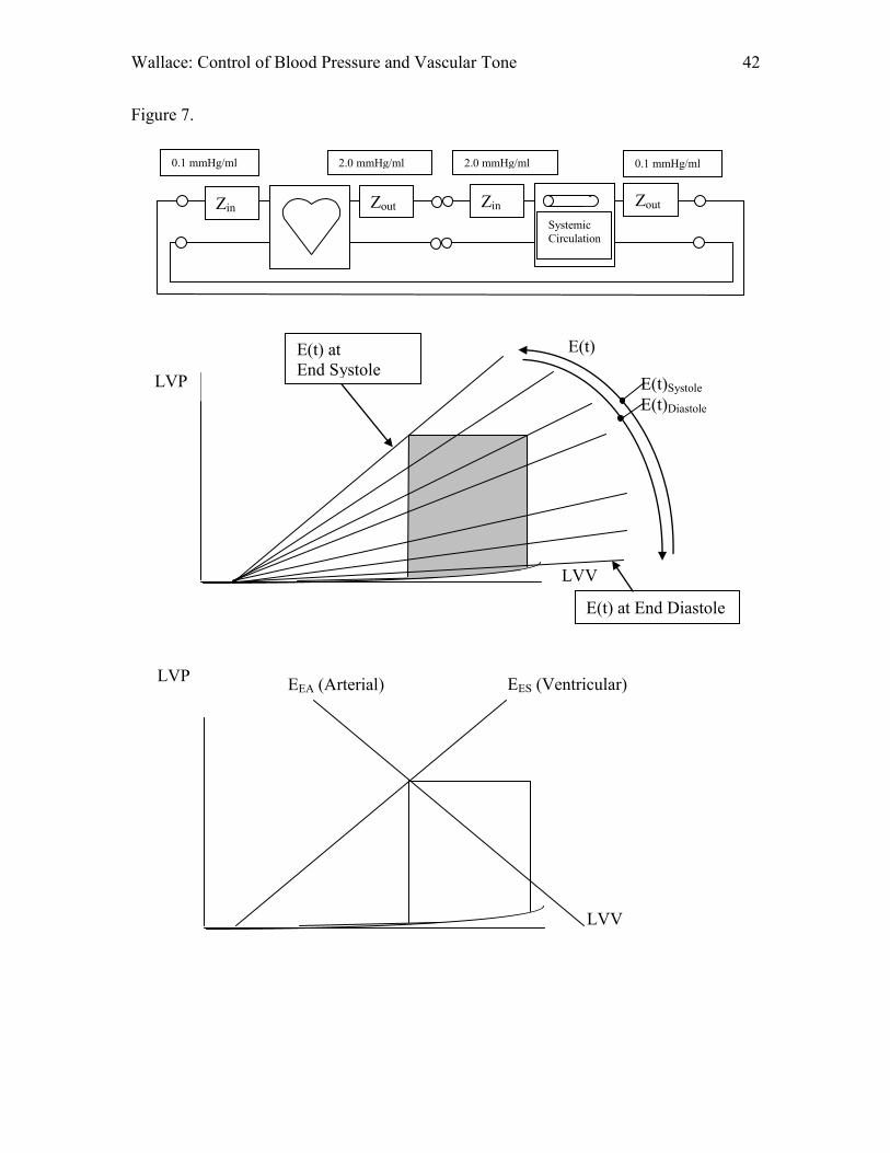
Figure 7 (top) gives a schematic of the cardiovascular system. The impedance of
the heart can be described by the pressure-volume relationship.31,32 The end-diastolic
pressure-volume relationship describes the input impedance to the heart. The end-systolic
pressure-volume relationship describes the output impedance of the heart. The end-
diastolic and end-systolic pressure-volume relationships describe the diastolic and
systolic elastances of the ventricle. The slope of the ventricular diastolic pressure-volume
relationship is about 0.1 mmHg/ml.10,37,38 The elastance of the venous system is about
0.1 mmHg/ml.2,15,39 The normal venous system has a very similar elastance to the
ventricular diastolic elastance, providing impedance matching between the venous system
and the ventricle in diastole.39 The slope of the systolic pressure-volume relationship
normally is about 2.0 mmHg/ml which is very similar to the arterial elastance of 2.0
mmHg/ml, once again matching the output systolic impedance of the heart to the input
impedance of the arterial system.10,37-39 The heart in essence changes it,s elastance from
that of the venous system (0.1 mmHg/ml) to that of the arterial system (2.0 mmHg/ml) on
each beat (Figure 7, middle).31,35,40 The contraction of the heart can be described as a
change in ventricular elastance between the elastance of the venous system and the
elastance of the arterial system. If the elastance of the venous system and the diastolic
elastance of the heart are equal, there will be maximal transfer of energy from the venous
system to the heart (i.e. optimal filling). If the elastance of the ventricle at end-systole

Wallace: Control of Blood Pressure and Vascular Tone 16
equals that of the arterial system, there will be optimal energy transfer (i.e. optimal
ejection).
H emodynamic Problems and Impedance
Solutions of hemodynamic problems should be thought of as optimizing energy
transfer from the venous system to the heart, and from the heart to the arterial system by
matching the impedance of the venous system to diastolic impedance of the heart, and
optimizing the impedance of the arterial system to the systolic impedance of the heart.39
Figure 7 (bottom) shows the maximization of cardiac function by matching the arterial
elastance EEA to the end-systolic pressure-volume relationship EES. Maximum cardiac
function with minimal energy consumption and maximum efficiency can only be
achieved when the venous impedance matches the diastolic elastance and the arterial
impedance matches the systolic elastance of the heart.32,39
Coronary Perfusion:
It is important to remember that coronary perfusion to the left ventricle occurs
during diastole. It is given by: CVR
LVEDPDBPCBF )( where, CBF is coronary blood
flow, DBP is diastolic blood pressure, LVEDP is left ventricular end diastolic blood
pressure, and CVR is coronary vascular resistance. For example, if the diastolic blood
pressure decreases from 60 to 45 and the left ventricular end diastolic blood pressure
increases from 10 to 20, then coronary perfusion pressure (DBP-LVEDP) decreases from
(60-10) = 50 mmHg to (45-20) = 25 mmHg, resulting in a 50% reduction in coronary
blood flow. Decreasing diastolic blood pressure by small amounts can have profound
implications for coronary blood flow. Appropriate afterload and preload are important to
optimal coronary perfusion and ventricular function.

Wallace: Control of Blood Pressure and Vascular Tone 17
H eart Rate:
There is a very important point to make about heart rate. Figure 8 illustrates the
effects of heart rate on coronary blood flow and oxygen demand. Oxygen demand is per
beat.34 Increases in heart rate increase oxygen consumption of the heart.41 Coronary blood
flow to the left ventricle occurs only during diastole. As heart rate increases, the systolic
component of the cardiac cycle changes little in duration but the diastolic period shortens,
shortening the time for coronary perfusion.42 Increases in heart rate increase oxygen
consumption, while decreasing coronary blood flow and oxygen supply. There is a heart
rate in all patients where oxygen supply will be inadequate for demand, resulting in
myocardial ischemia. The only question is what is the heart rate for the ischemic
threshold? In contrast, increases in blood pressure increase oxygen demand, but they also
increase coronary blood flow. There is no threshold high or low blood pressure that is
guaranteed to lead to myocardial ischemia. The next point in discussions of heart rate is
that the equation SVHRCO * is misleading. It suggests that increasing heart rate
causes an increase in cardiac output. Whether increases in heart rate change cardiac
output depend on the initial heart rate, the patient, and the medical condition. There are
heart rates that are too low, where increases in heart rate will increase cardiac output.
There are heart rates where increasing the heart rate will lower cardiac output, because of
inadequate time for diastolic filling. The relationship between heart rate and cardiac
output is not highly correlated (R2 = 0.29, p < 0.0010) as SVHRCO * would suggest.43
The following is true: HRC O
SV ; the later SVHRCO * is not strictly correct.43
Pharmacology:

Wallace: Control of Blood Pressure and Vascular Tone 18
Having established a fundamental understanding of the control of the
cardiovascular system including vascular function, cardiac mechanics, and autonomic
control, the next objective is to understand how pharmacologic agents can be used to
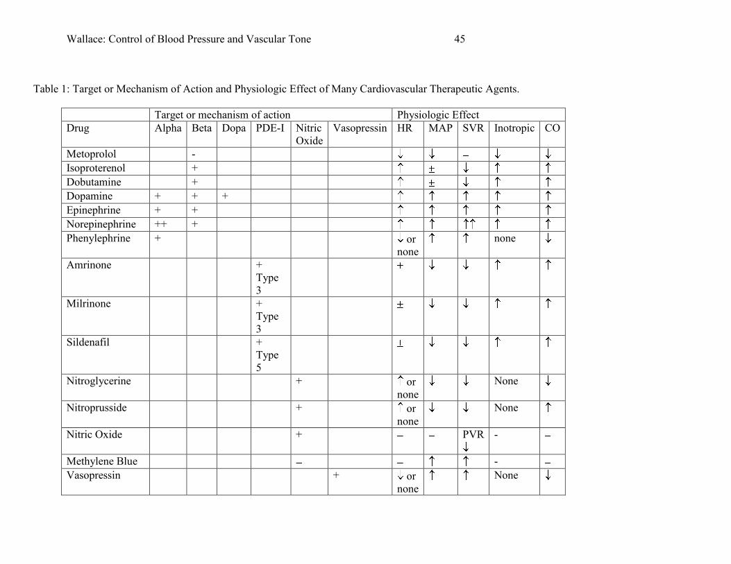
manipulate this system. Table 1 details the mechanism of action and physiologic effects
of some of the common pharmacologic agents used to control hemodynamics. In solving
hemodynamic problems, the first step is to identify the cause.
1. Volume
2. Systemic Vascular Resistance
3. Pulmonary Vascular Resistance
4. Venous Capacity
5. Inotropic State
a. Left Ventricle
b. Right Ventricle
6. Chronotropic State
7. Lusiotropic (Diastolic Function) State
8. Tamponade
9. Tension Pneumothorax
The next step is to fix the identified problem. Identification of the causal problem:
volume, resistance, capacity, inotropic state, chronotropic state, lusiotropic state, will
suggest possible solutions. The faster the cause of a hemodynamic problem is identified
and corrected, the fewer side effects such as myocardial ischemia and ventricular
stunning will occur, and the easier it will be to correct the problem. If hemodynamic
perturbations can be avoided through prophylactic therapy, the cardiovascular system

Wallace: Control of Blood Pressure and Vascular Tone 19
will be even more stable. Avoiding a problem is always superior to trying to correct the
sequellae of a problem.
Identification of the cause of hemodynamic problems should be done logically.
What is the most likely etiology: volume, vasodilation, or ventricular function? If a
single intervention does not immediately solve the problem, then additional data should
be obtained to identify the cause. If the cause is something other than vascular volume,
then a pharmacologic intervention may be needed. The simplest approach is to choose a
drug that specifically addresses the problem. It is rare that there is only a single
pharmacologic agent that is the correct solution to a hemodynamic problem, rather, there
are usually multiple acceptable drugs. There are, however, incorrect choices. If the
systemic vascular resistance is low, using a drug that lowers systemic vascular resistance
is a mistake (dobutamine, amrinone, milrinone). If the resistance is high, choosing a drug
that raises resistance even more (phenylephrine, norepinephrine, epinephrine) is a
mistake. If the heart rate is high, choosing a drug that raises it even more (beta agonist) is
a mistake. If the afterload is high, lowering afterload will commonly raise cardiac output
(Figure 7C). Use of arterial vasodilators such as nitroprusside in the face of elevated
systemic vascular resistance will improve ventricular efficiency, and lower oxygen
consumption, while raising cardiac output.
Vasoconstrictors and vasodilators:
Management of vascular tone through the use of vasodilators and vasoconstrictors
is essential to optimal pharmacologic control of blood pressure and vascular tone. Since
the most common reaction to induction of anesthesia is hypotension with vasodilation,

Wallace: Control of Blood Pressure and Vascular Tone 20
patients with risk for profound hypotension including those with aortic stenosis, coronary
artery disease, congestive heart failure, volume depletion, and pre-existing shock should
have invasive arterial pressure measurement placed prior to induction of anesthesia and
prophylactic hemodynamic management. Optimal hemodynamic control and stability can
be achieved through the prophylactic addition of a vasoconstrictor such as intravenous
infusion of phenylephrine prior to induction. Waiting for hypotension after induction,
prior to administering multiple boluses of phenylephrine, followed by a delay to mix and
start an infusion, causes prolongation of hypotension, and more hemodynamic instability.
One can scramble around trying to treat the 95% of these patients who develop
hypotension after induction or alternatively, start a prophylactic infusion of
phenylephrine prior to induction, and discontinue it 1/&'("&EF&C(*&#*/,'&/""#&1'5&
Prophylactic initiation of phenylephrine infusions prior to induction in patients at risk of
hypotension, reduces hemodynamic instability. The etiology of hypotension during
anesthesia should be identified and corrected. Volume depletion is the most common
etiology of hypotension, with vasodilation from lowered autonomic tone a close second.
Vasoconstriction with phenylephrine can correct hypotension, reduce the need for
excessive volume administration, and improve hemodynamics, all without stimulating the
heart with a risk of myocardial ischemia.
The most common vasodilator used in anesthesia is an inhaled agent. When
inhaled agents are inadequate to control blood pressure and vascular tone, or when a
patient needs pharmacologic hemodynamic management for hypotension after anesthesia,
there are multiple choices of vasodilators. The decision should begin with a diagnosis. Is

Wallace: Control of Blood Pressure and Vascular Tone 21
the hypertension from pain? A full bladder? Essential hypertension? Once pain has been
adequately controlled and other causes corrected, any tachycardia should be corrected. If
the heart rate is elevated, administration of beta blockade with intravenous metoprolol
should be the next step. Once pain and heart rate are adequately controlled and
hypertension is still an issue, then vasodilation with hydralazine, nitroprusside,
nitroglycerine, clonidine, nicardipine, or clevidipine is the next step. The choice depends
on timing and severity of hypertension. Intravenous hydralazine is excellent after
neurosurgery or in patients with persistent hypertension that are otherwise
hemodynamically stable. In patients after cardiac surgery or where hemodynamic
instability is expected, a short acting intravenous vasodilator may be more appropriate.
Nitroprusside is a nitric oxide donor and rapid arterial vasodilator which will increase
cardiac output through afterload reduction. Nitroglycerine is a nitric oxide donor which
requires metabolism to make NO. It has more of an effect on venous beds than
nitroprusside. Nitroglycerine can make patients more volume sensitive as it vasodilates
venous capacitance beds, reducing venous return and cardiac output. Nitroglycerine is
NOT a selective vasodilator. Intravenous nitroglycerine administration vasodilates most
vascular beds uniformly lowering systemic vascular resistance and blood pressure while
maintaining coronary blood flow constant. Relative myocardial oxygen availability, as
defined by the amount of oxygen utilized divided by the amount of oxygen available, is
improved. The amount of oxygen available is constant while the oxygen demand is
decreased. Intravenous nitroglycerine does not selectively increase coronary blood flow.
Nitroglycerine does improve relative oxygen availability while maintaining coronary
blood flow constant.

Wallace: Control of Blood Pressure and Vascular Tone 22
Optimal hemodynamic control of blood pressure requires rapid diagnosis of the
etiology of the hemodyanmic problem, control of blood volume, heart rate, and
vasoconstriction or vasodilation. Vasocontriction with phenylephrine boluses and
infusions can provide optimal hemodynamic control in the critically ill or patients with
multiple co-existing medical conditions such as coronary artery disease or aortic stenosis.
Inotropes:
Inotropes can be used in situations where cardiac output remains inadequate after
optimizing vascular function (resistance and blood volume). Adequacy of cardiac output
can be established through measurement using cardiac output monitoring or mixed
venous saturation. Inotropic agents such as epinephrine, norepinephrine, dobutamine,
milrinone, or amrinone should be selected based on the systemic vascular resistance,
pulmonary vascular resistance, and heart rate needed to optimize vascular function and
maximize ventricular efficiency. In situations where ventricular function is profoundly
depressed and infusions of epinephrine and/or norepinephrine are inadequate, and where
the heart rate and systemic vascular resistance are elevated, while cardiac output is
inadequate, combinations of agents can achieve synergistic increases in cardiac output
with fewer side effects. The combination of epinephrine or norepinephrine with a type-3
phosphodiesterase such as milrinone or amrinone, will provide synergistic increases in
inotropic state and cardiac output. Epinephrine or norepinephrine stimulate beta receptors
in the heart, increasing inotropic state but simultaneously vasoconstrict the vasculature,
raising systemic vascular resistance and afterload. The addition of milrinone or amrinone
to a beta agonist will 1) synergistically stimulate the heart to increase inotropic state by

Wallace: Control of Blood Pressure and Vascular Tone 23
prolonging the half life of cAMP by preventing its metabolism, 2) vasodilate peripherally
lowering afterload, and 3) increase cardiac output. The combination of a catecholamine
and a type-3 phosphodiesterase inhibitor causes synergistic increases in intropic state
while balancing the effects on systemic vascular resistance and afterload.
Phosphodiesterase inhibitors Types-1 through 4 work with cAMP dependent
phosphodiesterases and Type-5 works with cGMP. Very careful adjustments of systemic
vascular resistance are essential for optimal hemodyanamics. Placement of an intra-
aortic balloon pump is frequently helpful in these situations to increase diastolic blood
pressure and improve coronary blood flow.
Risks of Inotropes:
There are significant risks from inotropes which must be balanced against the
need to support cardiac output. Catecholamines can be used as inotropic agents to
increase cardiac output and improve hemodynamics,46 but have significant risks
including arrythmias,46-48 myocardial ischemia,49 infarction, hypokalemia,50 and
hyperglycemia.51 Administration of catecholamines has been shown to decrease survival
in patients with congestive heart failure,52 when compared to vasodilators.53 Rare trials
show improved survival with dobutamine in congestive heart failure,54 but most show
decreased survival.55,56 In contrast, beta blockaders, which are negative inotropes,
improve long term cardiac function57 and survival in congestive heart failure.58,59 Even in
patients with congestive heart failure after myocardial infarction, beta blockade improves
survival.60 Stimulation of the myocardium with catecholamines can be detrimental and
increase morbidity and mortality.
Wallace: Control of Blood Pressure and Vascular Tone 24
The standard therapy for congestive heart failure includes vasodilation and
afterload reduction because lowering the load on the heart improves cardiac output and
efficiency while reducing oxygen consumption (Figure 7C Bottom Panel). The standard
therapy during an acute myocardial infarction is to lower the load on the heart using beta
blockers.61-63 Increasing the load on the heart with catecholamines can be detrimental,
increasing morbidity and mortality.52 Blocking the end organ effect of catecholamines
with beta blockers,64-67 as well as lowering catecholamine production with the alpha-2
agonist clonidine,68 has been shown to reduce perioperative mortality. Catecholamines
should only be used to increase cardiac output when other measures, such as volume
administration and afterload optimization have failed, and cardiac output is clearly
inadequate.
Nitric Oxide:
The type-5 phosphodiesterase inhibitors (sildenafil, vardenafil) can be used to
lower pulmonary vascular resistance. Their use in combination with nitrates must be done
carefully, as there will be synergistic effects between the nitric oxide produced by nitrates
(nitroglycerine or nitroprusside) and the type-5 phosphodiesterase inhibitors. Figure 9
details the biochemical effects of type-5 phosphodiesteases. Type-5 phosphodiesterase
inhibitors will drop systemic blood pressures by 5 mmHg.44 Type-5 phosphodiesterase
inhibitors combined with nitrates will result in larger decreases in blood pressure unless
blood volume is increased slightly.44 Inhibition of nitric oxide effects can be achieved
with methylene blue which may normalize blood pressure in the face of vasoplegic
shock.45
Summary:

Wallace: Control of Blood Pressure and Vascular Tone 25
The present chapter has summarized the Control of Blood Pressure and Vascular
Tone covering the physiology, autonomic nervous system function, vascular mechanics,
ventricular mechanics, and pharmacology of commonly used vasoactive drugs.
Hemodynamic problems are usually quite simple to solve if the root cause is rapidly
identified. Vascular volume is the most common problem followed by systemic vascular
resistance. Hypotension and tachycardia are the result of volume depletion until proven
otherwise. Matching vascular impedance, both venous and arteriolar, to the heart
provides optimal energy transfer and maximal cardiac output. Cardiogenic causes of low
cardiac output such as diastolic dysfunction, systolic dysfunction, and myocardial
ischemia need to be rapidly identified and corrected. External causes such as tamponade
or tension pneumothorax should be viewed as essentially diastolic dysfunction, and must
be rapidly corrected. Pulmonary hypertension raises right ventricular afterload and can
lead to right ventricular failure and hemodynamic collapse. Anesthesia dramatically
influences the autonomic, vascular, and ventricular function. A primary role of
anesthesiology is correcting hemodynamic problems through appropriate pharmacologic
and non pharmacologic control of blood pressure and vascular tone.
Wallace: Control of Blood Pressure and Vascular Tone 26
References:
1. Greene AS, Shoukas AA: Changes in canine cardiac function and venous return curves by the carotid baroreflex. Am J Physiol 1986; 251: H288-96
2. Shoukas AA, Sagawa K: Control of total systemic vascular capacity by the carotid sinus baroreceptor reflex. Circ Res 1973; 33: 22-33
3. Katoh N, Sheriff DD, Siu CO, Sagawa K: Relative importance of four pressoregulatory mechanisms after 10% bleeding in rabbits. Am J Physiol 1989; 256: H291-6
4. Hosomi H, Sagawa K: Sinovagal interaction in arterial pressure restoration after 10% hemorrhage. Am J Physiol 1979; 237: R203-9
5. Daniel M, Weiskopf RB, Noorani M, Eger EI, 2nd: Fentanyl augments the blockade of the sympathetic response to incision (MAC-BAR) produced by desflurane and isoflurane: desflurane and isoflurane MAC-BAR without and with fentanyl. Anesthesiology 1998; 88: 43-9
6. Matsukawa T, Sessler DI, Sessler AM, Schroeder M, Ozaki M, Kurz A, Cheng C: Heat flow and distribution during induction of general anesthesia. Anesthesiology 1995; 82: 662-73
7. Latson TW, McCarroll SM, Mirhej MA, Hyndman VA, Whitten CW, Lipton JM: Effects of three anesthetic induction techniques on heart rate variability. J Clin Anesth 1992; 4: 265-76
8. Gravlee GP, Ramsey FM, Roy RC, Angert KC, Rogers AT, Pauca AL: Rapid administration of a narcotic and neuromuscular blocker: a hemodynamic comparison of fentanyl, sufentanil, pancuronium, and vecuronium. Anesth Analg 1988; 67: 39-47
9. Starr NJ, Sethna DH, Estafanous FG: Bradycardia and asystole following the rapid administration of sufentanil with vecuronium. Anesthesiology 1986; 64: 521-3
10. Wallace A, Lam HW, Mangano DT: Linearity, load dependence, hysteresis, and clinical associations of systolic and diastolic indices of left ventricular function in man. Multicenter Study of Perioperative Ischemia (McSPI) Research Group. J Card Surg 1995; 10: 460-7
11. Contreras RJ, Gomez MM, Norgren R: Central origins of cranial nerve parasympathetic neurons in the rat. J Comp Neurol 1980; 190: 373-94
12. Lipski J, McAllen RM, Spyer KM: The sinus nerve and baroreceptor input to the medulla of the cat. J Physiol 1975; 251: 61-78
13. Ciriello J, Calaresu FR: Projections from buffer nerves to the nucleus of the solitary tract: an anatomical and electrophysiological study in the cat. J Auton Nerv Syst 1981; 3: 299-310
14. Brunner MJ, Greene AS, Kallman CH, Shoukas AA: Interaction of canine carotid sinus and aortic arch baroreflexes in the control of total peripheral resistance. Circ Res 1984; 55: 740-50
15. Shoukas AA, Brunner MJ, Greene AS, MacAnespie CL: Aortic arch reflex control of total systemic vascular capacity. Am J Physiol 1987; 253: H598-603

Wallace: Control of Blood Pressure and Vascular Tone 27
16. Hirooka Y, Sakai K, Kishi T, Takeshita A: Adenovirus-mediated gene transfer into the NTS in conscious rats. A new approach to examining the central control of cardiovascular regulation. Ann N Y Acad Sci 2001; 940: 197-205
17. Ter Horst GJ, Postema F: Forebrain parasympathetic control of heart activity: retrograde transneuronal viral labeling in rats. Am J Physiol 1997; 273: H2926-30
18. Mtui EP, Anwar M, Gomez R, Reis DJ, Ruggiero DA: Projections from the nucleus tractus solitarii to the spinal cord. J Comp Neurol 1993; 337: 231-52
19. Antognini JF, Berg K: Cardiovascular responses to noxious stimuli during isoflurane anesthesia are minimally affected by anesthetic action in the brain. Anesth Analg 1995; 81: 843-8
20. Hickey RF, Sybert PE, Verrier ED, Cason BA: Effects of halothane, enflurane, and isoflurane on coronary blood flow autoregulation and coronary vascular reserve in the canine heart. Anesthesiology 1988; 68: 21-30
21. Sahlman L, Henriksson BA, Martner J, Ricksten SE: Effects of halothane, enflurane, and isoflurane on coronary vascular tone, myocardial performance, and oxygen consumption during controlled changes in aortic and left atrial pressure. Studies on isolated working rat hearts in vitro. Anesthesiology 1988; 69: 1-10
22. Cutfield GR, Francis CM, Foex P, Jones LA, Ryder WA: Isoflurane and large coronary artery haemodynamics. A study in dogs. Br J Anaesth 1988; 60: 784-90
23. Cason BA, Verrier ED, London MJ, Mangano DT, Hickey RF: Effects of isoflurane and halothane on coronary vascular resistance and collateral myocardial blood flow: their capacity to induce coronary steal. Anesthesiology 1987; 67: 665-75
24. Bunting HE, Kelly MC, Milligan KR: Effect of nebulized lignocaine on airway irritation and haemodynamic changes during induction of anaesthesia with desflurane. Br J Anaesth 1995; 75: 631-3
25. Schutz N, Petak F, Barazzone-Argiroffo C, Fontao F, Habre W: Effects of volatile anaesthetic agents on enhanced airway tone in sensitized guinea pigs. Br J Anaesth 2004; 92: 254-60
26. Sagawa K, Suga H, Shoukas AA, Bakalar KM: End-systolic pressure/volume ratio: a new index of ventricular contractility. Am J Cardiol 1977; 40: 748-53
27. Sagawa K: The end-systolic pressure-volume relation of the ventricle: definition, modifications and clinical use. Circulation 1981; 63: 1223-7
28. Sagawa K, Maughan L, Suga H, Sunagawa K: Cardiac Contraction and the Pressure-Volume Relationship. New York
Oxford, Oxford University Press, 1988 29. Suga H, Hisano R, Hirata S, Hayashi T, Yamada O, Ninomiya I: Heart rate-
independent energetics and systolic pressure-volume area in dog heart. Am J Physiol 1983; 244: H206-14
30. Denslow S: Relationship between PVA and myocardial oxygen consumption can be derived from thermodynamics. Am J Physiol 1996; 270: H730-40
31. Sunagawa K, Maughan WL, Sagawa K: Optimal arterial resistance for the maximal stroke work studied in isolated canine left ventricle. Circ Res 1985; 56: 586-95
32. Burkhoff D, Sagawa K: Ventricular efficiency predicted by an analytical model. Am J Physiol 1986; 250: R1021-7

Wallace: Control of Blood Pressure and Vascular Tone 28
33. Khalafbeigui F, Suga H, Sagawa K: Left ventricular systolic pressure-volume area correlates with oxygen consumption. Am J Physiol 1979; 237: H566-9
34. Suga H: Total mechanical energy of a ventricle model and cardiac oxygen consumption. Am J Physiol 1979; 236: H498-505
35. Sunagawa K, Sagawa K, Maughan WL: Ventricular interaction with the loading system. Ann Biomed Eng 1984; 12: 163-89
36. Maughan WL, Sunagawa K, Burkhoff D, Sagawa K: Effect of arterial impedance changes on the end-systolic pressure-volume relation. Circ Res 1984; 54: 595-602
37. Wallace A, Lam HW, Nose PS, Bellows W, Mangano DT: Changes in systolic and diastolic ventricular function with cold cardioplegic arrest in man. The Multicenter Study of Perioperative Ischemia (McSPI) Research Group. J Card Surg 1994; 9: 497-502
38. Wallace AW, Ratcliffe MB, Nose PS, Bellows W, Moores W, McEnany MT, Flachsbart K, Mangano DT: Effect of induction and reperfusion with warm substrate-enriched cardioplegia on ventricular function [In Process Citation]. Ann Thorac Surg 2000; 70: 1301-7
39. Rose WC, Shoukas AA: Two-port analysis of systemic venous and arterial impedances. Am J Physiol 1993; 265: H1577-87
40. Sunagawa K, Sagawa K: Models of ventricular contraction based on time-varying elastance. Crit Rev Biomed Eng 1982; 7: 193-228
41. Suga H, Yasumura Y, Nozawa T, Futaki S, Tanaka N, Uenishi M: Ventricular systolic pressure-volume area (PVA) and contractile state (Emax) determine myocardial oxygen demand. Adv Exp Med Biol 1988; 222: 421-30
42. Ferro G, Spinelli L, Duilio C, Spadafora M, Guarnaccia F, Condorelli M: Diastolic perfusion time at ischemic threshold in patients with stress-induced ischemia. Circulation 1991; 84: 49-56
43. Sasse SA, Chen PA, Mahutte CK: Relationship of changes in cardiac output to changes in heart rate in medical ICU patients. Intensive Care Med 1996; 22: 409-14
44. Parker JD, Bart BA, Webb DJ, Koren MJ, Siegel RL, Wang H, Malhotra B, Jen F, Glue P: Safety of intravenous nitroglycerin after administration of sildenafil citrate to men with coronary artery disease: a double-blind, placebo-controlled, randomized, crossover trial. Crit Care Med 2007; 35: 1863-8
45. Levin RL, Degrange MA, Bruno GF, Del Mazo CD, Taborda DJ, Griotti JJ, Boullon FJ: Methylene blue reduces mortality and morbidity in vasoplegic patients after cardiac surgery. Ann Thorac Surg 2004; 77: 496-9
46. Biddle TL, Benotti JR, Creager MA, Faxon DP, Firth BG, Fitzpatrick PG, Konstam MA, Krebs C, Walton L, Kershner RP, et al.: Comparison of intravenous milrinone and dobutamine for congestive heart failure secondary to either ischemic or dilated cardiomyopathy. Am J Cardiol 1987; 59: 1345-50
47. Aranda JM, Jr., Schofield RS, Pauly DF, Cleeton TS, Walker TC, Monroe VS, Jr., Leach D, Lopez LM, Hill JA: Comparison of dobutamine versus milrinone therapy in hospitalized patients awaiting cardiac transplantation: a prospective, randomized trial. Am Heart J 2003; 145: 324-9
48. Burger AJ, Elkayam U, Neibaur MT, Haught H, Ghali J, Horton DP, Aronson D: Comparison of the occurrence of ventricular arrhythmias in patients with acutely

Wallace: Control of Blood Pressure and Vascular Tone 29
decompensated congestive heart failure receiving dobutamine versus nesiritide therapy. Am J Cardiol 2001; 88: 35-9
49. McCance AJ, Forfar JC: Myocardial ischaemia and ventricular arrhythmias precipitated by physiological concentrations of adrenaline in patients with coronary heart disease. Br Heart J 1991; 66: 316-9
50. Reid JL, Whyte KF, Struthers AD: Epinephrine-induced hypokalemia: the role of beta adrenoceptors. Am J Cardiol 1986; 57: 23F-27F
51. Kreisman SH, Ah Mew N, Arsenault M, Nessim SJ, Halter JB, Vranic M, Marliss EB: Epinephrine infusion during moderate intensity exercise increases glucose production and uptake. Am J Physiol Endocrinol Metab 2000; 278: E949-57
52. O'Connor CM, Gattis WA, Uretsky BF, Adams KF, Jr., McNulty SE, Grossman SH, McKenna WJ, Zannad F, Swedberg K, Gheorghiade M, Califf RM: Continuous intravenous dobutamine is associated with an increased risk of death in patients with advanced heart failure: insights from the Flolan International Randomized Survival Trial (FIRST). Am Heart J 1999; 138: 78-86
53. Silver MA, Horton DP, Ghali JK, Elkayam U: Effect of nesiritide versus dobutamine on short-term outcomes in the treatment of patients with acutely decompensated heart failure. J Am Coll Cardiol 2002; 39: 798-803
54. Nanas JN, Tsagalou EP, Kanakakis J, Nanas SN, Terrovitis JV, Moon T, Anastasiou-Nana MI: Long-term intermittent dobutamine infusion, combined with oral amiodarone for end-stage heart failure: a randomized double-blind study. Chest 2004; 125: 1198-204
55. Stanek B, Sturm B, Frey B, Hulsmann M, Bojic A, Berger R, Rodler S, Locker G, Grimm M, Laufer G, Pacher R: Bridging to heart transplantation: prostaglandin E1 versus prostacyclin versus dobutamine. J Heart Lung Transplant 1999; 18: 358-66
56. Elis A, Bental T, Kimchi O, Ravid M, Lishner M: Intermittent dobutamine treatment in patients with chronic refractory congestive heart failure: a randomized, double-blind, placebo-controlled study. Clin Pharmacol Ther 1998; 63: 682-5
57. Gruner Svealv B, Tang MS, Waagstein F, Andersson B: Pronounced improvement in systolic and diastolic ventricular long axis function after treatment with metoprolol. Eur J Heart Fail 2007; 9: 678-83
58. Hjalmarson A, Fagerberg B: MERIT-HF mortality and morbidity data. Basic Res Cardiol 2000; 95 Suppl 1: I98-103
59. Maggioni AP, Sinagra G, Opasich C, Geraci E, Gorini M, Gronda E, Lucci D, Tognoni G, Balli E, Tavazzi L: Treatment of chronic heart failure with beta adrenergic blockade beyond controlled clinical trials: the BRING-UP experience. Heart 2003; 89: 299-305
60. Janosi A, Ghali JK, Herlitz J, Czuriga I, Klibaner M, Wikstrand J, Hjalmarson A: Metoprolol CR/XL in postmyocardial infarction patients with chronic heart failure: experiences from MERIT-HF. Am Heart J 2003; 146: 721-8
61. Randomised trial of intravenous atenolol among 16 027 cases of suspected acute myocardial infarction: ISIS-1. First International Study of Infarct Survival Collaborative Group. Lancet 1986; 2: 57-66

Wallace: Control of Blood Pressure and Vascular Tone 30
62. Metoprolol in acute myocardial infarction. Patients and methods. The MIAMI Trial Research Group. Am J Cardiol 1985; 56: 3G-9G
63. Herlitz J, Waldenstrom J, Hjalmarson A: Infarct size limitation after early intervention with metoprolol in the MIAMI Trial. Cardiology 1988; 75: 117-22
64. Wallace A, Layug B, Tateo I, Li J, Hollenberg M, Browner W, Miller D, Mangano DT: Prophylactic atenolol reduces postoperative myocardial ischemia. McSPI Research Group Anesthesiology 1998; 88: 7-17
65. Mangano DT, Layug EL, Wallace A, Tateo I: Effect of atenolol on mortality and cardiovascular morbidity after noncardiac surgery. Multicenter Study of Perioperative Ischemia Research Group [published erratum appears in N Engl J Med 1997 Apr 3;336(14):1039]. N Engl J Med 1996; 335: 1713-20
66. Poldermans D, Boersma E, Bax JJ, Thomson IR, Paelinck B, van de Ven LL, Scheffer MG, Trocino G, Vigna C, Baars HF, van Urk H, Roelandt JR: Bisoprolol reduces cardiac death and myocardial infarction in high-risk patients as long as 2 years after successful major vascular surgery. Eur Heart J 2001; 22: 1353-8
67. Poldermans D, Boersma E, Bax JJ, Thomson IR, van de Ven LL, Blankensteijn JD, Baars HF, Yo TI, Trocino G, Vigna C, Roelandt JR, van Urk H: The effect of bisoprolol on perioperative mortality and myocardial infarction in high-risk patients undergoing vascular surgery. Dutch Echocardiographic Cardiac Risk Evaluation Applying Stress Echocardiography Study Group. N Engl J Med 1999; 341: 1789-94
68. Wallace AW, Galindez D, Salahieh A, Layug EL, Lazo EA, Haratonik KA, Boisvert DM, Kardatzke D: Effect of Clonidine on Cardiovascular Morbidity and Mortality after Noncardiac Surgery. Anesthesiology 2004; 101: 284-293

Wallace: Control of Blood Pressure and Vascular Tone 31
FIGURE LEGENDS
Figure 1. Autonomic control of hemodynamics. The top panel shows the basics of the
closed loop control system. Blood pressure measured by the baroreceptors is fed back to
the NTS to achieved closed loop control and increased stability. The lower panel shows
the complete control system with multiple inputs to the NTS as well as the two output
systems (sympathetic and parasympathetic) and the multiple output variables in the
cardiovascular system.
Figure 2. Interaction of left and right carotid sinus baroreceptor systems demonstrating
inhibition of one carotid sinus baroreceptor reflex by high pressure in the contralateral
system. Denervation or low contralateral pressure allows full reflex control by the
ipsilateral reflex. Greene AS, Brunner MJ, Shoukas AA. Am J Physiol. 1986 Jan;250(1 Pt
2):H96-107
Figure 3. Effect of hemorrhage on blood pressure with and without inhibition of
autonomic function. A 10% hemorrhage has little effect on blood pressure if autonomic
function is intact. With inhibited autonomic function reduction in blood volume reduces
blood pressure.
Figure 4. Ventricular pressure-volume relationship. Gray area is stroke work. Black area
is potential energy. LVP: Left ventricular pressure. LVV: Left ventricular volume. SV:
stroke volume.

Wallace: Control of Blood Pressure and Vascular Tone 32
Figure 5. Ventricular Pressure-Volume Relationship. LVP: Left ventricular pressure.
LVV: Left ventricular volume. SV: stroke volume.
TOP: Top panel shows the pressure-volume relationship of an isovolumic contraction.
No external work is done. The black area represents the potential energy of an isovolumic
contraction with energy stored in the pressurized blood in the ventricle.
BOTTOM: Bottom panel shows the pressure-volume relationship of an ejecting beat. The
black area is the potential energy of the pressurized blood at the end of systole. The gray
area is the external work from the ejection. The sum of the gray and black area is the total
energy of the contraction and is proportional to the total oxygen consumed to pump
blood.
Figure 6. Ventricular Pressure-Volume Relationship with pharmacologic changes in
afterload. LVP: Left ventricular pressure. LVV: Left ventricular volume. SV: stroke
volume.
A: Top Panel: Baseline pressure-volume relationship for comparison. Black area is
potential energy. Gray area is external work. Total of gray and black area is total energy
consumed and is proportional to total oxygen consumed. Efficiency is gray area divided
by sum of gray and black areas.
B: Middle Panel: Effect of increased afterload by vasoconstriction with phenylephrine.
Notice the increase in total energy and total oxygen consumption with a reduction in
stroke volume and cardiac output. Total energy (black area + gray area) is increased.
Wallace: Control of Blood Pressure and Vascular Tone 33
Total oxygen consumption is increased. Efficiency is reduced (gray area/(black + gray
area)).
C: Bottom Panel: Effect of reduction in afterload by vasodilation with nitroprusside.
Notice the decrease in total energy and total oxygen consumption with an increase in
stroke volume and cardiac output. Efficiency is increased (gray area/(black + gray area)).
Cardiac output is increased while reducing oxygen consumption and improving
efficiency.
Figure 7. Optimization of cardiac function by impedance matching of vasculature to
ventricle.
A: Top Panel: Schematic diagram of cardiovascular system with input (Zin) and output
(Zout) impedance of heart as well as input (Zin) and output (Zout) impedance of
vasculature. Optimal energy transfer and cardiac function occurs when output impedance
(Zout) is equal to the input impedance (Zin). Output impedance of venous system should
equal the diastolic input impedance of heart. Output impedance of heart during systole
should equal input impedance of arterial vasculature.
B: Middle Panel: Ventricular Pressure-Volume Relationship with variation in elastance.
Ventricular elastance (pressure/volume) changes with the cardiac cycle varying from end-
diastole to end-systole and then back to end-diastole with each contraction.
C: Bottom Panel: Relationship between Ventricular End-Systolic Elastance (EES) and
Arterial Elastance (EEA). When arterial elastance EEA equals ventricular systolic elastance
EES, ventricular systolic output impedance equals arterial input impedance and there is
optimal energy transfer and optimal cardiac output.

Wallace: Control of Blood Pressure and Vascular Tone 34
Figure 8. Relationships between Oxygen Consumption and Coronary Blood Flow when
Heart Rate and Blood Pressure Vary.
A: Top Panel: Relationship between heart rate and oxygen consumption per beat and
coronary blood flow. Plot demonstrates ischemic threshold with increasing heart rate
secondary to reduction in diastolic time with reduction in coronary blood flow to left
ventricle.
B: Bottom Panel: Relationship between blood pressure and coronary blood flow and
oxygen consumption. Increases in blood pressure simultaneously increase coronary blood
flow and oxygen consumption. There is no ischemic threshold with increases in blood
pressure.
Figure 9: Biochemical mechanism of Type III (cAMP) and Type V (cGMP)
phosphodiesterase inhibitors (PDE-I). For cGMP systems, receptor activation causes
conversion of L-arginine into L-citrulline by nitric oxide synthetase with production of
nitric oxide. Nitric oxide then causes conversion of GTP into cGMP. cGMP is
metabolized into GMP by type V phospodiesterase. Type 1-4 phosphodiesterase work
with ATP based systems converting cAMP into AMP. Phosphodiesterase inhibitor blocks
action of phosphodiesterase, increasing levels of cAMP or cGMP, increasing effects of
receptor activation. Examples of Type 1 PDE-I: caffeine, Type 2 PDE-I: theophyline,
Type 3 PDE-I: amrinone and milrinone, Type 4 PDE-I unknown, Type 5 PDE-I:
sildenafil, vardenafil.

Wallace: Control of Blood Pressure and Vascular Tone 35
Wallace: Control of Blood Pressure and Vascular Tone 36
Figure 1
Cardiovascular Control System
Target Blood Pressure
Nucleus Tractus Solitarious (NTS)
Autonomics Sympathetic and Parasympathetic
Cardiovascular System
Baroreceptor
Left Carotid Sinus Baroceptor
Right Carotid Sinus Baroceptor
Aortic Arch Baroceptor
Left Atrial Baroceptor
Right Artial Baroceptor
Pulmonary Artery Baroceptor
Nucleus Tractus Solitarius
Dorsal Motor Nucleaus X
Intermediolateral Column
Parasympathetics
Sympathetics
Chronotrope
Inotrope
Systemic Vascular Resistance
Venous Capacity
Autonomic Control of Cardiovascular System

Wallace: Control of Blood Pressure and Vascular Tone 37
Baroreceptor Control of Arterial Pressure
Figure 2
Wallace: Control of Blood Pressure and Vascular Tone 38
Figure 3
Systemic Blood Pressure
Blood Volume
Normal Autonomic Function
Inhibited Autonomic Function

Wallace: Control of Blood Pressure and Vascular Tone 39
Figure 4:
Isovolumic Contraction
LVP
LVV
End Diastolic Volume End Systolic Volume
End Diastole
Ejection End-Systole
Isovolumic Relaxation
SV
End-Systolic Pressure-Volume Relationship

Wallace: Control of Blood Pressure and Vascular Tone 40
Figure 5
Isovolumic Beat: PVA = Potential Energy
LVP
LVP
External Work
Ejecting Beat: Potential Energy
End Systolic Volume
End-Systole
LVV
LVV
End Diastolic Volume
End Diastole
Isovolumic Contraction
Ejection

Wallace: Control of Blood Pressure and Vascular Tone 41
Figure 6
LVP
LVV
LVP
LVV
LVP
LVV
A
B
C
External Work
External Work
External Work
SV
SV
SV
Potential Energy
Potential Energy
Potential Energy
Wallace: Control of Blood Pressure and Vascular Tone 42
Figure 7.
LVP
LVV
EES (Ventricular) EEA (Arterial)
E(t)Systole E(t)Diastole
LVP
LVV
E(t) E(t) at End Systole
E(t) at End Diastole
Zout Zin Zin Zout Systemic Circulation
0.1 mmHg/ml 0.1 mmHg/ml 2.0 mmHg/ml 2.0 mmHg/ml

Wallace: Control of Blood Pressure and Vascular Tone 43
Figure 8
Oxygen Consumption
Coronary Blood Flow
Heart Rate
Oxygen Consumption
Coronary Blood Flow
Blood Pressure
Ischemic Threshold

Wallace: Control of Blood Pressure and Vascular Tone 44
Endothelium Cell Smooth Muscle Cell
Adenylate Cyclase
E,-AMP
Type III cAMP Phosphodiesterase cAMP
ATP Mg2+
Relaxation
GTP
cGMP
Mg2+
Methylene Blue Ca2+
E,-GMP Guanylate Cyclase
Type V cGMP Phosphodiesterase
Type III cAMP Phosphodiesterase Inhibitor (Amrinone, Milrinone)
Type V cGMP Phosphodiesterase Inhibitor (Zaprinast, Sildenafil)
Receptor
L-Arginine L-Citrulline
Nitric Oxide
Nitric Oxide Synthase
L-NMA, L-NAME, L-NMMA
Ca2+ NADPH Tetrahydrobiopterin Calmodulin
Basal Release
Stimulate
Receptor
Stimulate
Inhibit Reaction Pathway
Figure 9
Wallace: Control of Blood Pressure and Vascular Tone 45
Table 1: Target or Mechanism of Action and Physiologic Effect of Many Cardiovascular Therapeutic Agents. Target or mechanism of action Physiologic Effect Drug Alpha Beta Dopa PDE-I Nitric
Oxide Vasopressin HR MAP SVR Inotropic CO
Metoprolol - Isoproterenol + ! Dobutamine + ! Dopamine + + + Epinephrine + + Norepinephrine ++ + Phenylephrine + or
none none
Amrinone + Type 3
Milrinone + Type 3
Sildenafil + Type 5
Nitroglycerine + or none
!
!
None !
Nitroprusside + or
none !
!
None
Nitric Oxide + PVR !
-
Methylene Blue - Vasopressin + or
none None

Wallace: Control of Blood Pressure and Vascular Tone 46
Table 1: Alpha: Alpha Receptor Beta: Beta Receptor Dopa: Dopaminergic Receptor PDE-I: Phosphodiesterase inhibitor HR: Heart Rate MAP: Mean Arterial Pressure SVR: Systemic Vascular Resistance CO: Cardiac Output


![HIS 29 2Ph cology fbrnlytic drugs-1.ppt [Read-Only]ocw.usu.ac.id/.../his127_slide_pharmacology_of_fibrinolytic_drugs.pdf · Fibrinolytic drugs Lyse thrombi (i v administration: generalized](https://img.pdfslide.us/doc/110x75/6052879b38150a39ce5fb99a/his-29-2ph-cology-fbrnlytic-drugs-1ppt-read-onlyocwusuacidhis127slidepharmacologyoffibrinolyticdrugspdf.jpg)