Embed Size (px)
Citation preview
Page 1/19
Lyn inhibited BCR signal pathway mediates B-cellanergy induced by avian leukosis virus subgroup JShuhai He
Xinyang Agriculture and Forestry UniversityGaoying Zheng
Shandong Agricultural UniversityXiaoxia Yang
Shandong Agricultural UniversityJianguo Dong
Xinyang Agriculture and Forestry UniversityDefang Zhou
Shandong Agricultural UniversityNair Venugopal
Pirbright InstituteYongxiu Yao
Pirbright InstituteZiqiang Cheng ( [email protected] )
College of veterinary Medicine, Shandong Agricultural University https://orcid.org/0000-0003-4323-2541
Research
Keywords: avian leukosis virus subgroup J, B-cell anergy, immune tolerance, Lyn, Syk
Posted Date: January 24th, 2020
DOI: https://doi.org/10.21203/rs.2.21865/v1
License: This work is licensed under a Creative Commons Attribution 4.0 International License. Read Full License
Page 2/19
AbstractBackground: Immune tolerance induced by retrovirus is a prerequisite for tumorigeness. We had reportedthat B-cell anergy was the main reason for immune tolerance induced by avian leukosis virus subgroup J(ALV-J). However, the molecular mechanism remains unclear. Results: Initially, we found that Lyn showeddown-regulation in chick embryo �broblasts (CEF) and up-regulation in B-cells infected by ALV-J. So, wespeculated that tyrosine kinase Lyn plays a key role in B-cell anergy induced by ALV-J. Confocal laserscanning microscopy (CLSM) and co-immunoprecipitation (Co-IP) results demonstrated that ALV-Jindirectly regulated the expression of Lyn. To further investigate the role and regulatory mechanism ofLyn in B-cell anergy induced by ALV-J, the expression levels of Lyn and Syk at different phosphorylationsite, the Ca 2+ mobilization, and the expression levels of NF-κB p65 protein in vitro and vivo were detectedin B-cells. The result showed that Ca 2+ mobilization was delayed and p65 expression level wasdecreased in B-cells after ALV-J infection. Consistently, the retrieve of Ca 2+ mobilization, expressionlevels of NF-κB p65 were found after RNA interference of Lyn. Subsequently, we demonstrated that theactivation of phosphorylated Lyn protein at Tyr507 site played a critical role in B-cells anergy, which wereveri�ed by the fact of the signi�cantly up-regulation of the expression levels of phosphorylated Sykprotein at Tyr525/526 site when RNA interference for Lyn were performed in B-cells. Furthermore,immunohistochemical (IHC) staining results con�rmed that the expression levels of Lyn phosphorylatedprotein at Tyr507 site in bursal cells were increased, while the expression levels of Syk phosphorylatedprotein at Tyr525/526 sites were decreased. Conclusions: These �ndings suggested that Lyn inhibitedBCR signal pathway mediates B-cell anergy under ALV-J infection which will provide a new insight forrevealing the molecular mechanism of immune tolerance induced by ALV-J. Keywords: avian leukosisvirus subgroup J , B-cell anergy, immune tolerance, Lyn, Syk
BackgroundAvian leukosis virus subgroup J (ALV-J) is an oncogenic retrovirus, characterized by induction of immunetolerance and neoplasia [1–3]. Chickens infected with ALV-J often show persistent viremia due to the lackof effective neutralizing antibody in vivo [4, 5]. The adaptive immune system is tasked with producingantibodies that recognize and eliminate a wide scope of pathogens. Obviously, the acquired immunitysystem often fails to exert the role of neutralizing virus in the process of ALV-J infection [6]. Although thesuppressed effect of ALV-J on immune system of chickens has been studied extensively [7, 8], little wasstill known about the molecular pathogenesis of immune tolerance.
It has been proved that the process of lymphocyte anergy is an important tolerance mechanism wherebycells are functionally inactivated[9]. Experimental data have shown that the cause of immune toleranceinduced by some virus such as human immunode�ciency virus (HIV) could be attributed to the anergy oflymphocytes[10]. The existence and identity of anergic B-cell in animal models or human have beenknown for some time [11, 12], but the pathogenesis of B-cell anergy induced by some viruses has notbeen completely addressed. However, our previous studies has showed that the main cause of immunetolerance was associated with the B-cell anergy induced by ALV-J[13].
Page 3/19
Previous studies have shown that the anergic B-cell sending signals through the B-cell receptors (BCR)has inherent defects, and the intracellular calcium mobilization and tyrosine kinase phosphorylationlevels were also abnormal[11, 14]. In contrast, BCR signaling in normal B-cell is initiated by activation ofSrc family kinases(SFKs)after recognition of antigen, and then that leads to transduction andpropagation of BCR signals that induce expression of activation markers and prepare the cell to interactproductively with T-cells[15, 16]. It has been shown that strong BCR signal can cause Syk to be activatedthrough Lyn dependent pathway, but the negative regulatory function of tyrosine kinases Lyn can induceB-cell anergy [17, 18].
Here, to further study the molecular pathogenesis of B-cell anergy caused by ALV-J, the differentialexpression proteins in host cells of ALV-J were �rstly screened by proteomic analysis, and then themolecular mechanism of how key proteins regulated BCR signal transduction and thus induce B-cellanergy under ALV-J infection was further studied.
ResultLyn was signi�cantly down-regulated in CEF infected with ALV-J
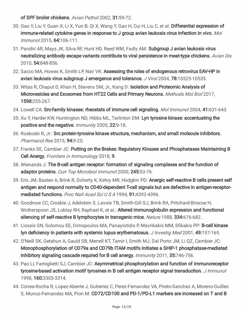
To screen differentially expressed proteins which plays a key role in cell signal transduction under ALV-Jinfection, the TMT-based proteomic analysis and hierarchical clustering method were used to visualizechanges in the abundance of differentially expressed proteins in the CEF(laboratory host cells of ALV-J)between ALV-J-infected and normal groups. GO functional analysis revealed that ALV-J infection resultedin signi�cant changes in protein expression associated with immune and developmental processes inCEF (Fig. 1a). We identi�ed the top 19 differentially proteins associated with immune and developmentalprocesses in CEF. Test results showed that the abundance of tyrosine protein kinase Lyn, which mediatesthe B-cell signal transduction, were signi�cantly down-regulated in the CEF (Fig. 1b and 1c).We furtherused the quantitative real-time PCR (qPCR) and the western blot (WB) to veri�ed the analysis results ofproteomics in CEF infected with ALV-J(Fig. 1d, 1e, and 1f).
Enhanced expression of Lyn in B-cells infected with ALV-J
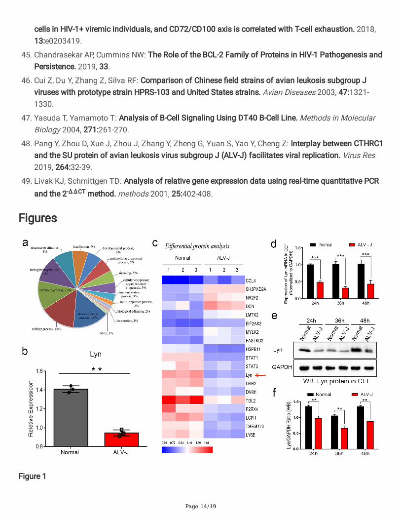
A series of research data showed that Lyn plays an important role in BCR signal transduction pathwaywhich associated with the development, differentiation, maturation, or tolerance (anergy) of B-cell [19–21]. Based on the proteomic results mentioned above, we investigated the Lyn expression in B-cellinfected by ALV-J. Confocal laser scanning microscopy (CLSM) analysis showed that ALV-J and Lyn wereboth located in the cytoplasm of chicken B-cell, indicating that ALV-J directly or indirectly affects Lyn inthe cytoplasm (Fig. 2a). The mRNA level and protein level of Lyn in chicken B-cells were up-regulated afterALV-J infection tested by qPCR and WB detection, different from the proteomic data of CEF (Fig. 2b, 2c,and 2d). As another molecular switch in BCR singling, tyrosine kinase Fyn also plays the active role inBCR singling regulation [22, 23]. However, the expression levels of Fyn showed no signi�cant difference inchicken B-cells before and after infection (Fig. 2b, 2c, and 2d). Furthermore, we also detected theexpression level of Lyn in bursal B-cells in vivo. The expression levels of Lyn were increased signi�cantly
Page 4/19
in B-cells of bursa of Fabricius of chicken infected with ALV-J in 14 and 28 days old tested byimmunohistochemistry (IHC) and WB (Fig. 2e, 2f, 2 g, and 2 h), which was consistent with the expressionlevel of Lyn in chicken B-cells infected with ALV-J in vitro.
Enhanced phosphorylation of Lyn but decreased phosphorylation of Syk in B-cells infected with ALV-J
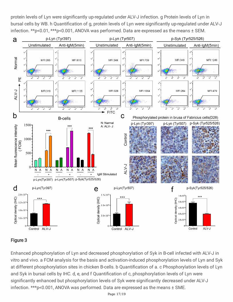
Studies on the molecular structure of Lyn have shown that there are many phosphorylation sites in Lyn,two of which have the opposite effect. The phosphorylation of Tyr397 site can activates the positiveregulation effect of Lyn, but the phosphorylation of Tyr507 can activates the negative regulation effect ofLyn[24]. Based on this, we further detected the expression levels of the two phosphorylation site thatdetermined whether Lyn was activated or not. As shown the detection results of �ow cytometry(FCM),although the two phosphorylated Lyn expressed signi�cantly higher than the housekeeping protein afterBCR signaling pathway was activated by anti IgM antibody, the Tyr525/526 phosphorylation level of Lyndownstream direct substrate Syk decreased signi�cantly compared with the same enhancedphosphorylation Lyn protein(Fig. 3a and 3b). These results indicate that Lyn actually plays an inhibitoryrole in BCR signal transduction of chicken B-cells infected with ALV-J.
Previous studies have shown that Lyn is mainly expressed in B-cells or other some blood cells except T-cells[25]. To further verify the actual effect of ALV-J on the expression levels of Lyn in vivo, thephosphorylation level of Lyn and Syk in bursal cells of chicken were measured by IHC and WB. Inaccordance with the results of in vitro assay, it was found that Tyr397 and Tyr507 phosphorylation levelsof Lyn increased signi�cantly in bursal cells of chicken infected with ALV-J, while the phosphorylationlevels of Syk decreased signi�cantly (Fig. 3c, 3d, 3e, and 3f). Above results suggest that ALV-J interferesthe expression and regulation of Lyn in the BCR signaling pathway.
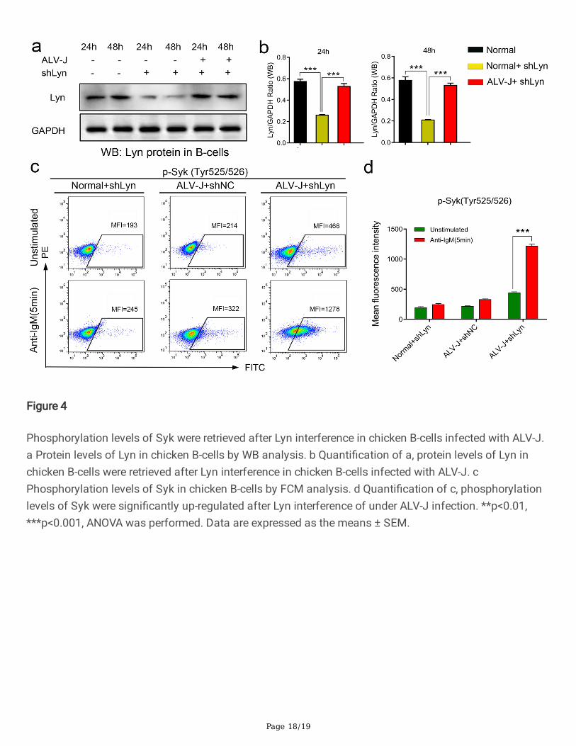
Phosphorylation levels of Syk were retrieved after Lyn interference in B-cells infected with ALV-J
To further verify the effect of ALV-J on the expression levels of Lyn in B-cells, the shRNA interference ofLyn was performed in chicken B-cells, and the expression levels of Lyn was detected. It was found thatthe protein levels of Lyn were signi�cantly inhibited 24 hours after Lyn interference on chicken B-cells ofnormal chickens, meanwhile, the protein level of Lyn in infection group chicken B-cells signi�cantlydecreased after Lyn interference, which was equivalent of the expression levels of Lyn in normal groupchicken B-cells (Fig. 4a and 4b). This �nding indicated that the expression levels of Lyn in chicken B-cellswere promoted by ALV-J. To verify whether Lyn inhibited the BCR signaling through the phosphorylationof Tyr507 under ALV-J infection, the phosphorylation levels of Syk in chicken B-cells were detected. TheFCM results showed that expression levels of phosphorylation Tyr525/525 site of Syk were signi�cantlyretrieved (enhanced) under activated by anti-IgM antibody after 48 hours of Lyn interference (Fig. 4c and4d). These �ndings further con�rmed that Lyn performed actually the negative regulatory effect in BCRsignal transduction through the phosphorylation of Tyr507 under ALV-J infection.
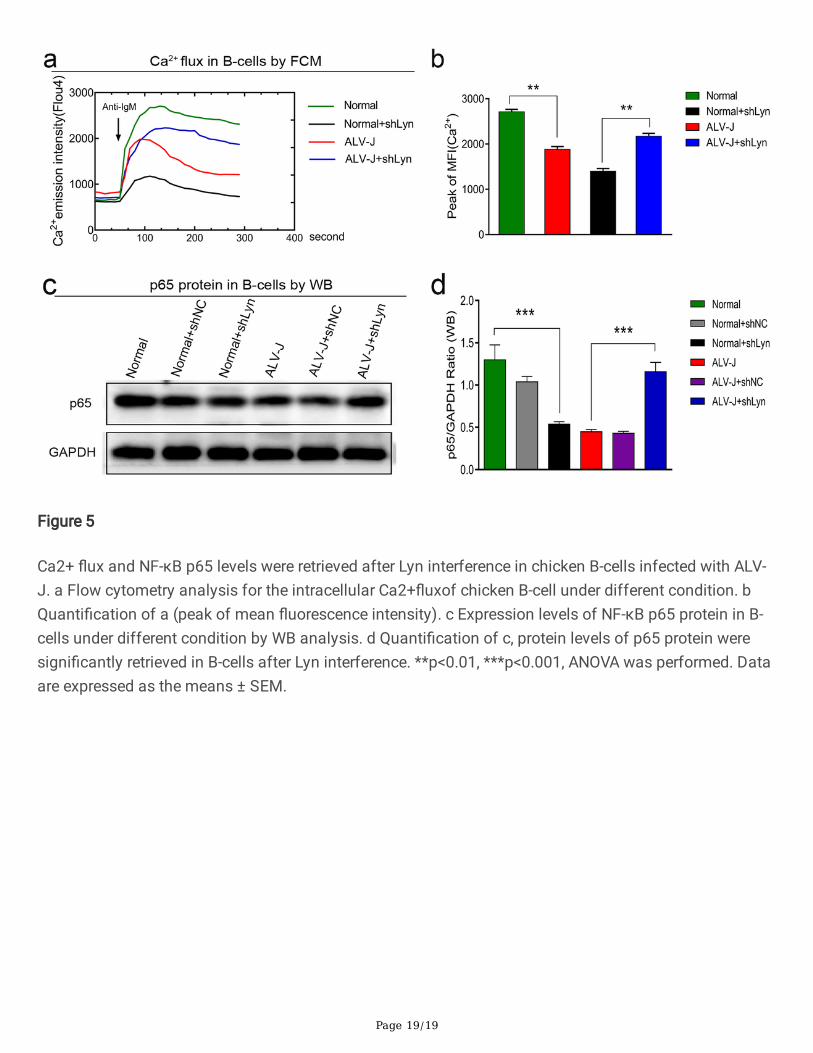
Ca 2+ �ux and NF-κB p65 levels were retrieved after Lyn interference in B-cells infected with ALV-J
Page 5/19
Data shows that many anergy B-cells are defective in intracellular calcium mobilization and transcriptionof nuclear transcription factors [26]. To further analyze, the effect of Lyn on BCR signaling cascadesunder ALV-J infection, the Ca2+ mobilization and protein expression of NF-κB p65 were detected inchicken B-cells under different conditions. Test results showed that the response time of intracellular Ca2+
mobilization was delayed, the intracellular Ca2+ �ux was decreased, and the protein level of NF-κBp65were decreased in chicken B-cells infected with ALV-J. However, the levels of the Ca2+ �ux and NF-κB p65were retrieved in chicken B-cells infected with ALV-J after Lyn interference (Fig. 5a, 5b, 5c, and 5d). Theseresults further indicated that Lyn played a negative regulatory role in BCR signal cascades in ALV-Jinfected B-cells.
DiscussionSince the phenomenon of chickens with congenital infection with ALV-J showing persistent viremia wasdiscovered and de�ned as immune tolerance, the pathogenesis of immune tolerance induced by ALV-Jhas been studied continuously. Because the immune tolerance caused by ALV-J, HBV, and HIV has manycommon features in the pathological process [27, 28], it has drawn a great attraction to study thepathogenesis of immune tolerance induced by these viruses. It has been found that the damage oflymphocytes [29], the abnormal secretion of cytokines [30], the variation of viral epitopes [31], and theexpression of EAV-HP gene in embryo [32] all play more or less roles in the induction of immune toleranceby ALV-J. In previous study, we demonstrated that B-cell anergy induce by ALV-J should take most of theresponsibility for immune tolerance[13]. After all, the antiviral neutralizing antibodies were produced bynormal B-cells. To screening the speci�c protein closely related to the immune tolerance induced by ALV-J, we selected primary cells (such as CEF) rather than cell lines (such as DF-1 cell lines or chicken B-cellslines) in process of proteomic analysis for investigating the practical regulation effect of ALV-J on proteinexpression [33]. As expected, we screened and con�rmed that Lyn, a key protein initiating BCR signaltransduction in B-cells[34], was signi�cantly affected by ALV-J in both CEF cells and chicken B-cells.
Tyrosine kinase Lyn is a member of SFKs expressed in various cells except for T lymphocytes, and exertsa unique dual role acting both as a positive and a negative regulatory molecule in BCR signaling [35, 36].The self-phosphorylation of Lyn protein triggers the assembly of the BCR signalosome and cascadereaction of BCR signal transduction[37]. In response B cells, BCR signaling is initiated by Lynphosphorylating the ITAM, which further recruit and activate Syk[38]. However, anergic B-cells arecharacterized by reduced ability to proliferate and secrete antibody after antigen stimulationaccompanied by reduced BCR-signaling responses which including delay of intracellular calciummobilization and activation of inhibitory tyrosine kinase phosphorylation[39, 40]. Antigen-inducedaggregation of the BCR on normal mature B-cells results in the initiation of BCR signal cascades thatculminate in proliferation, the increased of the Ca2+ in�ux and alterations in protein expression such asNF-κB p65. Our data showed that the response time of Ca2+ mobilization in chicken B-cells was delayed,and as one of BCR signal activation events, the intracellular Ca2+ in�ux level was also signi�cantlydecreased. Meanwhile, the protein level of NF-κB p65 was also decreased. In this context, these results
Page 6/19
indicated that Lyn appears to function as a driver of inhibitory signaling pathways of BCR under ALV-Jinfection.
The unique role of Lyn in down-modulating B-cell receptor (BCR) activation mainly is mainly realized bythe phosphorylation of inhibitory molecules and receptors [41]. Moreover, chronic antigen stimulationdrives biased monophosphorylation of CD79 immunoreceptor tyrosine-based activation motif (ITAM)leading to recruitment of Lyn instead of Syk, which ultimately leads to B-cell anergy [42, 43]. Presentstudy results showed that ALV-J signi�cantly up-regulated the protein expression level of Lyn in chickenB-cells and activated the phosphorylation of Tyr507 site in Lyn, which triggered the inhibitory effect of Lynin BCR signal transduction, as shown by the decrease of the phosphorylation level of Syk, a downstreamdirect substrate of Lyn. Therefore, we suggested that the inhibitory effect of Lyn on BCR signaltransduction is the crucial factor for the anergy of chicken B-cells induced by ALV-J.
In this study, the up-regulation of Lyn expression in chicken B-cells infected with ALV-J eventually led tothe suppression of BCR signaling and the down-regulation of the nuclear transcription factor NF-κBp65.We speculate that these anergic B-cells eventually undergo apoptosis and lead to the maintenance ofviremia. Interestingly, previous studies have shown that up-regulated of programmed cell death protein 1(PD-1) or down-regulated of anti apoptotic molecule Bcl-2 in immature B-cells in the peripheral blood ofHIV patients �nally made B-cells prone to apoptosis by endogenous apoptotic pathway[44, 45], whichwas the important pathological process of immune tolerance induced by HIV-1.Therefore, wehypothesized that ALV-J, as a chronic stimulant antigen, activated the negative regulation role of Lyn in B-cell signal transduction.
ConclusionsThese �ndings suggested that ALV-J activated the negative regulation role of Lyn in B-cell signaltransduction which induced B-cell anergy. Previous studies on the role of Lyn in B-cell signal transductionwere mainly carried out on transgenic animal models or gene knockout cells. Here, we investigated therole of Lyn in chicken B-cell in the context of virus infection, which will provide a new insight for studyingthe pathogenic mechanism of immune tolerance induced by retrovirology viruses. Admittedly, furtherstudies are necessary to investigate how ALV-J regulates the expression of Lyn.
Materials And MethodsVirus, cells and chickens
ALV-J (NX0101 strain) [46] were maintained in our laboratory and titered by TCID50 assays in CEF. TheCEF and the chicken B-cells line (DT40 cells) [47] were maintained in Dulbecco’s modi�ed Eagle’s medium(DMEM) supplemented with 10% fetal bovine serum (FBS), 1% penicillin/streptomycin, and 1% l-glutamine, in a 5% CO2 incubator at 37 °C. Cells were infected with ALV-J containing 10 − 4 TCID50, itwas needed to add 8 µg/mL polybrene when infected chicken B-cells.
Page 7/19
The 6-day-old embryos of Leghorn speci�c-pathogen-free (SPF) chickens (Jinan poultry technologycompany, China) were injected with ALV-J through the allantoic cavity to establish the infected group (n = 30).The control group (n = 30) was established by injecting DMEM instead of virus. The treatment of virusinoculation, chicken embryos incubation, and virus detection were all followed as our previousdescription[13].
Proteomics assay and bioinformatics analysis
In order to screen differentially expressed proteins that plays a key role in cell signal transduction underALV-J infection,protein lysates from ALV-J-infected CEF and normal CEF were collected for proteomicanalysis and bioinformatics analysis. The experimental process, iTRAQ labelled LC-MS/MS, andbioinformatics analysis were all followed as our previous description [48].
Confocal laser scanning microscopy
Chicken B-cells were cultured in �ask and infected with ALV-J. These cells were maintained for 48 h withDMEM containing 10% neonatal calf serum at 37 °C in a 5% CO2 incubator. The cells were �xed with theice-cold methanol for 10 min, and blocked with PBS containing 10% FBS for 10 min at room temperatureafter washed with PBS. The cells were then incubated with the rabbitanti-chicken ALV-J primaryantibodies (made in our laboratory, 1:200) and the mouse anti-chicken Lyn primary antibodies (NovusBiotech, Colorado, USA, 1:200) for 2 h at 37 °C, followed by incubation with �uorescein isothiocyanate(FITC)-labeled goat anti-rabbit secondary antibodies and the alexa�uor 647 (AF647)-labeled goat anti-mouse secondary antibodies (Bioss, Beijin, China, 1:1000) for 30 min at 37 °C. After staining cell nucleiwith 4′, 6-diamidino-2-phenylindole (DAPI), the cells were seeded on glass bottom dish and observed bySP8 CLSM (Leica,Germany).
Quantitative real-time PCR
Total RNA was extracted from CEF cells or chicken B-cells using TRIzol reagent according to themanufacturer’s instructions (Invitrogen, Carlsbad, USA). One microgram of total RNA was used as atemplate to synthesize cDNA using a reverse transcriptase kit (TaKaRa, Shiga, Japan) to generate �rst-strand cDNA. qPCR was performed by the LightCycler 96 system (Roche, Basel, Switzerland) with thediluted cDNA. The mRNA levels were analyzed using the 2−ΔΔCt method[49].
The primers were as follows: Lyn-F: 5’-TGCGTG CGTGGTTATTATTTC-3’,
Lyn-R: 5’-AATGGTGAGGTCGCTGACTGT-3’;
Fyn-F: 5’-TTGTTGAAGGCAAGCATCAG-3’,
Fyn-R: 5’-GAGGATAGCATCTGCCCTTT-3’;
Syk-F: 5’-G TCTCCATCCACCACACCTTCT-3’,
Page 8/19
Syk-R: 5’-ACAAGCAGTCCAAGGCAGTGA-3’.
Western blot
Tissues or cells were collected and then lysed in RIPA lysis buffer (BeyotimeBiotech, Shanghai, China).Proteins from the tissue lysates were separated by polyvinylidenedi�uoride (PVDF) membraneselectrophoresis. The PVDF membranes were incubated with primary antibodies after being blocked bydifco skim milk (Solarbio, Beijing, China). The primary antibodies included mouse anti-chicken Lynmonoclonal antibody (Novus Biotech, Colorado, USA,1:200), rabbit anti-Fyn polyclonal antibody (Jackson,Westgrove, USA,1:800),rabbit anti-NF-κB p65 polyclonal antibody (Bioss, Beijin, China,1:200), and mouseanti-GAPDH-loading control antibody(Bioss, Beijing, China,1:5000).Finally, the membranes were exposedto a chemiluminescence instrument.
Immunohistochemistry
At the age of 14 and 28 days, the bursa of Fabricius from infected chickens and control chickens weresampled, formalin-�xed, para�n embedded, and sectioned (5-µm thickness) sections for IHC. All chickenswere euthanized with sodium pentobarbital before the organs were removed. The perform of IHC testwere followed as our previous description.[13]Negative controls were also performed with the sametissues. Primary antibodies include mouse anti-chicken Lyn monoclonal antibodies (Novus Biotech,Colorado, USA,1:200), rabbit anti-phospho-Lyn(Try397)polyclonal antibody (Bioss, Beijing, China,1:200),rabbit anti-phospho-Lyn Try507 polyclonal antibody (Bioss, Beijing, China,1:200), rabbit anti-phospho-Syk Tyr525/526 polyclonal antibody (Bioss, Beijing, China,1:200). Secondary antibodies werehorseradish peroxidase-labelled goat anti-mouse/rabbit IgG polymer. Six randomly selected �elds ofpositive expression in each target tissue section were analysed in Image J software to accuratelycalculate the positive area and the mean optical density.
Flow cytometry for tyrosine phosphorylation
FCM analysis was performed to assess the levels of phosphorylated Lyn protein (Tyr397 or Tyr507) andphosphorylated Syk protein(Tyr525/526) in B-cells. 8 µg/mL polybrene were added into the DMEMmedium containing chicken B-cells to improve the infection e�ciency of ALV-J, and then the (SouthernBiotech, USA, 20 µg/ml), were added to activate chicken B-cells in logarithmic growth phase. Chicken B-cells were harvested in 2, 5, 10, 30 minutes after stimulation by anti-IgM antibody, and �xed in 100% ice-cold methanol solution. Then these chicken B-cells were divided into three groups and respectivelylabeled with rabbit anti-phospo-Lyn (Tyr397)-FITC polyclonal antibody(Bioss, Beijing, China,1:200), rabbitanti-phospo-Lyn (Tyr507)-FITC polyclonal antibody(Bioss, Beijing, China,1:200), rabbit anti-phospo-Syk(Tyr525/526) -FITC polyclonal antibody(Bioss, Beijing, China,1:200), incubated at 4 °C in dark for 30 min,washed with PBS buffer and analyzed with BD �ow cytometer (BD Biosciences,USA). The sameexperiment was repeated three times, and isotype control antibodies were also used. Data were analysedusing FlowJo (TreeStar) software.
Page 9/19
Short-hairpin RNA (shRNA) interference
The pgpu6 / GFP / Neo shRNA interference expression vector (three segments: Lyn-chicken-544:gcagttattctcttctgtcatcatcataa; Lyn-chicken-944: gcttcagcatgaagctagt; Lyn-chicken-1345:ggattctcctgtatatgaaaaatcg) targeted Lyn was constructed and then transfected into chicken B-cellsaccordance with the manufacturer’s instructions. The transfection e�ciency was observed by�uorescence microscope and cell growth after transfection with shLyn vector was determined by cellcounting kit-8 (CCK-8) test. The Lyn expression after transfection was detected by WB. Then the Syktyrosine phosphorylation was analysed by FCM as mentioned above.
Calcium mobilization measurement
For recording BCR-induced Ca2+ �ux, 106 chicken B-cells were loaded with 2.5 µL Fluo4-AM (MolecularProbes) in 200 µL hanks balanced salt solution containing 1% FBS and at 37 °C for 25 min. Thesechicken B-cellswere washed with PBS buffer saline for three times, and then kept it at 37 °C. Theintracellular calcium basal level was detected by FCM for one minute. Then, the anti-chickenIgM(Southern Biotech, USA, 2 µg/ml) was added to stimulate these chicken B-cells.The �uorescence ofFluo-4 at 516 nmwas continuously measured by the �ow cytometer to determine the change ofintracellular Ca2+ �ux.
Statistical analysis
Multiple sets of data comparisons were measured using one-way analysis of variance (ANOVA). Theunpaired t-test was used when two groups were compared. The results were accepted as signi�cantlydifferent when p ≤ 0.05, p ≤ 0.01, or p ≤ 0.001. Analysis and plotting of data were performed usingGraphPad Prism 6.0 and are expressed as the means ± SEM.
AbbreviationsALV-J: avian leukosis virus subgroup J; AF647: alexa�uor 647; BCR: B cell antigen receptor; CEF: chickenembryonic �broblasts; Co-IP: co-immunoprecipitation; CLSM: confocal laser scanning microscopy; FCM:�ow cytometry; FITC: �uoresceinisothiocyanate; HBV: hepatitis B virus; HIV: human immunode�ciencyvirus; iTRAQ: isobaric tags for relative and absolute quanti�cation; IHC: immunohistochemistry; ITAM:immunoreceptor tyrosine-based activation motif; LC-MS/MS: liquid chromatography-tandem massspectrometry; NF-κB: nuclear factor-κB; TCID50: 50%tissue culture infective dose; PVDF:polyvinylidenedi�uoride; qPCR: quantitative real-time polymoerase chain reaction; shRNA: short-hairpinRNA; SFK: Src family kinase; SPF: speci�c pathogen free; WB: western blot.
DeclarationsAcknowledgements
Page 10/19
We are grateful to Dr. Libo Huang and Ms. Li Zhang for their technical assistance. We thank Dr. GuihuaWang and Dr. Chengui Li for their helpful discussion and manuscript revision.
Authors’ contributions
ZC and VN conceived and designed the research. SH participated in the design of the study, performedthe research, and did the analysis and interpretation of data and the writing of the manuscript. GZ and DZperformed the research and interpretation of data, XY contributed reagents/materials/analysis tools. YYand JD participated in the interpretation of data and manuscript writing. All of the authors discussed theresults.
Funding
This study was supported by grants from the National Natural Science Foundation of China (31672521),the China-UK Partnership on Global Food Security: Combating Avian Leukosis Virus Subgroup J forSustainable Poultry Production (31761133002, BB/R012865/1), the Shandong Modern AgriculturalTechnology & Industry System (SDAIT-11-04), and the Xinyang Agriculture and Forestry UniversityScienti�c Research Funds for Young Teachers(2019LG012).
Availability of data and materials
The datasets used and/or analyzed during the current study are available from the corresponding authoron reasonable request.
Ethics approval and consent to participate
Approval for this study was obtained through the Committee on the Ethics of Animal Experiments ofShandong Province (Permit Number of Protocol: 20160124).
Consent for publication
All authors consented to the publication of this manuscript. The funding agencies had no role in thedecision to publish this manuscript.
Competing interests
The authors declare that they have no competing interests.
Author details
1 College of Veterinary Medicine, Shandong Agricultural University, No 61,Daizong Street, Tai’an City,Shandong Province, 271018, China.
2 College of Husbandry and Veterinary, Xinyang Agriculture and Forestry University, No 1,North Ring Road,Xinyang City, Henan Province, 464000, China.
Page 11/19
3 Hospital of Shandong Agricultural University, No 61,Daizong Street, Tai’an City, Shandong Province,271018, China.
4 The Pirbright Institute & UK-China Centre of Excellence on Avian Disease Research, Pirbright, Ash Road,Guildford, Surrey, GU24 0NF, UK.
References1. Rubin H, Fanshier L, Cornelius A, Hughes WF: Tolerance and immunity in chickens after congenital
and contact infection with an avian leukosis virus. Virology 1962, 17:143-156.
2. Payne LN, Gillespie AM, Howes K: Myeloid leukaemogenicity and transmission of the HPRS-103strain of avian leukosis virus. Leukemia 1992, 6:1167-1176.
3. Russell PH, Ahmad K, Howes K, Payne LN: Some chickens which are viraemic with subgroup J avianleukosis virus have antibody-forming cells but no circulating antibody. Research in VeterinaryScience 1997, 63:81.
4. Cui N, Wang Q, Shi W, Han L, Wang J, Ma X, Li H, Wang F, Su S, Zhao X: Synergy of subgroup J avianleukosis virus and Eimeria tenella to increase pathogenesis in speci�c-pathogen-free chickens. VetImmunol Immunopathol 2016, 177:42-47.
5. Wen Y, Huang Q, Yang C, Pan L, Wang G, Qi K, Liu H: Characterizing the histopathology of natural co-infection with Marek's disease virus and subgroup J avian leucosis virus in egg-laying hens. AvianPathol 2018, 47:83-89.
�. Russell PH, Ahmad K, Howes K, Payne LN: Some chickens which are viraemic with subgroup J avianleukosis virus have antibody-forming cells but no circulating antibody. Res Vet Sci 1997, 63:81-83.
7. Dai M, Feng M, Xie T, Li Y, Ruan Z: ALV-J infection induces chicken monocyte death accompaniedwith the production of IL-1β and IL-18. Oncotarget 2017, 8:99889-99900.
�. Hang B, Sang J, Qin A, Qian K, Shao H, Mei M, Ye J: Transcription analysis of the response of chickenbursa of Fabricius to avian leukosis virus subgroup J strain JS09GY3. Virus Res 2014, 188:8-14.
9. Yarkoni Y, Getahun A, Cambier JC: Molecular underpinning of B-cell anergy. Immunol Rev 2010,237:249-263.
10. Schroeder KM, Agazio A, Torres RM: Immunological tolerance as a barrier to protective HIV humoralimmunity. Current Opinion in Immunology 2017, 47:26-34.
11. Duty JA, Szodoray P, Zheng NY, Koelsch KA, Zhang Q, Swiatkowski M, Mathias M, Garman L, HelmsC, Nakken B, et al: Functional anergy in a subpopulation of naive B cells from healthy humans thatexpress autoreactive immunoglobulin receptors. J Exp Med 2009, 206:139-151.
12. Cyster JG, Hartley SB, Goodnow CC: Competition for follicular niches excludes self-reactive cellsfrom the recirculating B-cell repertoire. Nature 1994, 371:389-395.
13. He S, Zheng G, Zhou D, Li G, Cheng Z: Clonal anergy of CD117+chB6+ B cell progenitors induced byavian leukosis virus subgroup J is associated with immunological tolerance. Retrovirology 2019,
Page 12/19
16:1.
14. Cambier JC, Gauld SB, Merrell KT, Vilen BJ: B-cell anergy: from transgenic models to naturallyoccurring anergic B cells? Nat Rev Immunol 2007, 7:633-643.
15. Blake S, Hughes TP, Mayrhofer G, Lyons AB: The Src/ABL kinase inhibitor dasatinib (BMS-354825)inhibits function of normal human T-lymphocytes in vitro. Clin Immunol 2008, 127:330-339.
1�. Lamagna C, Hu Y, DeFranco AL, Lowell CA: B cell-speci�c loss of Lyn kinase leads to autoimmunity. JImmunol 2014, 192:919-928.
17. Hsueh RC, Scheuermann RH: Tyrosine kinase activation in the decision between growth,differentiation, and death responses initiated from the B cell antigen receptor. Adv Immunol 2000,75:283-316.
1�. Shahaf G, Gross AJ, Sternberg-Simon M, Kaplan D, DeFranco AL, Mehr R: Lyn de�ciency affects B-cell maturation as well as survival. Eur J Immunol 2012, 42:511-521.
19. Chu CL, Lowell CA: The Lyn tyrosine kinase differentially regulates dendritic cell generation andmaturation. J Immunol 2005, 175:2880-2889.
20. Hu Y, Liu Y, Pelletier S, Buchdunger E, Warmuth M, Fabbro D, Hallek M, Van Etten RA, Li S:Requirement of Src kinases Lyn, Hck and Fgr for BCR-ABL1-induced B-lymphoblastic leukemia butnot chronic myeloid leukemia. Nat Genet 2004, 36:453-461.
21. Gross AJ, Proekt I, DeFranco AL: Elevated BCR signaling and decreased survival of Lyn-de�cienttransitional and follicular B cells. Eur J Immunol 2011, 41:3645-3655.
22. Mkaddem SB, Murua A, Flament H, Titeca-Beauport D, Bounaix C, Danelli L, Launay P, Benhamou M,Blank U, Daugas E, Charles N: Lyn and Fyn function as molecular switches that controlimmunoreceptors to direct homeostasis or in�ammation. 2017, 8:246.
23. Gauld SB, Cambier JC: Src-family kinases in B-cell development and signaling. Oncogene 2004,23:8001-8006.
24. Donella-Deana A, Cesaro L, Ruzzene M, Brunati AM, Marin O, Pinna LA: Spontaneousautophosphorylation of Lyn tyrosine kinase at both its activation segment and C-terminal tail confersaltered substrate speci�city. Biochemistry 1998, 37:1438-1446.
25. Scapini P, Pereira S, Zhang H, Lowell CA: Multiple roles of Lyn kinase in myeloid cell signaling andfunction . Immunological Reviews 2009, 228:23-40.
2�. Szodoray P, Stanford SM, Molberg O, Munthe LA, Bottini N, Nakken B: T-helper signals restore B-cellreceptor signaling in autoreactive anergic B cells by upregulating CD45 phosphatase activity. JAllergy Clin Immunol 2016, 138:839-851.e838.
27. Payne LN: Retrovirus-induced disease in poultry. Poult Sci 1998, 77:1204-1212.
2�. Hong M, Bertoletti A: Tolerance and immunity to pathogens in early life: insights from HBV infection.Semin Immunopathol 2017, 39:643-652.
29. Landman WJ, Post J, Boonstra-Blom AG, Buyse J, Elbers AR, Koch G: Effect of an in ovo infectionwith a Dutch avian leukosis virus subgroup J isolate on the growth and immunological performance
Page 13/19
of SPF broiler chickens. Avian Pathol 2002, 31:59-72.
30. Gao Y, Liu Y, Guan X, Li X, Yun B, Qi X, Wang Y, Gao H, Cui H, Liu C, et al: Differential expression ofimmune-related cytokine genes in response to J group avian leukosis virus infection in vivo. MolImmunol 2015, 64:106-111.
31. Pandiri AR, Mays JK, Silva RF, Hunt HD, Reed WM, Fadly AM: Subgroup J avian leukosis virusneutralizing antibody escape variants contribute to viral persistence in meat-type chickens. Avian Dis2010, 54:848-856.
32. Sacco MA, Howes K, Smith LP, Nair VK: Assessing the roles of endogenous retrovirus EAV-HP inavian leukosis virus subgroup J emergence and tolerance. J Virol 2004, 78:10525-10535.
33. Witas R, Chaput D, Khan H, Stevens SM, Jr., Kang D: Isolation and Proteomic Analysis ofMicrovesicles and Exosomes from HT22 Cells and Primary Neurons. Methods Mol Biol 2017,1598:255-267.
34. Lowell CA: Src-family kinases: rheostats of immune cell signaling. Mol Immunol 2004, 41:631-643.
35. Xu Y, Harder KW, Huntington ND, Hibbs ML, Tarlinton DM: Lyn tyrosine kinase: accentuating thepositive and the negative. Immunity 2005, 22:9-18.
3�. Roskoski R, Jr.: Src protein-tyrosine kinase structure, mechanism, and small molecule inhibitors.Pharmacol Res 2015, 94:9-25.
37. Franks SE, Cambier JC: Putting on the Brakes: Regulatory Kinases and Phosphatases Maintaining BCell Anergy. Frontiers in Immunology 2018, 9.
3�. Wienands J: The B-cell antigen receptor: formation of signaling complexes and the function ofadaptor proteins. Curr Top Microbiol Immunol 2000, 245:53-76.
39. Eris JM, Basten A, Brink R, Doherty K, Kehry MR, Hodgkin PD: Anergic self-reactive B cells present selfantigen and respond normally to CD40-dependent T-cell signals but are defective in antigen-receptor-mediated functions. Proc Natl Acad Sci U S A 1994, 91:4392-4396.
40. Goodnow CC, Crosbie J, Adelstein S, Lavoie TB, Smith-Gill SJ, Brink RA, Pritchard-Briscoe H,Wotherspoon JS, Loblay RH, Raphael K, et al.: Altered immunoglobulin expression and functionalsilencing of self-reactive B lymphocytes in transgenic mice. Nature 1988, 334:676-682.
41. Liossis SN, Solomou EE, Dimopoulos MA, Panayiotidis P, Mavrikakis MM, S�kakis PP: B-cell kinaselyn de�ciency in patients with systemic lupus erythematosus. J Investig Med 2001, 49:157-165.
42. O'Neill SK, Getahun A, Gauld SB, Merrell KT, Tamir I, Smith MJ, Dal Porto JM, Li QZ, Cambier JC:Monophosphorylation of CD79a and CD79b ITAM motifs initiates a SHIP-1 phosphatase-mediatedinhibitory signaling cascade required for B cell anergy. Immunity 2011, 35:746-756.
43. Pao LI, Famiglietti SJ, Cambier JC: Asymmetrical phosphorylation and function of immunoreceptortyrosine-based activation motif tyrosines in B cell antigen receptor signal transduction. J Immunol1998, 160:3305-3314.
44. Correa-Rocha R, Lopez-Abente J, Gutierrez C, Perez-Fernandez VA, Prieto-Sanchez A, Moreno-GuillenS, Munoz-Fernandez MA, Pion M: CD72/CD100 and PD-1/PD-L1 markers are increased on T and B
Page 14/19
cells in HIV-1+ viremic individuals, and CD72/CD100 axis is correlated with T-cell exhaustion. 2018,13:e0203419.
45. Chandrasekar AP, Cummins NW: The Role of the BCL-2 Family of Proteins in HIV-1 Pathogenesis andPersistence. 2019, 33.
4�. Cui Z, Du Y, Zhang Z, Silva RF: Comparison of Chinese �eld strains of avian leukosis subgroup Jviruses with prototype strain HPRS-103 and United States strains. Avian Diseases 2003, 47:1321-1330.
47. Yasuda T, Yamamoto T: Analysis of B-Cell Signaling Using DT40 B-Cell Line. Methods in MolecularBiology 2004, 271:261-270.
4�. Pang Y, Zhou D, Xue J, Zhou J, Zhang Y, Zheng G, Yuan S, Yao Y, Cheng Z: Interplay between CTHRC1and the SU protein of avian leukosis virus subgroup J (ALV-J) facilitates viral replication. Virus Res2019, 264:32-39.
49. Livak KJ, Schmittgen TD: Analysis of relative gene expression data using real-time quantitative PCRand the 2-∆∆CT method. methods 2001, 25:402-408.
Figures
Figure 1
Page 15/19
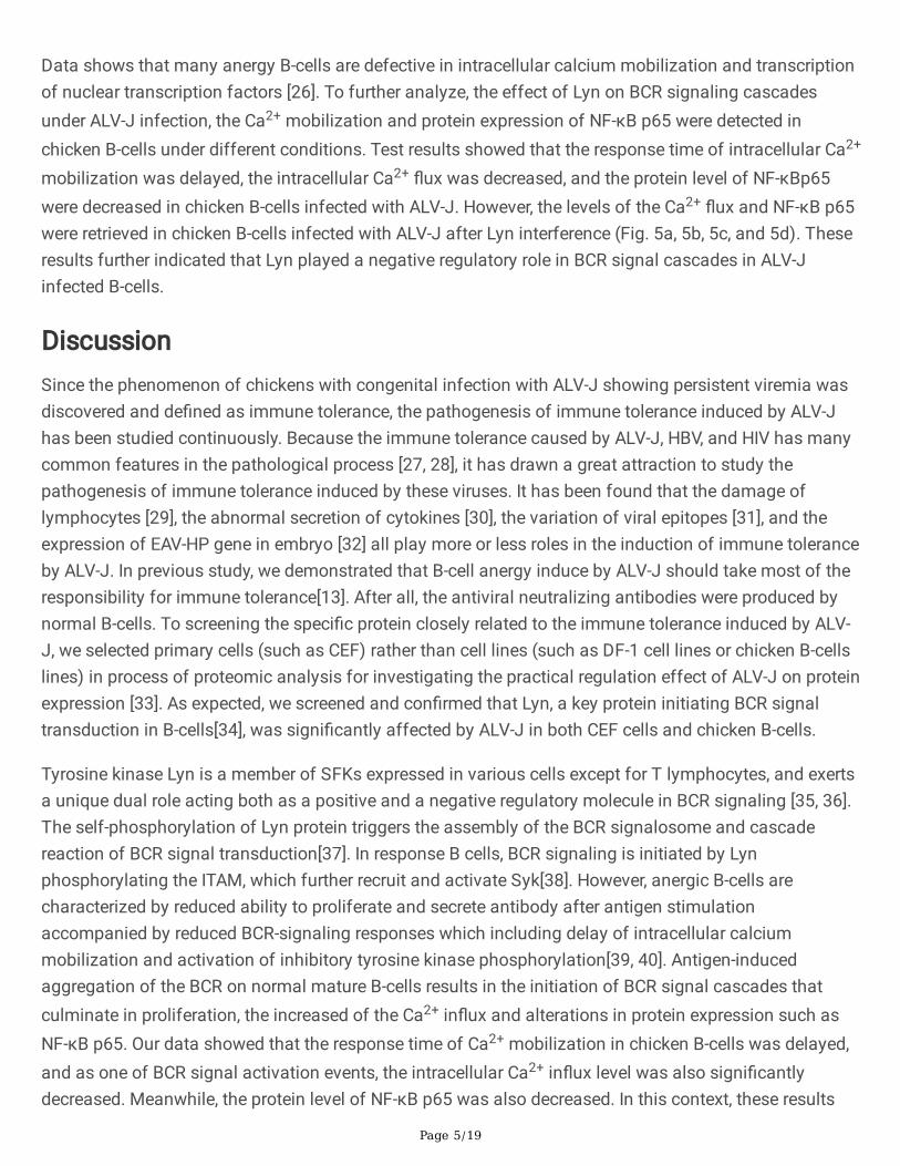
Expression levels of Lyn were signi�cantly down-regulated in CEF infected with ALV-J. a GO functionalanalysis revealed that ALV-J infection resulted in signi�cant changes in protein expression associatedwith immune and developmental processes in CEF. b Heat map of the top 19 differentially proteinsassociated with immune and developmental processes in CEF. Red color indicates up-regulated proteins,and blue color indicates down-regulated proteins. Arrow showed that the Lyn were signi�cantly down-regulated. c Protein levels of Lyn were signi�cantly down-regulated in ALV-J-infected CEF by LC-MS/MSanalysis. d mRNA levels of Lyn were signi�cantly down-regulated in CEF infected with ALV-J by qPCRanalysis. e Protein levels of Lyn in CEF infected with ALV-J by WB analysis. f Quanti�cation of e, proteinlevels of Lyn were signi�cantly down-regulated. ** p< 0.01, *** p < 0.001, ANOVA was performed. Data areexpressed as the means ± SEM.
Page 16/19
Figure 2
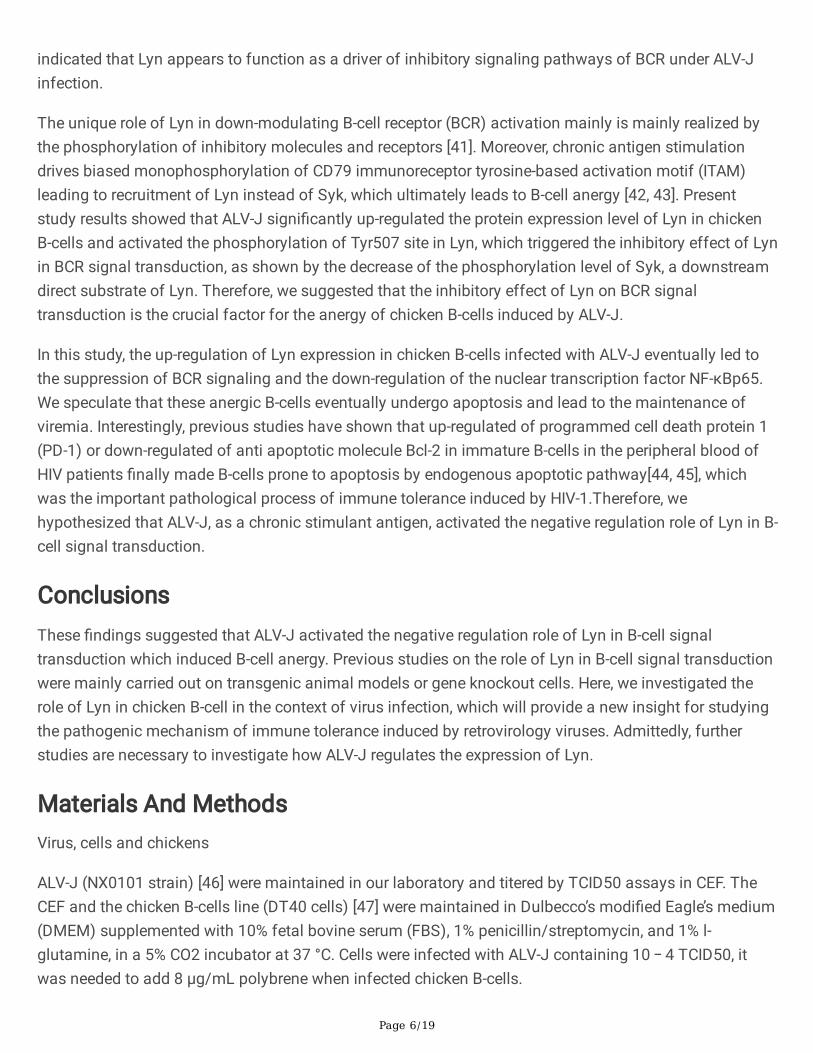
Expression of Lyn was activated in B-cell after ALV-J infection in vivo and vitro. a Immuno�uorescenceconfocal laser microscopy showed that ALV-J (gp85) and Lyn coexist in the cytoplasm of chicken B-cells.b Relative expression levels of Lyn and Fyn in chicken B-cell by qPCR. c Protein levels of Lyn in chicken B-cells by WB analysis. d Quanti�cation of c, protein levels of Lyn were signi�cantly up-regulated but Fynwas indifferent under ALV-J infection. e Protein levels of Lyn in bursal cells by IHC. f Quanti�cation of e,
Page 17/19
protein levels of Lyn were signi�cantly up-regulated under ALV-J infection. g Protein levels of Lyn inbursal cells by WB. h Quanti�cation of g, protein levels of Lyn were signi�cantly up-regulated under ALV-Jinfection. **p<0.01, ***p<0.001, ANOVA was performed. Data are expressed as the means ± SEM.
Figure 3
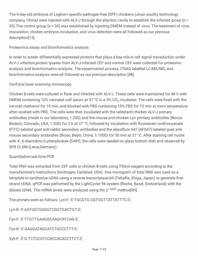
Enhanced phosphorylation of Lyn and decreased phosphorylation of Syk in B-cell infected with ALV-J invitro and vivo. a FCM analysis for the basis and activation-induced phosphorylation levels of Lyn and Sykat different phosphorylation sites in chicken B-cells. b Quanti�cation of a. c Phosphorylation levels of Lynand Syk in bursal cells by IHC. d, e, and f Quanti�cation of c, phosphorylation levels of Lyn weresigni�cantly enhanced but phosphorylation levels of Syk were signi�cantly decreased under ALV-Jinfection. ***p<0.001, ANOVA was performed. Data are expressed as the means ± SME.
Page 18/19
Figure 4
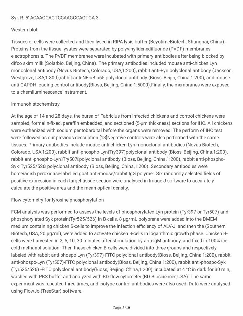
Phosphorylation levels of Syk were retrieved after Lyn interference in chicken B-cells infected with ALV-J.a Protein levels of Lyn in chicken B-cells by WB analysis. b Quanti�cation of a, protein levels of Lyn inchicken B-cells were retrieved after Lyn interference in chicken B-cells infected with ALV-J. cPhosphorylation levels of Syk in chicken B-cells by FCM analysis. d Quanti�cation of c, phosphorylationlevels of Syk were signi�cantly up-regulated after Lyn interference of under ALV-J infection. **p<0.01,***p<0.001, ANOVA was performed. Data are expressed as the means ± SEM.
Page 19/19
Figure 5
Ca2+ �ux and NF-κB p65 levels were retrieved after Lyn interference in chicken B-cells infected with ALV-J. a Flow cytometry analysis for the intracellular Ca2+�uxof chicken B-cell under different condition. bQuanti�cation of a (peak of mean �uorescence intensity). c Expression levels of NF-κB p65 protein in B-cells under different condition by WB analysis. d Quanti�cation of c, protein levels of p65 protein weresigni�cantly retrieved in B-cells after Lyn interference. **p<0.01, ***p<0.001, ANOVA was performed. Dataare expressed as the means ± SEM.