Embed Size (px)
Citation preview

HEMATOLOGY for Board Review
Joe Schwenkler, MD
Medical Director
UMDNJ‐ PA Program
June 7, 2013
This image is a work of the National Institutes of Health
AnemiasVitamin B12 deficiencyFolate deficiencyIron deficiencyG6PD deficiencyHemolytic and aplastic anemiaSickle cell anemiaThalassemia
Coagulation DisordersFactor VIII disordersFactor IX disordersFactor XI disorders
ThrombocytopeniaIdiopathic thrombocytopenic purpuraThrombotic thrombocytopenic purpuraVon Willebrand's disease
MalignanciesAcute/chronic lymphocytic leukemiaAcute/chronic myelogenous leukemaLymphomaMultiple myeloma
Anemia General Principles
• Anemia is a sign, not a disease. • Anemia's are a dynamic process.
• It is never normal to be anemic.• Correct use of lab tests is paramount.• Concomitant causes of anemia are common.• The diagnosis of iron deficiency anemia mandates further work‐up.

MORPHOLOGIC APPROACH TO ANEMIA
• Microcytic Anemia‐> MCV<80– Reduced iron availability — severe iron deficiency, the anemia of chronic disease, copper deficiency
– Reduced heme synthesis — lead poisoning, congenital or acquired sideroblastic anemia
– Reduced globin production — thalassemic states, other hemoglobinopathies
• Macrocytic Anemia‐> MCV>100– Megaloblastic anemias‐ Folic acid and Vitamin B12 deficiency
– alcohol abuse, liver disease, and hypothyroidism • Normocytic Anemia
– Anemia of chronic disease– Anemia of chronic renal failure
RBC Destruction/ Life Cycle
• Normal life span about 120 days• Destroyed by phagocytes
– spleen, liver, bone marrow, lymph nodes• heme biliverdin unconjugated (indirect) bilirubin
• liver converts to conjugated (direct) bilirubin which enhances elimination from the body
• globin and iron recycled• RBC destruction in blood vessels free Hb in urine (Hemoglobinuria vs. Hematuria which is whole red blood cells in urine due to kidney or tissue damage)
Reticulocyte Count
• Erythrocytes newly released from Bone Marrow
• Contain small amount of RNA• Stain with methylene blue• Increase in response to erythropoietin (EPO)
http://en.wikipedia.org/wiki/Image:Hematopoiesis_%28human%29_diagram.png

KINETIC APPROACH TO ANEMIA
Decreased Production (Low Retic count)Lack of nutrients…iron, Vitamin B12, FolateBone Marrow Suppression… Aplastic anemiaLow levels of trophic factors…chronic renal disease (low EPO), low thyroid, testosteroneAnemia of chronic disease
Increased destruction (High Retic count)Hemolytic Anemias
Inherited…sickle cell, thalassemiasAcquired…idiopathic, drug‐induced, and myelodysplasticsyndrome.
Algorithm using Retic. count, WBC, Platelet
Low Retic count suggests poorly functioning bone marrowNormal Platelets and WBC
Acute blood lossRenal diseaseInfectionsDrugs
Low platelets and WBCLeukemiaAplastic anemiaInfection
IRON METABOLISM
• Serum iron is free
• Transferrin binds iron in circulation– TIBC is identical–% Saturation is serum iron/TIBC
• Ferritin stores iron in liver and ReticuloEndothelial System (RES)

Case One76 yo female comes in c/o being “run down”for over a month
Only med is daily ibuprofen for chronic LBPPMH unremarkable, no previous hosp.Denies extra stress, problems sleeping except for restless legsRecently has been craving ice to chew (Pagophagia)
Physical exam unremarkable except angular stomatitis, glossitis, pale conjunctiva, 2/6 SEM at LUSB, and spoon nails as below
(From wikipedia commons)
Angular Stomatitis (Cheilosis) (from wikipedia commons)
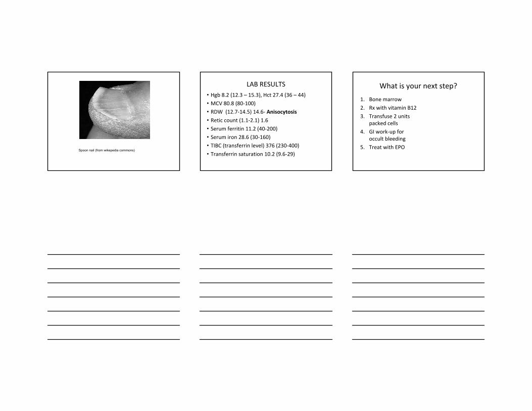
Spoon nail (from wikepedia commons)
LAB RESULTS• Hgb 8.2 (12.3 – 15.3), Hct 27.4 (36 – 44)• MCV 80.8 (80‐100)• RDW (12.7‐14.5) 14.6‐ Anisocytosis
• Retic count (1.1‐2.1) 1.6• Serum ferritin 11.2 (40‐200)• Serum iron 28.6 (30‐160)• TIBC (transferrin level) 376 (230‐400)• Transferrin saturation 10.2 (9.6‐29)
What is your next step?
1. Bone marrow2. Rx with vitamin B12
3. Transfuse 2 units packed cells
4. GI work‐up for occult bleeding
5. Treat with EPO
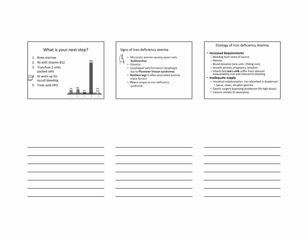
What is your next step?
1. Bone marrow2. Rx with vitamin B12
3. Transfuse 2 units packed cells
4. GI work‐up for occult bleeding
5. Treat with EPO
1 2 3 4 5
7% 9% 7%
74%
2%
Signs of iron deficiency anemia
• Microcytic anemia causing spoon nails (koilonychia).
• Glossitis• esophageal web formation (dysphagia
due to Plummer‐Vinson syndrome).• Restless legs is often associated anemia,
check ferritin!• Pica is unique to iron‐deficiency
syndrome.
Etiology of Iron deficiency Anemia
• Increased Requirements– Bleeding from some GI source– Menses– Blood donation (one unit= 250mg iron)– Growth periods, pregnancy, lactation– Infants fed cow’s milk suffer from reduced bioavailability iron and induced GI bleeding
• Inadequate supply• Intestinal malabsorption‐ iron absorbed in duodenum
• Sprue, celiac, atrophic gastritis• Gastric surgery bypassing duodenum (Rx high doses)• Calcium inhibits GI absorption

Treatment
• Ferrous sulfate 325mg b.i.d.– Beware constipation
• Recheck blood tests 6 weeks later– Continue oral iron until serum ferritin normalizes (up to 6 months)
• Iron salts not absorbed well if taken with food• Iron pills need to be given 2 hours before, or four hours after antacids
• Vitamin C helps absorption
Normal Fe deficiency without anemia
Fe deficiency with mild anemia
Severe Fe deficiency with severe anemia
Marrow iron 2+ to 3+ None None None
Serum iron 60 to 150 60 to 150 <60 <40
Iron binding capacity (transferrin)
300 to 360 300 to 390 350 to 400 >410
Saturation (SI/TIBC), percent
20 to 50 30 <15 <10
Hemoglobin Normal Normal 9 to 12 6 to 7
Red cell morphology
Normal NormalNormal or slight hypochromia
Hypochromiaand microcytosis
Plasma or serum ferritin
40 to 200 <40 <20 <10
Other tissue changes
None None NoneNail and epithelial changes
Case Two
47 yo male with 10 year h/o type 2 comes for PECurrently taking max doses metformin & glyburideBP 148/92, retinal exam shows cotton wool exudatesDiminished monofilament sensation on feetDiabetes poorly controlled with A1c=10.6%Microalbumin 300 (<20), creatinine 1.4CBC shows Hgb 9.1, MCV 85, normal plateletsStool guiac negative x 3Serum ferritin 170 (40‐200), Retic count .5%Serum iron 65 (60‐150), TIBC 320 (300‐360)

What can cause elevated ferritin AND low serum iron
1. Chronic inflammation2. Aplastic anemia3. Hemolysis
4. Hemoglobinopathies5. Acute leukemia
What can cause elevated ferritin AND low serum iron
1. Chronic inflammation2. Aplastic anemia3. Hemolysis
4. Hemoglobinopathies5. Acute leukemia
1 2 3 4 5
78%
7%0%
7%9%
What is the best treatment for this patient?
1. EPO
2. Transfuse 2 units pc3. Oral iron4. Parenteral iron

What is the best treatment for this patient?
1 2 3 4
64%
9%
23%
5%
1. EPO
2. Transfuse 2 units pc3. Oral iron4. Parenteral iron
ANEMIA OF CHRONIC DISEASE (ACD)(ANEMIA OF INFLAMMATION)
Second most common anemia after Iron DeficiencyInduced by inflammatory cytokines (IL‐6)Reduction in red blood cell (RBC) production by BMTrapping of iron in macrophagesreduced plasma iron levels making iron relatively unavailable for new hemoglobin synthesis
Erythroid precursors are impairedInterferons are potent inhibitors
Blunted erythropoietin response
chronic disease
iron deficiency
serum iron
TIBC (transferrin)
iron saturation
serum ferritin
nL or
Diagnosis of Anemia of Chronic Disease is often complicated…

ACUTE VARIANT (ANEMIA OF CRITICAL ILLNESS)
• Acute event‐related anemia– after surgery, major trauma, myocardial infarction, or sepsis
• Secondary to tissue damage and acute inflammatory changes
• Shares many of the features of ACD – low serum iron
– high ferritin
– blunted response to EPO
Underlying causes of ACD
Acute and chronic infectionsTBEndocarditisChronic UTI
MalignanciesMetastatic cancerLeukemiaLymphoma
Chronic arthritic conditionsChronic renal insufficiencyHypothyroidismANY CHRONIC INFLAMMATORY CONDITION!
DIAGNOSIS OF ANEMIA CHRONIC DISEASE
Generally mild/moderate anemia (Hb 8‐10)
Normochromic, normocytic (may be slightly low)
Low to normal reticulocyte count
Reduced serum iron and transferrin saturation
Reduced or normal TIBC/transferrin levels
Normal ferritin levels (acute phase reactant)
Need to exclude chronic renal failure, hyperthyroidism, hypothyroidism
May have concomitant iron deficiency anemia

TREATMENT OF ACD
Erythropoietin (EPO) is most effective therapy
Oral iron of little benefit unless also iron deficient
Transfusions only for short‐term if Hb<8
Who to treat with EPO?Hemoglobin <10
Additional risk factors (pulmonary, CV, renal)
What is goal of therapy?Hb 11 to 12 generally accepted
HEMOGLOBINOPATHIESSickle cell disease‐ homozygous
Autosomal recessive disease Substitution of the amino acid valine for glutamine8% to 10% of African Americans carry gene
Sickle cell trait‐ heterozygotesSplenic infarction can occur with hypoxia (altitude)Renal hematuria commonBeware bacteruria during pregnancy (pyelonephritis)
Thalassemias‐ imbalanced synthesis of normal globin chains
BetaAlpha
Normal Adult Hemoglobins
Name of Hemoglobin Distribution Structure
A 95%‐98% of adult Hb α2β2A2 1.5%‐3.5% of Adult Hb α2δ2F Fetal, 0.5%‐1.0% of
adult Hbα2γ2

Pathophysiology of SCD
• On deoxygenation, hemoglobinS polymers form, causing cell sickling and damage to the membrane
• Vasocclusive episodes result from a combination of vascular adhesion of young sickle cells and consequent trapping of dense sickle cells
• Functional asplenism
(From wikepedia commons)
SICKLE CELL ANEMIA
• Chronic hemolysis of sickle cell disease is usually associated with:– a mild to moderate anemia (hematocrit 20 to 30 percent)
– reticulocytosis of 3 to 15 percent (accounting for the high or high‐normal mean corpuscular volume [MCV])
– unconjugated hyperbilirubinemia– elevated serum lactate dehydrogenase
• Red cells are normochromic unless there is coexistent thalassemia or iron deficiency
• Hb electrophoresis high levels Hb F
The elongated and crescent-shaped red blood cells seen on this smear represent circulating irreversibly sickled cells. Target cells and a nucleated red blood cell are also seen. (Licensed under the Creative Commons Attribution-Share Alike 3.0 Unported license.)

Acute Pain EpisodesSickle cell “crisis”
– Precipitated by weather, infection, stress– Lasts 2 to 7 days– Often undertreated!– Low risk of narcotic addiction– Generate feelings of despair, depression
• ManagementHydrationPain managementSeek source of infection‐> Antibiotics?Hydroxyurea has promise‐> raises HbF levels
Clinical manifestations of SCD
• Hand & foot syndrome (dactylitis)‐ painful crisis in hands/feet‐ common children under four
• *Aplastic crisis can result from Parvovirus 19 infect.
• Splenic sequestration with enlarging spleen
• *Acute chest syndrome‐major cause of death‐– Fever, wheezing, chest pain, new pulmonary infiltrate
• *CVA risk increased if transcranial doppler slow(*Exchange transfusion indicated)
Clinical manifestations (cont.)
• Infections: Strep pneumonia and H. Influenza
• Gallstones
• Renal failure due to papillary infarcts– painless hematuria is common
• Chronic leg ulcers
• Priapism needs to be treated within 4 to 6 hours
• Aseptic necrosis in femoral and humeral heads
• Chronic osteomyelitis (salmonelli typhi)

Health Care MaintenanceRoutine visits with primary provider
Folic acid 1 mg daily
Transcranial doppler examDetect patients that would benefit from regular transfusions to prevent CVA
Retina exam to look for proliferative changes
Strep pneumonia vaccine below age 5 both 7 and 23‐valent, then 23‐valent every 7 years
H. flu, meningococcal, influenza starting age 6 months
Daily prophylactic oral penicillin until age 5
ß‐THALASSEMIAS
• Diminished production of ß‐globin chains– causing unmatched α‐globin chains to accumulate and aggregate
• ß‐Thalassemia minor (ß‐thalassemia trait)– Heterozygous condition
• ß‐Thalassemia major (Cooley anemia) – no ß chains are synthesized; only HbF and HbA2
– severe anemia that appears in the first year
ß‐THALASSEMIA MAJOR (HOMOZYGOUS BETA)
severe anemiablood film
pronounced variation in red cell size and shape (High RDW)pale red cells, target cells, basophilic stippling (ribosomal precipitates), nucleated red cells, moderately raised reticcount
infants well at birth but develop anemia in first few months when switch occurs from gamma (HbF) to beta globin chainsprogressive splenomegaly; iron loading; prone to infectionAllogenic Bone Marrow transplantation Rx of choice

ß‐THALASSEMIA TRAIT (HETEROZYGOUS CARRIER)
• mild hypochromic microcytic anemia– HGB 9‐11 g/dL
– MCV 50‐70 fL
– MCH 20‐22 pg
• no clinical features, patients asymptomatic
• often diagnosed on routine blood count• raised HbA2 level
ΑLPHA THALASSEMIA SYNDROMES
• α‐thalassemia‐2 trait (minima)– Loss of one of the four alpha globin genes– No abnormalities of blood testing
• α‐thalassemia‐1 trait (minor) – loss of two of the four alpha globin genes– MCV is often less than 80, but Hb electrophoresis is normal
• Hemoglobin H disease– Hemoglobin H, composed of four beta chains (beta4)– three of the four alpha globin loci are nonfunctional– chronic hemolytic anemia, due to the formation of inclusion bodies in circulating red cells as Hb H precipitates
• Hydrops fetalis with Hb Barts– none of the four alpha globin loci is functional
Disorder Genotypic Abnormality Clinical Phenotype
β‐Thalassemia
Thalassemia major (Cooley's anemia)
Homozygous β0‐thalassemia
Severe hemolysis, ineffective erythropoiesis, transfusion dependency, iron overload
Thalassemia intermedia Compound heterozygous β0‐ and β+‐thalassemia
Moderate hemolysis, severe anemia, but not transfusion dependent; main life‐threatening complication is iron overload
Thalassemia minor Heterozygous β0‐ or β+‐thalassemia
Microcytosis, mild anemia
α‐Thalassemia
Silent carrier α‐/αα Normal complete blood count
α‐Thalassemia trait αα/‐ ‐ (α‐thalassemia 1) OR Mild microcytic anemia
α‐/α‐ (α‐thalassemia 2)
Hemoglobin H α‐/‐ ‐ Microcytic anemia and mild hemolysis; not transfusion dependent
Hydrops fetalis ‐ ‐/‐ ‐ Severe anemia, intrauterine anasarcafrom congestive heart failure; death in utero or at birth

NORMOCYTIC ANEMIAS• Anemia of Chronic renal Insufficiency
• EPO is effective treatment • Acute blood loss
• Orthostatic Symptoms predominate• Resting tachycardia and hypotension• Can take 24 hr. for Hct to fall• 3‐5 days reticulocytosis elevates MCV
• Anemia of liver disease multifactorial:• Remodeling of RBC membranes• Hypersplenism• Folate deficiency• Co‐existing iron deficiency
HEMOLYTIC ANEMIACaused by premature breakdown of RBCs
Intracorpuscular Defects‐ RBC membrane defectsHeriditary Spherocystosis & Elliptocytosis
Extracorpuscular Defects‐Autoimmune Hemolytic Anemia
Positive coombs testRx prednisone high dose and taper slowly
G6PD Deficiency
Severity of anemia related to rate RBC destruction and ability of bone marrow to produce reticulocytesFree hemoglobin binds to haptoglobin
Removed by RES unless exceeds capacity (low haptoglobin)Excess filtered through kidney‐> dark urine
Typical case of Hemolytic anemia
• Acute onset pallor from anemia• Jaundice with high indirect bilirubin
• Increased serum LDH• Reduced (or absent) serum haptoglobin• Increased reticulocytes• Positive coombs test if autoimmune etiology

HEREDITARY SPHEROCYTOSISForms spherocytic cells that are destroyed in spleenPresent with jaundice and splenomegalyElevated retic countSpherocytes on smearSplenectomy often requiredmajor risk is bacterial sepsis: pneumococcus, H. Flu, meningococcusespecially in children younger than age 3need to immunize prior to surgery
GLUCOSE‐6‐PHOSPHATE DEHYDROGENASE
(G‐6‐PD) Deficiency• RBCs depend on anaerobic metabolism• First enzyme in pentose phosphate shunt
• Catalyzes conversion NADP+‐>NADPH• RBCs deficient if G‐6‐PD susceptible to hemolysis
• 10% of male blacks in the U.S. are affected • Gene carried on X‐chromosome
• Hemolysis occurs after exposure to a drug or substance that produces an oxidant stress
• Favism‐ Ingestion of, or exposure to, fava beans may cause a devastating intravascular hemolysis
DRUGS CAUSES HEMOLYSIS IN PATIENTS WITH G6PD DEFICIENCY
antimalarialsprimiquinepamaquine
analgesicsphenacetinacetyl salicylic acid
otherssulfonamidesnalidixic aciddapsone

APLASTIC ANEMIAPresent with recurrent infections (due to profound neutropenia)
Mucosal hemorrhage due to thrombocytopeniaFatigue and dyspneaPancytopenia, lack of reticulocytesMarrow is profoundly hypocellular with a decrease in all elements
Rx options: Hematopoietic cell transplantation if HLA compatible siblingImmunosuppressive regimens (cyclosporine)Antithymocyte globulin (ATG)‐ selectively destroys T‐cellsAntiserum from animals immunized against human thymocytes
Causes of Acquired Aplastic AnemiaIdiopathic
Cytotoxic drugs and Radiation
Chloramphenicol
Gold
NSAID ‐ phenylbutazone,indomethacin
Sulfonamides
Antiepileptic drugs ‐ felbamate
Arsenicals
Benzene
Lindane
Glue vapors
Non‐A, non‐B, non‐C hepatitis
HIV infection
Epstein‐Barr virus
Systemic lupus erythematosus
Graft versus host disease
Main Causes of MEGALOBLASTIC ANEMIAS
Alcoholism frequently causes elevated MCVVitamin B12 (cobalmin) deficiency due to:
Inadequate absorption due to Pernicious AnemiaGastric Disease/Removal of terminal ileumStrict Vegan
Folic Acid deficiency due to inadequate diet and/or alcoholismChemotherapeutic drugs can cause megaloblastic anemia

DIAGNOSTIC WORK‐UP of B12 deficiency
• Neurologic symptoms are related to lack of Cobalmin• Neuro symptoms often unrelated to degree of anemia• Up to 50% have normal MCV and no anemia• If you treat with folate, only anemia improves
• B12 Serum levels are helpful if low, but can be normal
• Schilling Test rarely needed‐measure absorption radioactive B12
• Methylmalonic Acid high with cobalmin deficiency• Homocysteine elevated in both B12 and folatedeficiency
• Use tests for follow‐up to confirm successful therapy
PERNICIOUS ANEMIA
Autoimmune gastritisAutoimmune attack on gastric intrinsic factor(IF)70% have elevated anti‐IF antibodiesIncreased risk gastric cancer
Gastric carcinoid tumors
25% have autoimmune thyroid disordersLab: RBC show macrocytosis (MCV>100)
Hypersegmented neutrophils
CLINICAL MANIFESTATIONS
Dementia or depression can be major symptom12% present with neuropathy but not anemiaProgressive cases develop peripheral neuropathyAtaxia, broad‐based gait, rhomberg, slow reflexesLoss of position sense, vibration, reduced skin sensationTreatment:
Old Rx: weekly 1000 micrograms cobalmin x 6 then monthly for lifetimeNew Rx: daily high dose 1‐2mg daily. At least 2% is absorbed and results look superior to parenteralroute
FOLIC ACID DEFICIENCY• Most common cause is nutritional
• Connected to alcohol abuse, malnutrition, faddism
• Clinical syndrome similar to pernicious anemia
• Diagnose with serum folic acid level
• Treat with 1mg daily supplement
• Homocysteine level is best way to monitor progress
• Pregnancy increases demand for folic acid
– Helps to prevent fetal neural tube defects
– All women of child‐bearing age daily .4 mg• Prescription Prenatal vitamins have 1 mg***
HEMORRHAGIC DISORDERS
Platelet AbnormalitiesThrombocytopenia due to decreased production
Aplastic anemia, drug reaction
Idiopathic Thrombocytopenic Purpura (ITP)
Thrombotic Thrombocytopenic Purpura (TTP)
Drugs (heparin 3‐5%), Viruses, SLE
Sequestration in enlarged spleen
Common in advanced liver disease
Coagulation Factor Deficiencies
Idiopathic Thrombocytopenia Purpura (ITP)
Self‐limitted in children (post virus) in 70%Petechial hemorrhage, mucosal bleeding, and thrombocytopenia, with counts often lower than 20,000/mcLAntiplatelet antibody test‐?Useful (many false +)Most clinicians prefer to treat children with steroids or intravenous immunoglobulin (IVIG) if platelet counts < 10,000

Idiopathic Thrombocytopenic Purpura (cont.)
Chronic in adults: treat if platelet count <10,000‐20,000Steroids first choice x 4 weeksIntravenous Immunoglobulin (IVIG)Splenectomy causes remission in 60%Immunosuppressive agents
FEATURES OF ACUTE AND CHRONIC ITP
Features Acute ITP Chronic ITP
Peak age Children (2-6 yrs) Adults (20-40 yrs)Female:male 1:1 3:1Antecedent infection Common RareOnset of symptoms Abrupt Abrupt-indolentPlatelet count at presentation <20,000 <50,000Duration 2-6 weeks Long-termSpontaneous remission Common Uncommon
Platelet Defect vs. Clotting factor deficiency
Clinical characteristic
Platelet defectClotting factor deficiency
Site of bleeding
Skin, mucous membranes (gingivae, nares, GI and genitourinary tracts)
Deep in soft tissues (joints, muscles)
Bleeding after minor cuts
Yes Not usually
Petechiae Present Absent
Ecchymoses Small, superficial Large, palpable
Hemarthroses, muscle hematomas
Rare Common
Bleeding after surgery Immediate, mild Delayed, severe

Differential diagnosis of ITP• Falsely low platelet counts
– In vitro platelet clumping caused by EDTA‐dependent agglutinins or giant platelets
• Common causes of thrombocytopenia
– Pregnancy • Gestational thrombocytopenia
• Preeclampsia
– Drug‐induced thrombocytopenia: Heparin, Quinidine, Quinine, Sulfonamides, Gold
– Viral infections: HIV, infectious mononucleosis, Hepatitis
– Hypersplenism due to chronic liver disease
The coagulation cascade LAB TESTS IN HEMORRHAGIC DISORDERS
Bleeding Time (BT): measures platelet functionPlatelet count: normal 150,000‐300,000Prothrombin Time (PT): test of extrinsic system (INR)Partial Thromboplastin time (aPTT): intrinsicsystemThrombin Time (TT): tests fibrinogen‐> fibrin Fibrinogen Level: DICD‐Dimer: specific to plasmin degradation seen in DIC , pulmonary embolus

VON WILLEBRAND DISEASE (VWD)
Most common bleeding disorder (1‐3% population)Majority asymptomatic
Autosomal dominant inheritanceVon Willebrand factor (vWF) is defective/deficient
Large multimetric protein from chromosome 12Forms adhesive bridge between platelets and endotheliumCarrier molecule for Factor VIII
Lab mostly normal:aPTT and bleeding time slightly elevatedvWF levels are lowRistocetin‐induced platelet aggregation test
TREATMENT OF VWD
• DDAVP (deamino-8-arginine vasopressin)– ↑ plasma VWF levels by stimulating secretion from
endothelium– Duration of response is variable– Dosage 0.3 µg/kg q 12 hr IV an hour before surgery
• Factor VIII concentrate– Contains large amount vWF
THROMBOTIC THROMBOCYTOPENIC PURPURA (TTP)
Rare disease of unknown cause
Severe thrombocytopenia
Hemolytic anemia with schistocytes and helmet cells
Neurologic abnormalitiesSeizuresClouded sensorium
Fever
Mild renal disease with creatinine <3.0
Minimal changes in coagulation tests
Rx large‐volume plasmapharesis

Case Three
• 26 yo female had a normal spontaneous vaginal delivery an hour ago
• Following the delivery the obstetrician had difficulty removing the entire placenta
• Patient now mildly hypotensive and confused
• Oozing around IV site, increased bloody discharge from vagina
• Lab showed Hb 10.3, prolonged PT, aPTT, Thrombin Time (TT ) and high levels of D‐dimer
What is the best treatment?
1. Heparin IV2. Warfarin po
3. Transfuse 2 units packed cells
4. DDAVP
5. Vitamin K subQ
What is the best treatment?
1. Heparin IV2. Warfarin po
3. Transfuse 2 units packed cells
4. DDAVP
5. Vitamin K subQ
1 2 3 4 5
13%
2%
9%
33%
42%

Disseminated Intravascular Coagulation (DIC)
Systemic disorder producing both:ThrombosisHemorrhage
Complicates about 1% hospital admissionsAcute DIC results from:Blood exposed to large amounts of tissue factorMassive generation of thrombinCoagulation triggered in overwhelming fashion
Chronic DIC is low grade disorder
DIC (cont.)
Procoagulant substances trigger systemic activation of coagulation system
Coagulation factors consumed faster than liver can produce new factorsPlatelets are consumed faster than BM can cope
Acute form is often severeChronic form associated with malignancies especially pancreatic
Thrombotic complications (Trousseau syndrome‐migratory thrombophlebitis)
Common manifestations of acute DIC
Bleeding (64 percent) Renal dysfunction (25 percent) Hepatic dysfunction (19 percent) Respiratory dysfunction (16 percent) Shock (14 percent) Thromboembolism (7 percent) Central nervous system involvement (2 percent)

Causes of DIC
• Sepsis– Meningococcemia– Gram + or ‐
• Trauma– Head injury– Fat embolism
• Malignancy– Solid cancers (pancreas)– Trousseau Syndrome-
• Migratory thrombophebitis
• Obstetrical complications– Amniotic fluid embolism– Abruptio placentae
• Vascular disorders• Reaction to toxin (e.g.
snake venom, drugs)
• Immunologic disorders– Severe allergic reaction– Transplant rejection
Activation of both coagulation and fibrinolysisTriggered by:
DIC Treatment Options• Treatment of underlying disorder
• Anticoagulation with heparin
• Platelet transfusion
• Fresh frozen plasma
• Coagulation inhibitor concentrate (ATIII)
HEMOPHILIASSex‐linked recessive
Genes on long arm of X chromosome
Hemophilia A affects one in 10,000 males deficient or defective clotting factor VIII
Hemophilia B‐ Factor IX Deficiency Factor XI Deficiency‐ Ashkenazi Jews Replacement therapy
Recombinant forms now available ($100,000/yr)Cryoprecipitate effective but risky

Acute Leukemias
• Acute Lymphocytic Leukemia (ALL)–Peak incidence age 3‐5–20% adult leukemia, most childhood cases
–Philadelphia chromosome 25% to 30% of all adult cases
• Acute Myeloid Leukemia (AML)–Peak incidence age 60
–Auer Rods formed by the aggregation of myeloid granules
Chronic Leukemia
• Chronic Lymphocytic Leukemia – most common form of leukemia in adults in Western countries
– median age at diagnosis is 62 years– therapy should be initiated only when indicated by one or more disease‐related symptoms, hepatosplenomegaly, or recurrent infections
• Chronic Myelogenous Leukemia– caused by the transforming capability of the protein products resulting from the Philadelphia translocation (Ph Chromosome)
– Average survival 5 years (until new therapies)
CML Natural History
• Chronic phase lasts 3 to 5 years– Asymptomatic with high WBC counts
• Accelerated phase with increasing symptoms– 10 to 20% blast cells on peripheral smear
• Blast crisis– Evolves to acute leukemia (2/3 AML, 1/3 ALL)– Death occurs within weeks to months
Gleevec (imatinib) is new treatment– 80% go into remission– Lifelong Rx needed
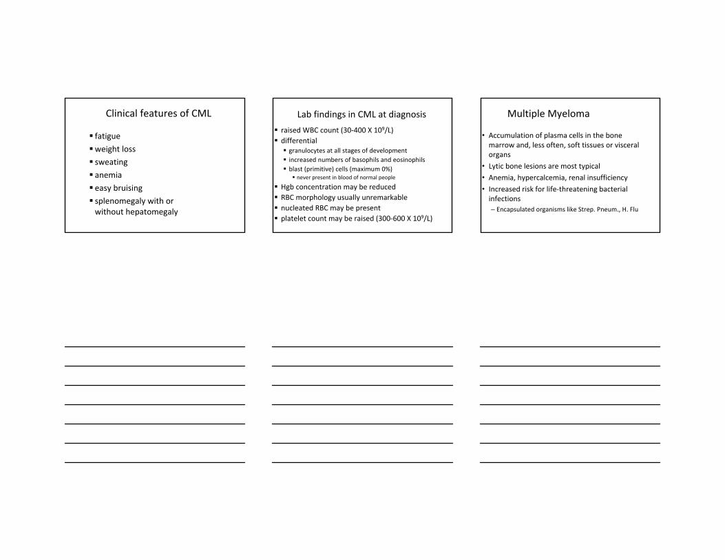
Clinical features of CML
fatigue
weight loss
sweating
anemia
easy bruising
splenomegaly with or without hepatomegaly
Lab findings in CML at diagnosis
raised WBC count (30‐400 X 109/L)differential
granulocytes at all stages of developmentincreased numbers of basophils and eosinophilsblast (primitive) cells (maximum 0%)
never present in blood of normal people
Hgb concentration may be reducedRBC morphology usually unremarkablenucleated RBC may be presentplatelet count may be raised (300‐600 X 109/L)
Multiple Myeloma
• Accumulation of plasma cells in the bone marrow and, less often, soft tissues or visceral organs
• Lytic bone lesions are most typical • Anemia, hypercalcemia, renal insufficiency• Increased risk for life‐threatening bacterial infections – Encapsulated organisms like Strep. Pneum., H. Flu

Diagnostic criteria for myeloma
• >10% plasma cells in Bone Marrow or plasmacytoma on biopsy
• clinical features of myeloma– bone pain, often in low back
• plus at least one of:– serum paraprotein spike (IgG.30g/L; IgA>20g/L)
• Seen on serum electrophoresis (SPEP)
– urine paraprotein (Bence Jones proteinuria)
– osteolytic lesions on skeletal survey‐ often cause Hypercalcemia
Lymphomas
• Lymphoma is the sixth most common type of cancer in the United States
• 15% Hodgkin’s Lymphomas• 85% Non‐Hodgkins lymphomas
Hodgkin’s Disease
Higher incidence in men than in womenOccurs in a bimodal age distribution
greatest peak in the third decadelesser peak in the seventh decade
Increased incidence of Hodgkin lymphoma in persons with a history of infectious mononucleosisNeoplastic cell of Hodgkin lymphoma is almost always a B celleither the Reed‐Sternberg cell or one of its mononuclear variants

Hodgkins Clinical Features
• Most common presenting feature is painless lymph node enlargement
• Mediastinal lymphadenopathy is common at presentation.
• Orderly spread from one lymph node region to contiguous nodal sites.
• The spleen and the lymph nodes in the celiac axis are often the first sites of subdiaphragmaticdisease
Systemic Symptoms (B symptoms)
• Drenching sweats at night, fever, and unexplained weight loss. – Pel‐Ebstein fevers are intermittent episodes of evening fevers that last for several days and alternate with afebrileperiods.
• Total body pruritus• A unique feature is pain at sites of lymphadenopathy immediately after ingestion of alcohol.
Hodgkins Disease Non‐HodgkinsLymphoma
Incidence Unchanged IncreasingAge Median 29 years Incidence increases
with age
Sites Mostly nodal:Supradiaphragmatic
No predictable pattern
Clinical Features
Mediastinal massPruritusAlcohol induces pain
Nothing specific
Prognoosis 70—80% cure Most incurable but very variable
Clinical Features Hodgkins vs. NHL