Embed Size (px)
Citation preview

Anemia
CHAPTER 2

Definition of Anemia
Deficiency in the oxygen-carrying capacity of the blood due to a diminished erythrocyte mass.May be due to:
Erythrocyte loss (bleeding)Decreased Erythrocyte production
low erythropoietinDecreased marrow response to erythropoietin
Increased Erythrocyte destruction(hemolysis)

Measurements of AnemiaHemoglobin = grams of hemoglobin per 100 mL of whole blood (g/dL)Hematocrit = percent of a sample of whole blood occupied by intact red blood cellsRBC = millions of red blood cells per microL of whole bloodMCV = Mean corpuscular volume
If > 100 → Macrocytic anemiaIf 80 – 100 → Normocytic anemiaIf < 80 → Microcytic anemia
RDW = Red blood cell distribution width= (Standard deviation of red cell volume ÷ mean cell volume) × 100 Normal value is 11-15%If elevated, suggests large variability in sizes of RBCs

Laboratory Definition of Anemia
Hgb:Women: <12.0Men: < 13.5
Hct:Women: < 36Men: <41
Symptoms of AnemiaDecreased oxygenation
Exertional dyspneaDyspnea at restFatigueBounding pulsesLethargy, confusion
Decreased volumeFatigueMuscle crampsPostural dizzinesssyncope

Special Considerations in Determining Anemia
Acute BleedDrop in Hgb or Hct may not be shown until 36 to 48 hours after acute bleed (even though patient may be hypotensive)
PregnancyIn third trimester, RBC and plasma volume are expanded by 25 and 50%, respectively.Labs will show reductions in Hgb, Hct, and RBC count, often to anemic levels, but according to RBC mass, they are actually polycythemic
Volume DepletionPatient’s who are severely volume depleted may not show anemia until after rehydrated

RBC Life CycleIn the bone marrow, erythropoietin enhances the growth of differentiation of burst forming units-erythroid (BFU-E) and colony forming units-erythroid (CFU-E) into reticulocytes.Reticulocyte spends three days maturing in the marrow, and then one day maturing in the peripheral blood.A mature Red Blood Cell circulates in the peripheral blood for 100 to 120 days.Under steady state conditions, the rate of RBC production equals the rate of RBC loss.

Normal Peripheral Smear

Causes of Anemia --Erythrocyte Loss
BleedingChronic (gastrointestinal, menstrual)Acute/Hemodynamically significant:
GastrointestinalRetroperitoneal

Anemia due to Low Erythropoietin
Kidney DiseaseNormochromic, normocyticLow reticulocyte countFrequently, peripheral smear in uremic patients show “burr cells” or echinocytesTarget hemoglobin for patients on dialysis is 11 to 12 g/dL
Administer erythropoietin or darbopoietin weeklyGood Iron stores must be maintained

Echinocytes (“burr cells”)

Anemia due to Decreased Response to Erythropoietin
Iron-DeficiencyVitamin B12 DeficiencyFolate DeficiencyAnemia of Chronic Disease

Anemia due to Decreased Response to Erythropoietin
Iron DeficiencyCan result from:
Pregnancy/lactationNormal growthBlood lossIntravascular hemolysisGastric bypassMalabsorption
Iron is absorbed in proximal small bowel; decreased abosrption in celiac disease, inflammatory bowel disease
May manifest as PICATendency to eat ice, clay, starch, crunchy materials
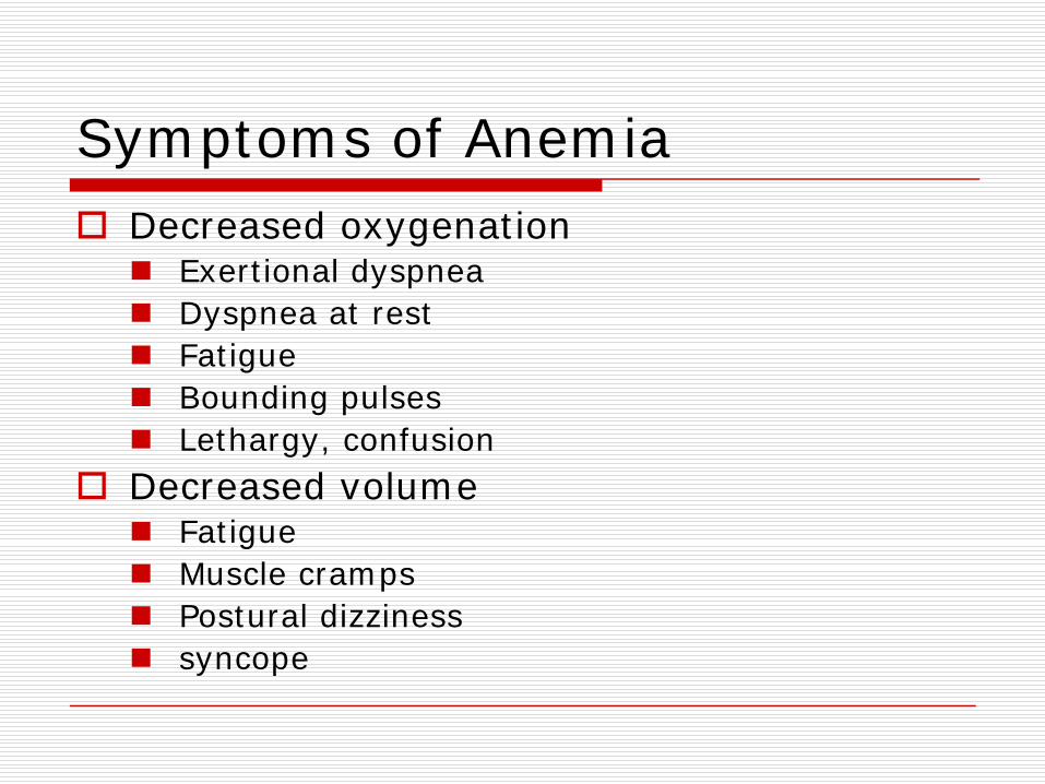
May have pallor, koilonychia of the nails, beeturiaPeripheral smear shows microcytic, hypochromic red cells with marked anisopoikilocytosis.

Iron Deficiency Anemia
Iron Deficiency Anemia -koilonychia

Iron Deficiency Anemia – Lab Findings
Serum IronLOW (< 60 micrograms/dL)
Total Iron Binding Capacity (TIBC)HIGH ( > 360 micrograms/dL)
Serum FerritinLOW (< 20 nanograms/mL)Can be “falsely”normal in inflammatory states

Treatment of Iron Deficiency Anemia
Oral iron saltsFerrous sulfate – 325 mg po Q Day
Side effects: constipation, black stools, positive hemmoccult test
Vitamin C can facilitate iron absorption.
Anemia due to Decreased Response to Erythropoietin
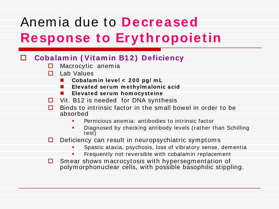
Cobalamin (Vitamin B12) DeficiencyMacrocytic anemiaLab Values
Cobalamin level < 200 pg/mLElevated serum methylmalonic acidElevated serum homocysteine
Vit. B12 is needed for DNA synthesisBinds to intrinsic factor in the small bowel in order to be absorbed
Pernicious anemia: antibodies to intrinsic factorDiagnosed by checking antibody levels (rather than Schilling test)
Deficiency can result in neuropsychiatric symptomsSpastic ataxia, psychosis, loss of vibratory sense, dementiaFrequently not reversible with cobalamin replacement
Smear shows macrocytosis with hypersegmentation of polymorphonuclear cells, with possible basophilic stippling.

Vitamin B12 Deficiency

Treatment of Vitamin B12 Deficiency
Vitamin B12 – 1000 micrograms intramuscularly monthly
-OR-
Vitamin B12 – 1000-2000 micrograms po QDaily

Anemia due to Decreased Response to Erythropoietin
Folate DeficiencyMacrocytic anemiaLab Values
Low folateIncreased serum homocystineNORMAL methylmalonic acid
Often occurs with decreased oral intake, increased utilization, or impaired absorption of folate
Folate is normally absorbed in duodenum and proximal jejunum –deficiency found in celiac disease, regional enteritis, amyloidosisDeficiency frequently in alcoholics, because enzyme required for deglutamation of folate is inhibited by alcohol.Deficiency often found in pregnant women, persons with desquamating skin disorders, patients with sickle cell anemia (and other conditions associated with rapid cell division and turnover)
Smear shows macrocytosis with hypersegmented neutrophils

Folate Deficiency

Treatment of Folate Deficiency
Folate – 1 to 5 mg po QdayVit. B12 deficiency must be excluded in folate-deficient patients, because supplemental folate can improve the anemia of Vit. B12 deficiency but not the neurologic sequelae.

Vitamin B12 Deficiency Versus Folate Deficiency
Vitamin B 12 Deficiency
Folate Deficiency
MCV > 100 > 100Smear Macrocytosis with
hypersegmented neutrophils
Macrocytosis with hypersegmented neutrophils
Pernicious anemia Yes NO
Homocystine Elevated Elevated
Methylmalonic Acid Elevated NORMAL
Anemia due to Decreased Response to Erythropoietin
Anemia of Chronic DiseaseUsually normocytic, normochromic (but can become hypochromic, microcytic over time)Occurs in people with inflammatory conditions such as collage vascular disease, malignancy or chronic infection.Iron replacement is not necessaryMay benefit from erythropoietin supplementation.

Anemia due to Decreased marrow response
ThalassemiaMicrocytic anemiaDefects in either the alpha or beta chains of hemoglobin, leading to ineffective erythropoiesis and hemolysis
α-thalassemia:Prevalent in Africa, Mediterranean, Middle East, Asia
β-thalassemia:Prevalent in Mediterranean, South East Asia, India, Pakistan
Smear shows microcytosis with target cells
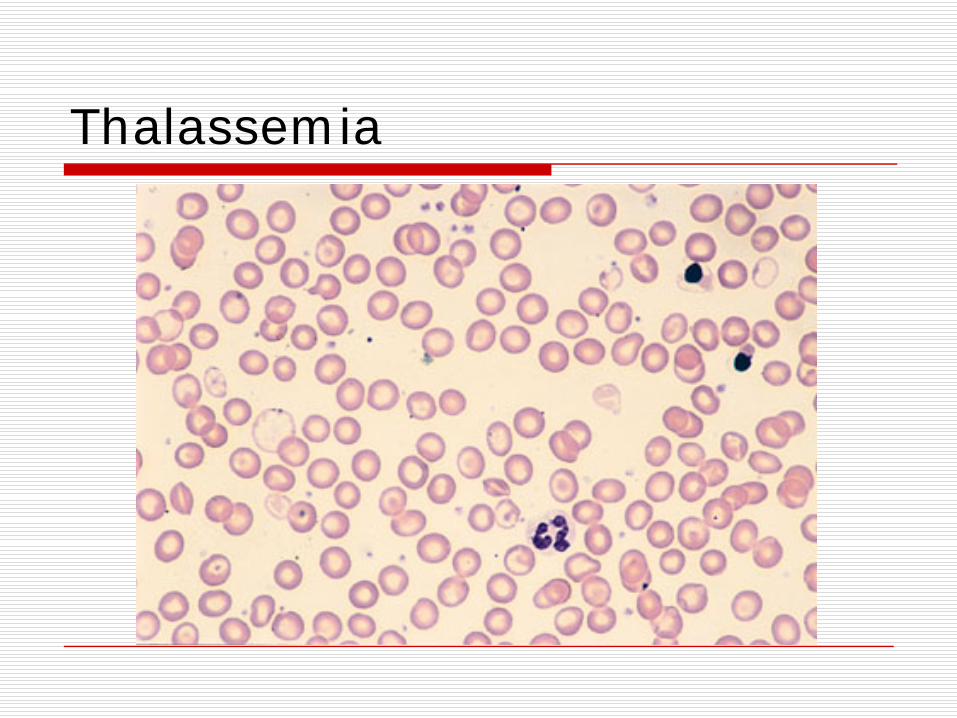
Thalassemia

Anemia due to Destruction of Red Blood Cells
HemoglobinopathiesSickle Cell Anemia
Aplastic AnemiaDecrease in all lines of cells – hemoglobin, hematocrit, WBC, plateletsParvovirus B19, EBV, CMVAcquired aplastic anemia
Hemolytic Anemia

Hemolytic AnemiasHereditary spherocytosisGlucose-6-phosphate dehydrogenase (G6PD) Deficiency
Most common enzyme defect in erythrocytesX-linkedBrisk hemolysis when patients exposed to oxidative stress from drugs, infections or toxins.
Thrombotic Thrombocytopenic Purpura (TTP)
Thrombocytopenia and microangiopathic hemolytic anemia, fever, renal insufficiency, neurologic symptomsSchistocytes on smear
Hemolytic Uremic SyndromeThrombocytopenia, Microangiopathic hemolytic anemia, renal insufficiency
Autoimmune Hemolytic AnemiaWarm-antibody mediated
IgG antibody binds to erythrocyte surfacemost commonDiagnosed by POSITIVE Coomb’s Test (detectgs IgG or complement on the cell surgace)Can be caused drugsTreated with corticosteroids or splenectomy if refractory
Cold agglutinin DiseaseIgM antibodies bind to erythrocyte surfaceDoes not respond to corticosteroids, but usually mild.
InfectionsMalariaBabesiosisSepsis
TraumaIncludes some snake, insect bites

Sickle Cell Anemia

Spherocytosis

TTP / HUS – microangiopathic hemolysis with schistocytes

Malaria
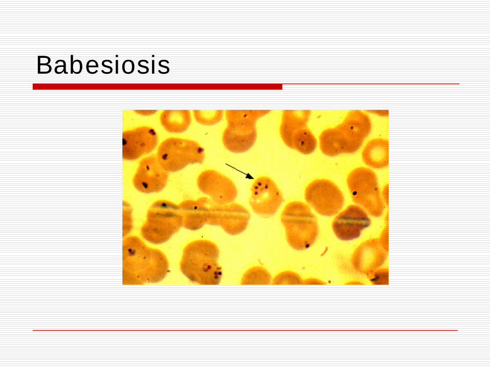
Babesiosis

Lab Analysis in Hemolytic Anemia
Increased indirect bilirubinIncreased LDHIncreased reticulocyte count
Normal reticulocyte count is 0.5 to 1.5%> 3% is sign of increased reticulocyte production, suggestive of hemolysis
Reduced or absent haptoglobin< 25 mg /dL suggests hemolysisHaptoglobin binds to free hemoglobin released after hemolysis

Evaluating the Patient with Anemia
Check Hemoglobin/HematocritIf female, is Hgb < 12 or Hct < 36?If male, is Hgb < 13.5 or Hct < 41?
If Yes, Patient has ANEMIA!If No, they are fine and this lecture was not necessary.

Evaluating the patient with Anemia
Any history of medical problems that could cause anemia?
Sickle cell Disease?Thalassemia?Renal Disease?Hereditary Spherocytosis?

Evaluating the Patient with Anemia
Are the other cell lines also low?If WBC and platelets are both low, consider APLASTIC ANEMIA!
Check medication listNSAIDS (phenylbutazone), Sulfonamides, Acyclovir, Gancyclovir, chloramphenicol, anti-epileptics (phenytoin, carbamazepine, valproic acid), nifedipineCheck parvovirus B19 IgG, IgMConsider hepatitis viruses, HIV
If Platelets are low consider TTP or HUS!Must check smear for schistocytes (for sign of microangiopathic hemolytic anemia)If renal failure, E. Coli O157:H7 exposure → HUSIf renal failure, neurologic changes, fever → TTP

Evaluating the Patient with Anemia
Is the patient bleeding?!Any bright red blood per rectum (hematochezia) or black tarry stools (melena)?
Check stool guaiac, may consider sigmoidoscopy or colonoscopy
Any abdominal pain, or recent femoral vein/artery manipulation?
Consider retroperitoneal hematoma

Evaluating the Patient with AnemiaIf other cell lines are okay, what is the MCV and RDW?
If MCV < 80, then it’s a microcytic anemiaCheck serum iron, ferritin, TIBC
If iron-deficiency anemia, look for sources of chronic bleeding –heavy menstrual bleeding, consider colonoscopy
Consider lead poisoning, copper deficiency, thalassemiasIf MCV 80-100, then it’s a normocytic anemia
Any inflammatory conditions that could result in anemia of chronic disease?Consider checking indirect bili, LDH, haptoglobin, reticulocyte count
If MCV > 100, then it’s a macrocytic anemiaCheck Vit. B 12, folateConsider liver disease, alcoholism, myelodysplastic syndromeCheck medications: hydoxyurea, AZT, methotrexate

Evaluating the Patient with Anemia
Any jaundice, elevated bilirubin, suspicious for hemolysis?
Check for increased indirect bilirubin, increased LDH, decreased haptoglobin, increased reticulocyte countAny sign of infection? Malaria? Babesiosis?Is Coombs test positive?
If yes, may be warm antibody hemolytic anemia; Consider drug as cause

Case #1
A 41-year old male with a history of HIV with a CD4 count of 150 who presents with a Hgb of 11, Hct of 33, which is down from a Hgb of 14 with a Hct of 42.

Case #1
Denies hematochezia, melena, any source of bleedingDenies any yellowing of the skinNo recent fevers, nausea or vomiting.

Case #1PMH: HIV/AIDS
No history of sickle cell disease, cancer, anemia
Allergies: SulfaMeds:
EfavirenzEmtricitabineTenofovirDapsone
Social History:No recent travel, no recent sick exposures, lives alone; occassional alcohol use, no tobacco use, no IV drug use; Works as attorney
Family History:No family history of cancer

Case #1
P.E.: 37.8, 123/68, 73, 16, 99% on RAGen: Alert and oriented x 3; in NAD;HEENT: no scleral icterus, no lymphadenopathyCV: RRRResp: LCTAAbd.: soft, nontenderExt.: no LE edema

Case # 1LABS:
WBC: 4.3Hgb: 11Hct: 33Platelets: 224Sodium: 137Potassium: 3.8Chloride: 101CO2: 25Glucose: 102
Tot. Protein: 5.3Albumin: 3.1Total Bili: 1.4Dir. Bili: 0.2AST: 23ALT: 42Alk. Phos: 122
Haptoglobin: 20Reticulocyte count: 3.2%

Case #1
What lab test do you want to make sure patient has had already or might you want to check?What might you see on peripheral smear if his total bilirubin was elevated, and his platelets were low?

Case #2
A 34- year old woman presents to your office with a 1-week history of generalized weakness, easy fatiguability and shortness of breath. One hour ago, she developed a headache a left hemiparesis. Two days ago, she noted easy bruisability and bleeding guyms. Three days ago, she developed a fever. A history reveals that she had no previous serious illnesses and review of systems is normal.

Case #2Physical Exam:
Temp: 40°, 120/70, 70, 16, 96% on RAGen: Alerti oriented, in NAD, but appears weakHEENT: petechiae on soft palate with some fresh blood on gingivaCV: RRR; II/VI high-pitched holosystolic murmurResp: LCTA bilaterallyNeuro: mild left hemiparesis with hyperactive reflexes and positive babinkski on the leftSkin: scattered pupuric lesions on lower extremities

Case # 2
Hgb: 6 g/dLMCV: 80RDW: 20%WBC: 15Reticulocyte count: 200Platelet: 9Creatinine: 1.0
Total Bili: 3.0Direct Bili: 0.2LDH: 3500UA: 2+ protein, 30-40 RBCs, 5 WBCs

Case #2

Case # 2
The most likely diagnosis of this patient’s disorder is:(A)Acute leukemia(B)Bacterial endocarditis(C)Thromboci thrombocytopenic purpura(D)Hemolytic uremic syndrome(E)Systemic Lupus erythematosus

Case # 3A 64-year old woman is hospitalized because of progressive SOB and palpitations over the past few weeks. She has also noticed a yellow tinge to her eyes during this time. She occasionally drinks wine excessively but says that she has abstained since the onset of her symptoms. For the last 6 months she has not eaten meat or fish, and her diet has consisted mostly of toast with margarine, tea, and an occassional banana. She says her social security checks do not stretch as far as they used to.

Case # 3
Physical Exam:Vitals: Pulse: 110, RR: 22General: pale, blue-eyed, gray-haired disheveled female with mild scleral icterus.CV: RRRResp: crackles that do not clear with coughing are heard at both lung basesExt: mild pitting edema at both anklesNeuro Exam: Normal

Case #3
Labs:Hgb: 5.1 g/dLMCV: 112RDW: 21%Platelets: 109WBC: 4.6

Case #3
Which of the following blood levels are most likely in this patient?
Vitamin B12
Folate Methylmalonic Acid
Homocysteine
(A) Low Normal High High
(B) Low Normal Normal High
(C) Normal Low High Normal
(D) Normal Low Normal High
(D) Normal Normal Normal Normal


![LeucocytosisandAsymptomaticUrinaryTractInfectionsinSickle ...downloads.hindawi.com/journals/anemia/2020/3792728.pdf · include blood vessel occlusion, erythrocyte sickling, and recurrentinfectionsduetoimmunecompromise[7–9]](https://img.pdfslide.us/doc/110x75/5f5a04e09899683224188ac7/leucocytosisandasymptomaticurinarytractinfectionsinsickle-include-blood-vessel.jpg)