Embed Size (px)
Citation preview
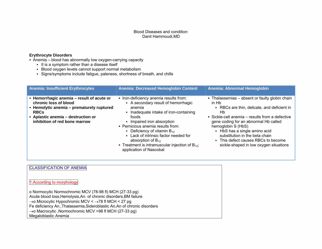
Blood Diseases and condition: Danil Hammoudi.MD
Erythrocyte Disorders Anemia – blood has abnormally low oxygen-carrying capacity
It is a symptom rather than a disease itself Blood oxygen levels cannot support normal metabolism Signs/symptoms include fatigue, paleness, shortness of breath, and chills
Anemia: Insufficient Erythrocytes
Anemia: Decreased Hemoglobin Content
Anemia: Abnormal Hemoglobin
Hemorrhagic anemia – result of acute or chronic loss of blood
Hemolytic anemia – prematurely ruptured RBCs
Aplastic anemia – destruction or inhibition of red bone marrow
Iron-deficiency anemia results from: A secondary result of hemorrhagic
anemia Inadequate intake of iron-containing
foods Impaired iron absorption
Pernicious anemia results from: Deficiency of vitamin B12 Lack of intrinsic factor needed for
absorption of B12 Treatment is intramuscular injection of B12; application of Nascobal
Thalassemias – absent or faulty globin chain in Hb
RBCs are thin, delicate, and deficient in Hb
Sickle-cell anemia – results from a defective gene coding for an abnormal Hb called hemoglobin S (HbS)
HbS has a single amino acid substitution in the beta chain
This defect causes RBCs to become sickle-shaped in low oxygen situations
CLASSIFICATION OF ANEMIA 1.According to morphology o Normocytic Normochromic MCV (78-98 fl) MCH (27-33 pg) Acute blood loss,Hemolysis,An. of chronic disorders,BM failure →o Microcytic Hypochromic MCV < →78 fl MCH < 27 pg Fe deficiency An.,Thalassemia,Sideroblastic An,An of chronic disorders →o Macrocytic ,Normochromic MCV >98 fl MCH (27-33 pg) Megaloblastic Anemia
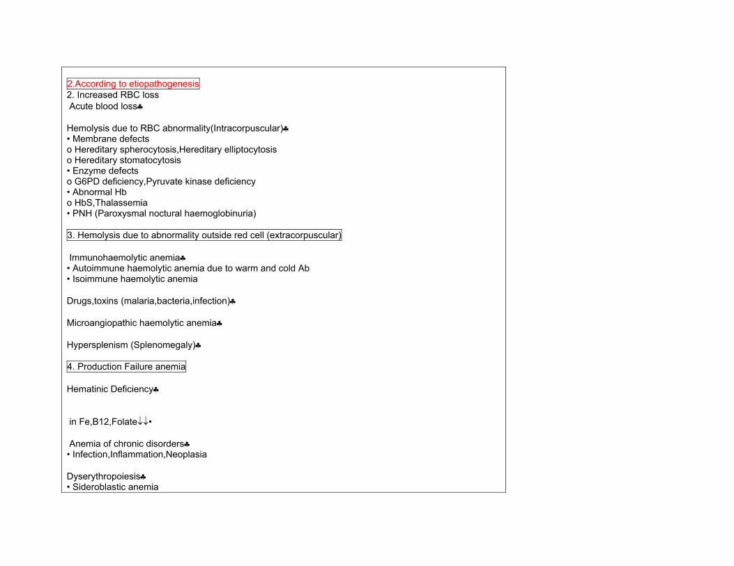
2.According to etiopathogenesis 2. Increased RBC loss Acute blood loss♣ Hemolysis due to RBC abnormality(Intracorpuscular)♣ • Membrane defects o Hereditary spherocytosis,Hereditary elliptocytosis o Hereditary stomatocytosis • Enzyme defects o G6PD deficiency,Pyruvate kinase deficiency • Abnormal Hb o HbS,Thalassemia • PNH (Paroxysmal noctural haemoglobinuria) 3. Hemolysis due to abnormality outside red cell (extracorpuscular) Immunohaemolytic anemia♣ • Autoimmune haemolytic anemia due to warm and cold Ab • Isoimmune haemolytic anemia Drugs,toxins (malaria,bacteria,infection)♣ Microangiopathic haemolytic anemia♣ Hypersplenism (Splenomegaly)♣ 4. Production Failure anemia Hematinic Deficiency♣ in Fe,B12,Folate↓↓• Anemia of chronic disorders♣ • Infection,Inflammation,Neoplasia Dyserythropoiesis♣ • Sideroblastic anemia

Hypoplasia♣ Marrow infiltration♣ • Leukamia,Myeloproliferative disorders,Neoplasms
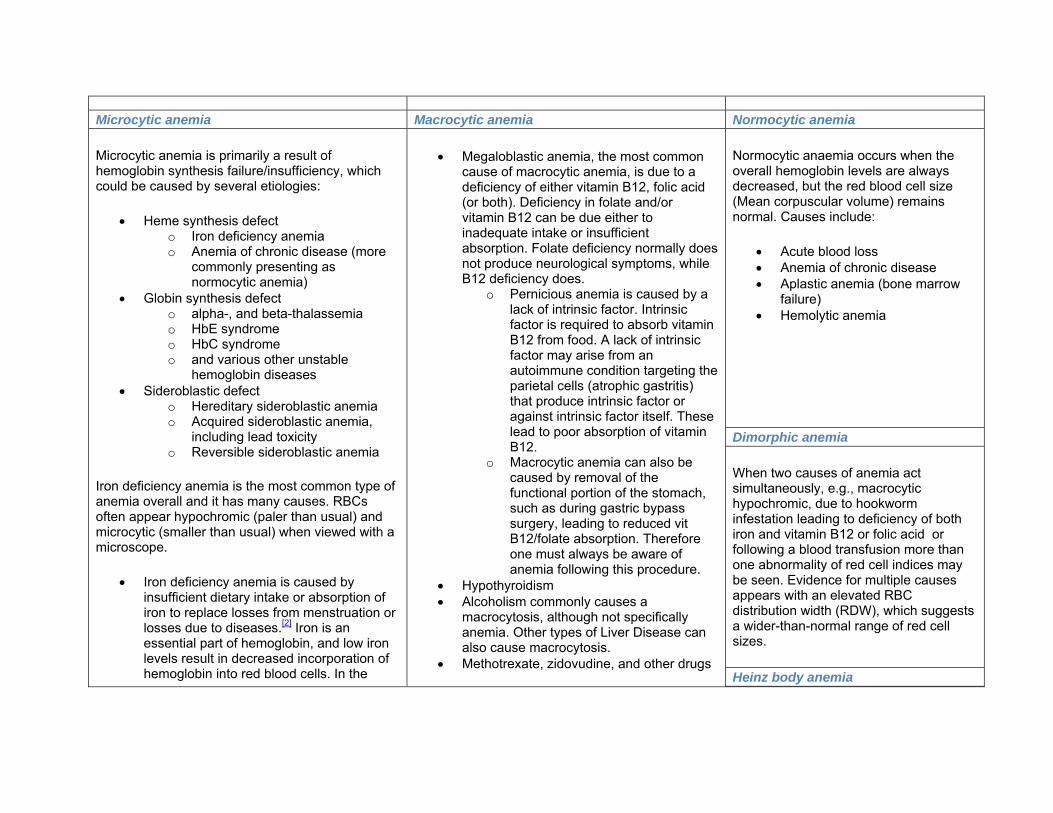
Microcytic anemia
Microcytic anemia is primarily a result of hemoglobin synthesis failure/insufficiency, which could be caused by several etiologies:
• Heme synthesis defect o Iron deficiency anemia o Anemia of chronic disease (more
commonly presenting as normocytic anemia)
• Globin synthesis defect o alpha-, and beta-thalassemia o HbE syndrome o HbC syndrome o and various other unstable
hemoglobin diseases • Sideroblastic defect
o Hereditary sideroblastic anemia o Acquired sideroblastic anemia,
including lead toxicity o Reversible sideroblastic anemia
Iron deficiency anemia is the most common type of anemia overall and it has many causes. RBCs often appear hypochromic (paler than usual) and microcytic (smaller than usual) when viewed with a microscope.
• Iron deficiency anemia is caused by insufficient dietary intake or absorption of iron to replace losses from menstruation or losses due to diseases.[2] Iron is an essential part of hemoglobin, and low iron levels result in decreased incorporation of hemoglobin into red blood cells. In the
Macrocytic anemia
• Megaloblastic anemia, the most common cause of macrocytic anemia, is due to a deficiency of either vitamin B12, folic acid (or both). Deficiency in folate and/or vitamin B12 can be due either to inadequate intake or insufficient absorption. Folate deficiency normally does not produce neurological symptoms, while B12 deficiency does.
o Pernicious anemia is caused by a lack of intrinsic factor. Intrinsic factor is required to absorb vitamin B12 from food. A lack of intrinsic factor may arise from an autoimmune condition targeting the parietal cells (atrophic gastritis) that produce intrinsic factor or against intrinsic factor itself. These lead to poor absorption of vitamin B12.
o Macrocytic anemia can also be caused by removal of the functional portion of the stomach, such as during gastric bypass surgery, leading to reduced vit B12/folate absorption. Therefore one must always be aware of anemia following this procedure.
• Hypothyroidism • Alcoholism commonly causes a
macrocytosis, although not specifically anemia. Other types of Liver Disease can also cause macrocytosis.
• Methotrexate, zidovudine, and other drugs
Normocytic anemia
Normocytic anaemia occurs when the overall hemoglobin levels are always decreased, but the red blood cell size (Mean corpuscular volume) remains normal. Causes include:
• Acute blood loss • Anemia of chronic disease • Aplastic anemia (bone marrow
failure) • Hemolytic anemia
Dimorphic anemia
When two causes of anemia act simultaneously, e.g., macrocytic hypochromic, due to hookworm infestation leading to deficiency of both iron and vitamin B12 or folic acid or following a blood transfusion more than one abnormality of red cell indices may be seen. Evidence for multiple causes appears with an elevated RBC distribution width (RDW), which suggests a wider-than-normal range of red cell sizes.
Heinz body anemia
United States, 20% of all women of childbearing age have iron deficiency anemia, compared with only 2% of adult men. The principal cause of iron deficiency anemia in premenopausal women is blood lost during menses. Studies have shown that iron deficiency without anemia causes poor school performance and lower IQ in teenage girls. Iron deficiency is the most prevalent deficiency state on a worldwide basis. Iron deficiency is sometimes the cause of abnormal fissuring of the angular
that inhibit DNA replication.
Macrocytic anemia can be further divided into "megaloblastic anemia" or "non-megaloblastic macrocytic anemia". The cause of megaloblastic anemia is primarily a failure of DNA synthesis with preserved RNA synthesis, which result in restricted cell division of the progenitor cells. The megaloblastic anemias often present with neutrophil hypersegmentation (6-10 lobes). The non-megaloblastic macrocytic anemias have diff t ti l i (i th i i i d DNA
Heinz bodies are an abnormality that form on the cells in this condition. This form of anemia may be brought on by taking certain medications; it is also triggered in cats by eating onions or acetaminophen (Tylenol). It can be triggered in dogs by ingesting onions or zinc, and in horses by ingesting dry red maple leaves.
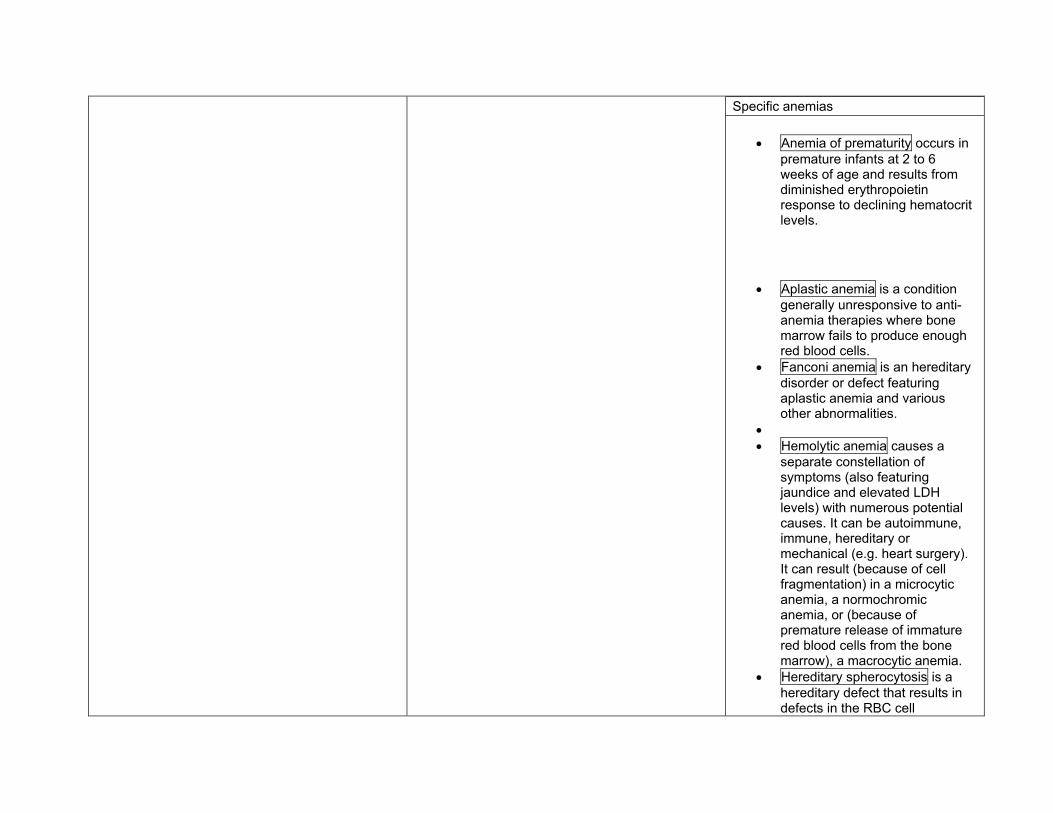
Specific anemias
• Anemia of prematurity occurs in premature infants at 2 to 6 weeks of age and results from diminished erythropoietin response to declining hematocrit levels.
• Aplastic anemia is a condition generally unresponsive to anti-anemia therapies where bone marrow fails to produce enough red blood cells.
• Fanconi anemia is an hereditary disorder or defect featuring aplastic anemia and various other abnormalities.
• • Hemolytic anemia causes a
separate constellation of symptoms (also featuring jaundice and elevated LDH levels) with numerous potential causes. It can be autoimmune, immune, hereditary or mechanical (e.g. heart surgery). It can result (because of cell fragmentation) in a microcytic anemia, a normochromic anemia, or (because of premature release of immature red blood cells from the bone marrow), a macrocytic anemia.
• Hereditary spherocytosis is a hereditary defect that results in defects in the RBC cell
membrane, causing the erythrocytes to be sequestered and destroyed by the spleen. This leads to a decrease in the number of circulating RBCs and, hence, anemia.
• Sickle-cell anemia, a hereditary disorder, is due to homozygous hemoglobin S genes.
• Warm autoimmune hemolytic anemia is an anemia caused by autoimmune attack against red blood cells, primarily by IgG.
• Cold agglutinin hemolytic anemia is primarily mediated by IgM.
• Pernicious anemia is a form of megaloblastic anemia due to vitamin B12 deficiency dependent on impaired absorption of vitamin B12.
• Myelophthisic anemia or Myelophthisis is a severe type of anemia resulting from the replacement of bone marrow by other materials, such as malignant tumors or granulomas.
Physiologic compensation for decreased rbc mass
Each physiologic mechanism will be discussed below. It should be noted that, although there are many adjustments that can be made, one that cannot is decrease in the tissue requirement for oxygen. Actually, overall body oxidative metabolism increases in anemia because of the energy requirement of the compensatory activities.
1. Decreased hemoglobin oxygen affinity
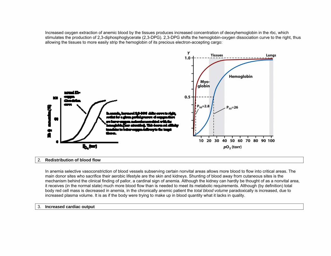
Increased oxygen extraction of anemic blood by the tissues produces increased concentration of deoxyhemoglobin in the rbc, which stimulates the production of 2,3-diphosphoglycerate (2,3-DPG). 2,3-DPG shifts the hemoglobin-oxygen dissociation curve to the right, thus allowing the tissues to more easily strip the hemoglobin of its precious electron-accepting cargo:
2. Redistribution of blood flow
In anemia selective vasoconstriction of blood vessels subserving certain nonvital areas allows more blood to flow into critical areas. The main donor sites who sacrifice their aerobic lifestyle are the skin and kidneys. Shunting of blood away from cutaneous sites is the mechanism behind the clinical finding of pallor, a cardinal sign of anemia. Although the kidney can hardly be thought of as a nonvital area, it receives (in the normal state) much more blood flow than is needed to meet its metabolic requirements. Although (by definition) total body red cell mass is decreased in anemia, in the chronically anemic patient the total blood volume paradoxically is increased, due to increased plasma volume. It is as if the body were trying to make up in blood quantity what it lacks in quality.
3. Increased cardiac output

The heart can respond to tissue hypoxia by increased cardiac output. The increased output is matched by decreased peripheral vascular resistance and decreased blood viscosity (thinner blood flows more freely than thick blood), so that cardiac output can rise without an increase in blood pressure. Generally, anemia must be fairly severe (hemoglobin < 7 g/dL) before cardiac output rises.
Clinical signs and symptoms of anemia
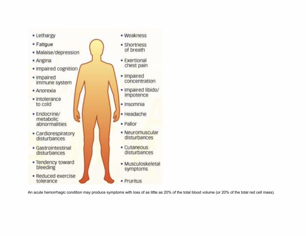
An acute hemorrhagic condition may produce symptoms with loss of as little as 20% of the total blood volume (or 20% of the total red cell mass).
Conversely, anemias developing over periods long enough to allow compensatory mechanisms to operate will allow much greater loss of rbc mass before producing symptoms.
It is not terribly uncommon to see a patient with a hemoglobin of 4 g/dL (hematocrit 12 cL/L), representing a loss of 70% of the rbc mass, being reluctantly dragged into a clinic by relatives concerned that he or she is looking a bit washed out.
When symptoms do develop, they are pretty much what you would expect given the precarious state of oxygen delivery to the tissues:
• dyspnea on exertion, • easy fatigability, • fainting, • lightheadedness, • tinnitus, and headache. • In addition, the hyperdynamic state of the circulatory system can produce palpitations and roaring in the ears. Pre-existing
cardiovascular pathologic conditions are, as you would expect, exacerbated by the anemia. o Angina pectoris, o intermittent claudication, o and night muscle cramps speak to the effect of anemia on already compromised perfusion.
Clinical signs of a slowly developed anemia are:
• pallor, • tachycardia, • and a systolic ejection murmur. • In rapidly developing anemia (as from hemorrhage and certain catastrophic hemolytic anemias), additional symptoms and signs
are noted: o syncope on rising from bed, o orthostatic hypotension (i.e., the blood pressure falls when the patient is raised from the supine to the sitting or
standing positions) a o nd orthostatic tachycardia.
Keep in mind that if anemia develops through rapid enough bleeding, the hematocrit and hemoglobin will be normal (since in hemorrhage the rbc's and plasma are lost in proportion).
Because of this, your appreciation of these clinical signs will serve you better in diagnosing this type of anemia than will the laboratory.
Classification of anemias
Anemias can be classified by cytometric schemes (i.e., those that depend on cell size and hemoglobin-content parameters, such as MCV and MCHC), erythrokinetic schemes (those that take into account the rates of rbc production and destruction), and biochemical/molecular schemes (those that consider the etiology of the anemia at the molecular level.
An example: sickle cell anemia
• Cytometric classification: normochromic, normocytic • Erythrokinetic classification: hemolytic • Biochemical/molecular classification: DNA point mutation producing amino acid substitution in hemoglobin beta chain
A. Cytometric classification
Because cytometric parameters are more easily and less expensively measured than are erythrokinetic and biochemical ones, it is most practical to work from the cytometric classification, to the erythrokinetic, and then (hopefully) to the biochemical. Your first job in working up a patient with anemia is to place the case in one of three major cytometric categories:
1. Normochromic, normocytic anemia (normal MCHC, normal MCV).
These include:
1. anemias of chronic disease 2. hemolytic anemias (those characterized by accelerated destruction of rbc's) 3. anemia of acute hemorrhage 4. aplastic anemias (those characterized by disappearance of rbc precursors from the marrow)
2. Hypochromic, microcytic anemia (low MCHC, low MCV).
These include:
1. iron deficiency anemia 2. thalassemias 3. anemia of chronic disease (rare cases)
3. Normochromic, macrocytic anemia (normal MCHC, high MCV).
These include:
1. vitamin B12 deficiency 2. folate deficiency
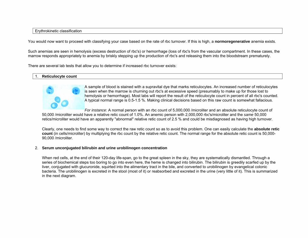
Erythrokinetic classification
You would now want to proceed with classifying your case based on the rate of rbc turnover. If this is high, a normoregenerative anemia exists.
Such anemias are seen in hemolysis (excess destruction of rbc's) or hemorrhage (loss of rbc's from the vascular compartment. In these cases, the marrow responds appropriately to anemia by briskly stepping up the production of rbc's and releasing them into the bloodstream prematurely.
There are several lab tests that allow you to determine if increased rbc turnover exists:
1. Reticulocyte count
A sample of blood is stained with a supravital dye that marks reticulocytes. An increased number of reticulocytes is seen when the marrow is churning out rbc's at excessive speed (presumably to make up for those lost to hemolysis or hemorrhage). Most labs will report the result of the reticulocyte count in percent of all rbc's counted. A typical normal range is 0.5-1.5 %. Making clinical decisions based on this raw count is somewhat fallacious.
For instance: A normal person with an rbc count of 5,000,000 /microliter and an absolute reticulocyte count of 50,000 /microliter would have a relative retic count of 1.0%. An anemic person with 2,000,000 rbc's/microliter and the same 50,000 retics/microliter would have an apparently "abnormal" relative retic count of 2.5 % and could be misdiagnosed as having high turnover.
Clearly, one needs to find some way to correct the raw retic count so as to avoid this problem. One can easily calculate the absolute retic count (in cells/microliter) by multiplying the rbc count by the relative retic count. The normal range for the absolute retic count is 50,000-90,000 /microliter.
2. Serum unconjugated bilirubin and urine urobilinogen concentration
When red cells, at the end of their 120-day life-span, go to the great spleen in the sky, they are systematically dismantled. Through a series of biochemical steps too boring to go into even here, the heme is changed into bilirubin. The bilirubin is greedily scarfed up by the liver, conjugated with glucuronide, squirted into the alimentary tract in the bile, and converted to urobilinogen by evangelical colonic bacteria. The urobilinogen is excreted in the stool (most of it) or reabsorbed and excreted in the urine (very little of it). This is summarized in the next diagram.
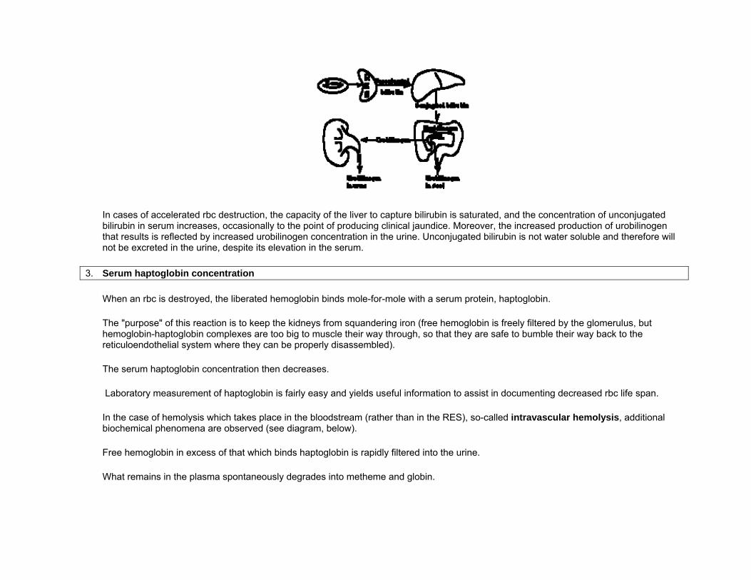
In cases of accelerated rbc destruction, the capacity of the liver to capture bilirubin is saturated, and the concentration of unconjugated bilirubin in serum increases, occasionally to the point of producing clinical jaundice. Moreover, the increased production of urobilinogen that results is reflected by increased urobilinogen concentration in the urine. Unconjugated bilirubin is not water soluble and therefore will not be excreted in the urine, despite its elevation in the serum.
3. Serum haptoglobin concentration
When an rbc is destroyed, the liberated hemoglobin binds mole-for-mole with a serum protein, haptoglobin.
The "purpose" of this reaction is to keep the kidneys from squandering iron (free hemoglobin is freely filtered by the glomerulus, but hemoglobin-haptoglobin complexes are too big to muscle their way through, so that they are safe to bumble their way back to the reticuloendothelial system where they can be properly disassembled).
The serum haptoglobin concentration then decreases.
Laboratory measurement of haptoglobin is fairly easy and yields useful information to assist in documenting decreased rbc life span.
In the case of hemolysis which takes place in the bloodstream (rather than in the RES), so-called intravascular hemolysis, additional biochemical phenomena are observed (see diagram, below).
Free hemoglobin in excess of that which binds haptoglobin is rapidly filtered into the urine.
What remains in the plasma spontaneously degrades into metheme and globin.
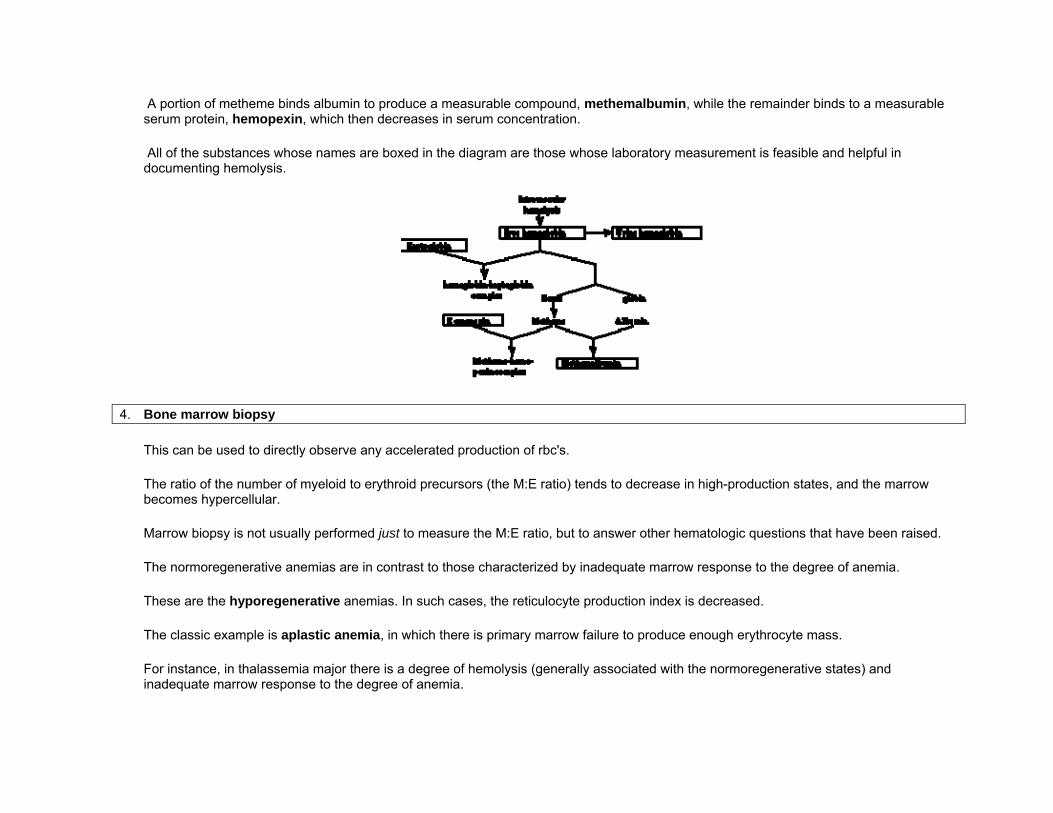
A portion of metheme binds albumin to produce a measurable compound, methemalbumin, while the remainder binds to a measurable serum protein, hemopexin, which then decreases in serum concentration.
All of the substances whose names are boxed in the diagram are those whose laboratory measurement is feasible and helpful in documenting hemolysis.
4. Bone marrow biopsy
This can be used to directly observe any accelerated production of rbc's.
The ratio of the number of myeloid to erythroid precursors (the M:E ratio) tends to decrease in high-production states, and the marrow becomes hypercellular.
Marrow biopsy is not usually performed just to measure the M:E ratio, but to answer other hematologic questions that have been raised.
The normoregenerative anemias are in contrast to those characterized by inadequate marrow response to the degree of anemia.
These are the hyporegenerative anemias. In such cases, the reticulocyte production index is decreased.
The classic example is aplastic anemia, in which there is primary marrow failure to produce enough erythrocyte mass.
For instance, in thalassemia major there is a degree of hemolysis (generally associated with the normoregenerative states) and inadequate marrow response to the degree of anemia.
C. Biochemical classification
. In some cases (e.g., iron deficiency), etiologic classification is easily attained;
in others (e.g.. aplastic anemia) the biochemical mechanism of disease may be hopelessly elusive.
Generally, biochemical tests are aimed at identifying a depleted cofactor necessary for normal hematopoiesis (iron, ferritin, folate, B12),
an abnormally functioning enzyme (glucose-6-phosphate dehydrogenase, pyruvate kinase),
or abnormal function of the immune system (the direct antiglobulin [Coombs'] test).
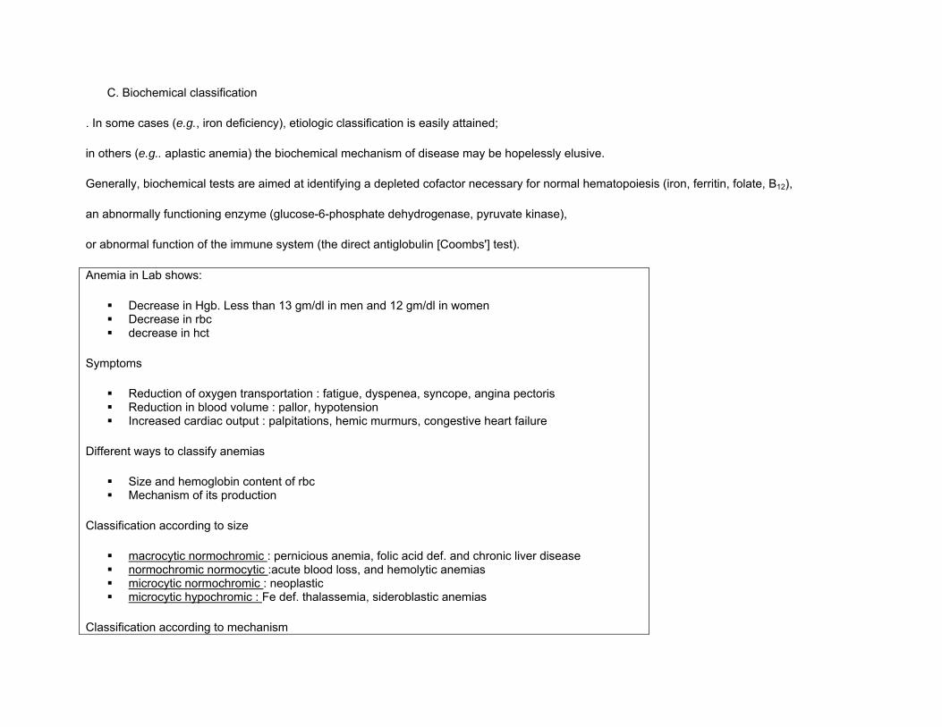
Anemia in Lab shows:
Decrease in Hgb. Less than 13 gm/dl in men and 12 gm/dl in women Decrease in rbc decrease in hct
Symptoms
Reduction of oxygen transportation : fatigue, dyspenea, syncope, angina pectoris Reduction in blood volume : pallor, hypotension Increased cardiac output : palpitations, hemic murmurs, congestive heart failure
Different ways to classify anemias
Size and hemoglobin content of rbc Mechanism of its production
Classification according to size
macrocytic normochromic : pernicious anemia, folic acid def. and chronic liver disease normochromic normocytic :acute blood loss, and hemolytic anemias microcytic normochromic : neoplastic microcytic hypochromic : Fe def. thalassemia, sideroblastic anemias
Classification according to mechanism
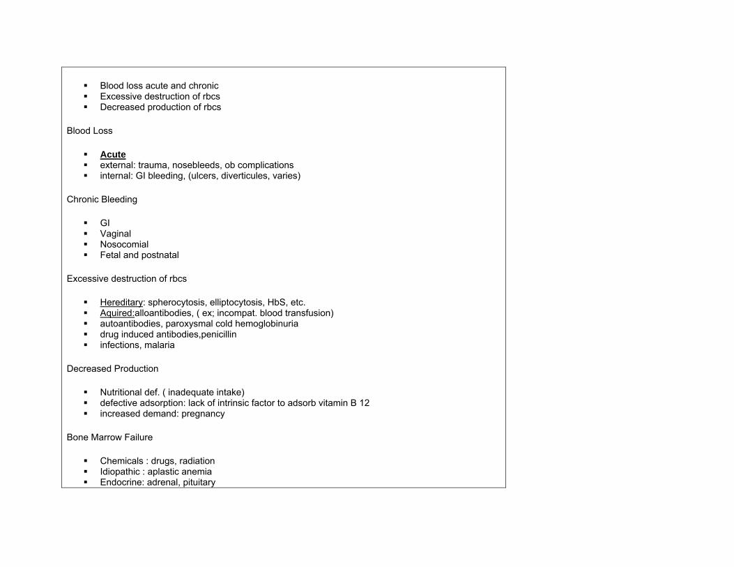
Blood loss acute and chronic Excessive destruction of rbcs Decreased production of rbcs
Blood Loss
Acute external: trauma, nosebleeds, ob complications internal: GI bleeding, (ulcers, diverticules, varies)
Chronic Bleeding
GI Vaginal Nosocomial Fetal and postnatal
Excessive destruction of rbcs
Hereditary: spherocytosis, elliptocytosis, HbS, etc. Aquired:alloantibodies, ( ex; incompat. blood transfusion) autoantibodies, paroxysmal cold hemoglobinuria drug induced antibodies,penicillin infections, malaria
Decreased Production
Nutritional def. ( inadequate intake) defective adsorption: lack of intrinsic factor to adsorb vitamin B 12 increased demand: pregnancy
Bone Marrow Failure
Chemicals : drugs, radiation Idiopathic : aplastic anemia Endocrine: adrenal, pituitary
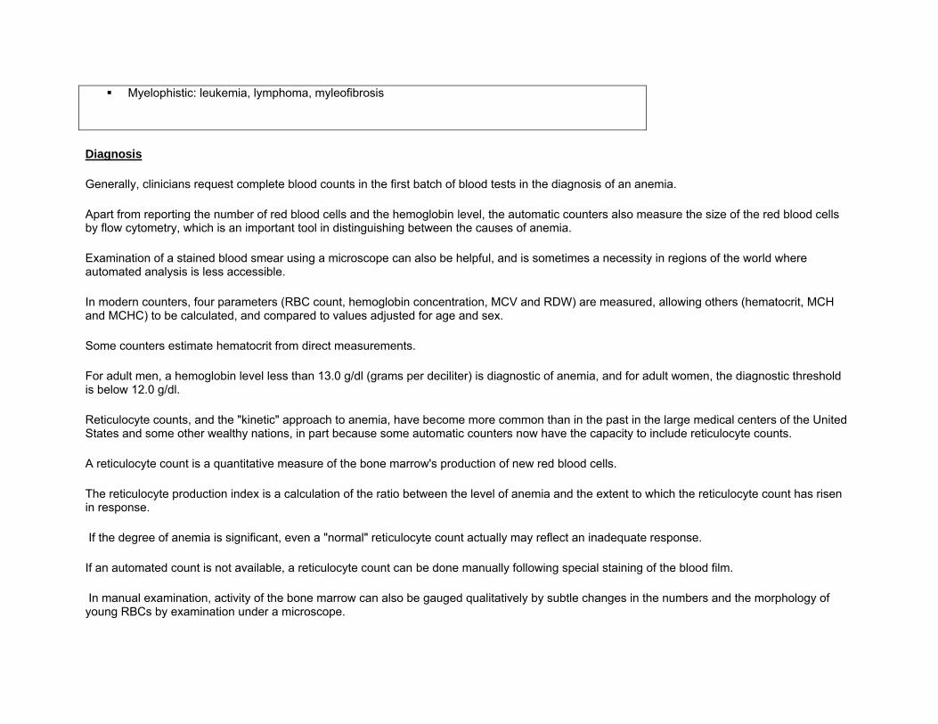
Myelophistic: leukemia, lymphoma, myleofibrosis
Diagnosis
Generally, clinicians request complete blood counts in the first batch of blood tests in the diagnosis of an anemia.
Apart from reporting the number of red blood cells and the hemoglobin level, the automatic counters also measure the size of the red blood cells by flow cytometry, which is an important tool in distinguishing between the causes of anemia.
Examination of a stained blood smear using a microscope can also be helpful, and is sometimes a necessity in regions of the world where automated analysis is less accessible.
In modern counters, four parameters (RBC count, hemoglobin concentration, MCV and RDW) are measured, allowing others (hematocrit, MCH and MCHC) to be calculated, and compared to values adjusted for age and sex.
Some counters estimate hematocrit from direct measurements.
For adult men, a hemoglobin level less than 13.0 g/dl (grams per deciliter) is diagnostic of anemia, and for adult women, the diagnostic threshold is below 12.0 g/dl.
Reticulocyte counts, and the "kinetic" approach to anemia, have become more common than in the past in the large medical centers of the United States and some other wealthy nations, in part because some automatic counters now have the capacity to include reticulocyte counts.
A reticulocyte count is a quantitative measure of the bone marrow's production of new red blood cells.
The reticulocyte production index is a calculation of the ratio between the level of anemia and the extent to which the reticulocyte count has risen in response.
If the degree of anemia is significant, even a "normal" reticulocyte count actually may reflect an inadequate response.
If an automated count is not available, a reticulocyte count can be done manually following special staining of the blood film.
In manual examination, activity of the bone marrow can also be gauged qualitatively by subtle changes in the numbers and the morphology of young RBCs by examination under a microscope.
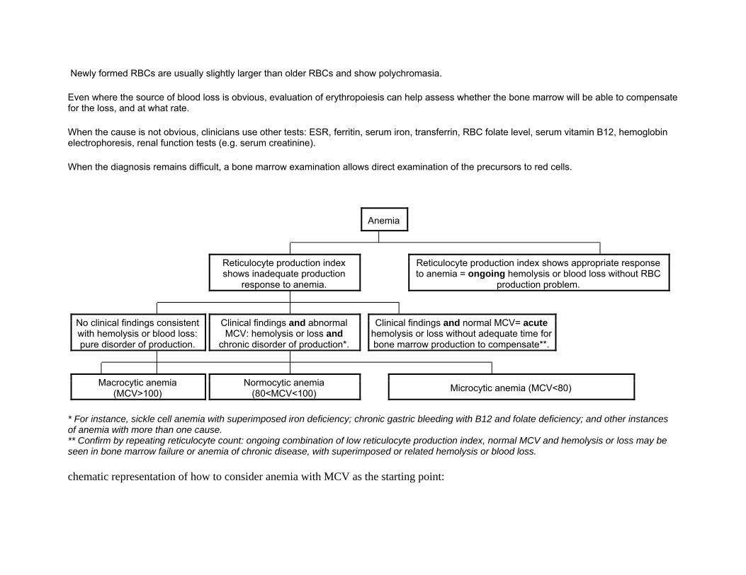
Newly formed RBCs are usually slightly larger than older RBCs and show polychromasia.
Even where the source of blood loss is obvious, evaluation of erythropoiesis can help assess whether the bone marrow will be able to compensate for the loss, and at what rate.
When the cause is not obvious, clinicians use other tests: ESR, ferritin, serum iron, transferrin, RBC folate level, serum vitamin B12, hemoglobin electrophoresis, renal function tests (e.g. serum creatinine).
When the diagnosis remains difficult, a bone marrow examination allows direct examination of the precursors to red cells.
Anemia
Reticulocyte production index shows inadequate production
response to anemia.
Reticulocyte production index shows appropriate response to anemia = ongoing hemolysis or blood loss without RBC
production problem.
No clinical findings consistent with hemolysis or blood loss: pure disorder of production.
Clinical findings and abnormal MCV: hemolysis or loss and
chronic disorder of production*.
Clinical findings and normal MCV= acute hemolysis or loss without adequate time for bone marrow production to compensate**.
Macrocytic anemia (MCV>100) Normocytic anemia
(80<MCV<100) Microcytic anemia (MCV<80)
* For instance, sickle cell anemia with superimposed iron deficiency; chronic gastric bleeding with B12 and folate deficiency; and other instances of anemia with more than one cause. ** Confirm by repeating reticulocyte count: ongoing combination of low reticulocyte production index, normal MCV and hemolysis or loss may be seen in bone marrow failure or anemia of chronic disease, with superimposed or related hemolysis or blood loss.
chematic representation of how to consider anemia with MCV as the starting point:
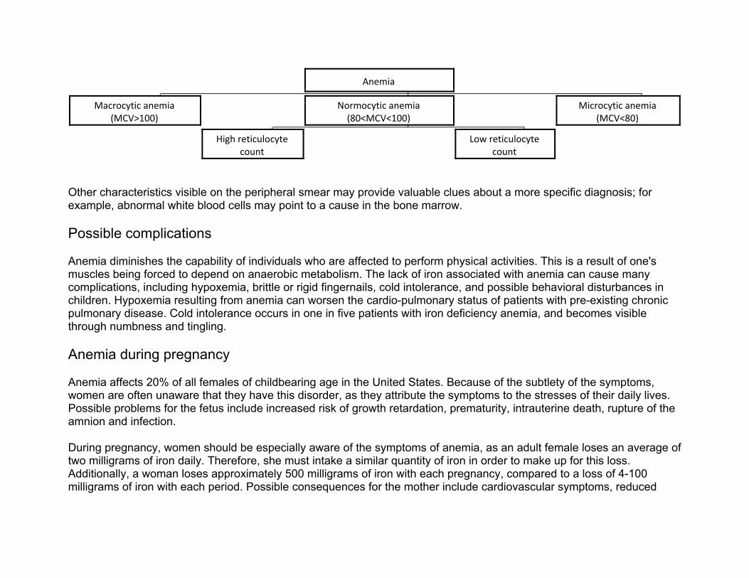
Anemia
Macrocytic anemia (MCV>100)
Normocytic anemia (80<MCV<100)
Microcytic anemia (MCV<80)
High reticulocyte count
Low reticulocyte count
Other characteristics visible on the peripheral smear may provide valuable clues about a more specific diagnosis; for example, abnormal white blood cells may point to a cause in the bone marrow.
Possible complications
Anemia diminishes the capability of individuals who are affected to perform physical activities. This is a result of one's muscles being forced to depend on anaerobic metabolism. The lack of iron associated with anemia can cause many complications, including hypoxemia, brittle or rigid fingernails, cold intolerance, and possible behavioral disturbances in children. Hypoxemia resulting from anemia can worsen the cardio-pulmonary status of patients with pre-existing chronic pulmonary disease. Cold intolerance occurs in one in five patients with iron deficiency anemia, and becomes visible through numbness and tingling.
Anemia during pregnancy
Anemia affects 20% of all females of childbearing age in the United States. Because of the subtlety of the symptoms, women are often unaware that they have this disorder, as they attribute the symptoms to the stresses of their daily lives. Possible problems for the fetus include increased risk of growth retardation, prematurity, intrauterine death, rupture of the amnion and infection.
During pregnancy, women should be especially aware of the symptoms of anemia, as an adult female loses an average of two milligrams of iron daily. Therefore, she must intake a similar quantity of iron in order to make up for this loss. Additionally, a woman loses approximately 500 milligrams of iron with each pregnancy, compared to a loss of 4-100 milligrams of iron with each period. Possible consequences for the mother include cardiovascular symptoms, reduced
physical and mental performance, reduced immune function, fatigue, reduced peripartal blood reserves and increased need for blood transfusion in the postpartum period.
Elevated hematocrit
• In cases of dengue fever a high hematocrit is a danger sign of an increased risk of dengue shock syndrome. • Polycythemia vera (PV), a myeloproliferative disorder in which the bone marrow produces excessive numbers of red cells, is associated
with elevated hematocrit. • Chronic obstructive pulmonary disease (COPD) and other pulmonary conditions associated with hypoxia may elicit an increased
production of red blood cells. This increase is mediated by the increased levels of erythropoietin by the kidneys in response to hypoxia. • Professional athletes' hematocrit levels are measured as part of tests for blood doping or Erythropoietin (EPO) use; the level of hematocrit
in a blood sample is compared with the long-term level for that athlete (to allow for individual variations in hematocrit level), and against an absolute permitted maximum (which is based on maximum expected levels within the population, and the hematocrit level which causes increased risk of blood clots resulting in strokes or heart attacks).
• If a patient is dehydrated, the hematocrit may be elevated. Repeat testing after adequate hydration therapy will usually result in a more reliable result.
Lowered hematocrit
Lowered hematocrit can imply significant hemorrhage (for example, in an ectopic pregnancy.)
The mean corpuscular volume (MCV) and the red cell distribution width (RDW) can be quite helpful in evaluating a lower-than-normal hematocrit, because it can help the clinician determine whether blood loss is chronic or acute. The MCV is the size of the red cells and the RDW is a relative measure of the variation in size of the red cell population. A low hematocrit with a low MCV with a normal RDW suggests a chronic iron-deficient erythropoiesis, but a high RDW suggests a blood loss that is more acute, such as a hemorrhage.
Groups of individuals who are at risk for developing anemia include:
• infants who may not have adequate iron intake • children going through a rapid growth spurt, during which the iron available cannot keep up with the demands for a growing red cell mass • women in childbearing years who have an excessive need for iron because of blood loss during menstruation • pregnant women, in whom the growing fetus creates a high demand for iron. • patients with chronic kidney disease, as their kidneys no longer secrete sufficient levels of the hormone erythropoietin, which stimulates
red blood cell production by the bone marrow.
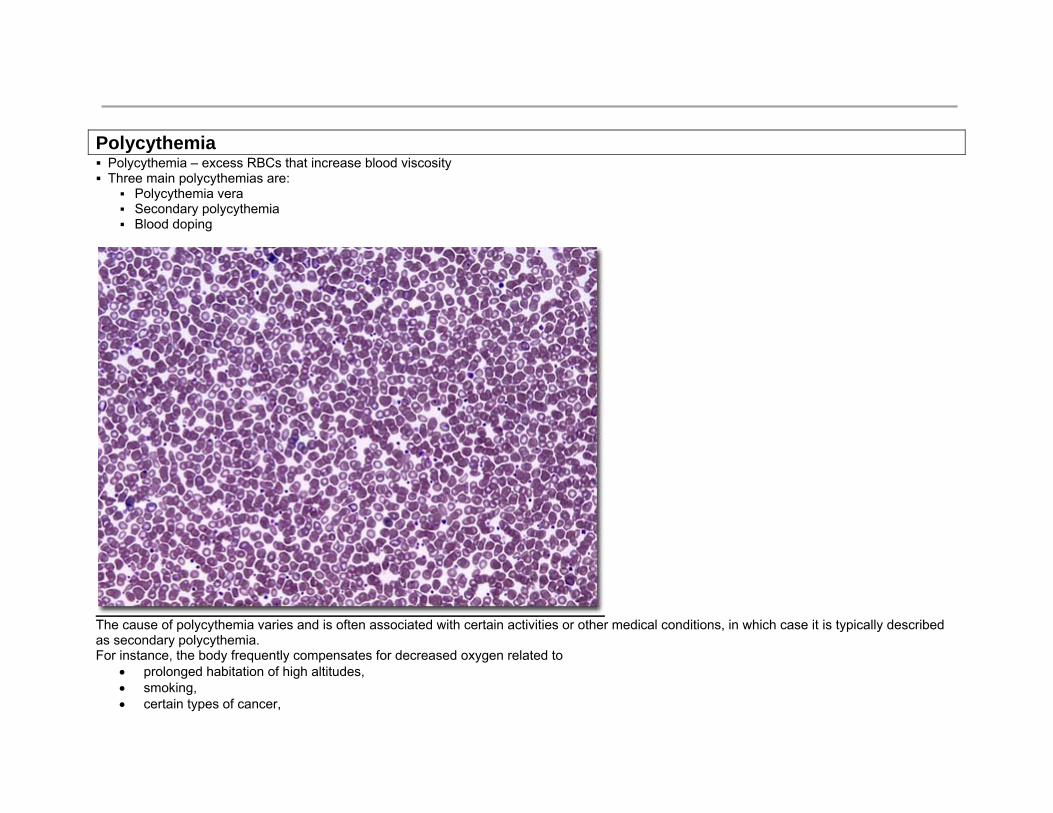
Polycythemia Polycythemia – excess RBCs that increase blood viscosity Three main polycythemias are:
Polycythemia vera Secondary polycythemia Blood doping
The cause of polycythemia varies and is often associated with certain activities or other medical conditions, in which case it is typically described as secondary polycythemia. For instance, the body frequently compensates for decreased oxygen related to
• prolonged habitation of high altitudes, • smoking, • certain types of cancer,
• pulmonary disease, • heart disorders, a • nd other conditions by increasing the production of red blood cells.
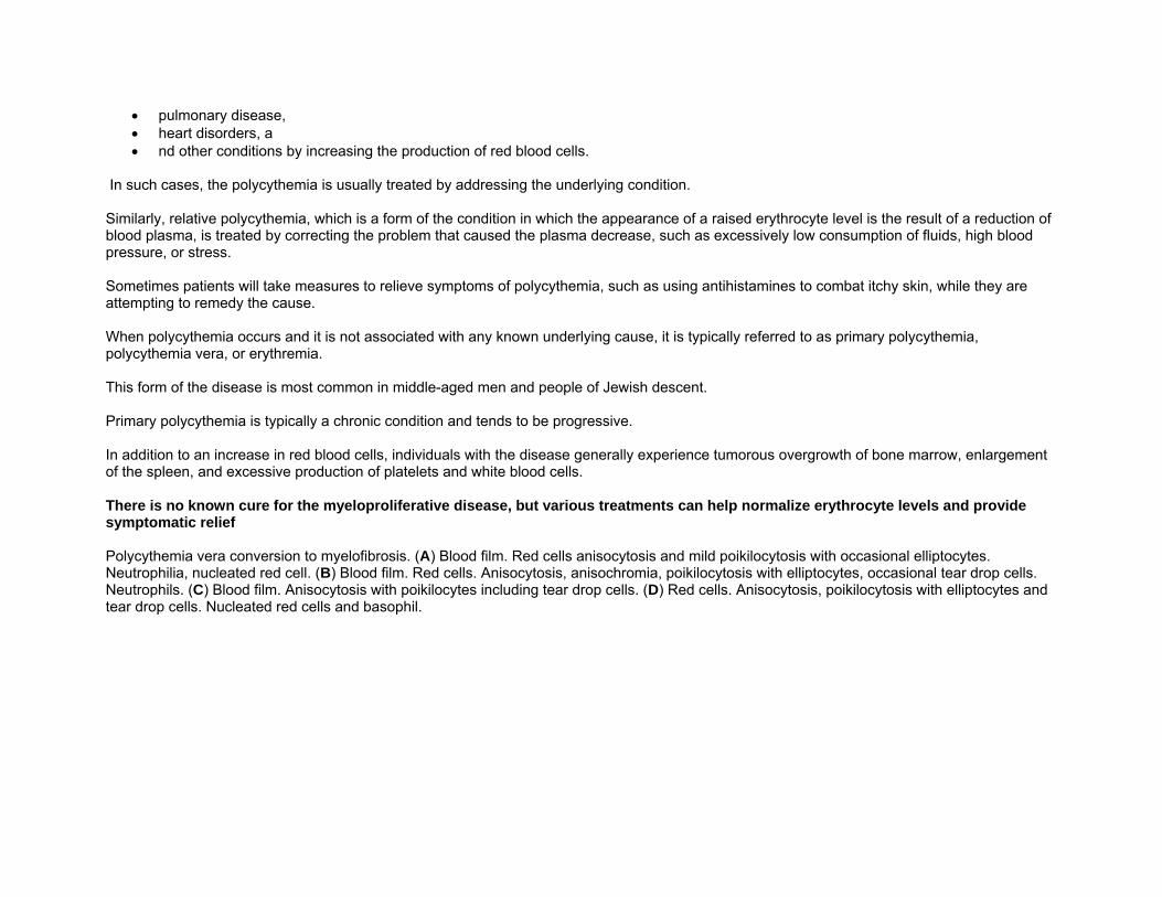
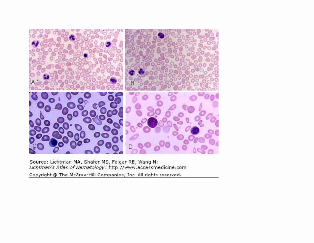
In such cases, the polycythemia is usually treated by addressing the underlying condition. Similarly, relative polycythemia, which is a form of the condition in which the appearance of a raised erythrocyte level is the result of a reduction of blood plasma, is treated by correcting the problem that caused the plasma decrease, such as excessively low consumption of fluids, high blood pressure, or stress. Sometimes patients will take measures to relieve symptoms of polycythemia, such as using antihistamines to combat itchy skin, while they are attempting to remedy the cause. When polycythemia occurs and it is not associated with any known underlying cause, it is typically referred to as primary polycythemia, polycythemia vera, or erythremia. This form of the disease is most common in middle-aged men and people of Jewish descent. Primary polycythemia is typically a chronic condition and tends to be progressive. In addition to an increase in red blood cells, individuals with the disease generally experience tumorous overgrowth of bone marrow, enlargement of the spleen, and excessive production of platelets and white blood cells. There is no known cure for the myeloproliferative disease, but various treatments can help normalize erythrocyte levels and provide symptomatic relief Polycythemia vera conversion to myelofibrosis. (A) Blood film. Red cells anisocytosis and mild poikilocytosis with occasional elliptocytes. Neutrophilia, nucleated red cell. (B) Blood film. Red cells. Anisocytosis, anisochromia, poikilocytosis with elliptocytes, occasional tear drop cells. Neutrophils. (C) Blood film. Anisocytosis with poikilocytes including tear drop cells. (D) Red cells. Anisocytosis, poikilocytosis with elliptocytes and tear drop cells. Nucleated red cells and basophil.
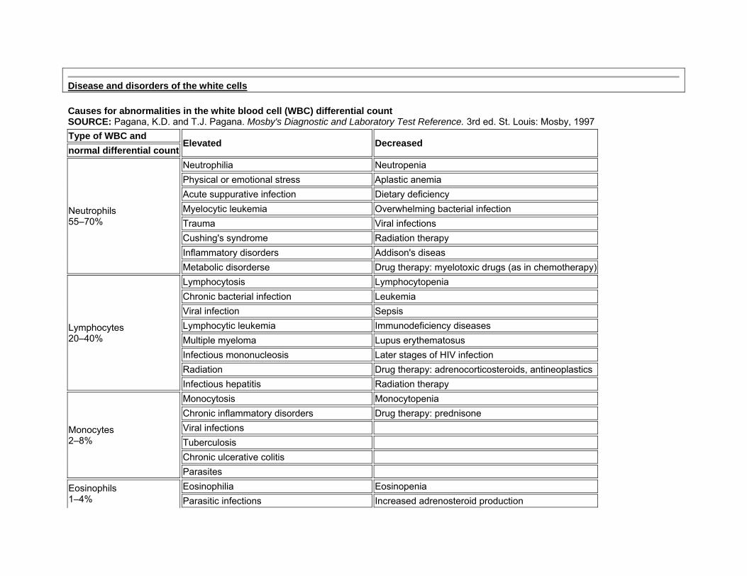
Disease and disorders of the white cells Causes for abnormalities in the white blood cell (WBC) differential count SOURCE: Pagana, K.D. and T.J. Pagana. Mosby's Diagnostic and Laboratory Test Reference. 3rd ed. St. Louis: Mosby, 1997Type of WBC and
Elevated Decreased normal differential count
Neutrophils 55–70%
Neutrophilia Neutropenia Physical or emotional stress Aplastic anemia Acute suppurative infection Dietary deficiency Myelocytic leukemia Overwhelming bacterial infection Trauma Viral infections Cushing's syndrome Radiation therapy Inflammatory disorders Addison's diseas Metabolic disorderse Drug therapy: myelotoxic drugs (as in chemotherapy)
Lymphocytes 20–40%
Lymphocytosis Lymphocytopenia Chronic bacterial infection Leukemia Viral infection Sepsis Lymphocytic leukemia Immunodeficiency diseases Multiple myeloma Lupus erythematosus Infectious mononucleosis Later stages of HIV infection Radiation Drug therapy: adrenocorticosteroids, antineoplasticsInfectious hepatitis Radiation therapy
Monocytes 2–8%
Monocytosis Monocytopenia Chronic inflammatory disorders Drug therapy: prednisone Viral infections Tuberculosis Chronic ulcerative colitis Parasites
Eosinophils 1–4%
Eosinophilia Eosinopenia Parasitic infections Increased adrenosteroid production
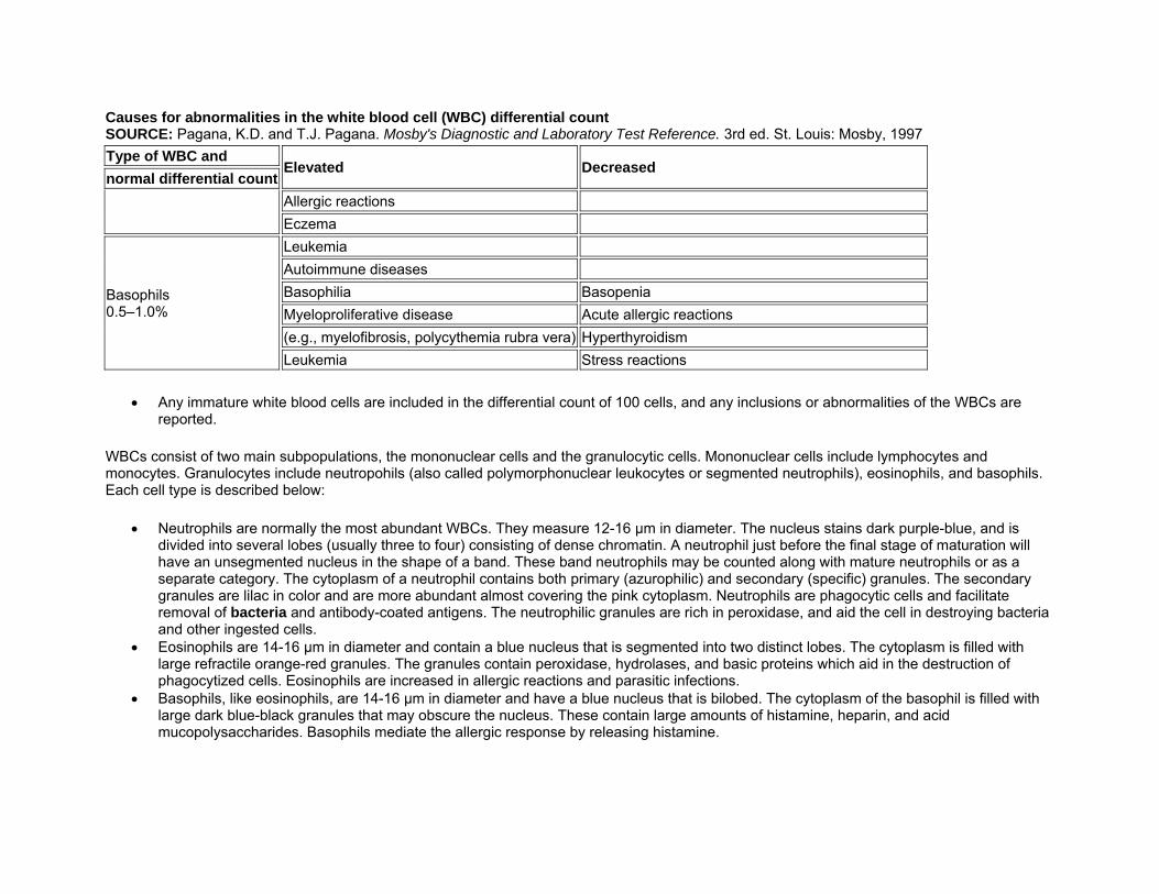
Causes for abnormalities in the white blood cell (WBC) differential count SOURCE: Pagana, K.D. and T.J. Pagana. Mosby's Diagnostic and Laboratory Test Reference. 3rd ed. St. Louis: Mosby, 1997Type of WBC and
Elevated Decreased normal differential count
Allergic reactions Eczema
Basophils 0.5–1.0%
Leukemia Autoimmune diseases Basophilia Basopenia Myeloproliferative disease Acute allergic reactions (e.g., myelofibrosis, polycythemia rubra vera) Hyperthyroidism Leukemia Stress reactions
• Any immature white blood cells are included in the differential count of 100 cells, and any inclusions or abnormalities of the WBCs are reported.
WBCs consist of two main subpopulations, the mononuclear cells and the granulocytic cells. Mononuclear cells include lymphocytes and monocytes. Granulocytes include neutropohils (also called polymorphonuclear leukocytes or segmented neutrophils), eosinophils, and basophils. Each cell type is described below:
• Neutrophils are normally the most abundant WBCs. They measure 12-16 μm in diameter. The nucleus stains dark purple-blue, and is divided into several lobes (usually three to four) consisting of dense chromatin. A neutrophil just before the final stage of maturation will have an unsegmented nucleus in the shape of a band. These band neutrophils may be counted along with mature neutrophils or as a separate category. The cytoplasm of a neutrophil contains both primary (azurophilic) and secondary (specific) granules. The secondary granules are lilac in color and are more abundant almost covering the pink cytoplasm. Neutrophils are phagocytic cells and facilitate removal of bacteria and antibody-coated antigens. The neutrophilic granules are rich in peroxidase, and aid the cell in destroying bacteria and other ingested cells.
• Eosinophils are 14-16 μm in diameter and contain a blue nucleus that is segmented into two distinct lobes. The cytoplasm is filled with large refractile orange-red granules. The granules contain peroxidase, hydrolases, and basic proteins which aid in the destruction of phagocytized cells. Eosinophils are increased in allergic reactions and parasitic infections.
• Basophils, like eosinophils, are 14-16 μm in diameter and have a blue nucleus that is bilobed. The cytoplasm of the basophil is filled with large dark blue-black granules that may obscure the nucleus. These contain large amounts of histamine, heparin, and acid mucopolysaccharides. Basophils mediate the allergic response by releasing histamine.
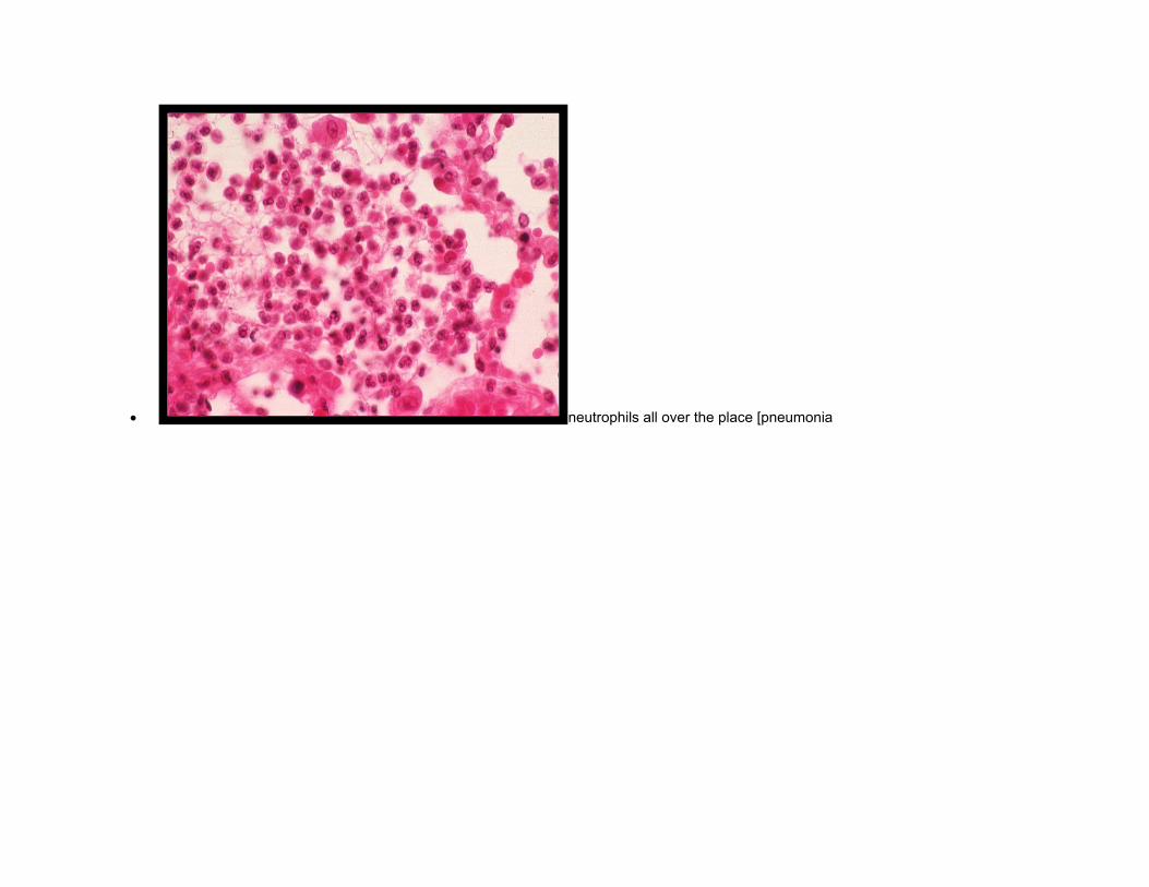
• neutrophils all over the place [pneumonia
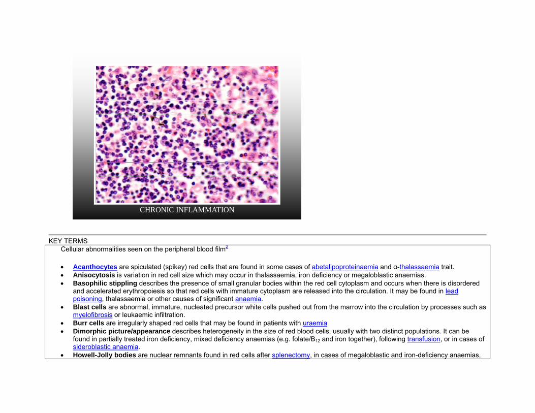
CHRONIC INFLAMMATION
KEY TERMS
Cellular abnormalities seen on the peripheral blood film2
• Acanthocytes are spiculated (spikey) red cells that are found in some cases of abetalipoproteinaemia and α-thalassaemia trait. • Anisocytosis is variation in red cell size which may occur in thalassaemia, iron deficiency or megaloblastic anaemias. • Basophilic stippling describes the presence of small granular bodies within the red cell cytoplasm and occurs when there is disordered
and accelerated erythropoiesis so that red cells with immature cytoplasm are released into the circulation. It may be found in lead poisoning, thalassaemia or other causes of significant anaemia.
• Blast cells are abnormal, immature, nucleated precursor white cells pushed out from the marrow into the circulation by processes such as myelofibrosis or leukaemic infiltration.
• Burr cells are irregularly shaped red cells that may be found in patients with uraemia • Dimorphic picture/appearance describes heterogeneity in the size of red blood cells, usually with two distinct populations. It can be
found in partially treated iron deficiency, mixed deficiency anaemias (e.g. folate/B12 and iron together), following transfusion, or in cases of sideroblastic anaemia.
• Howell-Jolly bodies are nuclear remnants found in red cells after splenectomy, in cases of megaloblastic and iron-deficiency anaemias,

and rarely in cases of leukaemia. Cabot's rings are circular or figure-of-eight structures in red cells that stain red with Wright's stain and are thought to represent nuclear membrane remnants; they are found in similar conditions to Howell-Jolly bodies.
• Hypochromia is impaired staining of red cells seen commonly in iron deficiency anaemia, and also in thalassaemia and sideroblastic anaemias.
• Left shift describes immature white blood cells that are released from the marrow when there is a cause of marrow outpouring, typically due to infection.
• Leptocytes are synonymous with target cells and mexican hat cells. They are red cells with a central area of increased staining, surrounded by a ring of hypodense staining and then a further ring of dense staining at the edge of the cell, giving an appearance akin to an archery target. They may be found in liver disease, thalassaemia, or sickle-cell disease. They occur occasionally in small numbers in iron deficiency anaemia.
• Leucoerythroblastic anaemia/picture describes the presence of immature cells such as myeloblasts and normoblasts in the film. It is seen in cases of marrow infiltration, for example in metastatic malignancy, prolonged hypoxia or severe infection.
• Leukaemoid reaction is a severe, reactive leucocytosis, usually consisting of granulocytes (polymorphonucleocytes). It is seen after burns, in cases of severe infection, following an acute haemolysis or prolonged hypoxia.
• Macrocytosis is the presence of abnormally large red cells found when erythropoiesis is disordered or when red cells are released prematurely from the marrow. They occur in alcohol excess, megaloblastic anaemia or as a consequence of haemolysis.
• Microcytosis is the presence of abnormally small red cells often found in association with hypochromia in iron deficiency anaemia. • Myelocytes, promeyelocytes, metamyelocytes and normoblasts are immature white cells seen in a leucoerythroblastic picture (see
above). • Normoblasts are immature, nucleated red cells seen in leucoerythroblastic anaemia, haemolysis, hypoxia and marrow infiltration. • Pappenheimer bodies are the granules seen in siderocytes found in carcinomatosis and after splenectomy. • Poikilocytosis is the variation in cell shape seen in iron deficiency anaemia. • Polychromasia is the heterogeneous staining of RBCs of different ages, with younger cells appearing blue, that occurs after
haemorrhage, haemolysis, dyserythropoiesis and treatment with haematinics such as iron and vitamin B12. • Reticulocytosis is the presence of >0.8–2% of total red cell count in the form of reticulocytes. They are young, oversized red cells that
are present when the marrow is actively producing red cells. They are present after haemorrhage, haemolysis or following treatment of deficient patients with haematinics.
• Right shift is characterised by the presence of hypersegmented polymorphonucleocytes (>5 lobes to their nucleus), seen in liver disease, uraemia and megaloblastic anaemia.
• Rouleaux are stacked/clumped groups of red cells caused by the presence of high levels of circulating acute-phase proteins which increase red cell 'stickiness'. They are often an indicator that a patient has a high ESR and are seen in infections, autoimmune conditions etc.
• Schistocytes are red cells fragmented by their passage through intravascular strands of fibrin, found in cases of intravascular haemolysis.• Spherocytes are overly-round or spheroid red cells that usually indicate active haemolysis. They are seen more rarely in cases of
hereditary spherocytosis.
L
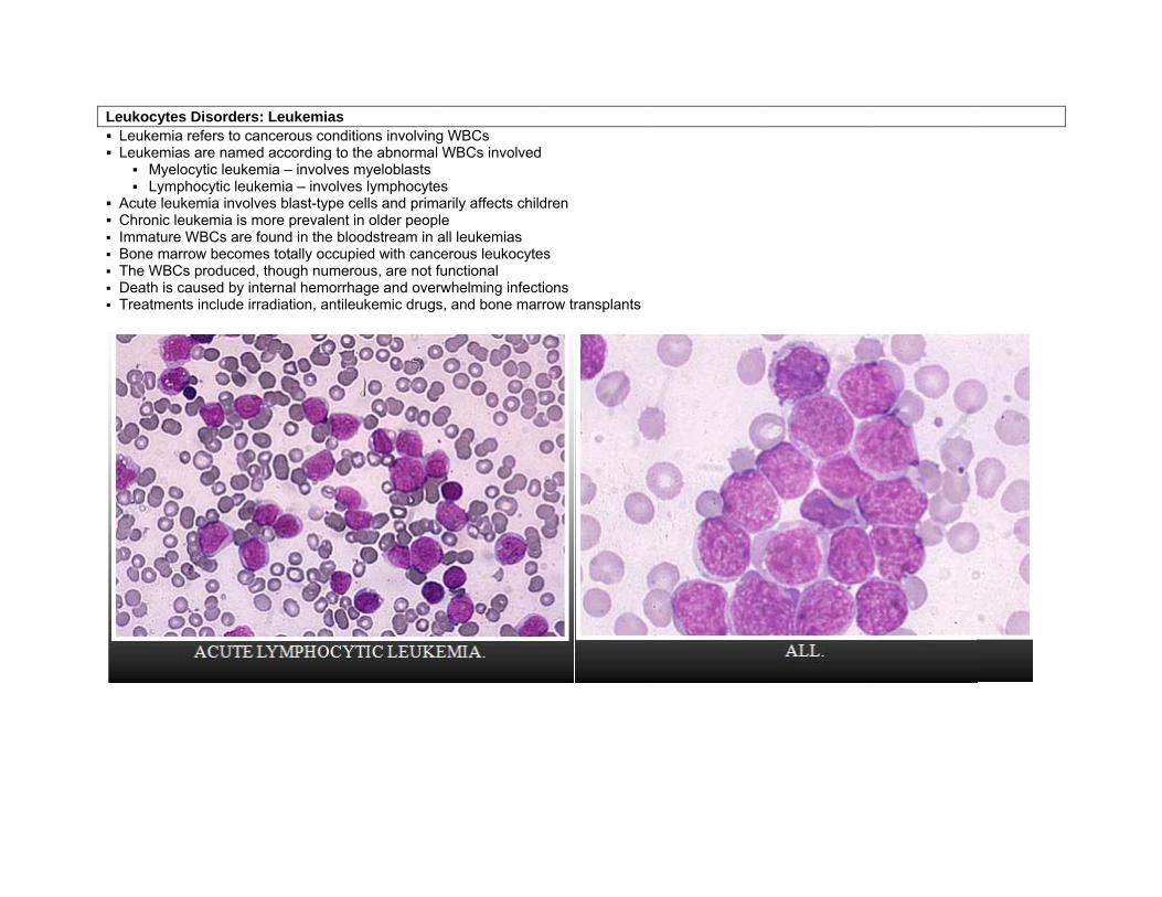
Leukocytes Diso Leukemia refer Leukemias are
Myelocytic Lymphocyt
Acute leukemia Chronic leukem Immature WBC Bone marrow b The WBCs prod Death is caused Treatments inc
orders: Leukemirs to cancerous conamed according leukemia – involtic leukemia – inv
a involves blast-tymia is more prevaCs are found in thebecomes totally ocduced, though nud by internal hemlude irradiation, a
ias onditions involving to the abnormaves myeloblasts
volves lymphocyteype cells and primlent in older peope bloodstream in ccupied with cancumerous, are not morrhage and oveantileukemic drug
ng WBCs l WBCs involved
es marily affects childple all leukemias
cerous leukocytefunctional
erwhelming infectigs, and bone marr
dren
s
ions row transplants
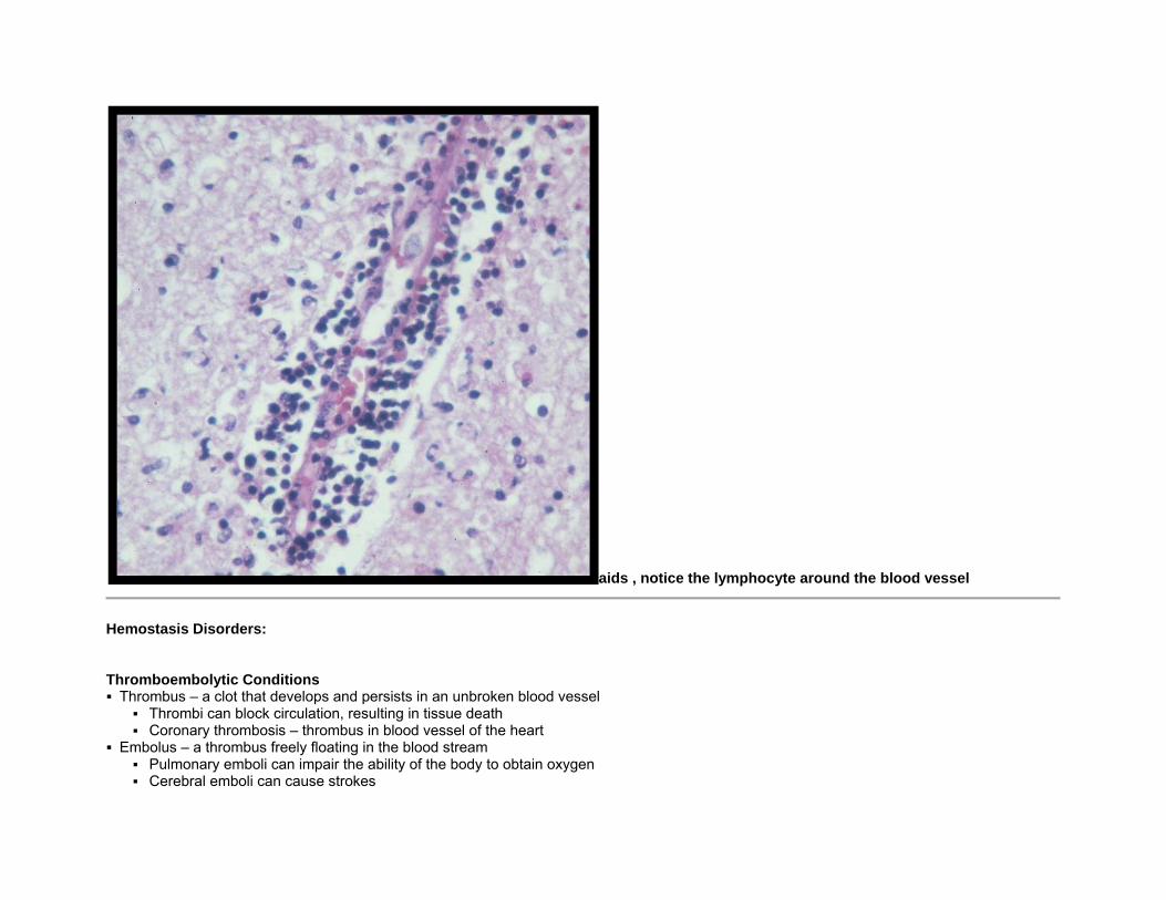
aids , notice the lymphocyte around the blood vessel
Hemostasis Disorders: Thromboembolytic Conditions Thrombus – a clot that develops and persists in an unbroken blood vessel
Thrombi can block circulation, resulting in tissue death Coronary thrombosis – thrombus in blood vessel of the heart
Embolus – a thrombus freely floating in the blood stream Pulmonary emboli can impair the ability of the body to obtain oxygen Cerebral emboli can cause strokes
Prevention of Undesirable Clots Substances used to prevent undesirable clots:
Aspirin – an antiprostaglandin that inhibits thromboxane A2 Heparin – an anticoagulant used clinically for pre- and postoperative cardiac care Warfarin – used for those prone to atrial fibrillation
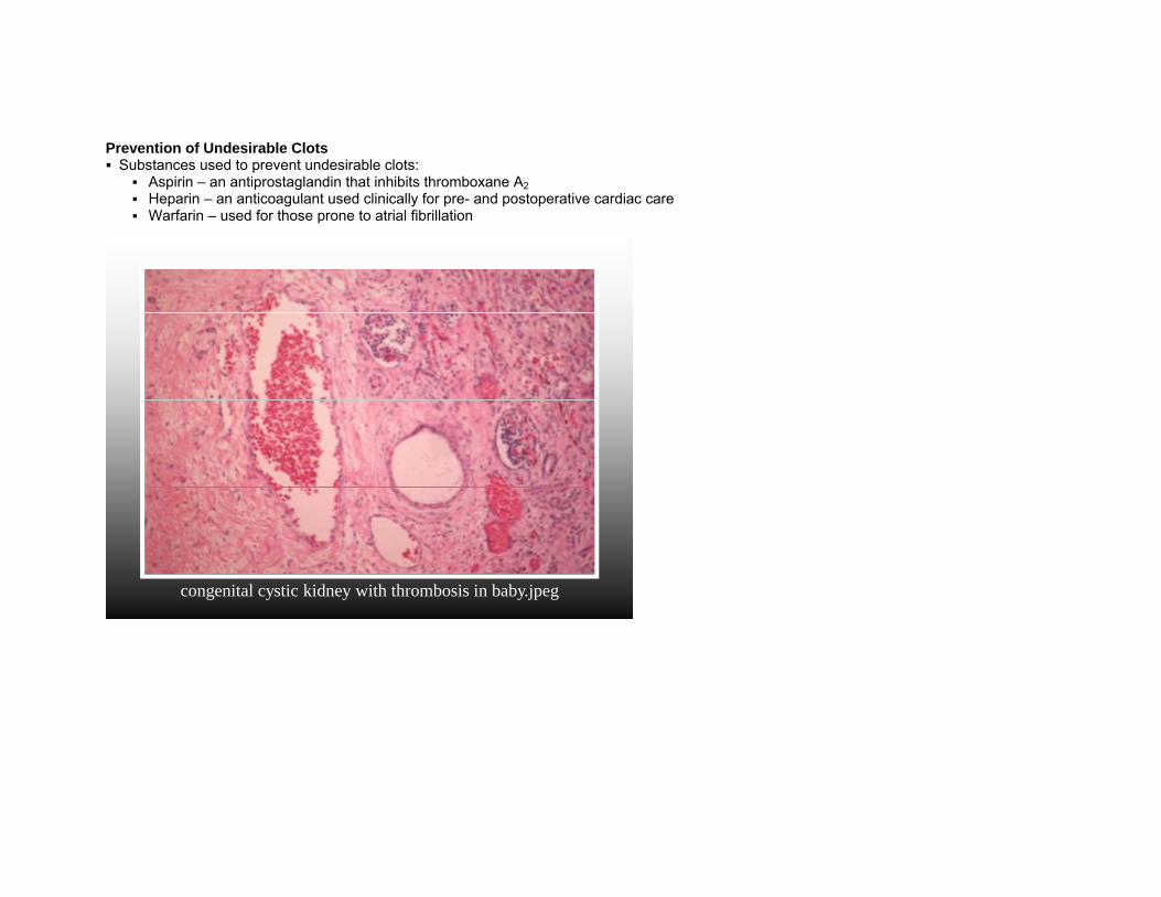
congenital cystic kidney with thrombosis in baby.jpeg
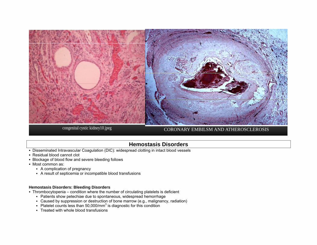
congenital cystic kidney10.jpeg CORONARY EMBILSM AND ATHEROSCLEROSIS
Hemostasis Disorders
Disseminated Intravascular Coagulation (DIC): widespread clotting in intact blood vessels Residual blood cannot clot Blockage of blood flow and severe bleeding follows Most common as:
A complication of pregnancy A result of septicemia or incompatible blood transfusions
Hemostasis Disorders: Bleeding Disorders Thrombocytopenia – condition where the number of circulating platelets is deficient
Patients show petechiae due to spontaneous, widespread hemorrhage Caused by suppression or destruction of bone marrow (e.g., malignancy, radiation) Platelet counts less than 50,000/mm3 is diagnostic for this condition Treated with whole blood transfusions
Inability to synthesize procoagulants by the liver results in severe bleeding disorders Causes can range from vitamin K deficiency to hepatitis and cirrhosis Inability to absorb fat can lead to vitamin K deficiencies as it is a fat-soluble substance and is absorbed along with fat Liver disease can also prevent the liver from producing bile, which is required for fat and vitamin K absorption
Hemophilias – hereditary bleeding disorders caused by lack of clotting factors
Hemophilia A – most common type (83% of all cases) due to a deficiency of factor VIII Hemophilia B – due to a deficiency of factor IX Hemophilia C – mild type, due to a deficiency of factor XI
Hemostasis Disorders: Bleeding Disorders Symptoms include prolonged bleeding and painful and disabled joints Treatment is with blood transfusions and the injection of missing factors
Hemolytic Disease of the Newborn Hemolytic disease of the newborn – Rh+ antibodies of a sensitized Rh– mother cross the placenta and attack and destroy the RBCs of an Rh+ baby
Rh– mother becomes sensitized when exposure to Rh+ blood causes her body to synthesize Rh+ antibodies The drug RhoGAM can prevent the Rh– mother from becoming sensitized Treatment of hemolytic disease of the newborn involves pre-birth transfusions and exchange transfusions after birth
Transfusion Reactions Transfusion reactions occur when mismatched blood is infused Donor’s cells are attacked by the recipient’s plasma agglutinins causing:
Diminished oxygen-carrying capacity Clumped cells that impede blood flow Ruptured RBCs that release free hemoglobin into the bloodstream
Circulating hemoglobin precipitates in the kidneys and causes renal failure Causes of Congenital Coagulation Factor Deficiencies* Congenital Deficient PT APTT Mode of
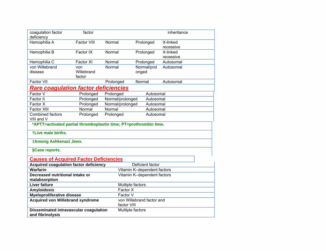
coagulation factor deficiency
factor inheritance
Hemophilia A Factor VIII Normal Prolonged X-linked recessive
Hemophilia B Factor IX Normal Prolonged X-linked recessive
Hemophilia C Factor XI Normal Prolonged Autosomal von Willebrand disease
von Willebrand factor
Normal Normal/prolonged
Autosomal
Factor VII Prolonged Normal Autosomal Rare coagulation factor deficiencies
Factor V Prolonged Prolonged Autosomal Factor II Prolonged Normal/prolonged Autosomal Factor X Prolonged Normal/prolonged Autosomal Factor XIII Normal Normal Autosomal Combined factors VIII and V
Prolonged Prolonged Autosomal
*APTT=activated partial thromboplastin time; PT=prothrombin time.
†Live male births.
‡Among Ashkenazi Jews.
§Case reports.
Causes of Acquired Factor Deficiencies Acquired coagulation factor deficiency Deficient factor Warfarin Vitamin K–dependent factors Decreased nutritional intake or malabsorption
Vitamin K–dependent factors
Liver failure Multiple factors Amyloidosis Factor X Myeloproliferative disease Factor V Acquired von Willebrand syndrome von Willebrand factor and
factor VIII
Disseminated intravascular coagulation and fibrinolysis
Multiple factors