Embed Size (px)
Citation preview

1
Structure and function of the respiratory musclesAuthors: N.G. Koulouris, I. DimitroulisKeywords: Histology, fatigue of respiratory muscles, anatomy, mechanical action, physiology
SUMMARY. The respiratory system consists essentially of two parts, a gas-exchanging organ, the lung, and a pump to pump gas in and out the gas exchanging part, consisting of the respiratory muscles and the chest wall. The lung and its diseases have traditionally been the focal point of interest, whereas pump disorders have received comparatively little attention. Research on respiratory muscles has accelerated during the last two decades. Respiratory muscles are all skeletal muscles having similar fibre composition to the limb muscles. Fibre composition of respiratory muscles is an important factor for their endurance and contractile properties. There are two fibre types, the fast (FT) and slow (ST) twitch fibres. Of the FT fibres two subgroups named FOG and FG have been identified. Human intercostals muscles appear to have mostly ST fibres. The diaphragm has a high percentage of fatigue-resistant fibres. The main respiratory muscles are the diaphragm, intercostal muscles and muscles of the abdominal wall. The accessory muscles of respiration include the sternomastoid and other muscles of the neck, back and shoulder girdle. The intercostals muscles are subdivided into two groups: the external and internal intercostals. The contraction of the diaphragm which is the most important respiratory muscle, decreases the intrathoracic pressure and increases the abdominal pressure in normal man by lowering the diaphragmatic dome. Intercostal muscles move the rib cage and can be inspiratory or expiratory. The scaleni is now believed to be true muscles of inspiration and not "accessory". The most important accessory muscles of inspiration are probably the sternocleidomastoid muscles. The respiratory muscles are the motive power for breathing and are subject to weakness from a variety of processes that affect the motor nerves, neuromuscular junction and muscle cell. Chronic neuromuscular disorders result in altered lung volumes. The effectiveness of cough is reduced in expiratory muscle weakness. Patients with respiratory muscle weakness breath faster and with a smaller tidal volume compared to healthy subjects. The main change in blood gases in patients with respiratory muscle weakness is usually a fall in PaO2. Hypercapnia may be a late event. Muscle fatigue, which is a reversible event, can be defined as the inability to sustain the required or expected force with continued contractions. The respiratory muscles, particularly those of inspiration, can fatigue and precipitate or intensify ventilatory failure. Pneumon 2001, 14 (2): 91-108
INTRODUCTION
Breathing like most other movements of the body depends on muscular action. Around the 3rd century BC one of the first known observers of this relationship, Erasistratus of Chios, who sometimes has been called the "Father of Physiology", believed the diaphragm to be the only muscle of breathing1-2. A few centuries later the remarkable Galen realized that not only the diaphragm but also intercostals and various accessory muscles were involved3. Until the Renaissance no advances were made, but for Leonardo da Vinci in Italy who made the analogy between the action of breathing and that of a pair of bellows.
Later in the 16th century, Vesalious the great Belgian anatomist, observed the movements of the lungs in the living dog by creating a pleural window with careful dissection4. During the 17th century in England, John Mayow wrote a clear description of the breathing mechanism5. In the
2
18th century two names stand out; G.E. Hamberger6, who presented a geometric model of the action of the intercostal muscles and Albrecht von Haller. Hamberger spent much of his efforts disputing with Haller mainly over the action of the internal intercostals. Controversies to the detailed action of the respiratory muscles, especially of the intercostals, continued throughout the 19th century. Guillaume Duchenne in 1846 studied muscle action by electrical stimulation in both animals and humans7. By this means he did much to clarify the action of the respiratory muscles, especially of the diaphragm. Since then these muscles have been increasingly neglected lying in a gray area, between anatomy and physiology. The description of their function in most textbooks of physiology and anatomy published in the last decade contain very little information and in some of them apologetically in small print8-10. The respiratory system consists essentially of two parts, a gas-exchanging organ, the lung, and a pump to pump gas in and out the gas exchanging part, consisting of the respiratory muscles and chest wall (figure 1). The lung and its diseases have traditionally been the focal point of interest, whereas pump disorders have received comparatively little attention. This is rather like a cardiologist who ignores the heart. It is surprising that this has happened, particularly because the ancient Greeks regarded the diaphragm as the seat of that part of the soul (phrenes) that is connected with emotions, desires and sensations, both pleasant and painful. However the respiratory muscles function as a pump, as vital as the heart. Physiologists have recently devoted more attention to the mechanics of breathing and the relevance of the respiratory muscles has become increasingly apparent. For this reason, research on respiratory muscles has accelerated during the last two decades.
Histology of the Respiratory Muscles
Respiratory muscles are all skeletal muscles having similar fibre composition to the limb muscles. Fibre composition of respiratory muscles is an important factor for their endurance and contractile properties (Table 1).

3
Fibre Composition
The classical distinction between red and white muscles12 was based on fibre darkness that is related to their content of myoglobin and mitochondria but a classification of fibres on this basis has limitations. The myosin ATP-ase reaction which has been widely accepted, separates muscle fibres into two groups13. Those staining dark have been shown to possess the largest myosin and actomyosin ATP-ase activities and furthermore to be the fastest contracting as shown in several human experiments. Thus, the two fibre types have been called fast (FT) and slow (ST) twitch fibres, respectively. Of the FT fibres, two subgroups named FOG and FG have been identified14. With few exceptions, ST and FT fibres are evenly represented in the muscles, however, with a large inter-individual variation15.
While ST fibres and one group of FT (FR=FOG) show little fatigue with repetitive stimulation, the tension developed by the other type of FT (FF=FR) fibres is almost eliminated after 3,000

4
contractions16. Histochemical and biochemical determinations of oxidative and glycolytic capacity have shown that the ST fibres have the higher capacity for aerobic metabolism and have more capillaries per fibre. With subdivision of the FT fibres in two groups, the FOG fibres appear to have the higher oxidative and lower glycolytic potential and to be surrounded by more capillaries. Athletes in endurance events tend to have a predominance of ST (SO) fibres and weight-lifters and sprinters to have a predominance of FT fibres. Indirect evidence using partially neuromuscular blocked muscles suggest that FT fibres have the larger electromyographic activity and also the higher mean power frequency when developing a given tension17. Human intercostal muscles appear to have approximately 60% ST fibres. In the external intercostal muscles, the number of capillaries and occurrence of FF fibres is similar to the other muscles. In contrast, the internal intercostal muscles placed in the mid-axilary line have no FF fibres and relatively many capillaries. Thus, these expiratory muscles appear to be extensively used. The diaphragms of most mammals, including humans, are composed of all three fibre types. The diaphragm has a high percentage of fatigue-resistant muscle fibres and most mammalian diaphragms are composed of approximately 60% ST fibres. For a given species, the diaphragm has a greater oxidative capacity and larger blood flow than those of limb muscles and is more resistant to fatigue. The contractile properties of the diaphragm are predictable from the muscle fibre composition. Adaptation to training and detraining, as has been observed in greater detail in limb muscles, also occurs in muscle fibres of the diaphragm18.
Anatomy of the Respiratory Muscles
The main respiratory muscles are the diaphragm, intercostal muscles and muscles of the abdominal wall. The accessory muscles of respiration include the sternomastoid and other muscles of the neck, back and shoulder girdle19.
THE DIAPHRAGM
Anatomy of the diaphragm
The diaphragm is a structural feature of the mammals9 and anatomically is a complicated muscle (figure 2). The main part dome-shaped with a large central fi brous tendon around which are arranged muscles of several fibre groups. These muscular fibres of the diaphragm are grouped into three parts: (i) the vertebral, (ii) the costal and (iii) the sternal. The vertebral fibres arise from the second and third lumbar vertebrae, from the medial arcuate ligaments (psoas) and from the lateral arcuate ligaments (quadratus lumborium). The costal fibres arise from the side and the upper margin of the lower six ribs, interdigitate with those of the transversus abdominis. The sternal fibres arise from the back of the xiphoid process. All the fibres converge on the central tendon. The costal fibres which constitute the larger part of the diaphragm run directly upwards, parallel and "apposed" to the inner surface of the rib cage. This area of apposition (figure 1) represents up to one third of the surface area of the rib cage at end-expiration (FRC), but diminishes during inspiration20. Both the origins and insertions of these costal fibres are mobile, and potentially move with respiration. By contrast the crural part does not move with respiration.

5
Innervation of the diaphragm
The cervical neuromeres contributing the motor fibres to the phrenic nerves are C3, C4 and C5 in man21. The fibres coming from the upper segments innervate mainly the ventromedial part, those coming from the lower segments innervate the dorso-lateral part (table 2). The diaphragm however contracts as a unit22.
Afferent fibres
The afferent innervation of the diaphragm is supplied via the phrenic nerves except for the

6
marginal part which is supplied by nerves coming from the T6 to T12 and most of the crura which are supplied by T1221-23. The diaphragm contains relatively fewer sensory end-organs, and there appear to be more tendon organs than muscle spindles24. There are, however, afferent fibres in the phrenic nerve of the cat and afferent traffic has been recorded. The paucity of end-organs may reflect the tendency of the diaphragm to contract uniformaly so that relatively few sensory endings are necessary to sample accurately its function25.
Blood supply to the diaphragm
The diaphragm receives arterial blood from three major sources: (i) the internal mammary, (ii) the intercostal, and (iii) the inferior phrenic arteries. The superior phrenic, a small artery that lies against the phrenic nerve, also provides a modest portion of the arterial supply. Recently Comptois and colleagues have shown that there is a rich network of anastomotic connections between these vessels26. These authors found that the internal mammary and phrenic arteries have head-to-head anastomoses, forming an internal arterial circle around the central tendon of the diaphragm. Collateral branches from this circle travel between diaphragm fibres, joining with branches of the intercostal arteries to form costophrenic arcades. Thus larger arteries run perpendicularly to the muscle fibres, and arterioles again run at right angles to the muscle fibres. The small muscular arteries form anastomotic loops, except in the costal portion of the diaphragm. The arrangement of large and intermediate size veins is similar to the arterial pattern27. The anatomical arrangement of the vessels appears to prevent kinking of the blood vessels during diaphragmatic contraction. It has been found that the configuration and branching of the small arteries, arterioles and capillaries of the diaphragm were identical to that of two other skeletal muscles, namely the triceps and intercostals28.
Lymphatic drainage of the diaphragm
The diaphragm is provided with an extensive system of lymphatic vessels that not only remove interstitial fluids and cells from the diaphragm, but also play a major role in the removal of fluids and cells from both pleural and peritoneal cavities. These serous cavities are continuous with the lymphatic vessels of the diaphragm via open pores (stomata) that exist on its surface. This organization of lymphatic vessels provides a system of open channels through which fluids and cells may be rapidly removed from the serous cavities. Once fluids and cells are within the lymphatic vessels, contraction of the diaphragmatic muscle fibres would also cause contraction of the lymphatic collecting vessels, compressing their walls and thereby causing the contents to be expelled toward the larger collecting vessels29.
Intercostal muscles
They are subdivided into two groups: (i) the external and (ii) internal intercostals (figure 3).

7
ANATOMY
The external intercostals extend from the tubercles of the ribs to the costochondral junction where they become continuous to the anterior intercostal membrane. They are thicker posteriorly than anteriorly30 and thicker than the internal intercostals31. Their fibres slope obliquely downwards and forwards from the upper rib to the one below. The internal intercostals extend from the anterior end of the intercostal space to the angles of the ribs posteriorly, where they become continuous with the posterior intercostal membrane. They are thicker anteriorly than posteriorly. The fibres slope obliquely downwards and backwards. The internal intercostals can be subdivided into a posterior or interosseous portion where the ribs slope downwards and forwards and an anterior or intercartilaginous portion (the parasternals) where the costal cartilages slope upwards and forwards.
INNERVATION OF THE INTERCOSTALS MUSCLES
The intercostal nerves arise from the first to the eleventh thoracic segments, and are derived from the ventral primary ramus (table 2). In man, each main intercostal nerve, which supplies filaments to the intercostal muscles, lies deep to the internal intercostal muscle, giving off a collateral branch early in its course, a lateral cutaneous branch and a terminal anterior cutaneous branch. The lower intercostal nerves, which supply the abdominal muscles after penetrating the diaphragm (to which they give a few sensory branches), freely communicate with one another over the abdominal wall32. The intercostal muscles are well supplied with proprioceptors. Histological studies have demonstrated muscle spindles, tendon organs and Pacinian corpuscles33.
Blood supply to the intercostal muscles

8
The intercostal muscles are supplied by intercostal arteries and branches of the internal mammary arteries, and drained by intercostal veins and branches of internal mammary veins. The intercostal veins drain into the azygos and hemizygous system. As these systems are connected, blood in the azygos vein, which enters the superior vena cava on the right side, represents drainage from the intercostal muscles on both sides.
Abdominal muscles
The abdominal cavity is bounded by anterior and posterior longitudinal muscles, which connect the thoracic cage to the pelvis and are contained within strong fascial sheaths, together with three muscle sheets which encircle the remainder of the abdomen (figure 3).
Anatomy of the abdominal muscles
External oblique. This muscle arises from the outer surfaces of the lower eight ribs. It is therefore superficial to the intercostal muscles in the lower spaces. The dorsal fibres pass downwards to the iliac crest. The rest of muscle slopes obliquely downwards and forwards, and passes into a fibrous aponeurosis which forms part of the rectus sheath and fuses with its fellow from the other side in the linea alba. The lower border of the aponeurosis forms the inguinal ligament. Internal oblique. The internal oblique muscle passes from the lumbar fascia, iliac crest, and lateral part of the inguinal ligament to an extensive attachment along the costal margin to an aponeurosis contributing to the rectus sheath down to the pubis. Transversus abdominis. The transversus abdominis muscle arises from the costal cartilages of the lower six ribs, the lumbar fascia, the iliac crest and the lateral part of the inguinal ligament. The main part of the muscle passes horizontally forwards into an aponeurosis similar in extent to that of the external oblique. The transversus abdominis is the most deep of these muscles. Rectus abdominis. This covers the external surface of the thoracic cage as it passes vertically downwards from its horizontal line of attachment to the fifth, sixth, and seventh costal cartilages (occasionally the third and the fourth) to its tapering tendinous attachment to the pubis. The rectus sheath enclose much of the muscle and is derived from aponeurosis of the three lateral muscles34.
Innervation of the abdominal muscles
The external oblique and the rectus abdominis are supplied by the lower 5 intercostal nerves (T7-11), the internal oblique and the transversus by the lower 5 intercostals, the subcostal (T12), the iliohypogastric (L1) and the ilioinguinal (L1) nerves (table 2).
Blood supply to the abdominal muscles
Much less information is known about the arterial supply and venous drainage of the abdominal as well as for the rest of the respiratory muscles.
Accessory muscles
Of all the so-called accessory muscles of respiration, only the sternomastoids, scaleni and
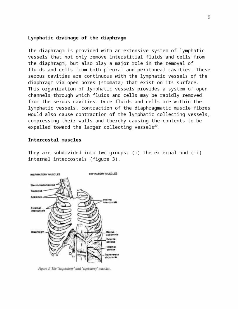
9
triangularis sterni show significant respiratory activity in man35,36.
The sternomastoids
The sternomastoid arises by two heads from themanubrium sternum and the medial part of the clavicle (figure 3). The fibres of these two heads fuse into a single one which is inserted into the mastoid process and the occipital bone. The muscle is supplied by the spinal accessory and second cervical nerves.
The scaleni
The scaleni arise from the transverse process of the lower five cervical vertebrae and pass downwards to be inserted into the upper surface of the first rib (scalenus anterior and medius) and second rib (scalenus posterior). The scalenus medius is the largest. They are supplied by the lower five cervical nerves (figure 3).
The triangularis sterni
The triangularis sterni fibres originate from the dorsal aspect of the caudal half of the sternum and insert into the inner surface of the costal cartilages of the 3rd to 7th rib. Its motor supply of the muscle comes from the intercostal nerves. It is a flat muscle that lies deep to the sternum and the parasternal intercostals36.
Other muscles
There are many other muscles that may participate in the respiratory act (figure 3). These are trapezius, pectoralis major, pectoralis minor, subclavius latissimus dorsi, serratus anterior, serratus posterior superior, serratus posterior inferior, quadratus lumborium and saccrospinalis. Most of these muscles are unlikely, on anatomical grounds, to be of importance in the mechanics of breathing8.
MECHANICAL ACTION OF THE RESPIRATORY MUSCLES
Action of the diaphragm
The contraction of the diaphragm decreases the intrathoracic pressure, and increases the abdominal pressure in normal man. It seems obvious that the lowering of the diaphragmatic dome generates these pressures. On the other hand it is difficult to assess whether these effects are facilitated or hindered by the direct action of the diaphragm on the ribs8. This problem is a classical cause of arguments. Galen3 considered that the diaphragm expands the rib cage, but this view was challenged by Borelli in 1680 who reasoned that the diaphragm constricts it. Duchenne in 1867 showed that the contraction of the diaphragm expands the rib cage provided that the normal relationships of the diaphragm are preserved and the abdomen is closed7. Experiments in dogs have shown that the costal and crural portions of the diaphragm have different actions on the rib cage but the details of segmental innervation and regional activation of the human diaphragm are not well known37. The shape of the normal human diaphragm at rest is an

10
elliptical cylindroid capped by a dome. The cylindrical portion of the diaphragm apposed to the inner aspect of the lower rib cage and constitutes the "zone of apposition" of the diaphragm to the rib cage20,38. The zone of apposition has important implications for diaphragmatic function because when the diaphragm contracts, increased tension within the diaphragmatic muscle fibres causes the diaphragmatic dome to move caudal relative to its insertions. The fibres are directed axially in the direction of movement of the dome so there is a piston-like axial displacement of the diaphragmatic dome39,40. At lung volumes below TLC in normal humans, the piston analogy probably holds, and the transdiaphragmatic pressure generated by the contracting diaphragm is a function principally of the neural activation of the diaphragm and the force-length and velocity force relationships of diaphragmatic muscle. These principles appear true for humans39 and dogs40. When the diaphragm has shortened enough to eliminate the zone of apposition from part of the circumference of the rib cage, or when the apposed diaphragmatic fibres are not parallel to the thoracic axis, the piston analogy breaks down (figure 4).
Under these circumstances the diaphragm force reflects its radius of curvature and anisotropic tension within the diaphragmatic dome. When the diaphragm contracts, it expands the thoracic cavity and tends to displace the abdominal viscera, resulting in lower pleural pressure and higher abdominal pressure. The diaphragm also exerts a force at the site of its attachment to the rib cage, a force directed cranially. The fall in pleural pressure has an expiratory effect on the upper rib cage. Inward displacement of the upper rib cage is seen during diaphragmatic inspiration in tetraplegic subjects41-43 and in subjects under spinal anaesthesia extending to T144. The diaphragmatic contraction has two inspiratory effects on the lower rib cage (figure 4). The first of these has been termed the "appositional" component of the diaphragm's inspiratory action on the rib cage20,37,45 and results from the increase in abdominal pressure acting on the lower rib cage. The second inspiratory effect has been termed the "insertional" component of the diaphragm' s action on the rib cage. This force is in the direction of its fibres, namely, towards the head. This force is inspiratory to the ribs, causing them to rotate up and out (figure 5).

11
Lung volume and diaphragmatic function The length of diaphragmatic muscle fibres is closely related to lung volume38 and therefore a change in lung volume is accompanied by a change in the operating length and hence the force of the diaphragm38. The action of the diaphragm on the rib cage is also strongly dependent on lung volume. With increasing lung volume, the fraction of

12
the rib cage exposed to pleural pressure increases as the fraction exposed to abdominal pressure decreases20,38 reducing the diaphragm's inspiratory action on the rib cage and increasing its expiratory action through pleural pressure45. At very high volumes, the zone of apposition disappears, and the fibres of the diaphragm can pull inward on their insertions, causing a direct expiratory action on the lower rib cage (Hoover's sign). These effects have been demonstrated in animals37,46,47.
Mechanical action of the rib cage muscles
To understand the action of particular muscles of the rib cage, we must understand the movement and articulation of ribs (figure 5). The ribs move around axes defined by their articulation with the vertebral bodies and the transverse processes48,49. The upper ribs rotate so that their anterior parts move cephalad and ventrally during inspiration, the so-called "pump-handle" motion of the ribs. The lower ribs have a prominent lateral as well as ventral movement during inspiration, the so-called "bucket-handle" motion. In the relaxed adult human, the ribs are angled down (Caudad) so that during inspiration, the lower and intermediate ribs move outward, away from the spine, providing an effective ventral and lateral expansion of the rib cage48,49. It is now well established by a variety of studies that intercostal muscles move the rib cage. Patients who have undergone destruction of intercostal nerves on one side of the thorax for treatment of pulmonary tuberculosis, show less respiratory movement of the ribs on the effected side50; in hemiplegic patients, outward displacement of the rib is reduced on the paralysed side51. In supine subjects with complete diaphragmatic paralysis, rib cage displacements are exaggerated and abdominal wall displacements are paradoxical when the abdominal muscles remain relaxed10,52,53. The function of the intercostal muscles has been a subject of controversy throughout medical history54. It is now commonly considered that external intercostal muscles and the interchondral part of the internal intercostal muscles (the parasternal muscles) are inspiratory and serve to raise the ribs, whereas the interosseous part of the internal intercostals are expiratory and act to lower the ribs.
Mechanical action of the abdominal muscles
The abdominal muscles have a number of functions: as rotators or flexors of the trunk, postural and respiratory functions. As respiratory muscles have expiratory functions as well as inspiratory ones. As expiratory muscles act in two ways: a) they pull the abdominal wall inward and produce an increase in introabdominal pressure. Because the abdominal contents are incompressible, this causes the diaphragm to move cranially into the thoracic activity. This contraction results in an increase in pleural pressure and a decrease in lung volume. Therefore the abdominal muscles are considered to be powerful expiratory muscles assisting in such activities as forced expiration and coughing. The other function of the abdominal muscles in relation to breathing is to displace the rib cage, acting to pull the lower ribs down and inward, again an expiratory action55. They assist inspiration in two ways: a) through a direct facilitation of diaphragmatic action by persistent abdominal contraction, as it occurs when normal humans adopt the standing position. Their activity is tonic, unrelated to phases of respiration and it is greatest in the dependent regions of the abdomen25,55,56 and b) through a second mechanism by which the abdominal muscles can assist inspiration is by contracting in phase with expiration. By contracting during expiration and forcing the diaphragm cranially into the thoracic cavity, these muscles can reduce lung volume

13
below the neutral position of the respiratory system. Hence, when they relax at end-expiration, they promote passive descent of the diaphragm, therefore lung volume can increase before the onset of inspiratory muscle contraction. Thus, great activity of the abdominal muscles appears when the ventilation reaches 70-90 l/min36,57,58.
Mechanical action of the accessory muscles
Many of theses muscles have prominently non-respiratory functions and many are relatively small or inaccessible. As a result, they have not been extensively studied by respiratory physiologists.
Scalene muscles
The importance of the scaleni as muscles of inspiration is disputed but it is now believed that they are true muscles of inspiration and should probably not be called "accessory"59,60. Studies using needle electrodes show that the scalenes are active during quiet breathing in upright and supine postures. Others classify them with the sternomastoid as accessory muscles. By their origins and insertions, these muscles must elevate the first two ribs, and therefore, they may be inspiratory but their exact mode of action is not well known, because these muscles rarely if ever act individually.
Sternocleidomastoid muscles
It has been suggested that the human sternocleidomastoid muscles have a predominantly "pump-handle" action on the rib cage, elevating the first rib and sternum and allowing the resultant decrease in transthoracic pressure to cause inward displacement of the lateral rib cage and abdomen41. The sternomastoids are probably the most important accessory muscles of inspiration and their participation in breathing with dyspnoea is a well known clinical observation.
The triangularis sterni
Most normal subjects when breathing at rest in the supine posture do not activate this muscle unlike cats and dogs61. It always contracts during vigorous expiratory efforts such as coughing, laughing and during expiration below FRC61,62. During such manoeuvres, the triangularis sterni acts to lower the ribs and increase pleural pressures.
Other muscles
There are other muscles whose origins and insertions suggest that they may have a respiratory function under the right conditions. In particular the trapezii and the platysma as well as some laryngeal muscles contract during inspiration. Despite that they have been considered unimportant to the breathing in normal man8,63.
PHYSIOLOGY OF THE RESPIRATORY MUSCLES
The respiratory muscles provide the motive power for breathing. Despite their central role in

14
ventilation their physiology has been relatively neglected, perhaps partly because of the complexity of their function, and the difficulties of studying them64. Statics and dynamics of contraction of these muscles are difficult to study in VIVO because force, initial length, velocity of shortening and magnitude of the neural drive of individual muscles are not measurable without invasive methods11. Understanding of the function of the respiratory muscles depends on the relationships of frequency-force, length-tension, force-velocity and fatigability-frequency.
Frequency-pressure relationship
The force developed by a skeletal muscle is a function of the frequency of stimulation (figure 6). The frequency-force relationships result from the summation of twitch tension during repeated stimulation. This relationship is useful in assessing force development by different muscles and for the evaluation of high and low frequency fatigue by the same muscle.
Since respiratory muscles are inaccessible for the measurement of force directly in vivo, their force is measured indirectly as measurement of the pressures generated by them. The frequency-force (pressure) curve for the respiratory muscles is similar to those of other human skeletal muscles. Using the technique of percutaneous stimulation over the motor point of the sternomastoid muscle, it is possible to describe the function of the sternomastoid in the same terms as those used to describe the function of limb skeletal muscles in humans. Similar frequency-force curves were recorded from a strain gauge applied to the mastoid process using a force transducer65. The recording of Pdi in response to electrical stimulation of the right phrenic nerve allows a similar myogram to be obtained from the diaphragm66. When the diaphragm becomes fatigued, its frequency-pressure curve is depressed at all stimulation frequencies.

15
Force-length relationship
The force-length relationship indicates that when a muscle is stimulated at its optimal resting length, it produces its maximum contractile force (figure 7). When the muscle is either stretched beyond the optimal resting length or alternatively is foreshortened prior to contraction, supramaximal stimulation of the muscle produces submaximal force. In the case of the diaphragm, there is little or no evidence for compromise of its contractile force by over stretching, but at lung volumes above normal FRC, the diaphragm and other inspiratory muscles are foreshortened and their contractile force is curtailed. In contrast, expiratory muscle contraction force is curtailed at low lung volumes39,67-73.

16
Force-velocity relationships
For any muscle length, the maximum contractile force is greatest when the muscle is prevented from shortening (figure 8). If the muscle is allowed to shorten during its contraction, its contractile force declines hyperbolically as a function of the velocity with which the muscle shortens. This is termed the "force-velocity relationship"75-76. When both the tension and velocity are normalised to the maximum value, slow muscles show greater curvature than fast muscles. This implies that type I (ST) fibres generate less power output than type II ( FT ). The force-velocity relationship of the diaphragm muscle is intermediate between those of slow (type I) and those fast (type II) skeletal muscles77.
RESPIRATORY MUSCLE WEAKNESS
The respiratory muscles are subject to weakness from a variety of processes which affect the motor nerves, neuromuscular junction and muscle cell per se (table 3).

17
Effect of respiratory muscle weakness on lung volumes
Weakness of these muscles reduces the capacity to generate the negative intrathoracic pressure to expand the lungs with a reduction in total lung capacity (TLC) and a parallel fall in vital capacity (VC). Weakness of the muscles of expiration, principally the abdominal musculature and the internal intercostals, reduces the capacity to generate positive intrathoracic pressures. This weakness reduces expiratory reserve volume or high, increases residual volume, and further reduces vital capacity. In these patients the FEV1/FVC ratio is normal, illustrating a pure restrictive ventilatory defect and the residual volume (RV)/total lung capacity (TLC) ratio is characteristically high. Gas transfer corrected for the reduced lung volume (KCO) is normal or high, and a low KCO implies that muscle weakness is unlikely to be the sole explanation of a respiratory problem79.
Effects of respiratory muscle weakness on lung mechanics
Moderate weakness of the inspiratory muscles prevents lung recoil pressure (Pst, L) from being developed at full inflation and therefore truncates the upper part of the static pressure-volume (PV) curve of the lung. However in patients with long-standing and severe respiratory muscle weakness pulmonary compliance is reduced also80-82, indicating that in these patients the elastic properties of the lung are altered. The cause of this reduced lung distensibility is not clear. Three

18
factors can theoretically affect lung compliance: (i) dispersed alveolar collapse, undetectable by radiographic techniques83, (ii) generalised increase in the surface tension of the alveolar lining layer84 and (iii) shortening and stiffening of the elastic fibres within the lungs.
Chest wall mechanics in respiratory muscle weakness
Several pathogenic mechanisms are involved in malfunction of the chest wall in patients with neuromuscular disorders. As noted before, FRC is frequently decreased in these patients. It has been suggested that the fall in FRC in these conditions is caused primarily by a decrease in the outward pull of the chest wall. Several studies have shown that chest wall compliance is decreased to about two-thirds of normal values in patients with long-standing neuromuscular disorders. These measurements apply to the entire chest wall, but it seems reasonable to speculate that these changes are primarily due to altered stiffness of the rib cage85,86. Further contributing factors can be the development of scoliosis, particularly, in patients with muscular dystrophy, and fibrotic changes and spasticity in the rib cage muscles, as it occurs in patients with tetraplegia. In conclusion, the alterations in lung volumes seen in patients with chronic neuromuscular disorders are attributable to a combination of muscle weakness and alterations of the mechanical propertles of the lungs and chest wall87.
Cough impairment-airway function
The effectiveness of cough is reduced in expiratory muscle weakness because the cough induces dynamic compression, affecting the linear velocity of airflow through the large intrathoracic airways. As a result, cough and clearance of secretions is defective in these patients, contributing to the high prevalence of bronchopulmonary infections. Respiratory muscle weakness would be expected to have larger effects on maximum inspiratory rates for two reasons: a) these flow rates depend on the ability of the inspiratory muscles to lower pleural pressure and b) a reduction in lung distensibility opposes inspiratory flow. Very few studies have been done on maximum inspiratory flow-rates in respiratory weakness88,89. Similarly, it might be expected that maximum voluntary ventilation (MVV) measured over 15 sec would be reduced disproportionally to changes in FEV1 in respiratory muscle weakness, but only a small trend in this direction has been found in myasthenia gravis90 and in myotonic dystrophy91.
Ventilatory drive
Patients with respiratory muscle weakness breath faster and with a smaller tidal volume than healthy subjects82,92,93. This tachypnoea may be related to the diffuse microatelectasis, reduction of lung compliance or different signals from the weakened muscles themselves92.
Alterations in the central control mechanisms of respiration have been repeatedly suggested in a number of patients with neuromuscular disorders94-98. However reductions in the ventilatory responses can be accepted as evidence of damaged medullary respiratory centres only if there are no accompanying abnormalities of lower motor neuron respiratory muscles or lung mechanics.
Ventilation and blood gases

19
The main change in blood gases in patients with respiratory muscle weakness is usually a fall in arterial PO2
99. Hypoxaemia without an elevated PaCO2 has been reported in patients with acute poliomyelitis during respiratory treatment100 and in a number of patients with other neuromuscular disorders82,101,102. In these cases decreased PaO2 coexists with an increase in alveolar-to-arterial tension difference for oxygen (A-a) PO2. Initially the tachypnea causes an increase in alveolar ventilation, resulting in alveolar and arterial hypocapnia99,101. Persistent hypercapnia may be a late and dramatic event and may occur only at a terminal stage of the disease, as in Duchenne's muscular dystrophy. However, hypercapnia may occasionally appear relatively early in the course of some chronic neuromuscular disorders, such as in limb-girdle dystrophy or in myotonic dystrophy.
RESPIRATORY MUSCLE FATIGUE
The question whether respiratory muscle fatigue causes respiratory failure is over 60 years old but we still have no definitive answer to this question103.
Physiological classification of fatigue
Muscle fatigue can be defined as the inability to sustain the required or expected force with continued contractions. When exercise ceases or it's intensity is reduced, the muscle will recover. Recovery from some forms of peripheral muscle fatigue is complete within seconds, but may be gradual with full recovery taking hours104. The command chain for voluntary muscular activity involves many steps and force failure i.e. fatigue - can occur as a result of impairment at any one or more links in the chain of command. As a simple practical analysis it is worth separating central from peripheral fatigue105. In the history of human muscle fatigue it was popular in the early years to attribute fatigue to failure of central neural processes. Comparisons between forces generated by maximum stimulated contractions and forces by maximum voluntary contraction at different stages of the experiment allowed central fatigue to be assessed106. The importance of peripheral fatigue was first clearly demonstrated by Merton (1954). He showed that force generation was impaired with twitches of the abductor pollicis muscle provided by supramaximal stimulation of the ulnar nerve at the wrist as a result of a sustained maximum voluntary contraction (MVC), during which the muscle became fatigued such that maximum force generation was less than 20% of the force obtained with an MVC with unfatigued muscles105.
Causes of peripheral muscle fatique
Several experimental models have been used to study fatigue: voluntary activation of the muscles, force output or electrical stimulation to determine their contractile properties. There are many possible sites and mechanisms with which fatigue may occur in a peripheral muscle. Changes in membrane electrical characteristics from efflux of potassium ions107,108, or increased cellular water content during exercise, may effect propagation of the action potential along the muscle membrane and the T-tubule, leading to a reduction in activation and therefore force generation. The excitation-contraction coupling process may be impaired by changes in the amount of calcium stored in or released from the sarcoplasmic reticulum109. Cossbridge formation can be affected and it may reduce force generation in several ways i.e. the sensitivity

20
of troponin for calcium may be reduced110 as may be the rate111 and power output112 for each cossbridge cycle. It is often thought that decreased intracellular pH is the cause of muscle fatigue but such relation is not clear. Whilst any increased hydrogen ion concentration reduces isometric force generation in vitro113 reductions in strength in excess of 50% can occur with low-intensity exercise in which no lactic acid is produced and pH is unchanged106. Patients suffering from McArdle's syndrome (myophorylase deficiency) can fatigue with no lactic acid production114. Other metabolic changes i.e. an increase in ADP in combination with a decrease in pH might plays key role in the development of fatigue as these changes have been associated with reductions in force. Furthermore as pH falls the concentrations of phosphocreatinine (Pcr) falls, adenosine diphosphate (ADP), adenosine monophosphate (AMP) and inorganic phosphate (Pi) in diprotonated form increase, the latter has been found to be directly related to force changes115. It has recently been proposed that the metabolic determinants of fatigue as well as the recovery from it are related to the nature of exercise used to induce it25,116. When work is sustained for 1-2 h, the point of exhaustion is related with the depletion of the glycogen stores of the working muscle117. During heavy exercise, large amounts of energy are converted to heat and the subsequent rise in body temperature and fluid loss may impair performance and enhance central fatigue. Eccentric contractions (where the muscle is forcibly extended during activation) produce more profound and longlasting fatigue despite having low metabolic cost118,119.
Respiratory muscle fatigue
By analogy with limb muscles, fatigue could develop in the respiratory muscles and contribute to hypercapnic ventilatory failure103. Patients with severe lung disease and hyperinflation have reduced force-generating capacity of the respiratory muscles. In contrast their ventilatory requirements are increased. These muscles, particularly the inspiratory are subject to large loads with every breath for prolonged periods with little opportunity to rest64. In these circumstances, it appears likely that the respiratory muscles, particularly those of inspiration, can fatigue and precipitate or intensify ventilatory failure66,103.
REFERENCES
1. Koulouris N. Strength and fatigue of the respiratory muscles in man. PhD thesis, University of London, 1989.2. Franklin, K J.: A short history of Physiology (2nd ed). London: Staples, 1949. 3. May M. T.: Galen on the Usefulness of the parts of the body: (De usu partium) Ithaca, N Y. Cornell Univ. Press, 1968.4. Foster M.: Lectures on the history of physiology London: Cambridge Univ. Press, 1901. 5. Mayow J.: Medico-Physical Works (Translation of Tractatus Quiinque Medico-Physici. 1674). Edinburgh: Alenbic Club, 1907.6. Hamberger, G.E.: Respirationis Mechanismo et Usu Genuino Dissertatio, Jena, Germany: Groeker, 1748. 7. Duchenne G.B.: Physiology of Motion. Translated by E.B. Kaplan. N.B. Saunders, London, 1959 pp 443-503. 8. Cambell E.J.M.: In: The respiratory muscles: Mechanics and neural control 2nd edition. Lloyd-Luke, London, 1970. 9. Agostoni E., Sant' Ambrogio G.: The diaphragm, In: The respiratory muscles, mechanics and

21
neural control. 2nd eds. LLoyd-Luke, 1970; pp 145-160. 10. Newsom-Davies J., Stagg D., Loh L., Casson M.: The effects of respiratory muscle weakness on some features of the breathing pattern. Clin. Sci. 1976;50:10p-11p. 11. Lockhart A.: A layman's view on respiratory muscles. Bull. Eur. Physiopathol Respir. 1984;20:395- 397. 12. Ranvier M L.: Proprietιs et structures differentes des muscles rouges et des muscles blancs, chez les Lapins et chex les Raies C R Acad Sci (Paris), 1873;77:1030- 1034. 13. Padikyla M.A, Herman E.: The specificity of the histochemical method of adenosine triphosphatase. J Histochem Cytochem 1955;3:170-195. 14. Brook M.H., Kaiser K.K.: Three myosin adenosine triphosphatase systems the nature of their PH liability and sulphydryl dependence. J Histochem Cytachem 1970; 18:670- 672. 15. Saltin B., Gollnick P.D.: Skeletal muscle adaptivity significance for metabolism and performance. In Handbook of Physiology, section 10: Skeletal muscle. L.D. Peachey et al. eds. American Physiological Society, Maryland 1983, pp 556-631. 16. Gamnet R.A.F., O'Donovan M.J, Stephens J.A.: Taylor A. Motor unit organisation of human medial gastrocnemious. J Physiol (Lond) 1979;287: 33-43. 17. Secher N.H., Koumi P.V.: Effect of tobucarine and dexamethonium on electromyographic activity during voluntary contraction in man. Acta Angesch Scand 1984. 18. Secher N.H., Mizuno M., Saltin B.: Adaptation of skeletal muscles to training. Bull Eur Physiopathol Respir 1984;20:453-457. 19. Green M., Moxham J.: Respiratory muscles: In recent advances in Respiratory Medicine. ed Churchill - Livingstone. London, 1983; pp 1-20. 20. Mead J.: Functional significance of the area of apposition of diaphragm to rib cage. Am Rev Respir Dis 1979; 119:31-32. 21. Hamilton W.S., Boyd J.D., Mossman H.W.: Human embryology, 3rd edit. Cambridge; Heffer; 1962. 22. Sant'Ambrogio G., Frazier D.T., Wilson M.F. and Agostoni E.: Motor innervation and pattern of activity of cat diaphragm. J Appl Physiol 1963; 48:43-46. 23. Rasmussen A.T.: The principal nervous pathways, New York: Macmillan 1952; p43. 24. Corda M., Von Euler C., Lennerstand G.: Proprioreceptive innervation of the diaphragm. Journal of Physiology (London), 1965; 178: 161-177. 25. Green M., Mead J., Sears T.A.: Muscle activity during chest wall restriction and positive pressure breathing in man. Respir Physiol 1978;35:283-300. 26. Comtois A., Gorczyca N., Grassino A.: Anatomy of diaphragmatic circulation. J Appl Physiol 1987; 62:238-244.27. Beck F. and Baxter J.S.: Some observations on diaphragmatic blood supply. J Anat 1960; 94: 224-230. 28. Schroufnagel D.E., Roussos C.H., Mackem P.T. and Wang, N.S.: The geometry of the microvascular bed of the diaphragm: Comparison of intercostals and triceps. Microvasc Res 1983;26:291-306. 29. Leek L.V. and Rahil K.: Permeability of the diaphragmatic mesothelium: The ultrastructural basis for "stomata". Am J Anat 1978; 151: 557. 30. Bryce T.H.: In Quain's Elements of Anatomy 11th edit. 1923;4 (pt 2) London: Longmans. Belman M.J. (ed). Clinics in Chest Medicine, 1988;9:175-193. 31. Johnston T.B. and Whillis: Gray's Anatomy, 30th edit. 1949; pp557-564. London: Longmans.

22
32. Davies F., Gladstone R.J. and Stibbe E.P.: The anatomy of the intercostal nerves. J Anat (London) 1932;66:323-333. 33. Barker D.: The structure and distribution of muscle receptors. In: Symposium on Muscle Receptors; 1962. 34. Osmond D.G.: Functional anatomy of the chest wall. In: The Thorax edt by Roussos C.H. and Macklem P.T. 1985; part A: pp 199-233, Marcel Dekker, Inc - New York. 35. Cambell E.J.M.: The mascular control of breathing in man (PhD Thesis, Univ of London), 1954. 36. De Troyer A., Estenne M.: Functional Anatomy of the Respiratory Muscles. In: Respiratory Muscles: Function in health and disease. Belman M.J. (ed). Clinics in Chest Medicine 1988;9:175-193. 37. De Troyer A., Sampsoll M., Singrist S. and Macklem P.T. Action of the costal and crural parts of the diaphragm on the rib cage in dog. J Appl Physiol 1982;53:30-39.38. Mead J. and Loring S.: Analysis of volume displacement and length of the diaphragm during breathing. J Appl Physiol 1982;53:750-755. 39. Braun N.M.T., Arora N.S., Rochester D.F.: Force length relationship of the normal human diaphragm. J Appl Physiol 1982;53:405-412. 40. Kim M.J., Druz W.S., Danon J., Machnach N., Sharp J.T.: Mechanics of the canine diaphragm. J Appl Physiol 1976;41:369-382. 41. Danon J., Druz W.S., Goldberg N.B., Sharp J.T.: Function of the isolated paced diaphragm and the cervical accessory muscles in C1 quadriplegics. Am Rev Respir Dis 1979;119:909-919. 42. Mortola J.P. and Sant' Ambrogio G.: Motion of the rib cage and the abdomen in tetraplegic patients. Clin Sci Med 1978;54:25-32. 43. Urmey W.F., Loring S.H., Mead J., Brown R. I Slutsky A.S., Sarkarati M., Rossier A.: Rib cage mechanics in quadriplegic subjects. Physiologist 1981;24:97 (abstract). 44. Eisele J., Trenchard D., Bruki N., Guz A.: The effect of chest wall block on respiratory sensation and control in man. Clin Sci 1968;35:23-33. 45. Loring S.H. and Mead J.: Action of the diaphragm on the rib cage inferred from a force-balance analysis. J Appl Physiol 1982b;53:756-760. 46. D' Angelo E., Sant'Ambrogio G.: Direct action of contracting diaphragm on the rib cage in rabbits and dogs. J. Appl. Physiol. 1974;36:715-719. 47. Sant' Ambrogio G., Saibene F.: Contractile properties of the diaphragm in some mammals. Respir Physiol 1970;10:349-359.48. Jordanoglou J.: Rib movement in health/kyphoscoliosis and ankylosing spondylitis. Thorax 1967;24:407-414. 49. Jordanoglou J.: Vector analysis of rib movement. Respir Physiol 1970;10:109-120. 50. Alexander J.: Multiple intercostal neurectomy of pulmonary tuberculosis. Am Rev Tuberc 1929;20:637-684. 51. Fluck D.C.: Chest movements in hemiplegia. Clin Sci 1966;31:383-388. 52. Kreitzer S.M., Feldman N.T., Sunders NeA., Ingram R.H.: Jr Bilateral diaphragmatic paralysis with hypercapnic respiratory failure: A physiological assessment. Am J Med 1978;65:89-95. 53. Skatrud J., Iber C., McHugh W., Rasmussen H., Nichols D.: Determinants of hypoventilation during wakefulness and sleep in diaphragmatic paralysis. Am Rev Respir Dis 1980;121:587-593.54. Cambell B.J.M., Newsom-Davis J.: The intercostal muscles and other muscles of rib cage. In: The respiratory muscles: mechanics and neural control. 2nd edition. Cambell E.J.M.,

23
Agostoni E., Newsom-Davis J. Eds. Lloyd-Luke Ltd, 1970; pp 161-174. 55. De Troyer A.: Mechanical role of the abdominal muscles in relation to posture. Respir Physiol 1983;53:341-353.56. De Troyer A.: Actions of the respiratory muscles or how the chest wall moves in upright man. Bull Eur Physiopathol Respir 1984;20:409-413.57. Cambell E.J.M. and Green J.H.:The variations in intra-abdominal pressure and the activity of the abdominal muscles during breathing and increased pulmonary ventilation. J Physiol (Lond.) 1953;122:282-290.58. Cambell E.J.M., Green J.H.: The behaviour of the abdominal muscles and the intra-abdominal pressure during quiet breathing and increased pulmonary ventilation. A study in man. J Physiol (Lond.) 1955;127:423-426. 59. Raper A.J., Thomson W.T.Jr., Shapiro N., Patterson J.L.J.: Scalene and sternomastoid muscle function. J Appl Physiol 1966;21:497-502. 60. De Troyer A., Estenne M.: Coordination between rib cage muscles and diaphragm during quiet breathing in humans. J Appl Physiol 1984;57:899-906. 61. De Troyer A., Ninane V., Gilmartin J.J.: Triangularis sterni muscle use during eupnea in humans: effect of posture. Respir Physiol 1988;74:151-162. 62. Ninane v., Decramer M., De Troyer A.: Coupling between triangularis sterni and parasternalis during breathing in dogs. J Appl Physiol 1986;61(2):539-544. 63. Delhez L., Petit J.M. Donnιes actuelles de l' Electromyographie respiratoire chez l' homme normal. Electromyography 1966;6:101-146. 64. Green M., Moxham J.: The respiratory muscles. Clin Sci 1985;68:1-10. 65. Moxham J., Wiles C.M., Newham D. I Edwards R.H.T.: Sternomastoid function and fatigue in man. Clin Sci 1980;59:433-468. 66. Moxham J., Morris A.J.R., Spiro S.G., Edwards R.H.T., Green M.: Contractile properties and fatigue of the diaphragm in man. Thorax 1981;36:154-168. 67. Marshall R.: Relationships between stimulus and work of breathing at different lung volumes. J Appl Physiol 1962;17(6):917-921. 68. Evanich M.J., Franco M.J, Lourenco R.V.: Force output of the diaphragm as a function of phrenic nerve firing rate and lung volume. J Appl Physiol 1973;35(2):208-212. 69. McCully R.K., Faulkner J.A.: Length-tension relationship of mammalian diaphragm muscles. J Appl Physiol 1983;54(6):1681-1686. 70. Farkas G.A., Roussos C.H.: Acute diaphragmatic shortening: In vitro Mechanics and Fatigue. Am Rev Respir Dis 1984;130:434-438. 71. Fitch s., McComas A.: Influence of human muscle length on fatigue. J Physiol 1985;363:205-213.72. Loring S.H., Mead J., Griscom N.T.: Dependence of diaphragmatic length on lung volume and thoracoabdominal configuration. J Appl Physiol 1985;59(6) :1961-1970. 73. Smith J., Bellemare F.: Effect of lung volume on in vivo contraction characteristics of human diaphragm. J Appl Physiol 1987;62(5):1893-1900. 74. Hill A.V.: The heat of shortening and the dynamic constants of muscle. Proceedings of Royal Society, London, B, 1938;126:136-195. 75. Wilkie D.R.: The relation between force and velocity in human muscle. J Physiol (London) 1950;110:240-280.76. Goldman M.D., Grassino A., Mead J., Sears T.A.: Mechanics of the human diaphragm during voluntary contraction dynamics. J Appl Physiol 1978;44(6):840-848.

24
77. Edwards R.H.T., Faulkner J.A.: Structure and function of the Respiratory Muscles Thorax: Roussos Ch. and Macklem P.T. ed Marcell Dekker Inc New York, 1985;1:297-326. 78. Pride N.B.: Interactions between chest wall, respiratory muscles and lung function in disease. Bull Eur Physiopathol Respir 1984;20:423-428. 79. Moxham J.: Function and fatigue of respiratory muscles In: Advanced medicine 18. Ed M Sarner London Pittman 1982;18:127-137. 80. De Troyer A., Borenstein Se, Cordier R.: Analysis of lung volume restriction in patients with respiratory muscle weakness. Thorax 1980;35:603-610. 81. De Troyer A., Deisser P.: The effects of intermittent positive pressure breathing on patients with respiratory muscle weakness. Am Rev Respir Dis 1981;124:132-137. 82. Gibson G.J., Pride N.B., Newsom-Davis J., Loh L.C.: Pulmonary mechanics in patients with respiratory muscle weakness. Am Rev Respir Dis 1977;115:389-395. 83. Prys-Roberts C., Nunn J.F., Dobson R.H., Robinson R.H. Greenbaum R., Harris R.S.: Radiologically undetectable pulmonary collapse in the supine position. Lancet 1967;2:399-401. 84. Young S.L., Tierney D.F., Clements J.A.: Mechanism of compliance change in excercised rat lungs at low transpulmonary pressure. J Appl Physiol 1970;29:780-785. 85. Affeldt J.E., Whittenberger J.L., Mead J., Ferris B.G.: Jr Pulmonary function in convalescent poliomyelitis patients. II. The pressure-volume relations of the thorax lungs of chronic respiratory patients. N Engl J Med 1952;247:43-47. 86. Ferris B.G., Mead J., Whittenberger J.L., Saxtoll G.A.: Pulmonary function in convalescent poliomyelitic patients. III. Compliance of the lungs and thorax. N Engl J Med 1952;247:390-393.
87. De Troyer A., Pride N.B.: The respiratory system in neuromuscular disorders In: Thorax. Roussos C. and Macklem P.T. eds Marcel Dekker Inc. N. York, 1985;2:1089-1121.
88. Gal T.J., Arora N.S.: Respiratory mechanics in supine subjects during progressive partial curarization. J Appl physiol 1982;52:57-63.89. De Troyer A., Borenstein S.: Acute changes in respiratory mechanics after pyridostigmine injection in patients with myasthenia gravis. Am Rev Respir Dis 1980; 121:629-638. 90. Ringqvist J., Ringqvist T.: Respiratory mechanics in untreated myasthenia gravis with special reference to the respiratory forces. Acta Med Scand 1971;190:499-508. 91. Serisier D.E., Mastaglia F.L., Gibson G.J.: Respiratory muscle function and ventilatory control. I. In patients with motor neuron disease. II. In patients with myotonic dystrophy. Q J M 1982;51:205-226. 92. Begin R., Bureau M.A., Lupien, Lemieux B.: Control and modulation of respiration in Steiner's myotonic dystrophy. Am Rev Respir Dis 1980;121:281-280. 93. Newsom Davis J., Goldman M., Loh L. I Casson M.: Diaphragm function and alveolar hypoventilation. Q J M 1976;45:87-100.94. Plum F., Swanson A.G: Abnormalities on central regulation of respiration in acute and convalescent poliomyelitis. Arch Neurol Psychiatry, 1958;80:267-285. 95. Kilburn K.H., Eagan J.T., Sieker H.O., Heyman A.: Cardiopulmonary insufficiency in myotonic and progressive muscular dystrophy. N Engl J Med 1959;261:1089-1096. 96. Rosenow E.C. and Engel A.G.: Acid-maltase deficiency in adults presenting as respiratory failure. Am J Med 1978;64:485-491.97. Bellamy D., Newsom-Davis J.M., Hickey B.D., Benataer S.R., Clark T.J.H.: A case of primary alveolar hypoventilation associated with mild proximal myopathy. Am Rev Respir Dis

25
1975;112:867-873 98. Cambell B.J.M., Agostoni E., Newsom-Davis J. Eds.: Loyd-Luke Ltd, 1970; pp 161-174. 99. Harrison B.D.N., Collins J.V., Brown K.G.E., Clark T.J.H.: Respiratory failure in neuromuscular diseases.100. Affeldt J.E.: Neuromotor paralysis In: Handbook at Physiology, Section 3. Respiration, Volume 2. Ed by Fenn W.O. and Rahn H. Washington D.C. American Physiological Society pp 1509-1518. 101. Hapke E.J., Meek J.C., Jacobs J.: Pulmonary function in progressive muscular dystrophy. Chest 1972;61:41-47.102. Newsom-Davies J., Loh L.: Alveolar hypoventilation and respiratory muscle weakness. Bull Eur Physiopathol Respir 1979;15:45-51. 103. Macklem P.T., Roussos C.S.: Respiratory muscle fatigue: A cause of respiratory failure. Clin Sci 1977;53:419-422. 104. Jones D.A., Bigland-Ritchie B.: Electrical and contractile changes in muscle fatigue. In: Saltin B. ed. Biochemistry of exercise. International series on sport sciences 1986;16:377-392. 105. Ewards R.H.T.: New techniques for studying human muscle function metabolism and fatigue. Muscle Nerve 1984;7:599-609. 106. Bigland-Ritchie B., Cafarelli E., Vollestad N.: Fatigue of submaximal static contractions. Acta Physiol Scand 1986;128 (Suppl 556)137-148. 107. Vyscocil F., Huik P, Rechfeldt H., Vejsada R., Ujec E.: The measurement of Ke- concentration changes in human muscles during volitional contractions. Pflugers Arch 1983;399:235-237.108. Sjogaard G.: Water and electrolyte fluxes during exercise and their relation to muscle fatigue. Acta Physiol Scand 1986;128 (Suppl 556): 129-136. 109. Jones D.A.: Muscle fatigue due to changes beyond the neuromuscular junction In: Porter R., Whelan J. eds. Human muscle fatigue: physiological mechanisms. Ciba Foundation symposium 82. London Pitman, London 1981;178-196. 110. Hermansen L.: Effect of metabolic changes on force generation in skeletal muscle during exercise. In: Porter R., Whelan J. eds. Human muscle fatigue physiological mechanics. Ciba Foundation symposium 82. London Pitman, 1981:75-88. 111. Cook R., Pate E.: The effects of ADP and phosphate on the contraction of muscle fibres. Biophysical J 1985; 48:789-798. 112. Kentish J.: The effects of inorganic phosphate and creatine phosphate on force production in skinned muscles from rat ventricle. J Physiol 1986;370:585-604. 113. Metzger J.M., Moss R.L.: Greater hydrogen ion induced depression of tension and velocity in skinned single fibres of fast rather than slow muscles. J Physiol 1987;393:727-742. 114. Edwards R.H.T., Jones D.A.: Disease of Skeletal muscle. In: Peachey D., Adrian R.H., Greiger S.R. eds. Handbook of physiology: Skeletal muscle. Baltimore: Williams and Wilkins, 1983;633-672. 115. Nosek T.M., Fender R.Y. I Godt R.E.: It is diprotonated inorganic phosphate that depresses force in skinned skeletal muscle fibres. Science 1987;236:191-193. 116. Hultman E., Shjoholm H.: Biochemical causes of fatigue. In: Jones N.L., McCarthy N., McLomas A.J., eds Human muscle power. Campaign. Human Kinetics Publisherse 1986;215-238. 117. Bergstrom J.,, Hermansenl L., Hultman E., Saltin B.: Diet, muscle glycogen and physical performance. Acta Physiol. Scande 1967;71:140-150.

26
118. Knuttgen H.G., Bonde-Peterson F. I Klausen R.: O2 uptake and heart rates responses to exercise performed with concentric and eccentric muscle contractions. Med Sci Sports 1971;3:1-5. 119. Newham D.J.: The consequences of eccentric contractions and their relationship to delayed onset muscle pain. Eur J Appl Physiol 1988;57:353-359.

27