Embed Size (px)
Citation preview
Universidade do Algarve
Departamento de Ciências Biomédicas e Medicina
Androgen receptor expression in
Triple-negative Breast Cancer
Orientadora: Doutora Sofia Braga, Hospital CUF Descobertas Lisboa
Discente: Filipa Nascimento Rodrigues
Dissertação de Mestrado de Ciências Biomédicas da
Universidade do Algarve
Faro, Outubro de 2014
2
Androgen receptor expression in
Triple-negative Breast Cancer
Declaração de autoria de trabalho:
Declaro ser o autor deste trabalho, que é original e inédito. Autores e trabalhos
consultados estão devidamente citados no texto e constam da listagem de
referências incluída.
Filipa Nascimento Rodrigues
Copyright
A Universidade do Algarve tem o direito, perpétuo e sem limites geográficos, de
arquivar e publicitar este trabalho através de exemplares impressos reproduzidos
em papel ou de forma digital, ou por qualquer outro meio conhecido ou que venha a
ser inventado, de o divulgar através de repositórios científicos e de admitir a sua
cópia e distribuição com objetivos educacionais ou de investigação, não
comerciais, desde que seja dado crédito ao autor e editor.
3
Index
Acknowledgments ........................................................................................................................... 5
Abbreviations .................................................................................................................................. 6
Abstract ........................................................................................................................................... 8
Resumo ......................................................................................................................................... 10
1. Introdution ................................................................................................................................ 13
1.1 Female breast ...................................................................................................................... 13
1.2 What is Breast Cancer?......................................................................................................... 19
1.3 Incidence.............................................................................................................................. 20
1.4 Breast Cancer Stages ............................................................................................................ 21
1.5 Clinical and histopathological prognostic factors .................................................................. 26
1.6 Risk factors ........................................................................................................................... 30
1.7 Diagnosis Tests ..................................................................................................................... 31
1.8 Treatment ............................................................................................................................ 35
2. Triple-negative breast cancer (TNBC) ......................................................................................... 36
2.1 TNBC subtypes ..................................................................................................................... 38
2.2 Androgen receptor ............................................................................................................... 41
3. Objective ................................................................................................................................... 44
4. Materials and methods .............................................................................................................. 45
4.1 Clinical-pathological method ................................................................................................ 45
4.2 Histopathological method .................................................................................................... 46
4.3 Immunohistochemical method ............................................................................................. 47
5. Statistic Analysis......................................................................................................................... 51
6. Results ....................................................................................................................................... 52
7. Discussion .................................................................................................................................. 61
8. Conclusion ................................................................................................................................. 67
9. Future perspectives.................................................................................................................... 70
References ..................................................................................................................................... 71
Appendix ....................................................................................................................................... 75

4
"A natureza é frequentemente escondida, algumas vezes dominada, mas
raramente extinta."
Francis Bacon

5
Acknowledgments
Os meus agradecimentos começam por se dirigir à Dra. Sofia Braga pela
oportunidade que me apresentou de desenvolver a dissertação em Lisboa. De
seguida, agradeço à Dra. Paula Borralho por toda atenção e ajuda que me facultou
durante o ano para a realização da tese de mestrado. Um obrigado dirigido à
Técnica Catarina Talina pois sem a sua ajuda não conseguiria realizar o trabalho
experimental. No seguimento um obrigado ao Dr. Francisco Tortosa, que se
mostrou disposto ajudar na aquisição dos anticorpos necessários na elaboração do
trabalho laboratorial. A toda a equipa do Hospital CUF Descobertas, por me ter
acolhido da melhor forma, eu dirijo o meu sincero obrigado.
De seguida os meus agradecimentos vão para os meus pais, pilar e força
constante de vida. Sem eles, realmente não conseguiria chegar onde estou.
Agradeço também, ao meu namorado Fernando, que se manteve sempre
pronto apoiar-me sem nunca me deixar desistir.
Ao meu melhor amigo, eu deixo os mais sinceros agradecimentos por toda a
caminhada que fizemos e que mesmo longe, não há nada que separe uma
verdadeira amizade.
Os meus restantes agradecimentos vão para todos aqueles que direta ou
indiretamente possibilitaram que eu conseguisse chegar aqui, que se mostraram
prontos ajudar-me, que me incentivaram e acreditaram ate ao fim que seria capaz
de alcançar mais uma etapa da minha vida.
A mudança não transforma só uma pessoa num melhor profissional, a
mudança transforma uma pessoa num melhor ser humano.
Bem-hajam!
6
Abbreviations
% - Percentage
ºC - Degrees Celsius
μL - Microliter
μm - Micrometer
AR - Androgen receptor
BC - Breast Cancer
BRCA1 - breast cancer 1, Gene
BRCA 2 - breast cancer 2, Gene
cm- Centimeter
CK- Cytokeratin
CK5/6- Cytokeratin 5/6
DAB- 3,3′-Diaminobenzidine tetrahydrochloride hydrate
DFS- Disease Free Survival
DNA - Deoxyribonucleic acid
e-cadherin- Epithelial caderin
EGFR- Epidermal growth factor receptor
ER - Estrogen receptor
g- Gram
GE- Gene Expression
HE- Hematoxylin & Eosin
7
HER2 – Human epithelial receptor 2
HCD- Hospital CUF Descobertas
IC 95%- Confidence interval of 95%
IHC - Immunohistochemical
µm - Micrometer
min – Minutes
mL- Milliliter
WHO- World Health Organization
OS- Overall Survival
PR - Progesterone receptor
PHH3- Phosphohystone H3
p53 - Tumor suppressor p53
p-cadherin- Placental cadherin
TIL- Tumor Infiltrating Lymphocytes
TMA - TissueMicroarray
TNBC – Triple-negative Breast Cancer
8
Abstract
Worldwide, 3 million the women are diagnosed with breast cancer which is the
most common malignancy in female gender. Breast Cancer is a heterogeneous
disease and clinically it is evaluated using three markers: the estrogen receptor
(ER), the progesterone receptor (PR) and the epidermal growth factor receptor type
2 (HER2).
The tumors that do not express any of these receptors are called triple-negative
breast cancers (TNBC). TNBC represent approximately 20% of BC.
These tumors constitute an important clinical challenge, as they do not respond
to endocrine treatment and to anti HER2 directed therapy. As a group they harbor
an aggressive clinical phenotype with early development of visceral metastases and
a poor long-term prognosis. The only systemic treatment for TNBC is
chemotherapy. Numerous experiments and trials have been done to try to
understand what is targetable in TNBC and if so, if the clinical trials achieve their
endpoints. Today, the trend in clinical practice is individualized treatment based on
molecular biology markers of tumor and patient.
Lehmann et. al. attempted to characterize TNBC and put forward a
microarray signature that divides these tumors in 6 groups, according to differential
gene expression profiles: basal-like 1 (BL1); basal-like 2 (BL2); immunomodulatory
(IM); mesenchymal (M); mesenchymal stem–like (MSL) and luminal androgen
receptor (LAR).
Androgen receptor is member of the steroid hormone receptor family, expressed
in more than 70% of breast cancers and has been implicated in breast cancer
pathogenesis. The role of AR is of particular interest in patients with TNBC.
In this project, immunohistochemistry was used to characterize the AR
expression in a consecutive cohort of TNBC cases from Hospital CUF Lisboa. The
immunohistochemical panels of markers used to characterize this subgroup of
breast cancer, along with AR, were the CK 5/6, p-cadherin, PHH3 and EGFR in an
attempt to further characterize TNBC subtypes.
Of all the TNBC cases, 56% (28/50) were CK5/6, 0; 22% (11/50) were CK5/6, 1+;
12% (6/50) were CK5/6, 2+; and 10% (5/50) were CK5/6, 3+. In 96% (48/50) the
expression of p-cadherin was positive while only 4% (2/50) was negative.
9
Regarding EGFR, 38% (19/50) were EGFR, 0; 26% (13/50) were EGFR, 1+; 24%
(12/50) were EGFR, 2+; and 12% (6/50) were EGFR, 3+. The expression of AR was
considered positive in 62% of cases of TNBC.
We have known from the literature that AR status is a significant independent
prognostic factor, although we cannot show this in this sample. In this study was not
possible to implement the Lehmann classification because it was not possible to
separate the different TNBC by Immunohistochemical staining into 6 different
categories of the Lehmann classification.
The functional role of AR in breast cancer remains unclear, further exploration of
this area could expand the repertoire of potential treatments for patients with AR+
TNBC.
Key words: Breast Cancer, Triple-negative Breast Cancer, prognostic markers,
Immunohistochemistry, Androgen Receptor, Therapeutic Target.
10
Resumo
O cancro da mama assume uma grande relevância na sociedade, a sua
incidência na Europa ocidental é de 90 novos casos por ano em cada 100.000
habitantes e em Portugal é semelhante. O cancro da mama apresenta-se como
uma doença heterogénea, não só clínica e histologicamente, como também no seu
perfil de expressão genética.
As modalidades terapêuticas têm evoluído, minimizando as cirurgias mais
mutilantes, e a implementação de terapêuticas dirigidas, tanto anti-estrogénicas
como anti-HER2. No seguimento deste conceito, a pesquisa de marcadores
moleculares com potencial alvo terapêutico, assume um crescente interesse no
seio da comunidade científica. Neste contexto temos como órfãos de terapêuticas
alvo os tumores da mama triplo-negativo (TMTN), porque não expressam
receptores de estrogénio, progesterona e do fator de crescimento epidérmico
humano tipo 2.
Lehmann et. al. apresentou uma classificação de seis subtipos histológicos
para identificar os TMTN, com implicações genéticas, epidemiológicas, terapêuticas
e práticas. Esta classificação foi obtida com recurso a expressão genética de
milhares de genes, através de “microarrays”. Um dos objetivos deste trabalho é
adaptar a classificação de Lehmann a uma técnica de utilização corrente nos
hospitais que recorre a proteínas (anticorpos) pré-definidas com sistema de alta
sensibilidade de deteção, a técnica de imunohistoquímica (IHQ). A classificação de
Lehmann divide em 6 subtipos o cancro da mama triplo-negativo (CMTN). “Basal-
like” 1 e 2 (BL1 e BL2) correspondem a um grupo de tumores susceptíveis à
quimioterapia clássica, porque eles têm mutações nos genes de reparação do
ADN, estes são, também, os tumores que respondem bem à quimioterapia
neoadjuvante e desenvolvem-se frequentemente em doentes portadores da
mutação BRCA1 e BRCA2; em seguida o grupo com evidências genómicas de
modulação imunológica, grupo de bom prognóstico; o terceiro grupo em que se
divide ao cancro CMTN é o subgrupo classificado como mesenquimal. Estes
tumores têm características mesenquimais e expressão de marcadores de células
tronco, como a p-caderina. Estes tumores são quimioresistentes e poderão explicar
a associação com mau prognóstico. Por fim o subgrupo de CMTN que expressam o
11
receptor de androgénio, descritos histologicamente, como tumores apócrinos, com
tendência a recidiva local e que poderão ser susceptíveis à terapêutica com anti-
androgénios.
Apesar de uma década de investigação clínica, continuamos sem ter qualquer
modalidade terapêutica alvo para tratar os TMTN, recorremos ainda e só à cirurgia,
quimioterapia (QT) e radioterapia (RT). Neste trabalho usámos um método auxiliar
de diagnóstico na anatomia patológica que se tem mostrado de extrema
importância, na prática clínica, que é o método da imunohistoquímica. O crescente
uso desta técnica deve-se à necessidade de diagnósticos precisos para
prognóstico e tratamento, principalmente de neoplasias. Vários avanços,
nomeadamente um de fundamental importância, a chamada técnica de
recuperação antigênica (sistemas de recuperação de epítopos através do calor
(radiação ou calor húmido), e o uso de um número crescente de anticorpos
disponíveis em tecidos fixados em formaldeído e incluídos em parafina, permitiram
uma progressão na técnica. Estratégias utilizadas para obter uma melhor qualidade
na marcação de anticorpos em tecidos pré- fixados, são também executadas pela
IHQ. A utilização de enzimas específicas que amplificam uma grande quantidade
de moléculas propiciadoras de visualização, são uma mais-valia pelo menor
dispêndio do anticorpo primário, permitindo o aumento de diluições sem
comprometer a intensidade de marcação antigénio-anticorpo.
Neste trabalho será determinado, como objetivo principal a expressão do
receptor de androgénio (RA), a partir do método imunohistoquímico. Na clínica o
desdobramento de diferentes subtipos de CMTN não pode ser determinada, por
esse motivo em conjunto com o RA, foram utilizados quatro anticorpos:
citoqueratina 5/6 (CK5/6), o factor de crescimento epidérmico (EGFR), p-caderina e
a fosfohistona H3 (PHH3). O nosso objetivo é dividir BL1 e BL2 através da
avaliação do EGFR e CK 5/6 e um marcador de proliferação (PHH3); TMTN com
fenótipo mesenquimal através da expressão de p-caderina; a infiltração linfocitária
tumoral (ILT) permite a descrição de TMTN com fenótipo descrito como
imunomodulador e os tumores que expressam receptores de androgénio, também
chamados de tumores apócrinos, consequentemente com RA.
O estudo abrange uma amostra de 80 casos de pacientes, previamente
selecionados. Cerca de 80 casos de cancro da mama triplo negativo foram
12
analisados e feita a sua associação, posterior, com características clinico-
patológicas. Os casos CMTN foram conservados em blocos de parafina,
selecionados consecutivamente no Serviço de Anatomia Patológica do Hospital
CUF Descobertas de Lisboa.
Nos casos de CMTN, a expressão de CK5/6 foi em 56% (28/50) de 0, em 22%
(11/50) de 1+, em 12% (6/50) de 2+ e em 10% (5/50) de 3+. Em 96% dos casos de
CMTN (48/50) a expressão de p-caderina foi positiva enquanto em apenas 4%
(2/50) foi negativa. A expressão de EGFR foi em 38% (19/50) dos casos de 0, 26%
(13/50) dos casos de 1+, 24% (12/50) dos cacos de 2+ e 12% (6/50) dos casos de
3+. A expressão de RA foi elevada em 62% dos casos de CMTN. O RA é um fator
de prognóstico independente significativo, embora não tenha sido possível
demostra-lo nesta amostra. Neste estudo não foi possível implementar
classificação de Lehmann à clínica porque não era possível separar os diferentes
CMTN pelo método imunohistoquímico nos 6 subgrupos desta classificação.
O papel funcional da RA no cancro da mama ainda não está claro. Um
tratamento individualizado, tendo por base os novos conhecimentos moleculares,
irá permitir a selecção de terapias efectivas com menores efeitos adversos.
Todavia, mais estudos e ensaios clínicos são necessários para determinar o seu
benefício.
Palavras-chave: cancro de mama, cancro de mama triplo-negativo,
marcadores prognósticos, imunohistoquímica, receptor de androgénio, terapêutica
dirigida.
13
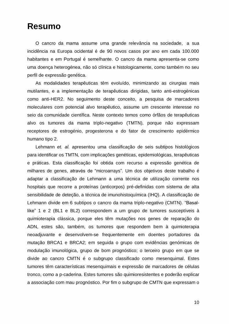
1. Introdution
1.1 Female breast
The breast of mammals is important for milk production that ensures the
survival of the newborn and the development of this species.
The breast consists of three tissue types: fatty tissue, connective tissue and
mammary gland tissue. The glandular tissue is a specialized tissue whose function
is to produce milk. The structures related to milk production are the lobules. Lobules
are structures organized in 15 to 20 sections. In each lobule there is a secondary
structure called lobe, where milk is produced. The structures responsible for
directing the milk to the nipple are the ducts. The dark area of skin around the
nipple is called the areola (figure 1) [1].
Figure 1. Illustration of female breasts. The main components are the lobules
and ducts, conductors and producers of breast milk, respectively; fat tissue,
connective tissues and lymphatic vessels or blood. Adapted from National Cancer
Institute (NCI) http://www.cancer.gov/images/cdr/live/CDR415520-750.jpg
14
The rest of the breast is composed of connective tissue (collagen and elastin),
adipose tissue (fat) and an aponeurosis called Cooper ligament. The breast
consists as well of skin, nerves, blood vessels, lymph vessels and lymph nodes [1].
The ducts are coated with 1 to 2 layers of cylindrical luminal cells. The lobular-
alveolar portion is coated with two layers of cuboidal cells with secretory capacity. A
layer of myoepithelial cells is located in close contact with the basement membrane
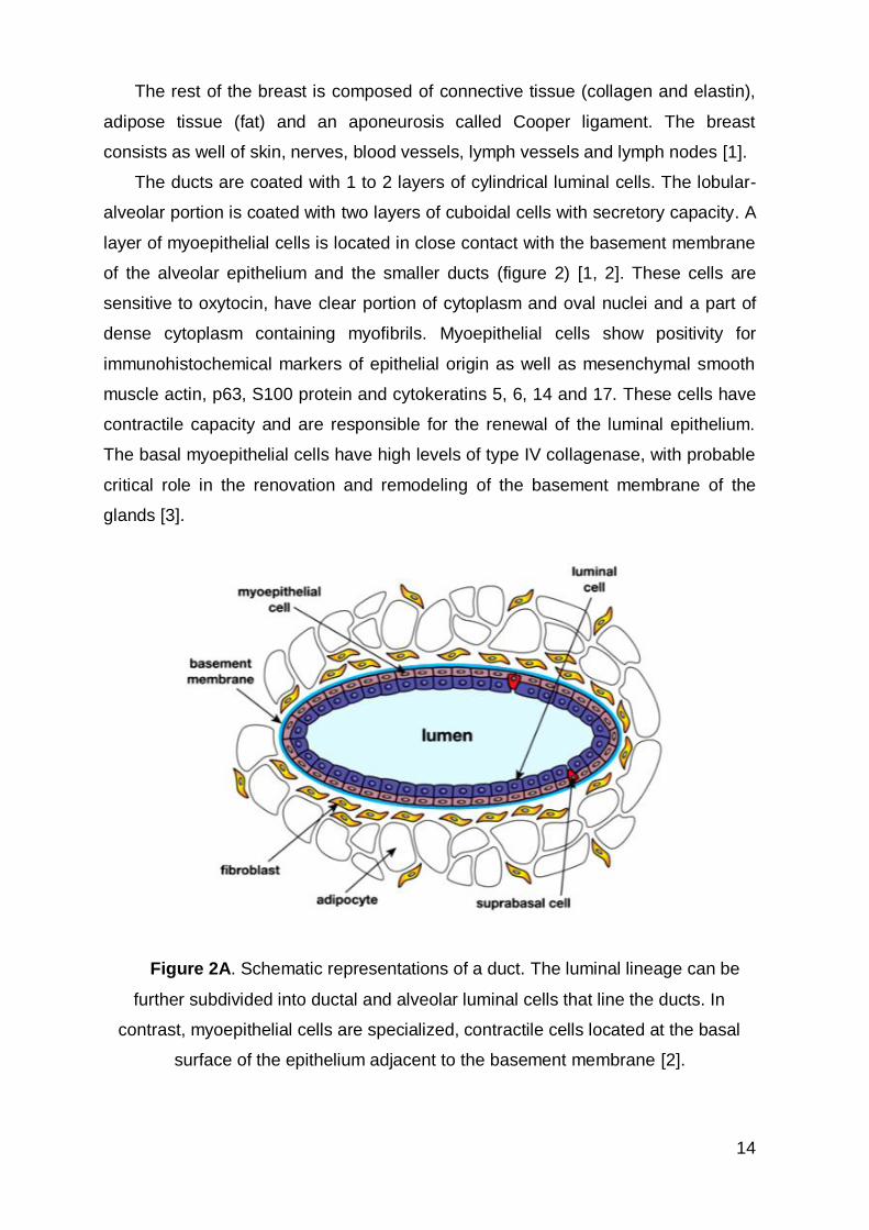
of the alveolar epithelium and the smaller ducts (figure 2) [1, 2]. These cells are
sensitive to oxytocin, have clear portion of cytoplasm and oval nuclei and a part of
dense cytoplasm containing myofibrils. Myoepithelial cells show positivity for
immunohistochemical markers of epithelial origin as well as mesenchymal smooth
muscle actin, p63, S100 protein and cytokeratins 5, 6, 14 and 17. These cells have
contractile capacity and are responsible for the renewal of the luminal epithelium.
The basal myoepithelial cells have high levels of type IV collagenase, with probable
critical role in the renovation and remodeling of the basement membrane of the
glands [3].
Figure 2A. Schematic representations of a duct. The luminal lineage can be
further subdivided into ductal and alveolar luminal cells that line the ducts. In
contrast, myoepithelial cells are specialized, contractile cells located at the basal
surface of the epithelium adjacent to the basement membrane [2].
15
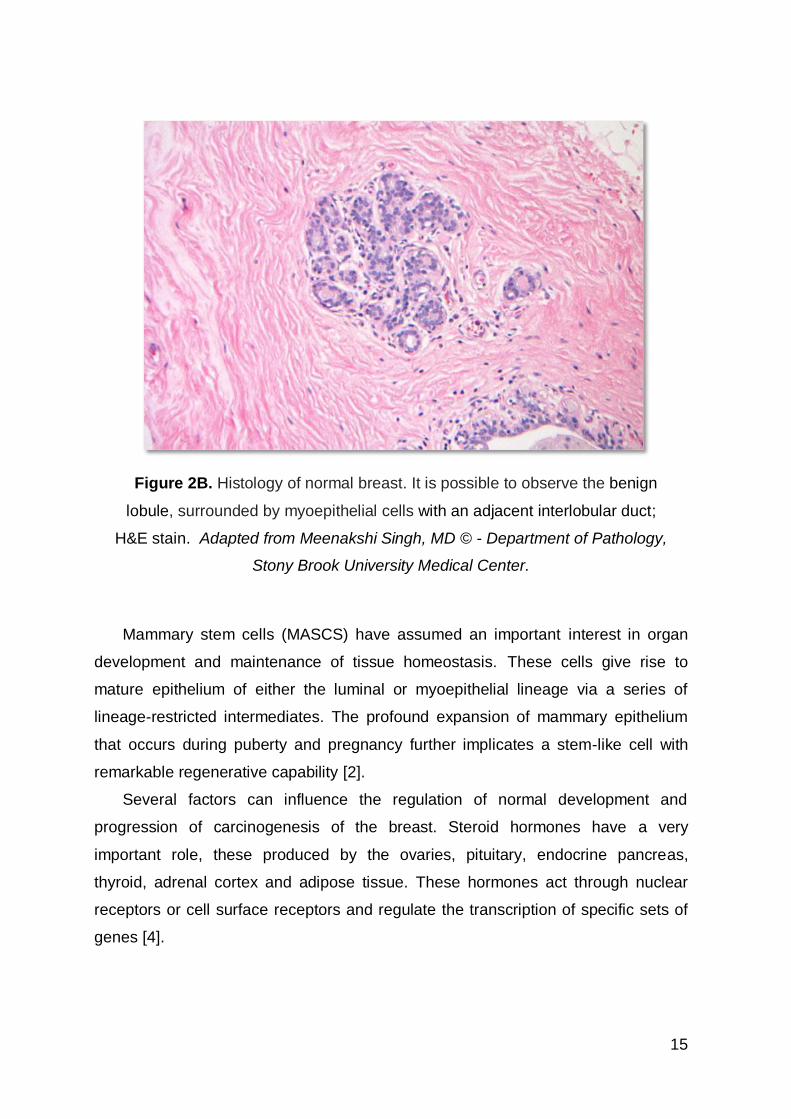
Figure 2B. Histology of normal breast. It is possible to observe the benign
lobule, surrounded by myoepithelial cells with an adjacent interlobular duct;
H&E stain. Adapted from Meenakshi Singh, MD © - Department of Pathology,
Stony Brook University Medical Center.
Mammary stem cells (MASCS) have assumed an important interest in organ
development and maintenance of tissue homeostasis. These cells give rise to
mature epithelium of either the luminal or myoepithelial lineage via a series of
lineage-restricted intermediates. The profound expansion of mammary epithelium
that occurs during puberty and pregnancy further implicates a stem-like cell with
remarkable regenerative capability [2].
Several factors can influence the regulation of normal development and
progression of carcinogenesis of the breast. Steroid hormones have a very
important role, these produced by the ovaries, pituitary, endocrine pancreas,
thyroid, adrenal cortex and adipose tissue. These hormones act through nuclear
receptors or cell surface receptors and regulate the transcription of specific sets of
genes [4].
16
In normal breast tissue there are three main groups of cells, namely epithelial,
myoepithelial/basal and stromal. Paracrine factors play a role in their interactions,
and between the epithelial cells, autocrine mechanisms also play an important role.
In normal conditions, the human body produces various types of hormones
responsible, for example, by the growth of several types of cells. One of these
hormones is estrogen which is responsible for female development, systematic and
organized with cell multiplication. If this cell proliferation stimulated by several
factors, including hormonal, occurs in an uncontrolled manner, this can result in the
appearance of cancers - particularly breast cancer. With this assumption, one way
to inhibit the growth of neoplastic mammary cells (cancer) is to eliminate the
production of female hormones, especially estrogen and/or blocking the action of
hormones on the cells. Only cells that have receptors for estrogen and/or
progesterone in its surface are grow in response to a hormonal stimulus. The
hypothalamus is the initial route of estrogen production, which "sends" the pituitary
gland signals to order production of several hormones, including the gonadotropic
hormones FSH (follicle stimulating hormone) and LH (luteinizing hormone). These
hormones will act on the female sexual glands - ovaries – stimulating the production
of estrogens. Estrogen may also be produced on a smaller scale, by another gland,
the adrenal. Blocking this pathway production, in its different stages, is the main
target of endocrine therapy for breast carcinoma. In all women the adipose tissue
(fat cells) is a site of estrogen production. When in childbearing age (menstrual
cycles), the production of female hormones is made primarily in the ovary, however,
after menopause the adrenal gland is responsible for this function, producing
hormones which will then be "converted" into estrogens in adipose tissue [1].
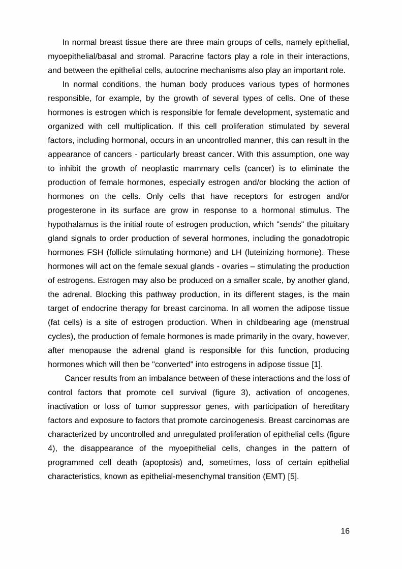
Cancer results from an imbalance between of these interactions and the loss of
control factors that promote cell survival (figure 3), activation of oncogenes,
inactivation or loss of tumor suppressor genes, with participation of hereditary
factors and exposure to factors that promote carcinogenesis. Breast carcinomas are
characterized by uncontrolled and unregulated proliferation of epithelial cells (figure
4), the disappearance of the myoepithelial cells, changes in the pattern of
programmed cell death (apoptosis) and, sometimes, loss of certain epithelial
characteristics, known as epithelial-mesenchymal transition (EMT) [5].
17
Figure 3. Normal growth requires a balance between the activity of genes that
promote and suppress cell proliferation. It also relies on the activities of genes that
signal when damaged cells should undergo apoptosis. Cancer cells do not respond
to many of the signals that control cellular growth and death. Cancer cells escape
programmed cell death (apoptosis). Over time, these cells become increasingly
resistant to the controls that maintain normal tissue. As a result, cancer cells divide
more rapidly and become less dependent on signals from other cells. In the late
stages of cancer, cells break through normal tissue boundaries and metastasize
(spread) to other organ sites in the body. Adapted from oncologiacuf.
Both normal breast and neoplastic tissues are able to produce similar
substances and hormones by its own cells, which act locally (paracrine hormones),
and modulate the function of the epithelial, stromal and vascular/endothelial
adjacent cells. The tumor cells are capable of producing polypeptides that act as
autocrine growth hormone factors regulating gene transcription by acting in its own
surface receptors [1]. Therefore, it is important to understand the origin of cancer
and the factors behind this so that biological processes can help find targetable
drugs for clinical success.
18
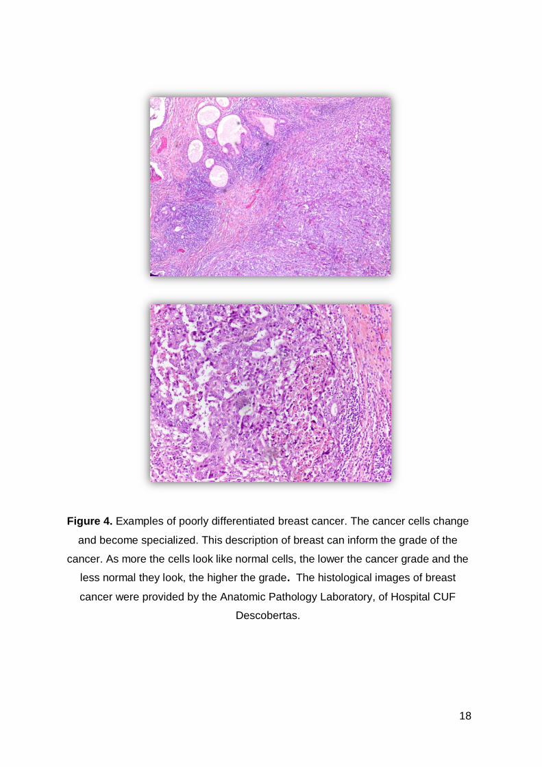
Figure 4. Examples of poorly differentiated breast cancer. The cancer cells change
and become specialized. This description of breast can inform the grade of the
cancer. As more the cells look like normal cells, the lower the cancer grade and the
less normal they look, the higher the grade. The histological images of breast
cancer were provided by the Anatomic Pathology Laboratory, of Hospital CUF
Descobertas.
19
1.2 What is Breast Cancer?
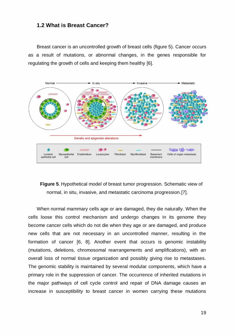
Breast cancer is an uncontrolled growth of breast cells (figure 5). Cancer occurs
as a result of mutations, or abnormal changes, in the genes responsible for
regulating the growth of cells and keeping them healthy [6].
Figure 5. Hypothetical model of breast tumor progression. Schematic view of
normal, in situ, invasive, and metastatic carcinoma progression.[7].
When normal mammary cells age or are damaged, they die naturally. When the
cells loose this control mechanism and undergo changes in its genome they
become cancer cells which do not die when they age or are damaged, and produce
new cells that are not necessary in an uncontrolled manner, resulting in the
formation of cancer [6, 8]. Another event that occurs is genomic instability
(mutations, deletions, chromosomal rearrangements and amplifications), with an
overall loss of normal tissue organization and possibly giving rise to metastases.
The genomic stability is maintained by several modular components, which have a
primary role in the suppression of cancer. The occurrence of inherited mutations in
the major pathways of cell cycle control and repair of DNA damage causes an
increase in susceptibility to breast cancer in women carrying these mutations
20
(BRCA1 and 2). On the other hand, mutations of the p53 gene are frequent in
sporadic breast carcinomas.
Breast cancer occurs when malignant tumors develop in the breast. These cells
can spread by breaking away from the original tumor and entering blood vessels or
lymph vessels, which branch into tissues throughout the body. When cancer cells
circulate and seed in distant organs and damage tissues and organs, the process is
called metastasis. The most common sites of cancer metastasis are (in alphabetical
order ) the bone, brain, liver, and lung [9].
1.3 Incidence
Breast cancer is a problem for global public health and is the non-cutaneous
malignancy more common in females, with increasing incidence in developed
countries.
About 12.7 million cancer cases and 7.6 million cancer deaths are estimated to
have occurred in 2008 worldwide, with 56% of the cases and 64% of the deaths in
the developing world [10].
Breast cancer is the second most common cancer in the world and, by far, the
most frequent cancer among women with an estimated 1.67 million new cancer
cases diagnosed in 2012 (25% of all cancers) [11].
In Portugal BC remains the leading cause of cancer death in women with about
5513 new cases per 100 000 persons per year [12].
More than half of these cases occur in industrialized countries. In the European
Union, 367,000 new cases of BC were diagnosed in 2012 (about 28.8 % of cancer
in women) [13]. The incidence of BC has increased more in developed countries but
there has been an increase in incidence in developing countries, particularly in
Africa, Latin America and Asia [14] [15].
Breast cancer is the leading cause of death in women between the ages of 25
and 55 years in developed countries [16].
The factors that contribute to the international variation in incidence rates are
largely originated from differences in reproductive and hormonal factors and the
availability of early detection services [17, 18].
21
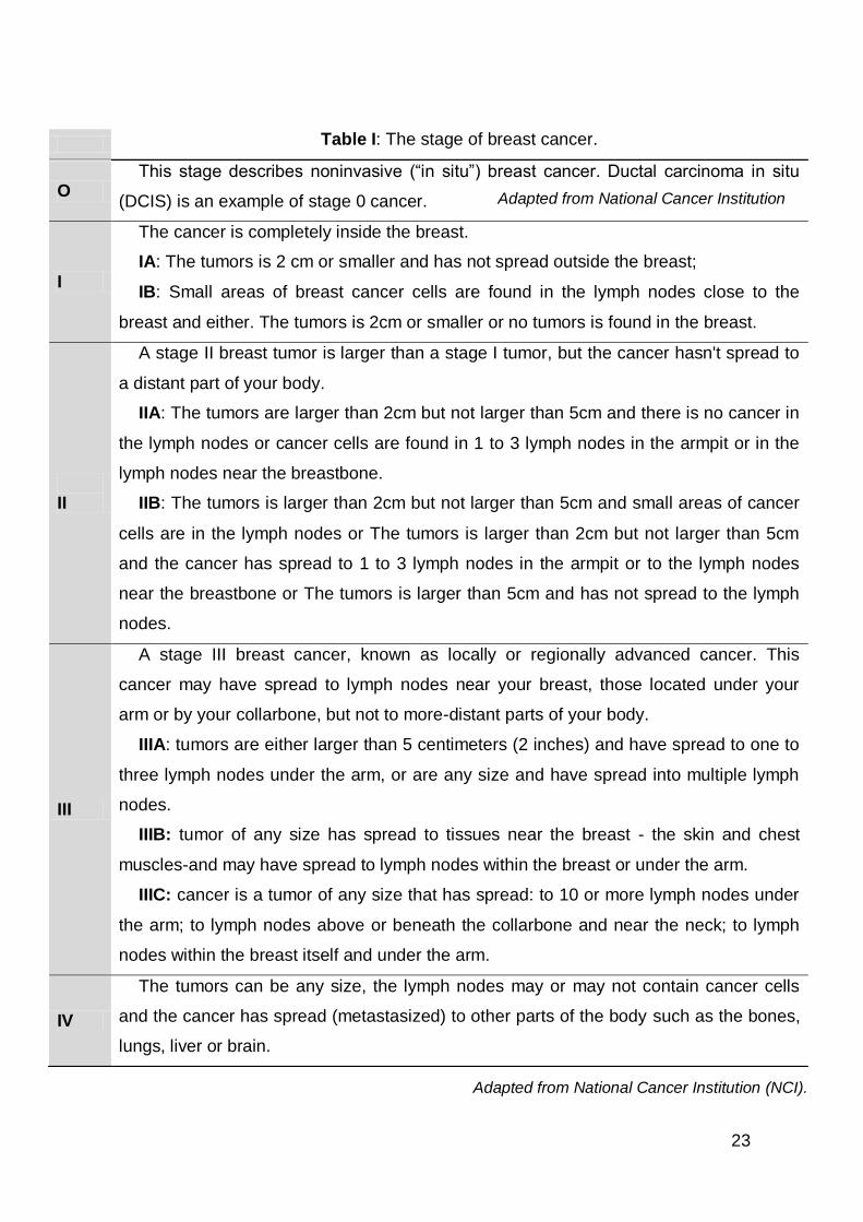
1.4 Breast Cancer Stages
Clinical staging (designated cTNM or TNM) accordding to the AJCC system
(American Joint Committee on Cancer) is based on all information available prior
the first definitive treatment and includes the findings on pathological examination,
imaging studies, operative findings on physical examination of the breast or other
tissue [19, 20]. The extent of tissue examined pathologically for clinical staging
includes tumor size (T), involvement of lymph nodes in the homolateral axilla (N)
and whether the cancer is invasive or non-invasive. The presence or absence of
distant metastases (M) is assessed through imaging [21]. Pathologic staging is
based on a pathologist’s study of the lymph nodes and tumor tissue removed during
surgery. The pathological classification requires the resection and examination of at
least the low axillary lymph nodes (level I). If the lymph nodes are negative, but the
number ordinarily examined is not met, classify as pN0. Often this is surgery to
remove the cancer and nearby lymph nodes, but sometimes surgery may be done
to just look at how much cancer is in the body and take out tissue samples. The
pathological stage gives the health care team more precise information that can be
used to predict treatment response and outcomes (prognosis) [21] (appendix I).The
invasive nature of cancer is defined by the rupture of the basal membrane. The term
"locally advanced" or "regionally advanced" is used to refer large tumors involving
the skin of the breast, underlying chest structures, altering the shape of the breast
and lymph nodes that are visible or palpable during clinical examination.
The stage of breast cancer is essential to determine prognosis and therapy.
Breast cancer is classified into 4 stages (Table I) [1].
Most breast malignancies arise from epithelial elements and are categorized
as carcinomas. The invasive breast carcinomas consist of several histologic
subtypes. Breast carcinomas are a diverse group of lesions that differ in
microscopic appearance and biologic behavior, although these disorders are often
discussed as a single disease. The in situ carcinomas of the breast are either ductal
(also known as intraductal carcinoma) or lobular. This distinction is primarily based
upon the growth pattern and cytologic features of the lesions, rather than their
anatomic location within the mammary ductal-lobular system [22].

22
BC is also classified by its histopathologic characteristics [22] which are
presented in table II and figure 6 .
Figure 6. Histological images of different types of breast cancer.
23
Table I: The stage of breast cancer.
O This stage describes noninvasive (“in situ”) breast cancer. Ductal carcinoma in situ
(DCIS) is an example of stage 0 cancer.
I
The cancer is completely inside the breast.
IA: The tumors is 2 cm or smaller and has not spread outside the breast;
IB: Small areas of breast cancer cells are found in the lymph nodes close to the
breast and either. The tumors is 2cm or smaller or no tumors is found in the breast.
II
A stage II breast tumor is larger than a stage I tumor, but the cancer hasn't spread to
a distant part of your body.
IIA: The tumors are larger than 2cm but not larger than 5cm and there is no cancer in
the lymph nodes or cancer cells are found in 1 to 3 lymph nodes in the armpit or in the
lymph nodes near the breastbone.
IIB: The tumors is larger than 2cm but not larger than 5cm and small areas of cancer
cells are in the lymph nodes or The tumors is larger than 2cm but not larger than 5cm
and the cancer has spread to 1 to 3 lymph nodes in the armpit or to the lymph nodes
near the breastbone or The tumors is larger than 5cm and has not spread to the lymph
nodes.
III
A stage III breast cancer, known as locally or regionally advanced cancer. This
cancer may have spread to lymph nodes near your breast, those located under your
arm or by your collarbone, but not to more-distant parts of your body.
IIIA: tumors are either larger than 5 centimeters (2 inches) and have spread to one to
three lymph nodes under the arm, or are any size and have spread into multiple lymph
nodes.
IIIB: tumor of any size has spread to tissues near the breast - the skin and chest
muscles-and may have spread to lymph nodes within the breast or under the arm.
IIIC: cancer is a tumor of any size that has spread: to 10 or more lymph nodes under
the arm; to lymph nodes above or beneath the collarbone and near the neck; to lymph
nodes within the breast itself and under the arm.
IV
The tumors can be any size, the lymph nodes may or may not contain cancer cells
and the cancer has spread (metastasized) to other parts of the body such as the bones,
lungs, liver or brain.
Adapted from National Cancer Institution A
Adapted from National Cancer Institution (NCI).

24
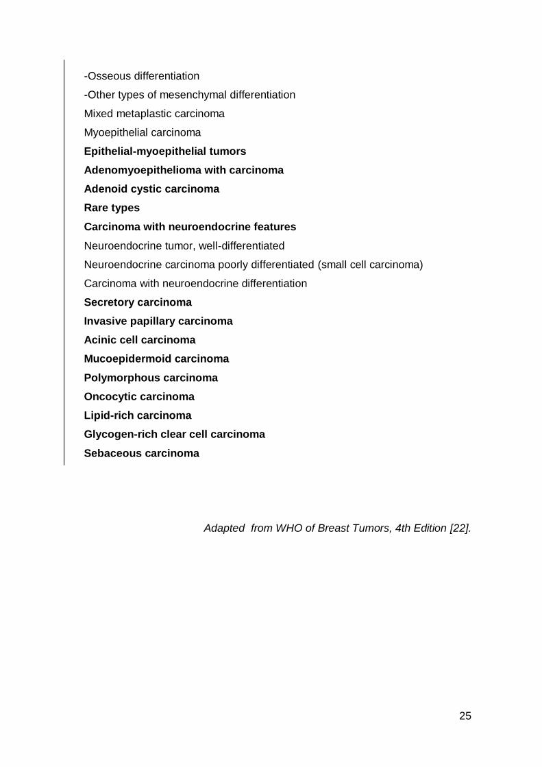
TableII. Breast Cancer Types.
Invasive carcinoma of no special type (NST)
Pleomorphic carcinoma
Carcinoma with osteoclast-like stromal giant cells
Carcinoma with choriocarcinomatous features
Carcinoma with melanotic features
Invasive lobular carcinoma
Classic lobular carcinoma
Solid lobular carcinoma
Alveolar lobular carcinoma
Pleomorphic lobular carcinoma
Tubulolobular carcinoma
Mixed lobular carcinoma
Tubular carcinoma
Cribriform carcinoma
Mucinous carcinoma
Carcinoma with medullary features
Medullary carcinoma
Atypical medullary carcinoma
Invasive carcinoma NST with medullary features
Carcinoma with apocrine differentiation
Carcinoma with signet-ring-cell differentiation
Invasive micropapillary carcinoma
Metaplastic carcinoma of no special type
Low-grade adenosquamous carcinoma
Fibromatosis-like metaplastic carcinoma
Squamous cell carcinoma
Spindle cell carcinoma
Metaplastic carcinoma with mesenchymal differentiation
-Chondroid differentiation
25
-Osseous differentiation
-Other types of mesenchymal differentiation
Mixed metaplastic carcinoma
Myoepithelial carcinoma
Epithelial-myoepithelial tumors
Adenomyoepithelioma with carcinoma
Adenoid cystic carcinoma
Rare types
Carcinoma with neuroendocrine features
Neuroendocrine tumor, well-differentiated
Neuroendocrine carcinoma poorly differentiated (small cell carcinoma)
Carcinoma with neuroendocrine differentiation
Secretory carcinoma
Invasive papillary carcinoma
Acinic cell carcinoma
Mucoepidermoid carcinoma
Polymorphous carcinoma
Oncocytic carcinoma
Lipid-rich carcinoma
Glycogen-rich clear cell carcinoma
Sebaceous carcinoma
Adapted from WHO of Breast Tumors, 4th Edition [22].
26
1.5 Clinical and histopathological prognostic factors
Breast cancer is the most common cancer in women and its incidence is
increasing. Breast cancer is a heterogeneous disease [23-26] which comprises a
number of distinct biological entities that are associated with morphological and
immunohistochemical outcomes and clinical characteristics [23, 26].
The invasive breast carcinomas were for decades, only classified according to
histological grade and hormone receptor expression [23]. The most important
histopatological prognostic factors are the expression of ER and PR. These have
enabled clinicians to treat BC in the last half a decade with anti-estrogens,
significantly reducing BC mortality. Anti-estrogenic therapy is not toxic, well
tolerated and cheap. Recently, after the success of clinical trials involving adjuvant
trastuzumab, expression of the human epidermal growth factor receptor 2 (HER2)
has become an integral part of the pathological workup for patients with breast
cancer. HER2-positive breast cancer had worse prognosis compared with HER2-
negative tumors [27]. HER2 amplification is widely known to indicate an aggressive
tumor behavior and a poor clinical outcome in breast cancer patients [28].
Simultaneously with the development of trastuzumab as a targeted therapy for
breast cancer, some results of genome microarray began to be reported [29]. There
are several types of clinical breast cancer, defined by amplification of specific
markers. The over expression of steroid hormone (like estrogen and progesterone
receptors) defines the most abundant type of breast cancer, accounting for about
70%. (ER and PR positive) [24, 30, 31]. Therefore, several studies demonstrate that
breast cancers can be divided according to hormone receptor (HR) expression
(negative or positive) and/or epithelial cellular origin (basal or luminal), that have
clinical implication [32]. Breast cancers can be divided into three main groups: (1)
hormone-positive breast tumors; (2) HER2 –positive breast cancer and (3) basal or
triple-negative breast tumors (classification summarized in table III). The overlap
between basal and triple negative BC is not complete [32]. Generally, there are 20%
of classifications in either direction i.e. there are 20% of basal like BC that are not
TNBC and similarly there are 20% of TNBC that are not basal-like BC.
27
The tumors that are HR-positive are luminal A e B. This subtypes originate
from inner (“luminal”) cells that line the mammary ducts, and they are dissimilar in
their expression of HER2 (luminal A, which is negative and luminal B which is
positive). Most carcinomas not related to genetic mutations, are characterized by
the luminal A type [25]. The luminal B type is more likely to be lymph node-positive
and to have high proliferation. Unlike the luminal A which tends to have a better
prognosis, they often diagnosed in young women, the luminal B tumors tend to have
a higher tumor grade, poorer prognosis and probably associated to genetic
mutations. These phenotypes are associated with elevated gene expression by
luminal epithelial cells, of molecules such as cytokeratins (CK) 7,8,18 and 19 [24,
33, 34].
The tumors that are HR-negative cannot be treated with anti-estrogens, are
associated with a higher recurrence rate and a decreased overall survival. These
tumors are more likely to be poorly differentiated, with higher histological grade [32].
HER2 tumors tend to be HR-negative and lymph node-positive. HER2 has been
a major target for the development of the new cancer therapies in the last 20 years.
Its greatest value as a predictive marker lies in the prediction of response to
therapies that target HER2, such as trastuzumab (Herceptin) and with neoadjuvant
anthracycline/taxane-based chemotherapy [35]. The HER2 oncogene amplification
and concomitant overexpression of its protein, is currently implicated as an
important prognostic biomarker in breast cancer [36].
Basal-like tumors originate in the outer (“basal”) cells that line the mammary
ducts. This subtype has a gene expression profile similar to the genes that are
identified in normal basal/myoepithelial cells of the mammary ducts and acini. The
gold standard for the identification of basal-like carcinomas as a particular class of
molecular breast cancer in the clinic is TNBC [33].
Their incidence has been estimated to be between 13% and 25% and they
occur more frequently in young women and are associated with hereditary BRCA1-
related breast cancers. Their metastatic pattern includes early dissemination to the
axillary nodes and to viscera (liver, lung, soft tissue and brain) and less frequently to
bone. According to the most recent publications, this phenotype shows positivity for
28
CK5/6, CK14, receptor for epidermal growth factor (EGFR), p-cadherin, p63 and
CK17 proteins that are expressed in the basal/myoepithelial cells [24, 30, 37]. This
profile is associated with numerous genetic mutations [30].
The original microarray classification published in the year 2000 [24], defined a
fourth group called normal-like tumors that account 6%-10% of all breast cancers.
These tumors do not fall into any other categories, usually small, typically have a
good prognosis and they are more common in postmenopausal than in
premenopausal women. Perou et. al. [24] identified these group by increasing the
expression of many genes known to be expressed by adipose tissue and other non-
epithelial cells. These tumors also showed strong expression of basal epithelial
genes and low expression genes to the luminal epithelium. Nevertheless, it is still
not clear if this distinction is of clinical value [24, 30].
Estrogen receptor and progesterone receptor are the most studied markers in
breast tissue. The results of ER and PR have been scored as ‘‘positive’’ and
‘‘negative’’ although receptor protein concentration (in biochemical assays) and the
percentage of cells stained and staining intensity (in Immunohistochemical assays)
range widely [26]. Several studies report that the ER/PR status is an independent
predictor of outcome in breast cancer tumors. The association of this status with the
mortality is observed in tumors ER+/PR-, ER-/PR+, and ER-/PR-tumors compared
to women with ER+/PR+ tumors. The prognostic usefulness of the expression of
steroids hormone receptors is partially independent from the association in clinical
characteristics and demographic characteristics of tumors. The higher relative
mortality identified among ER-/PR- patients with small or low-grade tumors, raise
the question of whether there may be a beneficial role for adjuvant chemotherapy in
this population [38].
The problem of steroid receptor testing inaccuracy and non reproducibility is
extremely serious because we are inadequately classifying BC but also because we
are depriving women of anti-estrogenic therapy. For the determination of prognosis
and therapy of BC some other proteins such as HER2, Ki-67, p53 are sought in
addition to hormone receptors[39]. When BC does not express ER, PR and HER2 it
will not benefit from the currently available receptor-targeted systemic therapy and
these tumors are called TN. This study will be focused on TNBC.
29
One of the most important challenges ahead is to identify specific molecules
alterations in tumors and to validate targeted therapies for them. This is sometimes
called precision/personalized therapy [24]. With the development of primary and
secondary resistance to hormonal treatment, new substances and new patterns of
association with blockers of growth factor receptors were created, enhancing the
effect of drugs and recovering the responsiveness to endocrine therapy. Some of
these substances are aromatase inhibitors (anastrozole, letrozole), antagonist of
estrogen and progesterone receptors (fulvestrant), pan-EGFR inhibitors
(GW572016), mTOR inhibitors (rapamycin RAD001-inhibit tumor growth central
controller and angiogenesis), anti-HER2 (trastuzumab), and drug that blocks cell
growth by stopping mitosis (cell division) [40].
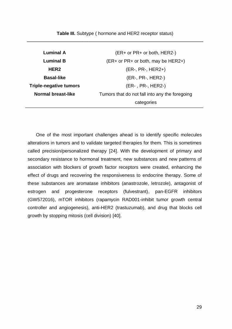
Table III. Subtype ( hormone and HER2 receptor status)
Luminal A
(ER+ or PR+ or both, HER2-)
Luminal B (ER+ or PR+ or both, may be HER2+)
HER2 (ER-, PR-, HER2+)
Basal-like (ER-, PR-, HER2-)
Triple-negative tumors (ER- , PR-, HER2-)
Normal breast-like Tumors that do not fall into any the foregoing
categories
30
1.6 Risk factors
Risk factors for breast cancer are age, race/ethnicity, age of menarche and
menopause, multiparity, radiation mantle before Hodgkin lymphoma, oral
contraceptive use and body mass index.
Family history of breast cancer, particularly having one or more first degree
relatives with breast cancer (although most women with breast cancer do not have
a family history of the disease) increases the risk of breast cancer. Inherited
mutations in breast cancer susceptibility genes account for approximately 5% to
10% of all cancers of the female breast and an estimated 4% to 40% of all male
breast cancers, but are very rare in the general population (much less than 1%).
Most of these mutations are located in BRCA1 and BRCA2 genes, although
mutations in other known genes have also been identified [18] [31].
Increased risk of hormone receptor–positive tumors was also associated with
postmenopausal obesity, which probably increases estrogen exposure via different
mechanisms. Obesity is associated with increased aromatization of circulating
androgens to estrogens in adipose tissue and reduced levels of sex hormone
binding globulin, thereby increasing both total and bioavailable estrogens [31]. A lot
of studies summarized the risks of hormone receptor-positive and hormone
receptor-negative BC. The majority of know BC risk factors are associated with
hormone receptor-positive disease. Risk factors for HR-negative BC are young
age, African origin and BRCA1/2 germline mutation [31]. In addition, the effect of
hormone-related risk factors on hormone content within the breast is unknown.
Although many factors have been shown to contribute to elevated systemic levels of
estrogens, a relationship between high serum levels and the development of
hormone receptor positive tumors has not been established [31].
Potentially modifiable risk factors that can be minimized include overweight,
lack of exercise, smoking cigarettes, eating unhealthy food, alcohol consumption.

31
Two drugs (tamoxifen and raloxifene), have been approved to reduce breast
cancer incidence in high risk women. Raloxifene appears to have a lower risk of
certain side effects, such as uterine cancer and blood clots; however, it is only
approved for use in postmenopausal women.
Women and society have the empowerment to lower BC risk by making
healthiest lifestyle options possible.
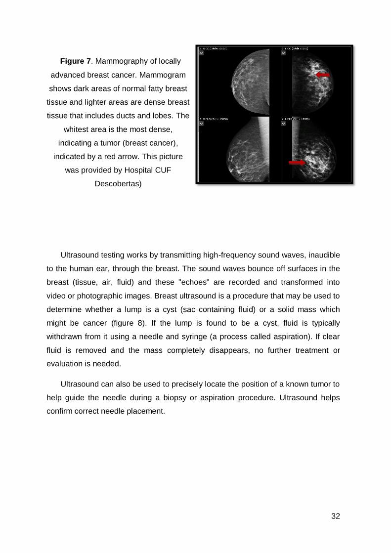
1.7 Diagnosis Tests
Several tests can help distinguish a benign (noncancerous) lump from a
malignant (cancerous) tumor. Because malignant and benign lumps tend to have
different physical features, imaging tests such as mammography (figure 7) and
ultrasonography can often rule out cancer. The only way to confirm cancer is to
perform a needle aspiration or a biopsy to be analyzed. Before BC screening was
implemented, breast cancer detection was limited to diagnosis based on physical
examination by the woman herself or her physician. The concept of screening is to
detect non palpable lesions.
When breast cancer is found at an early stage, the cure rate is high, the aim of
screening is to detect non palpable disease and increase survival. Such cancers
may be detectable only in mass population screening endeavors. Mammograph
was suggested by Salomon, a German pathologist in 1913, as a modality to detect
breast cancer and in 1959, Egan at the MD Anderson Hospital in Houston published
results from a series of 1000 cases emphasizing the detection of non palpable
lesions. In a practical, cheap and feasible, mammography has been implemented
as an important method for early detection of BC, reducing mortality.
32
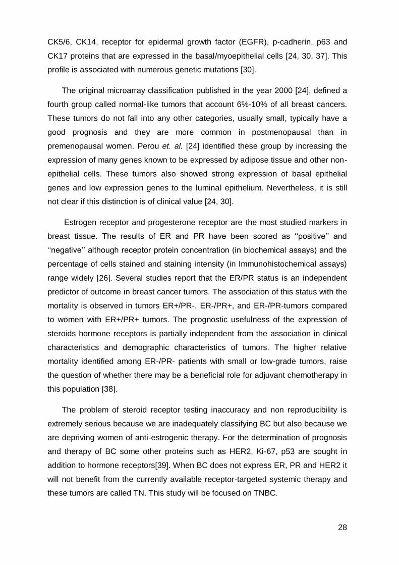
Figure 7. Mammography of locally
advanced breast cancer. Mammogram
shows dark areas of normal fatty breast
tissue and lighter areas are dense breast
tissue that includes ducts and lobes. The
whitest area is the most dense,
indicating a tumor (breast cancer),
indicated by a red arrow. This picture
was provided by Hospital CUF
Descobertas)
Ultrasound testing works by transmitting high-frequency sound waves, inaudible
to the human ear, through the breast. The sound waves bounce off surfaces in the
breast (tissue, air, fluid) and these "echoes" are recorded and transformed into
video or photographic images. Breast ultrasound is a procedure that may be used to
determine whether a lump is a cyst (sac containing fluid) or a solid mass which
might be cancer (figure 8). If the lump is found to be a cyst, fluid is typically
withdrawn from it using a needle and syringe (a process called aspiration). If clear
fluid is removed and the mass completely disappears, no further treatment or
evaluation is needed.
Ultrasound can also be used to precisely locate the position of a known tumor to
help guide the needle during a biopsy or aspiration procedure. Ultrasound helps
confirm correct needle placement.
33
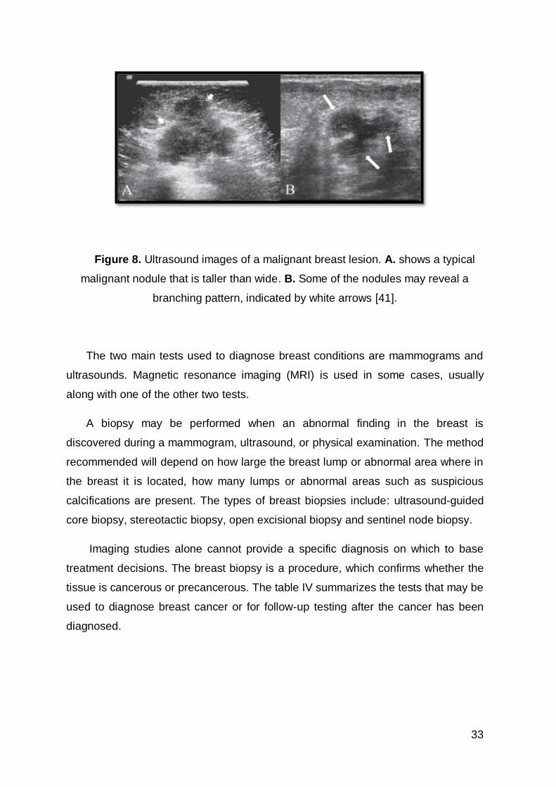
Figure 8. Ultrasound images of a malignant breast lesion. A. shows a typical
malignant nodule that is taller than wide. B. Some of the nodules may reveal a
branching pattern, indicated by white arrows [41].
The two main tests used to diagnose breast conditions are mammograms and
ultrasounds. Magnetic resonance imaging (MRI) is used in some cases, usually
along with one of the other two tests.
A biopsy may be performed when an abnormal finding in the breast is
discovered during a mammogram, ultrasound, or physical examination. The method
recommended will depend on how large the breast lump or abnormal area where in
the breast it is located, how many lumps or abnormal areas such as suspicious
calcifications are present. The types of breast biopsies include: ultrasound-guided
core biopsy, stereotactic biopsy, open excisional biopsy and sentinel node biopsy.
Imaging studies alone cannot provide a specific diagnosis on which to base
treatment decisions. The breast biopsy is a procedure, which confirms whether the
tissue is cancerous or precancerous. The table IV summarizes the tests that may be
used to diagnose breast cancer or for follow-up testing after the cancer has been
diagnosed.
34
Table IV. Tests and diagnostic procedures for breast cancer.
Imaging tests
Mammography
Untrasounds
MRI
Surgical tests Biopsy
Molecular testing of the tumor
ER and PR, HER2; KI-67
Tests and procedures used to stage breast cancer
-Blood tests, such as a complete blood count
-Mammogram of the other breast to look for signs of cancer
-Breast MRI
-Bone scan
-Computerized tomography (CT) scan
-Positron emission tomography (PET) scan
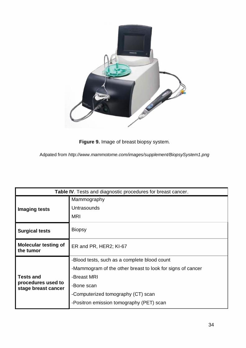
Figure 9. Image of breast biopsy system.
Adpated from http://www.mammotome.com/images/supplement/BiopsySystem1.png
35
1.8 Treatment
Treatment involves conservative surgery (surgical removal of the tumor and
surrounding tissue) or mastectomy (surgical removal of the breast). The tumor size
and breast size grade are the patient characteristics that will be important to choose
type of treatment to adopt.
Numerous studies have shown that for early breast cancer (cancer that has not
spread to the skin, chest wall, or distant organs), the long-term survival for women
treated with breast-conserving surgery plus radiotherapy is similar to those treated
with mastectomy. For women undergoing mastectomy, significant advances in
reconstruction techniques provide several options for breast reconstruction,
including several different options on the timing of the process.
In women with early stage disease, the sentinel lymph node biopsy, a
procedure in which only the first lymph nodes to which cancer is likely to spread due
to lymph node anatomical drainage system are removed, has a lower chance of
long-term side effects and is as effective as a full axillary node dissection, in which
numerous nodes are removed, to determine whether the tumor has spread beyond
the breast.
Treatment may also involve radiation therapy, chemotherapy (before or after
surgery), hormone therapy or targeted therapy.
Tumor cells viable after neoadjuvant chemotherapy (NAC), which is
chemotherapy administered before surgery, are a population of cancer cells that is
intrinsically resistant to chemotherapy. These tumor cells likely reflect the
component of micrometastatic disease, which is responsible for distant metastases,
and is unlikely to be sensitive to adjuvant chemotherapy, if so metastases will one
day become evident. Specifically, this phenomenon has been observed in patients
with TNBC who have residual disease after NAC [40].
36
2. Triple-negative breast cancer (TNBC)
TNBC has a growing recognition by oncologists, pathologists and geneticists
since 2005, when it began to be referred by the term "triple-negative" [42]. TNBC
comprises 12 to 17% of BC cases. This terminology reflects a heterogeneous
population with a much more complex molecular transcriptome than is suggested by
the triple-negative (TN) immunohistochemical (IHC) expression [43]. Furthermore
this definition is misleading because it tells us what these cancers are not and not
what they are, it is a negative definition.
TNBC is a distinct subtype defined by the lack of immunohistochemical
expression of the estrogen receptor (ER) and progesterone receptor (PR), human
epidermal receptor growth factor 2 (HER2) amplification. TNBC generally has
expression of genes normally found in the basal, myoepithelial cells or normal
breast [25, 42-44] .
The frequency of the triple-negative breast cancer increases with age, it
comprises approximately 15% to 20% of all breast cancers [25, 45] although TNBC
are more common than ER positive BC in younger patients (<50 years) and African
American women [42, 44]. TNBC is associated with an advanced stage, increased
risk of visceral disease and worse outcome. Several studies have been carried with
the purpose of demonstrating these associations [24, 25, 37, 46-48]. These tumors
are generally larger in size, are of high histological grade (III) [23], with lymph node
involvement at diagnosis, and are biologically more aggressive with worse
prognosis [24]. The majority of TNBC are grade III or poorly differentiated, infiltrating
ductal carcinoma not otherwise specified (IDC NOS). The few remaining cases are
rare histological types like adenoid-cystic, medullary, apocrine, metaplastic or
inflammatory BC [23].
The aggressiveness of this subtype of cancer is illustrated by the fact that the
peak risk of recurrence is between the first and third year of follow-up in patients
diagnosed with TNBC and most deaths occur in the first 5 years after therapy.
When compared with patients who have other subtypes of breast cancer [24]. Less
than 30% of women with metastatic TNBC survive five years and all die of the
37
disease, despite having undergone adjuvant chemotherapy and subsequently
chemotherapy for advanced disease [24] [49].
Patients with triple negative breast cancer are more likely to develop distant
metastases earlier than non-triple negative breast cancer patients, develop brain
metastases sooner [28]. The prevalence of genetic mutations among women with
TNBC referred for genetic counseling is high and differs significantly by
ethnicity/race and age [45].
This molecular subtype of breast cancer is characterized by a profile of gene
expression similar to that found in basal/myoepithelial cells. The multiplicity of
names reflects an underlying uncertainty about the true nature of this entity.
The breast cancer literature includes a large number of reports on prospective
clinical trials examining the effects of endocrine therapy, chemotherapy, targeted
therapy combinations, but none has validated a specific systemic therapy for TNBC
[42]. Although effective tailored therapies have been developed for patients with
HER2 positive or hormone receptor positive, chemotherapy is the only modality of
systemic therapy for patients with BC that lack the expression of these three
markers [44] [43].
One of the first molecular insights into TNBCs was the finding that they are
likely to arise in patients with germline mutations in the BRCA1 gene and have gene
expression profiles (GE) similar to those tumors which are not TNBC but arise in
germline BRCA1 mutated women [44]. Germline BRCA1 or BRCA 2 mutations
among women are the most frequent hereditary breast cancer syndromes and are
associated with a 90% lifetime risk of developing BC. Studies have shown the
prevalence of BRCA mutations in women with TNBC is high, therefore the finding of
a woman with a TNBC and positive family history of BC is a reliable indicator of
possible BRCA germline mutation [45]. BRCA1 behaves as an important gene in
DNA repair of double strand DNA breaks, similarly contributing to the maintenance
and stability of DNA [44]. Among a population of patients with TNBC referred for
genetic counseling and genetic testing between 2000 and 2012, the prevalence of
BRCA mutation carriers exceeded 30% [45].
38
2.1 TNBC subtypes
There are several studies that research the molecular profile that distinguishes
subtypes of TNBC. Until recently, most studies on TNBC aimed to identify markers
that separate TNBC from other BCs, and it is likely that these studies identify
molecules that differentiate amongst subtypes of TNBC.
Since 2005, when the widespread use of anti-HER2 therapy started to change
HER2 positive BC, TNBC finally became the only subtype of BC for which there is
no targeted therapy. This is a very important unmet need in BC. Since then a lot of
experiments and clinical trials have been done to try to understand what is
targetable in TNBC, and if the clinical trials meet their endpoints, so we might
validate usable drugs for daily practice. Besides the therapeutic challenge, TNBC
entails a diagnostic challenge and Lehman et. al. after several experiments have
put forward a convincing classification of six subtypes with histological, genetic,
epidemiological, therapeutic, and, therefore, practical implications (appendix II).
TNBC subtypes were characterized on the basis of differential GE and gene
ontologies and subsequently labeled TNBC as follows: basal-like (basal-like 1
(BL1); basal-like 2 (BL2)); immunomodulatory (IM); mesenchymal (M);
mesenchymal stem–like (MSL); luminal androgen receptor (LAR) [44].
The top biological processes present in the BL1 subtype are cell cycle and cell
division components and pathways. Elevated DNA damage response (ATR/BRCA)
pathways accompany the proliferation pathways in the BL1 subtype. Increased
proliferation and cell-cycle checkpoint loss are consistent with the elevated
expression of the DNA damage response genes observed. The highly proliferative
nature of this subtype is further supported by the finding of high Ki-67 mRNA
expression (MKI67). Enrichment of proliferation genes and increased Ki-67
expression in basal-like TNBC tumors suggest that this subtype would preferentially
respond to antimitotic agents such as taxanes (paclitaxel or docetaxel). The BL2
subtype displays unique gene ontologies involving growth factor signaling as well as
glycolysis and gluconeogenesis. Likewise, the BL2 subtype is uniquely enriched in
growth factor receptor expression such as EGFR, MET (proto-oncogene, receptor
tyrosine kinase), and EPHA2 (member of the ephrin-A receptor subfamily of
39
receptor tyrosine kinases). This subtype has features suggestive of
basal/myoepithelial origin as demonstrated by higher expression levels of p63 and
MME (CD10) [34].
The IM subtype is enriched in immune cell processes. The IM signaling is
evidenced by immune signal transduction GE, in addition to immune cell-surface
antigens, cytokine signaling, complement cascade, chemokine receptors and
ligands, and antigen presentation genes. This subtype of TNBC substantially
overlaps with the gene signature for medullary breast cancer, a rare, histologically
distinct form of TNBC that despite its high-grade histology is associated with a
favorable prognosis.
The M subtype (M and MSL subtypes) displays a variety of unique gene
ontologies that are heavily enriched in components and pathways involved in cell
motility. The difference between the M and MSL subtypes is that the MSL subtype
expresses high levels of proliferation genes. This subtype was associated with a
highly dedifferentiated type of breast cancer called metaplastic breast cancer, which
is characterized by mesenchymal/sarcomatoid or squamous features is
chemoresistant and, therefore, carries severe prognosis.
The LAR group is the most distinct among the TNBC subtypes. This subtype is
ER negative, but gene ontologies are heavily enriched in hormonally regulated
pathways including steroid synthesis, porphyrin metabolism, and androgen/estrogen
metabolism. Others have previously described a breast cancer subgroup
expressing AR termed molecular apocrine and this BC has good prognosis.
The identification of different subtypes of TNBC had focused on regulatory
molecules, both extracellularly the immune system and at the level of the plasma
membrane, a gene set comprised epithelial growth factors. The immune regulation
has previously been proposed to play a role in BC and, in particular the ER positive
BC. Intracellularly, these gene products bind to the molecules proliferation that
control, such as cyclins. The activation of the immune response may preclude a
good prognosis; this would be similar to the good prognosis associated with
immune activation in melanoma, ovarian and kidney cancer.
P53 expression is a predictor of shorter time to relapse in independent
populations diagnosed with TNBC. Similarly, increased expression of EGFR
40
appears to predict adverse prognosis in TNBC. In the latter report, loss of c-Kit and
BRCA1 expression were also reported to be predictive of poor outcome in TNBC.
The cell lines representative of the TNBC subtypes display different sensitivities
to a variety of agents, and importantly, these differences can be attributed to distinct
expression of cellular components and presence of mutations in key oncogenes and
tumor suppressors [44].
In the seventies, the early tests of the presence of AR protein in breast cancer
specimens were performed, by the McGuire laboratory [50]. Currently, most of the
studies were performed using specific anti-AR antibodies in IHC studies. This study
will focus more on the expression of this protein in samples of patients with TNBC.
41
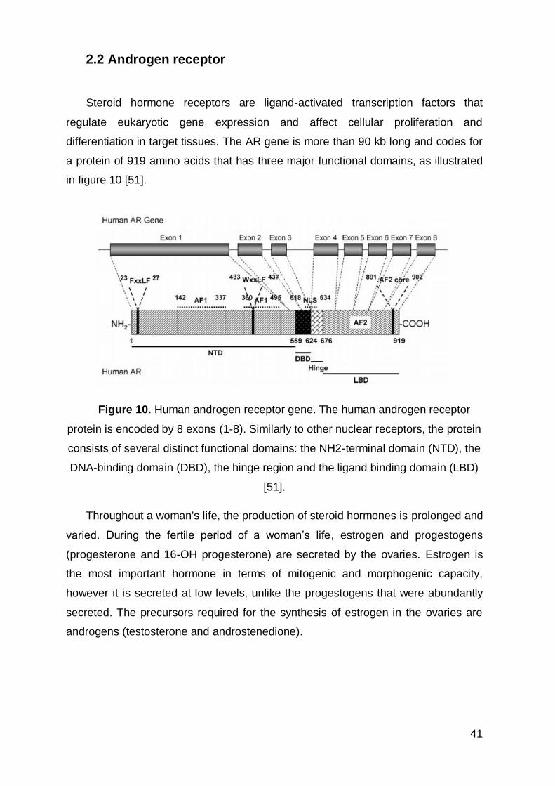
2.2 Androgen receptor
Steroid hormone receptors are ligand-activated transcription factors that
regulate eukaryotic gene expression and affect cellular proliferation and
differentiation in target tissues. The AR gene is more than 90 kb long and codes for
a protein of 919 amino acids that has three major functional domains, as illustrated
in figure 10 [51].
Figure 10. Human androgen receptor gene. The human androgen receptor
protein is encoded by 8 exons (1-8). Similarly to other nuclear receptors, the protein
consists of several distinct functional domains: the NH2-terminal domain (NTD), the
DNA-binding domain (DBD), the hinge region and the ligand binding domain (LBD)
[51].
Throughout a woman's life, the production of steroid hormones is prolonged and
varied. During the fertile period of a woman’s life, estrogen and progestogens
(progesterone and 16-OH progesterone) are secreted by the ovaries. Estrogen is
the most important hormone in terms of mitogenic and morphogenic capacity,
however it is secreted at low levels, unlike the progestogens that were abundantly
secreted. The precursors required for the synthesis of estrogen in the ovaries are
androgens (testosterone and androstenedione).
42
Androgens are also secreted by both the ovaries and adrenal glands and
circulate in a similar estradiol concentration during the pre-ovulatory peak and in a
higher during the remainder of the menstrual cycle. Androgens are sexual steroids
which are dominant throughout a woman's life. After menopause, there is a survival
adaptation to an environment with a low concentration of estrogen. This adaptation
requires increased capacity of local estrogen synthesis and increased cellular
response to these hormones. Together, this change may cause the epithelial cell to
have increased levels of aromatase and of receptor coactivators of estrogen.
Estrogen drives carcinogenesis because it induces cell survival. The endogenous
sex steroids have been implicated in the development of breast cancer with
evidence of increased risk among women who had early menarche and decreased
in women who experience early natural menopause or had a bilateral ovariectomy
at young age.
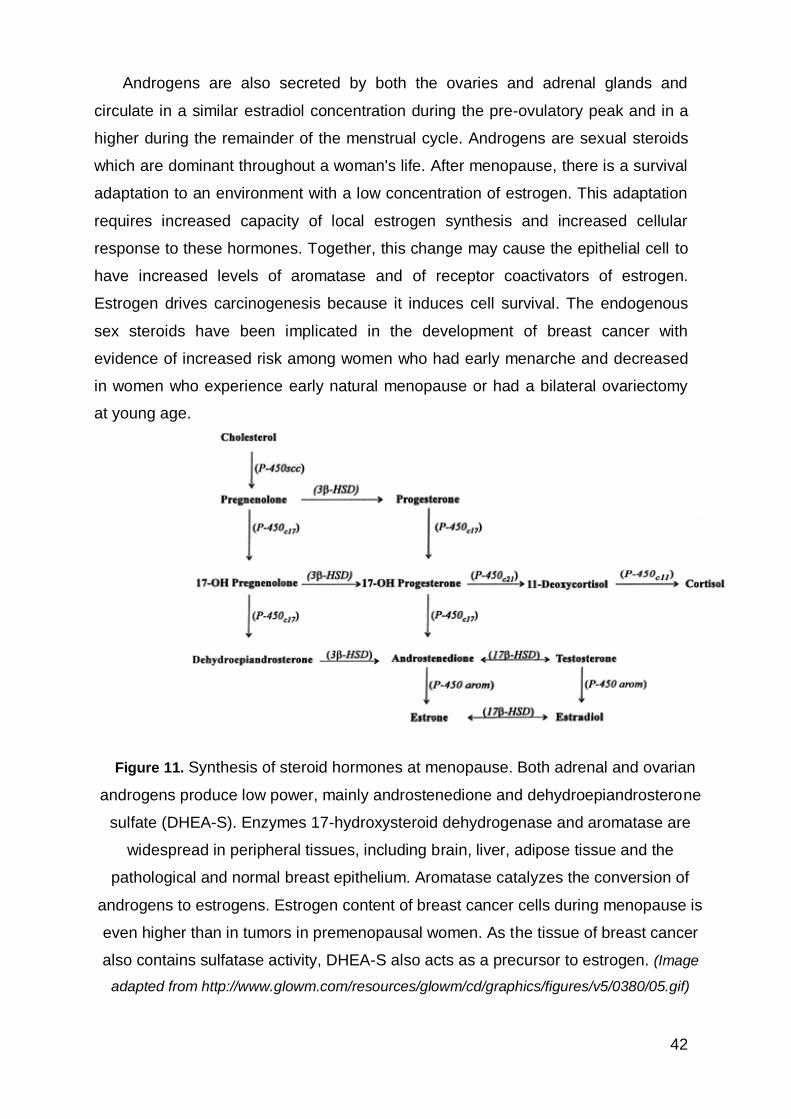
Figure 11. Synthesis of steroid hormones at menopause. Both adrenal and ovarian
androgens produce low power, mainly androstenedione and dehydroepiandrosterone
sulfate (DHEA-S). Enzymes 17-hydroxysteroid dehydrogenase and aromatase are
widespread in peripheral tissues, including brain, liver, adipose tissue and the
pathological and normal breast epithelium. Aromatase catalyzes the conversion of
androgens to estrogens. Estrogen content of breast cancer cells during menopause is
even higher than in tumors in premenopausal women. As the tissue of breast cancer
also contains sulfatase activity, DHEA-S also acts as a precursor to estrogen. (Image
adapted from http://www.glowm.com/resources/glowm/cd/graphics/figures/v5/0380/05.gif)
43
The androgen receptor is a member of the family of nuclear receptors that are
dependent on the binding of transcription factors. The AR has a higher affinity for
dihydrotestosterone (DHT) than for testosterone, the two most potent natural
androgens.
The process of binding of the hormone results in a conformational change in
the molecule that promotes the AR to spill cytosolic heat shock proteins, and
translocate to the nucleus. Once bound to a hormone, AR is able to form
homodimers that are associated with chaperones and nuclear coactivators and is
the active form of the receptor. The probability of accumulating, uncorrected DNA
replication errors and mutations, which are the basis of estrogen-related
tumorigenesis thus increases. Cells adapted to a low estrogenic environment
increase the number of cell cycles per year, these changes include androgen
aromatization to produce estradiol which is mitogenic in epithelial breast cells.
Hormone-related breast carcinogenesis after menopause possibly is a
consequence of subtle changes that allow the epithelial cells to survive in a low
estrogenic environment. Older age at diagnosis has previously been reported in
patients with AR-positive BC [44].
Besides being able to act as an estrogen precursor, androgen can directly act
on epithelial breast cells by activating the androgen receptor which modulates a
number of genes at the transcriptional level.
Information on of AR expression in breast cancer is sparse. Studies have
focused on the role of androgens and the androgen receptor and they have
demonstrated that androgens can lead to proliferation of AR expressing breast
cancer cell lines and promote tumor formation in animal models. AR is a marker of
good prognosis and this effect appeared independent of coexpression of ER. New
drugs targeting AR have been used in cases of BC. Despite an association with
good outcome, targeting of breast tumors expressing AR may be beneficial, similar
to the effects of pharmacologic targeting of ER (for example, bicalutamide, a non-
steroidal anti-androgen).
44
3. Objective
The main purpose of this study consisted in the evaluation of the expression of
androgen receptor in triple-negative breast cancer. The sample was collected at
Hospital CUF Descobertas, Lisbon, and consisted of all consecutive TNBC treated
and followed in the Institution in the years between 2005 to 2012.
TNBC due to its heterogeneous profile presents a specific problem in detecting
targets for systemic therapy.
Thus, in this study, we had a second objective. The relevance of the Lehmann
classification of TNBC cannot be ascertained because this classification has not
been adapted to IHC. We used, together with the androgen receptor, the following
antibodies: CK5/6 (cytokeratin 5/6), p-cadherin, EGFR (epidermal growth factor
receptor) and PHH3 (phosphohystone H3) to further separate TNBC.
Gene expression profiles signature derived from the most differentially
expressed genes found in the TNBC training set and used to predict which TNBC
subtype was best-fit for each of the tumors in the validation set.
In this study, distinct genes ontologies were used to each TNBC subtype. BL1
and BL2 subtypes were determined through the assessment of EGFR and CK5 /6
and a proliferation marker PHH3. The BL1 subtype expressed higher levels of basal
cytokeratin expression; enriched in cell cycle, cell division components and
pathways and the highly proliferative nature. The BL2 subtype displays unique gene
ontologies involving growth factor signaling. Expression of p-cadherin was used for
the identification of mesenchymal. This subtype displays a variety of unique gene
ontologies that are heavily enriched in components and pathways involved in cell
motility; epithelial-mesenchymal transition is associated. The immunomodulatory
TNBC subtype will be assess through the evaluation of TIL (tumor infiltration
lymphocytic). And for last, the LAR (luminal androgen receptor) subtype was
determined by the androgen receptor.

45
4. Materials and methods
Patients diagnosed with triple-negative breast cancer, between the years 2005-
2012, were included in this study from Hospital CUF Descobertas e Hospital Infante
Santo.
The initial sample included 80 cases of TNBC. Because of lack of information
from the patients and paraffin blocks from the Hospital CUF Infante Santo (patients
with an incomplete record and/or patients without tissue available to be studie), the
sample includes only patients from Hospital CUF Descobertas and comprises 66
patients with TNBC.
4.1 Clinical-pathological method
Clinicopathological parameters including age, type of surgery, tumor size,
histological grade, node involvement, adjuvant treatment, disease free survival
(DFS) and Overall survival (OS) were reviewed.
The patients' age categorized in less than 35 years, from 35 to 50 years, from
50 to 65 years and over 65 years.
Ppathological data correspond to the size of the tumor, lymph node involvement
and histological grade were also analysed. The size of tumor was categorized as <
2cm, between 2cm and 5cm, >5cm the diameter and/or not determined. For lymph
node involvement was described as positive or negative involvement, and/or not
determined. The histological grade was classified in 1 (well), 2 (moderate), 3
(poorly) and/or not determined (Scarff-Bloom-Richardson grading system).
Adjuvant treatment refers to the administration of both chemotherapy (CT) and
radiotherapy (RT) treatments.
DFS was defined as the interval (mouths) from primary surgery data until the
first relapse of disease.
46
OS was the time, in months, from the date of the primary surgery data to the
time of breast cancer related death.
The information from patients was collected from electronic patient records and
hospital charts. Before study initiation, Institutional ethics committee approval was
obtained.
4.2 Histopathological method
All the material collected had be fixed in 10% formaldehyde included in paraffin
(type 6, Richard said AllScientic ®), following the usual methodology. After the
inclusion, slices of 2,5 -3 μm thick were performed in a manual Minot microtome,
manual (RM2255 leica Microssystems). Then, the sections were placed on slides
and held drying in an oven (60 min at 60ºC or overnight at 37ºC). After drying in a row
in decreasing alcohol concentration (70%, 96% and absolute alcohol -100%),
proceeded to staining with Hematoxylin (Harris) and Eosin (alcoholic), about 40min-
1h.
The histopathologic review was made by pathologist with experience in breast
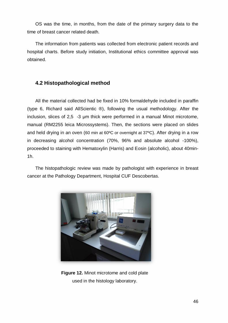
cancer at the Pathology Department, Hospital CUF Descobertas.
Figure 12. Minot microtome and cold plate
used in the histology laboratory.
47
4.3 Immunohistochemical method
The blocks for Immunohistochemical analysis of patients with TNBC subtype
were obtained from the Pathology files, Hospital CUF Descobertas. After obtaining
high quality paraffin blocks, the study comprised only 50 cases. The experimental
work was conducted at the Faculty of Medicine, University of Lisbon (FMUL) at the
Institute of Pathology, in collaboration with the Technical Catarina Isabel Talina.
After obtaining the blocks of patients with triple-negative breast cancer, 2μm
thick slices were made and placed in adherent SuperFrost®Plus slides, which then
are putted in the oven to dry for 60 min.
Immunohistochemistry is a set of methodologies that use antibodies as specific
reagents that are able to identify and connect with tissue constituents that act as
antigens. This connection allows locating and identifying the presence of various
substances in cells and tissues by means of color that is associated with the
antigen-antibody complexes formed [52].
The possibility of combining a marker with an antibody without causing any
damage to the specific connection established between antibody and antigen
makes this technology area heavily used in Pathology. This provides the
microscopic observation of the location where is the antibody and hence the
antigen. Apart from the quality of the antibody, the marking quality also depends on
the pre-analytical phase (highlighting tissue fixation and processing), the recovery of
antigenic epitopes and sensitivity of the detection system.
In this study, the immunohistochemical method used was the method of indirect
polymer. This method applies a primary antibody directed against the desired
antigen, and subsequently applies a polymer to which they are coupled "secondary"
antibody substances and enables visualization of HRP (Horseradish Peroxidase). It
is a quick and easy method, showing a decrease of error factors.
This method enables amplification of weak signals i.e. proteins of low
abundance can be visualized. It is however, a very expensive method.
48
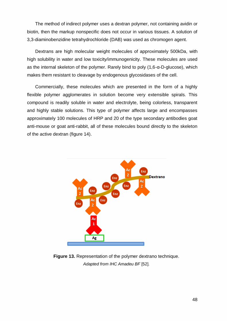
The method of indirect polymer uses a dextran polymer, not containing avidin or
biotin, then the markup nonspecific does not occur in various tissues. A solution of
3,3-diaminobenzidine tetrahydrochloride (DAB) was used as chromogen agent.
Dextrans are high molecular weight molecules of approximately 500kDa, with
high solubility in water and low toxicity/immunogenicity. These molecules are used
as the internal skeleton of the polymer. Rarely bind to poly (1,6-α-D-glucose), which
makes them resistant to cleavage by endogenous glycosidases of the cell.
Commercially, these molecules which are presented in the form of a highly
flexible polymer agglomerates in solution become very extensible spirals. This
compound is readily soluble in water and electrolyte, being colorless, transparent
and highly stable solutions. This type of polymer affects large and encompasses
approximately 100 molecules of HRP and 20 of the type secondary antibodies goat
anti-mouse or goat anti-rabbit, all of these molecules bound directly to the skeleton
of the active dextran (figure 14).
Figure 13. Representation of the polymer dextrano technique.
Adapted from IHC Amadeu BF [52].
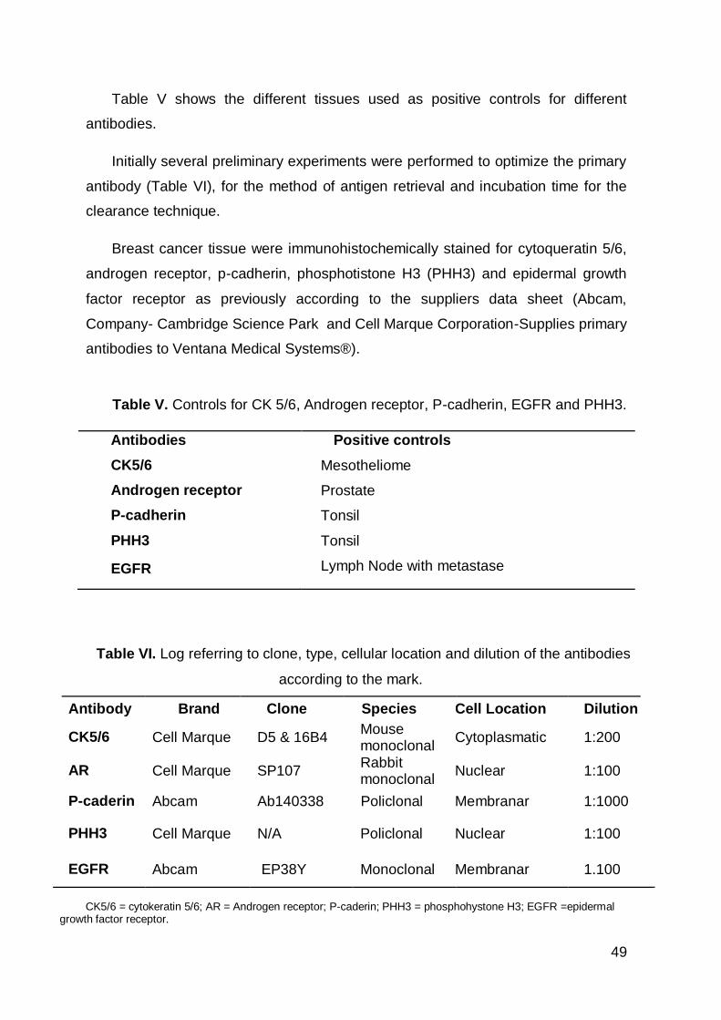
49
Table V shows the different tissues used as positive controls for different
antibodies.
Initially several preliminary experiments were performed to optimize the primary
antibody (Table VI), for the method of antigen retrieval and incubation time for the
clearance technique.
Breast cancer tissue were immunohistochemically stained for cytoqueratin 5/6,
androgen receptor, p-cadherin, phosphotistone H3 (PHH3) and epidermal growth
factor receptor as previously according to the suppliers data sheet (Abcam,
Company- Cambridge Science Park and Cell Marque Corporation-Supplies primary
antibodies to Ventana Medical Systems®).
Table V. Controls for CK 5/6, Androgen receptor, P-cadherin, EGFR and PHH3.
Antibodies Positive controls
CK5/6 Mesotheliome
Androgen receptor Prostate
P-cadherin Tonsil
PHH3 Tonsil
EGFR Lymph Node with metastase
Table VI. Log referring to clone, type, cellular location and dilution of the antibodies
according to the mark.
Antibody Brand Clone Species Cell Location Dilution
CK5/6 Cell Marque D5 & 16B4 Mouse monoclonal
Cytoplasmatic 1:200
AR Cell Marque SP107 Rabbit monoclonal
Nuclear 1:100
P-caderin Abcam Ab140338 Policlonal Membranar 1:1000
PHH3 Cell Marque N/A Policlonal Nuclear 1:100
EGFR Abcam EP38Y Monoclonal Membranar 1.100
CK5/6 = cytokeratin 5/6; AR = Androgen receptor; P-caderin; PHH3 = phosphohystone H3; EGFR =epidermal
growth factor receptor.
50
The immunohistochemical analysis followed the following protocol:
Dewaxing: Place slides in the sections marked with study at 60°C for 60min. Allow
to dry prior to antigen retrieval for 20 minutes (90°C)
Antigen retrieval: place the rack with the blades filled by antigen retrieval buffer PT
module (15ml Buffer Cell Marque Trillogy R 1: 400 in 1485 ml of distilled water) in
Electric Pressure camera to immunohistochemistry for 1 hour on high / low
pressure; at the end, removed and allowed to cool approximately 37 min in distilled
water.
Dehydrate until absolute alcohol ( 70%, 96%, 100%)
Blocking of endogenous peroxidase: the blades were passe by 4 ml bath of
hydrogen peroxide diluted in 200mL methanol for 15 min, rinse in distilled water.
Washes with TBS: two washes with TBS, 5 min each.
Dilution of antibodies that will be used: with the solution (antibody diluents;
Diamond) as the blades are in phase antigen retrieval.
Application of the primary antibody (for 60 minutes)
Washes with TBS: two washes with TBS, 5 min each.
Immunoenzymatic labeling method (HRP): 10 minutes applies an amplifier twice,
five minutes each; applies then the polymer for 10 minutes twice, five minutes each.
Revelation with DAB ((3,3′-Diaminobenzidine tetrahydrochloride hydrate) HRP
substrate; Cell Marque chromogens) for 3 minutes solution 50 ml of chromogen
more 1000 ml substrate.
Washing with tap water to remove excess
Counter staining with Mayer's Hematoxylin for a few seconds
Wash in warm water
Dehydration of the cuts to the absolute alcohol - xylene.
Clearing and mounting the slides in xylene
51
5. Statistic Analysis
The statistical analyzes was performed using SPSS for Windows (Version 17).
Survival analysis was performed with the Kaplan-Meier method. The Kaplan-Meier
estimate is also called as “product limit estimate” and it is the simplest way of
computing the survival over time in spite of all the variability associated with cancer
patients and cancer follow-up. In clinical trials, the effect of an intervention is
assessed by measuring the number of subjects whose survival increased or
decreased after that intervention over a period of time. The survival curve provides
a measure of mortality of patients with TNBC.
The results of the immunohistochemical analysis of the expression of Ck 5/6
were defined by intensity of staining and percentage of tumor cells. The expression
of stained cells was divided into four categories: absence or weak labeling (0); weak
staining and less than 1/3 of the cells (1+); moderate intensity staining of 2/3 of cells
(2+) and strong staining of 3/3 (3+). The same was applied to the methodology of
EGFR protein (score 0-3+). The marking was checked for EGFR membrane
labeling. The expression of p-cadherin was staining according to positivity or
negativity of the marked tissue. Expression of the androgen receptor was
determined by nuclear expression of this protein (≥ 10%).
Proliferation index is calculated the counting of PHH3 by expression of nuclear
tumor cells. The percentage of tumors cells expressing the proliferation marker
PHH3 determined the growth rate of tumors. The proliferation index was calculated
by the total number of PHH3- positive nuclear profiles. Due to poor tissue fixation,
some cases were excluded.
Inflammatory infiltration was classified in absent, moderate, slight, intense and
not determined.
52
6. Results
The clinical characteristics of the patients included in this study are presented in
table VII. We studied samples of 66 patients with TNBC obtained from Hospital CUF
Descobertas, Lisbon, between 2005 and 2012.The clinical chart data were reviewed
and analyzed. The majority of patients were middle aged between 50 and 65 years
old. The median age was 60 years. About 89% of patients underwent surgery. The
majority of these patients also presented a tumor grade II or III. The tumor size had
a mean 2,1 cm and a median of 1,8 cm.
TNBC patients had positive axillary nodal involvement was found 74%. In total,
44% of the patients underwent chemotherapy and radiotherapy, 79% underwent
chemotherapy.
Subsequently, the median follow-up time for the time diagnosis until the disease
free survival (DFS) was 12 months (range, 0-24months). Overall survival for that
death population was 3 years (range, 1-6 years). TNBC phenotype was associated
with poorer outcome in terms of overall survival and disease-free interval when
compared to publish cohorts of BC patients.
The Kaplan-Meier method was used to estimate time-to-event functions.
Approximately 75% of patients remain without evidence of disease (Figure 14A).
The calculation was made in a range of time in months, we found a median of 12
months and a mean of 11 months to relapse of TNBC. The calculation of OS
(Figure 14B) was made in a range of time, in years, with a median of 3 years and a
median of 3.28 years from the diagnosis to date of death. DFS and the OS were
obtained in a confidence interval of 95%.
Most patients received additional neoadjuvant chemotherapy following FEC
(Fluorouracil (5-FU), epirubicin, cyclophosphamide) plus docetaxel.
The most common site of initial relapse was bone. Other sites involved in
relapse are lymph nodes, central nervous, system lung and liver.
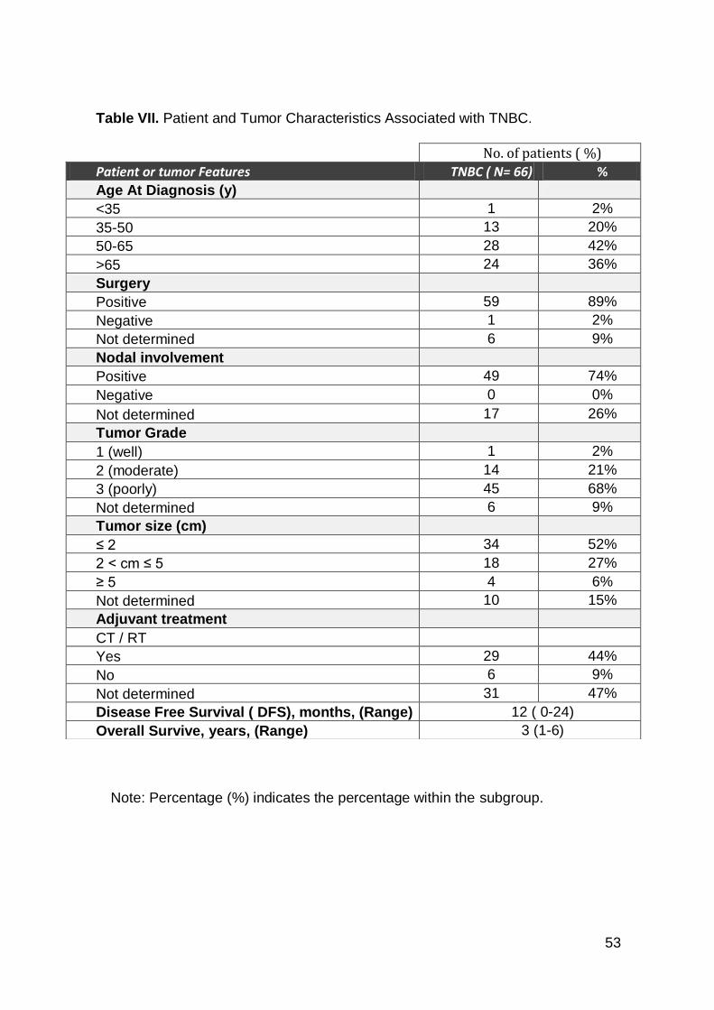
53
Note: Percentage (%) indicates the percentage within the subgroup.
Table VII. Patient and Tumor Characteristics Associated with TNBC.
No. of patients ( %)
Patient or tumor Features TNBC ( N= 66) %
Age At Diagnosis (y)
<35 1 2%
35-50 13 20%
50-65 28 42%
>65 24 36%
Surgery
Positive 59 89%
Negative 1 2%
Not determined 6 9%
Nodal involvement
Positive 49 74%
Negative 0 0%
Not determined 17 26%
Tumor Grade
1 (well) 1 2%
2 (moderate) 14 21%
3 (poorly) 45 68%
Not determined 6 9%
Tumor size (cm)
≤ 2 34 52%
2 < cm ≤ 5 18 27%
≥ 5 4 6%
Not determined 10 15%
Adjuvant treatment
CT / RT
Yes 29 44%
No 6 9%
Not determined 31 47%
Disease Free Survival ( DFS), months, (Range) 12 ( 0-24)
Overall Survive, years, (Range) 3 (1-6)

54
A
Figure 14. A. Disease-free survival curve for patients with TNBC.
Probability of survival estimated using the Kaplan-Meier method. B.
Overall-survival curve of patients with TNBC. Probability of survival
estimated using the Kaplan-Meier method.
B
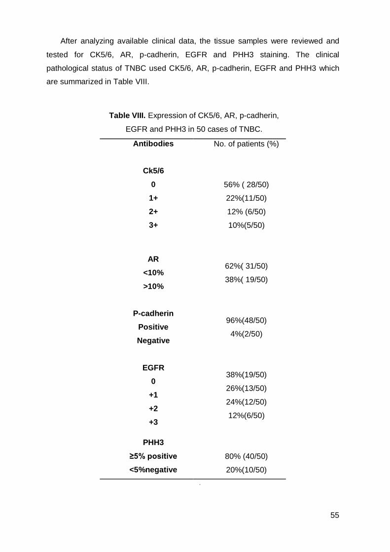
55
After analyzing available clinical data, the tissue samples were reviewed and
tested for CK5/6, AR, p-cadherin, EGFR and PHH3 staining. The clinical
pathological status of TNBC used CK5/6, AR, p-cadherin, EGFR and PHH3 which
are summarized in Table VIII.
Table VIII. Expression of CK5/6, AR, p-cadherin,
EGFR and PHH3 in 50 cases of TNBC.
Antibodies No. of patients (%)
Ck5/6
0
1+
2+
3+
56% ( 28/50)
22%(11/50)
12% (6/50)
10%(5/50)
AR
<10%
>10%
62%( 31/50)
38%( 19/50)
P-cadherin
Positive
Negative
96%(48/50)
4%(2/50)
EGFR
0
+1
+2
+3
38%(19/50)
26%(13/50)
24%(12/50)
12%(6/50)
PHH3
≥5% positive
<5%negative
80% (40/50)
20%(10/50)
.
56
Of all the TNBC cases, 56% (28/50) were CK5/6, 0; 22% (11/50) were CK5/6,
1+; 12% (6/50) were CK5/6, 2+; and 10% (5/50) were CK5/6, 3+. The expression of
AR was calculated by upper or lower than 10% of the labeled cells staining. In some
cases with higher expression of AR, we attributed scores of 50%, 90% and/or
100%. Thus, there were 62% of cases being lower than 10% and 38% of cases
where there was more than 10% AR labeling. In 96% (48/50) the expression of p-
cadherin was positive while only 4% (2/50) was negative. The 38% (19/50) were
EGFR, 0; 26% (13/50) were EGFR, 1+; 24% (12/50) were EGFR, 2+; and 12%
(6/50) were EGFR, 3+.
In this work we assessed proliferation through labeling our samples with the
PHH3 antibody. PHH3 is a core histone protein, which together with other histones
forms the major protein constituents of the chromatin in eukaryotic cells.
Immunohistochemical studies performed with anti-PHH3 antibody have shown that
the antibody detects specifically the core protein histone H3 only when is
phosphorylated at serine 10 or serine 28. The phosphorylation of histone H3 is a
rare event in interphase cells but is a process almost exclusively occurring during
mitosis. Studies have also revealed no phosphorylation on the histone H3 during
apoptosis. Therefore, PHH3 can serve as a very effective mitotic marker.
A Ki-67 cut-off point of 15% was defined according to the experience of different
pathologists as well as national and international recommendations at present [53].
In this study, less than 5% staining with the antibody is considered low for the
proliferation index and greater than or equal to 5% is considered high for PHH3
proliferation index. About 80% (40/50) of the sample of TNBC have a high nuclear
staining of PHH3 and around 20% (10/50) are considered low.
Inflammatory infiltration (IF) has been considered as a likely prognostic factor in
malignant neoplasms, and in this study was classified as absent, slight, moderate,
intense and not determined (table IX). This rating was assessed in the samples
used for PHH3 staining with the analysis of the antibody PHH3. In 34% (17/50) of
TNBC cases IF was classified as absent; in 16% (8/50) of TNBC cases IF was
classified as moderate; in 22% (11/50) of TNBC cases IF was classified as slight
and in 8% (4/50) TNBC cases IF was classified as intense inflammatory infiltration.
57
The remaining cases were called “not evaluable” for above mentioned reasons and
represent 20% (10/50) of cases TNBC.
In this series of TNBC, basal phenotype was defined by expression of CK5/6,
EGFR and PHH3 in tumor cells; mesenchymal phenotype of TNBC was defined by
expression of p-cadherin protein; immunomodulatory phenotype of TNBC was
defined by the evidence of lymphocytic infiltration and luminal receptor androgen
phenotype was defined for the expression of AR. The table X summarizes TNBC
subtypes and markers used in this study.
Microphotographs showing the morphology and immunohistochemical results
for CK5/6, p-cadherin, EGFR, AR and PHH3 in breast tissue (figures 15-18).
Immunohistochemical expression of CK5/6 was observed by cytoplasmatic staining
(figure 15), p-cadherin (figure 16A) and EGFR (figure 16B) positive cells were
observed by membranar staining and the AR (figure 17) and PHH3 (figure 18)
observed by nuclear staining.
Table IX. Lymphocytic infiltrate
absent 17 34%
slight 11 22%
moderate 8 16%
intense 4 8%
Not available 10 20%
Table X. TNBC subtypes and markers.
Subtype Markers
Basal -like CK5/6, EGFR, PHH3
Mesenchymal (M,MSL) P-cadherin
Immunomodulatory Lymphocytic infiltration
Luminal androgen
receptor AR
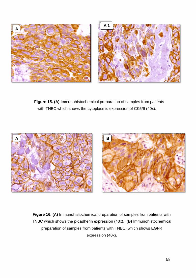
58
Figure 16. (A) Immunohistochemical preparation of samples from patients with
TNBC which shows the p-cadherin expression (40x). (B) Immunohistochemical
preparation of samples from patients with TNBC, which shows EGFR
expression (40x).
A
B
A.1
Figure 15. (A) Immunohistochemical preparation of samples from patients
with TNBC which shows the cytoplasmic expression of CK5/6 (40x).
A
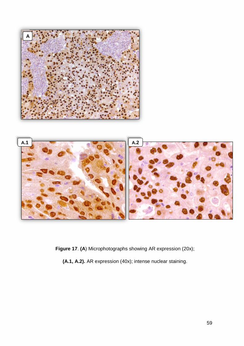
59
A
A.1
Figure 17. (A) Microphotographs showing AR expression (20x);
(A.1, A.2). AR expression (40x); intense nuclear staining.
A.2
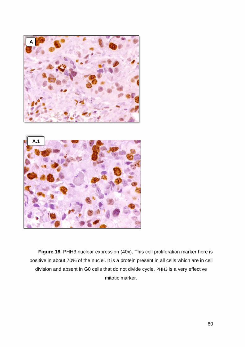
60
Figure 18. PHH3 nuclear expression (40x). This cell proliferation marker here is
positive in about 70% of the nuclei. It is a protein present in all cells which are in cell
division and absent in G0 cells that do not divide cycle. PHH3 is a very effective
mitotic marker.
D.2
A
A.1
61
7. Discussion
Breast cancer is a heterogeneous disease. Currently, breast cancer, relies on
traditional prognostic factors including nodal status, tumor histological grade and
primary tumor size, in addition on expression to ER, PR and HER2. The molecular
identification has allowed the classification of subtypes of BC, however it remains
difficult to predict response to therapy. Surgery, radiotherapy and chemotherapy
individually or in combination, are the only available modalities for patients with
TNBC. Patients with TNBC do not benefit from hormonal or trastuzumab-based
therapies, because of the loss of target receptors such as ER, PR and HER2 [32].
TNBC tumors were mainly of high histological grade, high mitotic index and
were found more frequently in old age women [32]. The most common histological
types were carcinomas of no specific type, metaplastic and apocrine carcinomas,
the majority of these tumors were grade III. 80%-90% of TNBC truly have the basal
molecular phenotype, which is known to be associated with worse OS [25].
This study shows the expression of five antibodies in cases of TNBC. These
results attempted to apply to IHC the Lehamnn classification of TNBC subtypes
performed in with genomics.
The expression profile of cytokeratin intermediate filaments is used in clinical
practice and has been applied for the differentiation of metastatic tumors of
unknown origin and currently is being used with interest to mark the cells of the
mammary gland [54]. Cytokeratins have an important role in the cell motility, cell
membrane support, forming pathways for the transport of organelles and other
elements in the cytosol, under constant rearrangements that can produce cell
movements [55] [56]. Cytokeratins type II (CK5 and 6) and type I (CK14) stain basal
and / or myoepithelial cells, while antibodies to cytokeratins type II (CK7 and 8) and
type I (CK18 and 19) are associated with the luminal/glandular epithelial cells of the
breast [54].
The expression profile of cytokeratin CK5/6 and CK14 is interesting for studying
BC. These markers have been used in the identification of basal-like TNBC. Since
the early work of Sorlie et. al. [30], the terminology and definition (morphological
62
and genomic) of basal-like cancers, remains controversial. Nielsen et. al. [37]
proposed an immunohistochemical panel of four antibodies (ER, HER2, HER1 and
CK5/6), although there is still no exact correlation with the genomic expression
profiles determined by DNA microarrays. Recent European studies propose the use
basal expression of cytokeratins (CK5/6 and/or CK/14) as the defining element of
the phenotype of basal-like BC. To identify the basal TNBC, Rakha et. al. recently
proposed to employ the criteria [57]: expression of CK5/6 and/or CK/14. In this
study, for characterization the basal TNBC we chose CK5/6, EGFR and PHH3
markers.
In this work, the expression of CK5/6 is negative in 56% of the case is positive
in 44% of these cases. The expression of EGFR is positive in 62% of cases and the
expression of PHH3 is high (80% the cases of TNBC). This finding allows us to
suggest that the subtype that is more dominant in the set, the group BL2 because
the high percentage of EGFR and proliferation marker.
EGFR is a cell surface protein which, is activated by binding of epidermal
growth factor (EGF), through tyrosine-kinase activity. EGFR gene amplification has
been described in different tumors, especially in the central nervous system, lung
and stomach [58]. Recently, protein expression of EGFR has been described in
breast carcinomas especially in metaplastic carcinoma, which although less
frequent, is inserted into the differentiation pathway of progenitor/myoepithelial cells
of the mammary carcinogenesis [59] [60]. EGFR immunoreactivity in TNBC IDC
(triple-negative breast cancer, invasive ductal carcinoma) of the breast emerges as
a clinically relevant prognostic parameter. Recently reported studies already
indicated that EGFR expression is associated with significantly worse DFS in early
breast cancer [61]. A significant proportion of the patients with TNBC IDC might
have tumors concomitantly showing intratumoral necrosis [28], as well as EGFR
and basal cytokeratin immunoreactivity [46]. This may explain why these closely
associated factors are not all independently predicting DFS or OS when taken
together in the full regression models [61].
The adherens-type junctions are related in normal development, to the role of
cell sorting during embryogenesis and in the maintenance of specific organ and
adult tissue architecture. Any disorders involving dysfunction of classical
63
cadherins/catenins are related to various disease states, including cancer. P-
cadherin is transiently expressed in various tissues during development and its
permanent expression is limited to adult epithelial tissues, at cell-cell boundaries.
The use of particular markers that are expressed only in basal cells (or
myoepithelial cells) of the mammary gland have led to a recently developed strategy
to improve the diagnosis, prognosis and treatment in these tumors. In various
human tumors, p-cadherin expression is altered, but its role in the carcinogenic
process remains unclear because it behaves differently depending on the cancer
cell model studied [62].
The expression of p-cadherin is well established as an indicator of poor
prognosis in human breast cancer [62]. P-cadherin stimulated our interest in
studying its role in TNBC and the results were consistent with what was reported in
literature. The p-cadherin is over expressed in TNBC, 96% of cases were positive
for this marker.
Several studies analyze lymphocyte infiltration in breast cancer and in fact they
show that inflammatory cell infiltration of tumors contributes either positively or
negatively to tumor invasion, growth, metastasis, and patient outcomes, which
creates a conundrum when examining mechanisms of action. TIL (tumors infiltrative
lymphocytes) has shown to be predictive of prognosis in breast cancer and it has
been taking emphasis. The correlation between intratumoral immune responses
and clinical outcomes in TNBC is potentially related to the role of immune cells in
the activity of cytotoxic chemotherapeutics. In mice, chemotherapy with
anthracyclines requires priming of IFNγ- producing CD8+ T cells [63]. In humans,
correlative studies have reported that high intratumoral levels of IFNγ and CD8+ T
cells [64], or TILs, are associated with better clinical responses to anthracycline-
based chemotherapy [65].
The immunomodulatory subtype of TNBC was determined by the expression of
lymphocytic infiltration. This assessment in our work was not technically optimized
and was not planned because many of the cases don´t have good tissue
preservation. Evaluation of TIL is thus compromised.
64
More recently, attention has focused in androgens and androgen receptor (AR)
as prognostic markers and therapeutic targets in breast cancer [66]. The
significance of AR expression is less well characterized when compared with the
prognostic significance of ER and PR expression, in breast cancer and their roles in
predicting therapy.
The implication of androgens in breast carcinogenesis was tested in animal
models and this showed that testosterone in combination with estrogen can induce
mammary carcinomas. The mammary gland density values were significantly higher
in animals treated with testosterone alone or in combination with 17β-oestradiol
than in those treated with 17β-oestradiol alone or in controls. The strong expression
of androgen receptor in epithelium increased the amount of perialveolar and
interlobular connective tissue and the proliferation rate of fibroblast-like cells in the
stroma and had decreased the surrounding adipose tissue. All these changes were
blocked by simultaneous implantation of flutamide, indicating that androgens play a
crucial role in the process despite the absence of androgen receptors in stromal
cells [67].
Several studies have focused on ER-positive tumors and have shown some
association between AR status and disease-free survival, as well as response to
endocrine therapy, but these findings have not been confirmed in AR positive and
ER negative [68] [69, 70]. The ER-negative tumors characteristically have a poorer
prognosis and are thought to respond poorly to endocrine therapy [71].
In one of the largest studies of AR status in breast cancer has found a highly
significant association between AR status and survival, that remained significant
even when taking ER status into account [72]. AR status was a significant
prognostic factor for disease-free survival in univariate analysis, in multivariate
analysis, only lymph node status, tumor size, and ER status were independent
prognostic variables [68].
Curiously, in our study, the expression of androgen receptor is low. The sample
reveals 38% positive case for AR expression. This observation shows that AR is not
the most frequently expressed marker. Although statistical significance was lacking
in our univariate analysis, a larger sample and additional research may help clarify
whether AR status is a significant independent prognostic factor. The functional role
65
of AR in breast cancer remains unclear, further exploration of this area could
expand the repertoire of potential treatments for patients with TNBC.
In this sample, IHC identified the subtypes of TNBC. The biologic validation of
all the cases studied by conventional immunohistochemical techniques is laborious,
expensive and time consuming. Given the high degree human error and difficulties
to maintain its performance under identical conditions of temperature, concentration
of reactants, even with the automation of some of the steps involved, there are
numerous reports of conflicting results in the literature.
Additional markers [24, 32] are being sought to further refine those
classifications, especially in patient subgroups such as TNBC, whose response
therapy cannot be predicted accurately by conventional parameters. In this study
we obtained the expression of the five markers in our work the expression of a
single marker cannot be quantified due to very frequently overlap by the others
markers. In clinical practice, this is what happens. Lehamnn starting from an
analysis of gene expression was able to individualize each marker and accurately
classify the subtypes of TNBC. It is important to note, the level of difficulty in clinical
practice to get a correct diagnosis, for better targeted therapeutics. TNBC is an
unclarified entity, despite all the advances in trying to respond to this subtype of BC,
it requires more research for better pathological, clinical and therapeutic
clarification.
Clinical trials evaluating the efficacy of certain receptors as targets for new
therapeutic drugs have been performed, for example [40]:
1- Anti-EGFR and C-kit [37] the expression of EGFR has been reported in 66%
of BC patients with TNBC tumors cells and basal-like tumors cells. Clinical trials
evaluating the efficacy of humanized anti-EGFR monoclonal antibodies and EGFR
tyrosine kinase inhibitors in TNBC are currently under way;
2- In vitro studies have indicated that breast tumor cells from BRCA1 germline
mutations are extremely sensitive to a damaging high importance of these agents is
based on the BRCA1 pathway and DNA repair dysfunction seen in TNBC, which
may confer enhanced sensitivity to agents that damage DNA; Cells null for BRCA1
were shown to be incapable of repairing DNA double strand breaks [73];
66
3- The agents that inhibit PARP1 have successfully completed phase II and
phase III studies have begun [74]. The PARP1 inhibitors effectively impair the ability
of cancer cells to repair DNA damage causing the death on these cells;
4- Several genes have been reported to be amplified in TNBC. Many
components of the proliferation pathways (Ras, MEK/ ERK) are overexpressed or
mutated in cancer cells [40]. This can represent potential targets for TNBC. Genetic
aberrations, such as gain of 1q, 3q, 7q, 8q and 10p and loss of 4p, 5q, 17p and 8p,
have also been found in tumors arising in BRAC1 mutations carriers [75].
In this study we predicted that TNBC tumors could belong to the mesenquimal
subtype by high expression of p-cadherin and/or, a sizable part of the sample could
belong to the basal-like subtype, through the expression of CK5/6, EGFR and
PHH3. However this observation is inconclusive, given the fact that we obtained a
lot of overlap between the classifiers we used. The scientific advances with a view
to clinical practice should move in this direction as future goals to better
characterize subtypes of TNBC.
67
8. Conclusion
Since the sequencing of the human genome in 2000, the ability to predict and
treat cancer has increased. Genomic date will be generated for all tumors in the
future and we will know the genetic abnormalities between normal and tumor tissue,
to be able to design effective targeted therapies [76].
In the era of precision medicine, enabled by affordable next generation
sequencing oncologists are often faced with patients requiring treatment and reports
describing mutations, amplifications or deletions of oncogenes and tumor
suppressor genes. The vision for the treatment of TNBC focuses on potential
molecular targets and biomarkers that can be designed or analyzed for the
prediction of response of TNBC patients to chemotherapy, radiotherapy and/or
biologic targeted therapy.
TNBC is characterized by the absence of ER, PR and HER2 expression these
are very aggressive tumors with poor prognosis. This aggressiveness is best
illustrated by the fact that the peak risk of recurrence is between the first and third
years and the majority of deaths occur in the first 5 years following therapy. An ideal
scenario would allow hypotheses around resistance pathway activities, optimal
target identification and target combination strategies to be studied driven by
pharmacodynamic and short term efficacy biomarker endpoints in accessible
residual disease tissue and in blood. Trial designs and platform approaches that are
in development to address these points [77].
Currently applied regimens are mostly based on a combination of
anthracyclines and taxanes which suggests again the importance of further
comparative studies evaluating the efficacy with these standard approaches based
or other therapies in TNBC[74].
It is apparent that patients with AR positive TNBC tumors are likely to
demonstrate limited benefit from the current standard-of-care chemotherapy
regimens for TNBC, and then it will be necessary to study with targeted
investigational combination treatments [71].
68
The molecular apocrine breast cancer subset has recently been identified. LAR
cells are in part dependent on AR signaling as siRNA-mediated AR knockdown or
pharmacological inhibition of AR by bicalutamide (CDX) greatly decreases cell
viability and tumor growth- hormonally responsive cancers. CDX as a single agent
in metastatic AR positive, ER/PR negative breast cancers (NCT00468715/
TBCRC011) has demonstrated some efficacy with a clinical benefit rate of 19% [78].
Tumors with AR expression are more likely to harbour PIK3CA mutations. The
combination of AR antagonism and PI3K inhibition has found an additive or
synergistic effect on AR positive TNBC in cell growth. AR is described as a
biomarker for the selection of TNBC patients for clinical trials, which would
investigate the efficacy of therapeutic combinations that simultaneously target AR
and PI3K [79].
TNBC patients when treated with anthracyclines, cyclophosphamide and
taxanes had overall pathological complete response (pCR) of 28% and patients with
the AR subtype had a poor response with only 10% achieving pCR prior to surgery.
Clinically, AR negative patients have a higher likelihood of achieving pCR with
neoadjuvant chemotherapy than AR positive patients [80].
The effective inhibition of sex hormones can be a therapeutic target in patients
that present AR expression. Seen that oestrogens and androgens act together in
mammary carcinogenesis, with estrogen could possibly be the predominant initiator
whereas the androgen acts as a great promoter of carcinogenesis of the mammary
gland [67].
Paracrine influences of factors secreted by epithelial cells, such as transforming
growth factor (TGF) β is an important growth factor involved in epithelial–stromal
interaction. The important point concerning TGF-β1 in epithelial–stromal interaction
in carcinogenesis is its various effects on tumors host tissue, particularly with
respect to angiogenesis, immunosuppression, fibroblast activation and restructuring
of the tumors extracellular matrix. Although TGF-β1 is a powerful inhibitor of normal
epithelial cell proliferation, its effect on epithelial tumor cells is slight in adenomas
and totally lost in carcinomas. T cell stimulation may act through the AR on
mammary epithelial cells. Upon stimulation by T cells, the epithelial cells secrete

69
TGF-β1, which can act on stromal cells to stimulate growth of the stroma. This may
explain growth of the stroma on androgen stimulation [67].
Emerging technological advances in proteomics and transcriptomics requiring
complex bioinformatics approaches in the future will pose further challenges and
exciting opportunities. AR positive TNBC tumors are likely to demonstrate limited
benefit from the current standard-of-care chemotherapy regimens for TNBC. The
combination therapy would be the first trial in which TNBC patients are divided
based on AR expression and, as a result, aligned to a targeted investigational
combination treatment.
70
9. Future perspectives
As future perspectives, it will be necessary to increase the study sample to
clarify a few points, namely to be able to generate better statistical, pathological,
clinical and therapeutic results.
A major challenge would bring to the routine clinical practice a more
technological and accurate approach, as did Lehamnn.
Analysis of a genomic profile could classify the various subtypes of BC and a
better treatment would be applied, including hormone therapy. To validate genomic
results with IHC, the use of tissue matrix arrangements tissue microarrays (TMA)
has greatly facilitated the rapid and reproducible verification of the relevant
candidate genes and molecules. IHC is a relatively simple technique, whose
validation over conventional techniques is increasingly consolidated, having quickly
spread in major global research and clinical centers.
TMA and IHC have the potential to generate predictive and prognostic markers
in data sets of large numbers of samples.
71
References
1. Harris J , L.M., ,Morrow M, et al., ed. , Diseases of the Breast: Philadelphia:
Lippinoott Williams & Wilkins. 2. Visvader, J.E., Keeping abreast of the mammary epithelial hierarchy and breast
tumorigenesis. Genes Dev, 2009. 23(22): p. 2563-77. 3. STERNBERG, S.S., Histology for Pathologists. Pennsylvania: Lippincott-Raven
Publishers. 1991. 4. Stuart J. , N.H.G.e.a., Molecular Oncology of Breast Cancer. 5. Yu, M., et al., Circulating breast tumor cells exhibit dynamic changes in epithelial
and mesenchymal composition. Science, 2013. 339(6119): p. 580-4.
6. BreastCancer.org. http://www.breastcancer.org/symptoms/understand_bc/what_is_bc; Access in September 2013.
7. Polyak, K., Breast cancer: origins and evolution. J Clin Invest, 2007. 117(11): p.
3155-63. 8. OncologiaCUF. http://www.oncologiacuf.pt/; Access in January 2014.
9. National Breast Cancer Foundation. 2003;http://www.nationalbreastcancer.org/what-is-breast-cancer ; Access in January 2014.
10. Jemal, A., et al., Global cancer statistics. CA Cancer J Clin, 2011. 61(2): p. 69-90. 11. WHO. International Agency for Research on Cancer (IARC),Epidemiological data on
breast cancer in Portugal (2012);http://globocan.iarc.fr/Pages/fact_sheets_cancer.aspx?cancer=breast, access in September 2013
12. WHO and International Agency for Research on Cancer (IARC),Epidemiological
data on breast cancer in Portugal (2012);http://globocan.iarc.fr/Pages/fact_sheets_population.aspx?country=620; access in September 2013.
13. WHO. International Agency for Research on Cancer (IARC), Epidemiological data on breast cancer in Europe (2012);http://globocan.iarc.fr/Pages/fact_sheets_population.aspx?country=968 access in September 2013.
14. Ginsburg, O.M. and R.R. Love, Breast cancer: a neglected disease for the majority of affected women worldwide. Breast J, 2011. 17(3): p. 289-95.
15. Ferlay, J., et al., Estimates of worldwide burden of cancer in 2008: GLOBOCAN 2008. Int J Cancer, 2010. 127(12): p. 2893-917.
16. Li, C.I., J.R. Daling, and K.E. Malone, Incidence of invasive breast cancer by hormone receptor status from 1992 to 1998. J Clin Oncol, 2003. 21(1): p. 28-34.
17. Global Cancer Statistics: CA Cancer J Clin. American Cancer Society, Inc., 2011. 18. Cancer, S.A., Cancer Facts & Figures Atlanta: American Cancer Society, 2013. 19. Singletary, S.E., et al., Revision of the American Joint Committee on Cancer staging
system for breast cancer. J Clin Oncol, 2002. 20(17): p. 3628-36. 20. Fleming ID, C.J., Henson DE, et al (eds), AJCC Cancer Staging Manual (ed 5).
1997: Philadelphia, PA, Lippincott-Raven 21. Edge, S.B. and C.C. Compton, The American Joint Committee on Cancer: the 7th
edition of the AJCC cancer staging manual and the future of TNM. Ann Surg Oncol, 2010. 17(6): p. 1471-4.
72
22. Sinn, H.P. and H. Kreipe, A Brief Overview of the WHO Classification of Breast Tumors, 4th Edition, Focusing on Issues and Updates from the 3rd Edition. Breast Care (Basel), 2013. 8(2): p. 149-54.
23. Reis-Filho, J.S. and A.N. Tutt, Triple negative tumours: a critical review. Histopathology, 2008. 52(1): p. 108-18.
24. Perou, C.M., et al., Molecular portraits of human breast tumours. Nature, 2000. 406(6797): p. 747-52.
25. Stagg, J. and B. Allard, Immunotherapeutic approaches in triple-negative breast cancer: latest research and clinical prospects. Ther Adv Med Oncol, 2013. 5(3): p.
169-81. 26. Althuis, M.D., et al., Etiology of hormone receptor-defined breast cancer: a
systematic review of the literature. Cancer epidemiology, biomarkers & prevention :
a publication of the American Association for Cancer Research, cosponsored by the American Society of Preventive Oncology, 2004. 13(10): p. 1558-68.
27. Slamon, D.J., et al., Human breast cancer: correlation of relapse and survival with amplification of the HER-2/neu oncogene. Science, 1987. 235(4785): p. 177-82.
28. Yonemori, K., et al., Disruption of the blood brain barrier by brain metastases of triple-negative and basal-type breast cancer but not HER2/neu-positive breast cancer. Cancer, 2010. 116(2): p. 302-8.
29. Stanford, J.L. and N.S. Weiss, Hormone replacement therapy and breast cancer. Arch Fam Med, 1996. 5(6): p. 349-50.
30. Sorlie, T., et al., Gene expression patterns of breast carcinomas distinguish tumor subclasses with clinical implications. Proc Natl Acad Sci U S A, 2001. 98(19): p.
10869-74. 31. Althuis, M.D., et al., Etiology of hormone receptor-defined breast cancer: a
systematic review of the literature. Cancer Epidemiol Biomarkers Prev, 2004. 13(10): p. 1558-68.
32. Rakha, E.A., et al., Prognostic markers in triple-negative breast cancer. Cancer, 2007. 109(1): p. 25-32.
33. Sotiriou, C. and L. Pusztai, Gene-expression signatures in breast cancer. N Engl J Med, 2009. 360(8): p. 790-800.
34. Lehmann, B.D., et al., Identification of human triple-negative breast cancer subtypes and preclinical models for selection of targeted therapies. J Clin Invest, 2011. 121(7): p. 2750-67.
35. Darb-Esfahani, S., et al., Identification of biology-based breast cancer types with distinct predictive and prognostic features: role of steroid hormone and HER2 receptor expression in patients treated with neoadjuvant anthracycline/taxane-based chemotherapy. Breast Cancer Res, 2009. 11(5): p. R69.
36. Tanner, M., et al., Chromogenic in situ hybridization: a practical alternative for fluorescence in situ hybridization to detect HER-2/neu oncogene amplification in archival breast cancer samples. Am J Pathol, 2000. 157(5): p. 1467-72.
37. Nielsen, T.O., et al., Immunohistochemical and clinical characterization of the basal-like subtype of invasive breast carcinoma. Clin Cancer Res, 2004. 10(16): p. 5367-
74. 38. Dunnwald, L.K., M.A. Rossing, and C.I. Li, Hormone receptor status, tumor
characteristics, and prognosis: a prospective cohort of breast cancer patients. Breast Cancer Res, 2007. 9(1): p. R6.
39. Ahn, S.H., et al., Effect Modification of Hormonal Therapy by p53 Status in Invasive Breast Cancer. J Breast Cancer, 2013. 16(4): p. 386-94.
40. Balko, J.M., et al., Molecular profiling of the residual disease of triple-negative breast cancers after neoadjuvant chemotherapy identifies actionable therapeutic targets. Cancer Discov, 2014. 4(2): p. 232-45.
41. Gokhale, S., Ultrasound characterization of breast masses. Indian J Radiol Imaging, 2009. 19(3): p. 242-7.
73
42. Foulkes, W.D., I.E. Smith, and J.S. Reis-Filho, Triple-negative breast cancer. The New England journal of medicine, 2010. 363(20): p. 1938-48.
43. Gelmon, K., et al., Targeting triple-negative breast cancer: optimising therapeutic outcomes. Ann Oncol, 2012. 23(9): p. 2223-34.
44. Lehmann, B.D., et al., Identification of human triple-negative breast cancer subtypes and preclinical models for selection of targeted therapies. The Journal of clinical investigation, 2011. 121(7): p. 2750-67.
45. Greenup, R., et al., Prevalence of BRCA mutations among women with triple-negative breast cancer (TNBC) in a genetic counseling cohort. Ann Surg Oncol, 2013. 20(10): p. 3254-8.
46. Carey, L.A., et al., Race, breast cancer subtypes, and survival in the Carolina Breast Cancer Study. JAMA, 2006. 295(21): p. 2492-502.
47. Liedtke, C., et al., Response to neoadjuvant therapy and long-term survival in patients with triple-negative breast cancer. J Clin Oncol, 2008. 26(8): p. 1275-81.
48. Dent, R., et al., Pattern of metastatic spread in triple-negative breast cancer. Breast Cancer Res Treat, 2009. 115(2): p. 423-8.
49. Dent, R., et al., Triple-negative breast cancer: clinical features and patterns of recurrence. Clin Cancer Res, 2007. 13(15 Pt 1): p. 4429-34.
50. Allegra, J.C., et al., Relationship between the progesterone, androgen, and glucocorticoid receptor and response rate to endocrine therapy in metastatic breast cancer. Cancer Res, 1979. 39(6 Pt 1): p. 1973-9.
51. Gao, W., C.E. Bohl, and J.T. Dalton, Chemistry and structural biology of androgen receptor. Chem Rev, 2005. 105(9): p. 3352-70.
52. Amadeu, B.F. Imunocitoquímica;http://amadeuferro.webs.com/documentos/texto%20total%20v2.4a.pdf. 2013.
53. Inwald, E.C., et al., Ki-67 is a prognostic parameter in breast cancer patients: results of a large population-based cohort of a cancer registry. Breast Cancer Res Treat, 2013. 139(2): p. 539-52.
54. Bocker, W., et al., Common adult stem cells in the human breast give rise to glandular and myoepithelial cell lineages: a new cell biological concept. Lab Invest, 2002. 82(6): p. 737-46.
55. Alberts, B.e.a., Molecular biology of the cell, ed. t. ed. 2002, New York: Garland
Science,. p.907-982. 56. LODISH, H.e.a., Molecular cell biology, ed. t. ed. 2000, New York: : W. H. Freeman
p.795-847. 57. Rakha, E.A., et al., Breast carcinoma with basal differentiation: a proposal for
pathology definition based on basal cytokeratin expression. Histopathology, 2007. 50(4): p. 434-8.
58. DiGiovanna, M.P., et al., Relationship of epidermal growth factor receptor expression to ErbB-2 signaling activity and prognosis in breast cancer patients. J Clin Oncol, 2005. 23(6): p. 1152-60.
59. Reis-Filho, J.S., et al., Novel and classic myoepithelial/stem cell markers in metaplastic carcinomas of the breast. Appl Immunohistochem Mol Morphol, 2003. 11(1): p. 1-8.
60. Reis-Filho, J.S., et al., EGFR amplification and lack of activating mutations in metaplastic breast carcinomas. J Pathol, 2006. 209(4): p. 445-53.
61. Viale, G., et al., Invasive ductal carcinoma of the breast with the "triple-negative" phenotype: prognostic implications of EGFR immunoreactivity. Breast Cancer Res Treat, 2009. 116(2): p. 317-28.
62. Paredes, J., et al., P-cadherin expression in breast cancer: a review. Breast Cancer Res, 2007. 9(5): p. 214.

74
63. Ghiringhelli, F., et al., Activation of the NLRP3 inflammasome in dendritic cells induces IL-1beta-dependent adaptive immunity against tumors. Nat Med, 2009. 15(10): p. 1170-8.
64. Mattarollo, S.R., et al., Pivotal role of innate and adaptive immunity in anthracycline chemotherapy of established tumors. Cancer Res, 2011. 71(14): p. 4809-20.
65. Denkert, C., et al., Tumor-associated lymphocytes as an independent predictor of response to neoadjuvant chemotherapy in breast cancer. J Clin Oncol, 2010. 28(1): p. 105-13.
66. Vera-Badillo, F.E., et al., Androgen receptor expression and outcomes in early breast cancer: a systematic review and meta-analysis. J Natl Cancer Inst, 2014. 106(1): p. djt319.
67. Xie, B., S.W. Tsao, and Y.C. Wong, Sex hormone-induced mammary carcinogenesis in female noble rats: the role of androgens. Carcinogenesis, 1999. 20(8): p. 1597-606.
68. Kuenen-Boumeester, V., et al., Immunohistochemical determination of androgen receptors in relation to oestrogen and progesterone receptors in female breast cancer. Int J Cancer, 1992. 52(4): p. 581-4.
69. Soreide, J.A., et al., Androgen receptors in operable breast cancer: relation to other steroid hormone receptors, correlations to prognostic factors and predictive value for effect of adjuvant tamoxifen treatment. Eur J Surg Oncol, 1992. 18(2): p. 112-8.
70. Isola, J.J., Immunohistochemical demonstration of androgen receptor in breast cancer and its relationship to other prognostic factors. J Pathol, 1993. 170(1): p. 31-
5. 71. Agoff, S.N., et al., Androgen receptor expression in estrogen receptor-negative
breast cancer. Immunohistochemical, clinical, and prognostic associations. Am J Clin Pathol, 2003. 120(5): p. 725-31.
72. Bryan, R.M., et al., Androgen receptors in breast cancer. Cancer, 1984. 54(11): p. 2436-40.
73. Quinn, J.E., et al., BRCA1 functions as a differential modulator of chemotherapy-induced apoptosis. Cancer Res, 2003. 63(19): p. 6221-8.
74. Bouchalova, K., et al., Triple negative breast cancer--current status and prospective targeted treatment based on HER1 (EGFR), TOP2A and C-MYC gene assessment. Biomed Pap Med Fac Univ Palacky Olomouc Czech Repub, 2009. 153(1): p. 13-7.
75. Turner, N., et al., Integrative molecular profiling of triple negative breast cancers identifies amplicon drivers and potential therapeutic targets. Oncogene, 2010. 29(14): p. 2013-23.
76. DeVita, V.T., Jr. and S.A. Rosenberg, Two hundred years of cancer research. N Engl J Med, 2012. 366(23): p. 2207-14.
77. Bauer, K.R., et al., Descriptive analysis of estrogen receptor (ER)-negative, progesterone receptor (PR)-negative, and HER2-negative invasive breast cancer, the so-called triple-negative phenotype: a population-based study from the California cancer Registry. Cancer, 2007. 109(9): p. 1721-8.
78. Gucalp, A., et al., Phase II trial of bicalutamide in patients with androgen receptor-positive, estrogen receptor-negative metastatic Breast Cancer. Clin Cancer Res, 2013. 19(19): p. 5505-12.
79. Lehmann, B.D., et al., PIK3CA mutations in androgen receptor-positive triple negative breast cancer confer sensitivity to the combination of PI3K and androgen receptor inhibitors. Breast Cancer Res, 2014. 16(4): p. 406.
80. Masuda, H., et al., Differential response to neoadjuvant chemotherapy among 7 triple-negative breast cancer molecular subtypes. Clin Cancer Res, 2013. 19(19): p.
5533-40.

75
Appendix
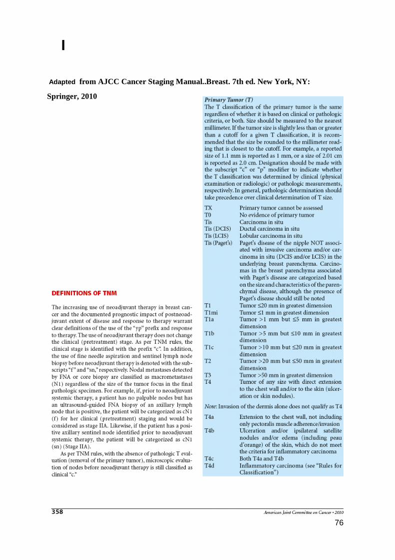
76
I
Adapted from AJCC Cancer Staging Manual..Breast. 7th ed. New York, NY:
Springer, 2010
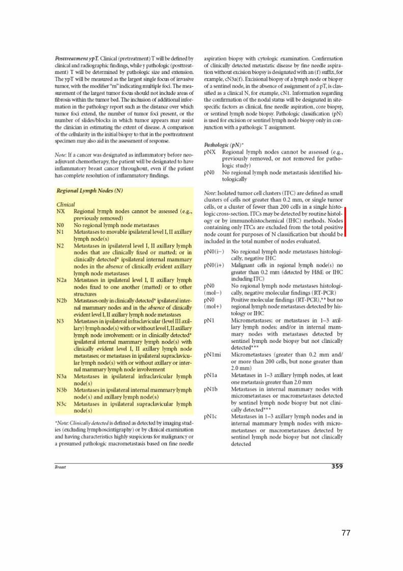
77
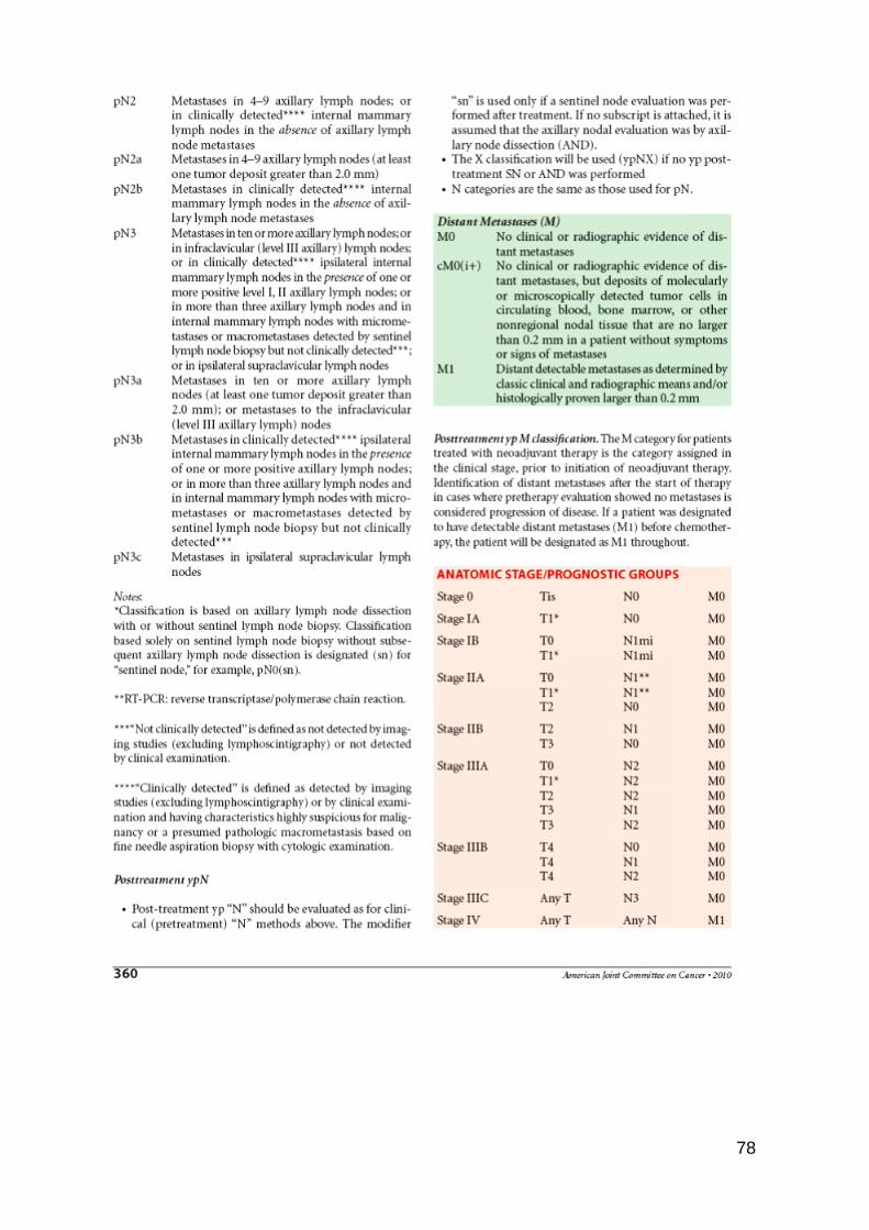
78
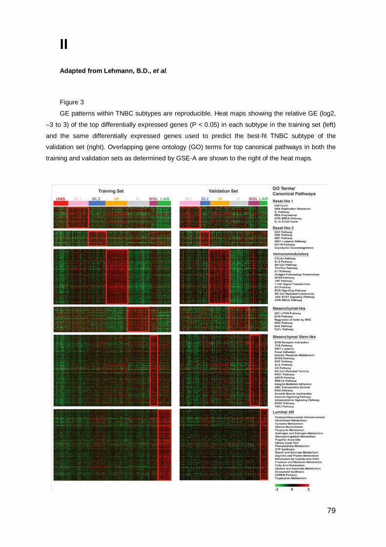
79
II
Adapted from Lehmann, B.D., et al.
Figure 3
GE patterns within TNBC subtypes are reproducible. Heat maps showing the relative GE (log2,
–3 to 3) of the top differentially expressed genes (P < 0.05) in each subtype in the training set (left)
and the same differentially expressed genes used to predict the best-fit TNBC subtype of the
validation set (right). Overlapping gene ontology (GO) terms for top canonical pathways in both the
training and validation sets as determined by GSE-A are shown to the right of the heat maps.

80