Embed Size (px)
Citation preview

Dr Gary Moore
Recent advances in lupus anticoagulant detection
and the ISTH SSC guideline – time for an update?


Variables
• Plasma preparation
• Activator
• Content & type of phospholipid
• Expression of results
• Cut-off derivation
• Antibody heterogeneity


Jennings I, Kitchen S, Woods TA, Preston FE, Greaves M. Potentially clinically important inaccuracies in testing for the lupus anticoagulant: an analysis of results from three surveys of the UK national external quality control scheme (NEQAS) for blood coagulation. Thromb Haemost 1997; 77: 934-937
Arnout J, Meijer P, Vermylen J. Lupus anticoagulant testing in Europe: An analysis of results from the first European Concerted Action on Thrombophilia (ECAT) survey using plasmas spiked with monoclonal antibodies against human β2-glycoprotein I. Thromb Haemost 1999; 81: 929-934
Lawrie AS, Mackie IJ, Purdy G, Machin SJ. The sensitivity and specificity of commercial reagents for the detection of lupus anticoagulant show marked differences in performance between photo-optical and mechanical coagulometers. Thromb Haemost 1999; 81: 758-762
Gardiner C, Mackie IJ, Malia RG, Jones DW, Winter M, Leeming D, Taberner SA, Machin SJ, Greaves M. The importance of locally derived reference ranges and standardized calculation of dilute Russell’s viper venom time results in screening for lupus anticoagulant. Br J Haematol 2000; 111: 1230-1235
Moore GW, Savidge GF, Smith MP. Improved detection of lupus anticoagulants by the dilute Russell’s Viper venom time. Blood Coagul Fibrinolysis 2000; 11: 767-774
Triplett DA. Use of the dilute Russell’s viper venom time (DRVVT): its importance and pitfalls. J Autoimm 2000; 15: 173-178
Jennings I, Greaves M, Mackie IJ, Kitchen S, Woods TA, Preston FE. UKNEQAS for Blood Coagulation. Lupus anticoagulant testing: improvements in performance in a UK NEQAS proficiency testing exercise after dissemination of national guidelines on laboratory methods. Br J Haematol 2002; 119: 364-369
Tripodi A, Biasiolo A, Chantarangkul V, Pengo V. Lupus anticoagulant (LA) testing: performance of clinical laboratories assessed by a national survey using lyophilised affinity-purified immunoglobulin with LA activity. Clin Chem 2003; 49: 1608 -1614
Moore GW & Savidge GF. Heterogeneity of Russell’s viper venom affects the sensitivity of the dilute Russell’s viper venom time to lupus anticoagulants. Blood CoagulFibrinolysis 2004; 15: 279-282
Moore GW, Tugnait S, Savidge GF. Evaluation of a new generation dilute Russell’s viper venom time assay system for lupus anticoagulant detection utilising frozen reagents and controls. Br J Biomed Sci 2005; 62: 127-131
Pengo V, Biasiolo A, Gresele P, Maronqui, F, Erba N, Veschi F, Ghirarduzzi A, de Candia E, Montaruli B, Testa S, Barcellona D, Tripodi A; participating centres of Italian Federation of Thrombosis Centres (FCSA). Survey of lupus anticoagulant diagnosis by central evaluation of positive plasma samples. J Thromb Haemost 2007; 5: 925-930
Moffat KA, Ledford-Kraemer MR, Plumhoff EA, McKay H, Nichols WL, Meijer P, Hayward CP. Are laboratories following published recommendations for lupus anticoagulant testing? An international evaluation of practices. Thromb Haemost 2009; 101: 178-184
McGlasson DL & Fritsma GA. Comparison of six dilute Russell Viper venom time lupus anticoagulant screen/confirm assay kits. Semin Thromb Hemost 2013; 39: 315-319
Tripodi A, Chantarangkul V, Cini M, Devreese K, Dlott JS, Giacomello R, Gray E, Legnani C, Martinuzzo ME, Pradella P, Siegemund A, Subramanian S, Suchon P, Testa S. Variability of cut-off values for the detection of lupus anticoagulants: results of an international multicenter multiplatform study. J Thromb Haemost 2017; 15: 1180-1190
Moore GW, Peyrafitte M, Dunois C, Amiral J. Newly developed dilute Russell's viper venom reagents for lupus anticoagulant detection with improved specificity. Lupus2017 Jan 1:961203317711773
Depreter B, Devreese KM. Dilute Russell's viper venom time reagents in lupus anticoagulant testing: a well-considered choice. Clin Chem Lab Med 2017; 55: 91-101
dRVVT variation
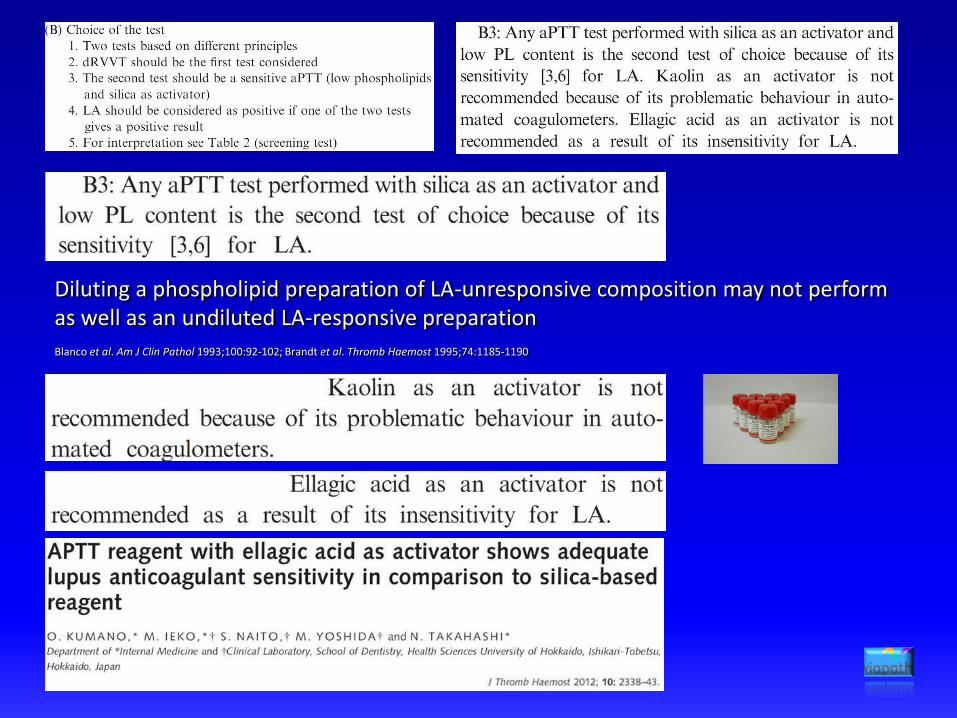
Diluting a phospholipid preparation of LA-unresponsive composition may not perform as well as an undiluted LA-responsive preparation
Blanco et al. Am J Clin Pathol 1993;100:92-102; Brandt et al. Thromb Haemost 1995;74:1185-1190

Much less variation with recombinant thromboplastinsArnout J, Vanrusselt M, Huybrechts E, Vermylen J. Optimisation of the dilute prothrombin time for the detection of lupus anticoagulant by use of a recombinant tissue thromboplastin. Br J Haematol. 1994;87:94–99.
Liestøl S, Jacobsen EM, Wisløff F. Dilute prothrombin-time based lupus ratio test. Integrated LA testing with recombinant tissue thromboplastin. Thromb Res. 2002;105:177–182.
Favourable evaluations of standardised assay - detects ‘other’ LAMackie IJ, Lawrie AS, Greenfield RS, Guinto ER, Machin SJ. A new lupus anticoagulant test based on dilute prothrombin time. Thromb Res. 2004;114:673–674
Lawrie AS, Mackie IJ, Purdy G, Greenfield RS, Guinto ER, Machin SJ. Lupus anticoagulant testing using a dilute prothrombin time with confirm procedure. J ThrombHaemost. 2005;3(Suppl 1):Abstract P1817
Dlott JS, Belter M, Ercolano E, Santulli C, Guinto ER, Greenfield RS. Evaluating the role of a new dilute prothrombin time assay ACTICLOT dPT in lupus anticoagulant testing. Blood 2007 110:3629
Devreese KMJ. Evaluation of a new commercial dilute prothrombin time in the diagnosis of lupus anticoagulants. Thromb Res. 2008;123:404–411
Nakai K, Wada H, Nakatani K, Kamiruka Y, Mastumoto T, Kobayashi T, Tonomura H, Tono Y, Ohyabu M, Ota S, Yamada N, Besho Y, Yamada E, Ikejiri M, Abe Y, Npbori T. Usefulness of a diluted prothrombin time for accurately diagnosing antiphospholipid syndrone. Vasc Dis Prev 2009;6:25-29

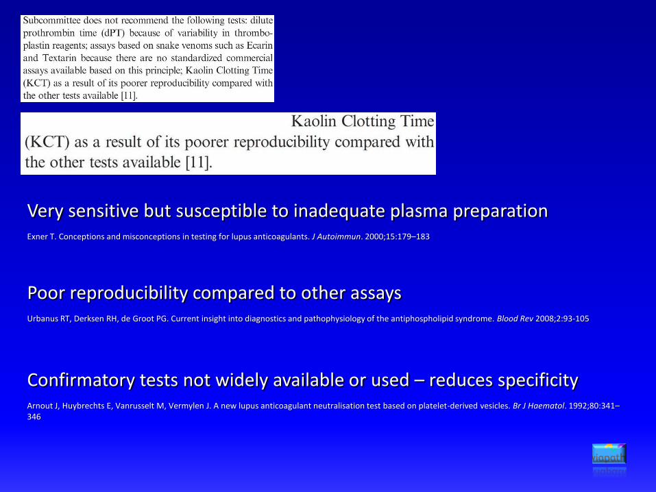
Very sensitive but susceptible to inadequate plasma preparationExner T. Conceptions and misconceptions in testing for lupus anticoagulants. J Autoimmun. 2000;15:179–183
Poor reproducibility compared to other assaysUrbanus RT, Derksen RH, de Groot PG. Current insight into diagnostics and pathophysiology of the antiphospholipid syndrome. Blood Rev 2008;2:93-105
Confirmatory tests not widely available or used – reduces specificityArnout J, Huybrechts E, Vanrusselt M, Vermylen J. A new lupus anticoagulant neutralisation test based on platelet-derived vesicles. Br J Haematol. 1992;80:341–346


PT (s)LA insensitive
APTT (s) TT (s)dRVVTscreen (ratio)
dRVVTconfirm (ratio) % correction
Normalised S/C ratio
dRVVTmix (ratio)
10 - 13 30 - 40 10 - 15 0.82 – 1.18 0.82 – 1.18 >10 >1.20 0.89 – 1.11
11 70 60 1.45 1.04 28.3 1.39 1.29
APTT of 70s equates to a ratio of 2.0 - dRVVT screen ratio not so elevated
Normal dRVVT confirm ratio confirms (i) heparin neutralisation(ii) LA
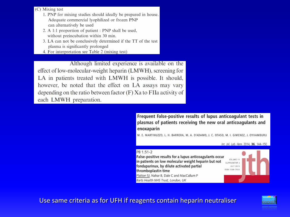
Use same criteria as for UFH if reagents contain heparin neutraliser


HEADLINES
Dabigatran prolongs screen, confirm and mixing tests
• dose dependent• LA assays largely unreliable• normal thrombin time may help
Direct FXa inhibitors prolong screen, confirm and mixing tests
• dose dependent• APTT assays generally less sensitive• dRVVT can be markedly affected
less so with apixaban than rivaroxaban or edoxaban
dRVVT screen commonly more elevated than confirm – leads to false-positive interpretations
false-positives even at trough levels
dRVVTscreen (ratio)
dRVVTconfirm (ratio) % correction
Normalised S/C ratio
dRVVTmix (ratio)
0.82 – 1.18 0.82 – 1.18 >10 >1.20 0.89 – 1.11
2.98 2.04 31.5 1.46 1.88

What can we do?

Prothrombin activators bypass direct FXa inhibitors
No assay will detect all lupus anticoagulants
• Positive TSVT/ET analysis is diagnostic
• Negative TSVT does not fully exclude LA

Similar screen and confirm results in patients on DOACs with new dRVVT
Far fewer false-positive interpretations
No false-positive interpretations in spiking with rivaroxaban, apixaban and dabigatran
New dRVVT less LA-sensitive than established dRVVT
No false-positive interpretations with new dRVVT in patients on rivaroxaban or warfarin
New dRVVT less LA-sensitive but more specific than established dRVVT
Improve performance of established dRVVT by raising cut-offs
Normalised screen/confirm ratio correction of screen ratio by confirm ratioStandard cut-offs 1.17 >10%Warfarin-specific cut-off 1.70 -Rivaroxaban-specific cut-offs 1.60 >40%

INR dRVVTscreen
dRVVT confirm
Mixing test
(screen)
Mixing test
(confirm)
MixS/C
Mix % correction
2.0 – 4.5 0.86 – 1.19 0.83 – 1.15 0.90 – 1.12 0.91 – 1.13 >1.17 >10
3.51 2.87 2.39 1.46 1.07 1.36 26.7
• Positive mixing tests are diagnostic
• Negative mixing test do not exclude LA due to dilution effect


Triplett DA, Stocker KF, Unger GA, Barna LK. The Textarin/Ecarin ratio: a confirmatory test for lupus anticoagulants. Thromb Haemost. 1993;70:925-931
Rooney AM, McNally T, Mackie IJ, Machin SJ. The Taipan snake venom time: a new test for lupus anticoagulant. J Clin Pathol 1994;47:497-501
Forastiero RR, Cerrato GS, Carreras LO. Evaluation of recently described tests for detection of the lupus anticoagulant. Thromb Haemost 1994;72:728-783
Luddington R, Scales C, Baglin T. Lupus anticoagulant testing with optical end point automation. Thromb Res 1999;96:197-203
Lawrie AS, Mackie IJ, Purdy G, Machin SJ. The sensitivity and specificity of commercial reagents for the detection of lupus anticoagulant show marked differences in performance between photo-optical and
mechanical coagulometers. Thromb Haemost. 1999;81:758-62.
Moore GW, Smith MP, Savidge GF. The Ecarin time is an improved confirmatory test for the Taipan snake venom time in warfarinised patients with lupus anticoagulants. Blood Coagul Fibrinolysis 003;14:307-312
Parmar K, Connor P, Hughes GRV, Hunt B J. Validation of the Taipan snake venom assay in routine practice to assess lupus anticoagulant status in patients being assessed for lupus anticoagulant and not
receiving oral anticoagulant. J Thromb Haemost 2003;1 Suppl 1 : PI553
Moore GW, Kamat AV, Gurney DA, O'Connor O, Rangarajan S, Carr R, Savidge GF. Alteration in the laboratory profile of a lupus anticoagulant in a patient with non-Hodgkin’s lymphoma. Clin Lab Haematol.
2004;26:429-34.
Moore GW, Savidge GF. The dilution effect of equal volume mixing studies compromises confirmation of inhibition by lupus anticoagulants even when mixture specific reference ranges are applied. Thromb Res
2006;118:523-528
Moore GW. Combining Taipan snake venom time/Ecarin time screening with the mixing studies of conventional assays increases detection rates of lupus anticoagulants in orally anticoagulated patients. Thromb J
2007;5:12
Moore GW, Rangarajan S, Savidge GF. The activated seven lupus anticoagulant assay detects clinically significant antibodies. Clin Appl Thromb Hemost 2008;14:332-337
Parmar K, Lefkou E, Doughty H, Connor P, Hunt BJ. The utility of the Taipan snake venom assay in assessing lupus anticoagulant status in individuals receiving or not receiving an oral vitamin K antagonist. Blood
Coagul Fibrinolysis 2009;20:271-275
van Os GM, de Laat B, Kamphuisen PW, Meijers JC, de Groot PG. Detection of lupus anticoagulant in the presence of rivaroxaban using Taipan snake venom time. J Thromb Haemost. 2011; 9:1657-1659
Sciascia S, Breen K, Hunt BJ. Rivaroxaban use in patients with antiphospholipid syndrome and previous thromboembolism. Blood Coagul Fibrinolysis 2015;26:476-477
Moore GW, Culhane AP, Maloney JC, Archer RA, Breen KA, Hunt BJ. Taipan snake venom time coupled with ecarin time testing enhances lupus anticoagulant detection in non-anticoagulated patients. Blood
Coagul Fibrinolysis 2016;27:477-480
Cohen H, Hunt BJ, Efthymiou M, Arachchillage DR, Mackie IJ, Clawson S, Sylvestre Y, Machin SJ, Bertolaccini ML, Ruiz-Castellano M, Muirhead N, Doré CJ, Khamashta M, Isenberg DA; RAPS trial investigators.
Rivaroxaban versus warfarin to treat patients with thrombotic antiphospholipid syndrome, with or without systemic lupus erythematosus (RAPS): a randomised, controlled, open-label, phase 2/3, non-inferiority
trial. Lancet Haematol 2016;3:e426-436
Arachchillage DR, Mackie IJ, Efthymiou M, Chitolie A, Hunt BJ, Isenberg DA, Khamashta M, Machin SJ, Cohen H. Rivaroxaban limits complement activation compared with warfarin in antiphospholipid syndrome
patients with venous thromboembolism. J Thromb Haemost. 2016;14:2177-2186
Pouplard C, Vayne C, Berthomet C, Guery EA, Delahousse B, Gruel Y. The Taipan snake venom time can be used to detect lupus anticoagulant in patients treated by rivaroxaban. Int J Lab Hematol 2017;39:e60-e63
Taipan, Textarin & Ecarin venoms
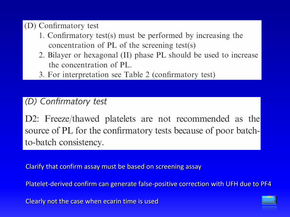
Clarify that confirm assay must be based on screening assay
Platelet-derived confirm can generate false-positive correction with UFH due to PF4
Clearly not the case when ecarin time is used

99th percentile
• Increases specificity• Statistically inevitable loss of sensitivity• Unreliable on only 40 donors

Can generate ±2SD of x from minimum 39 samples
Transference
• Old and new method comparison• If similar imprecision, interferences, comparable calibrators &
between-method results – transfer existing RI
Validation
• 20 normal donors – check for outliers
• No more than 2 out of 95% reference limits – accept RI
Locally derived cut-offs
95th vs 99th percentile
Lower cut-off increases sensitivity

The ability of a given concentration of antibody to prolong a clotting time above the reference range may depend on the clotting time an individual plasma would have had without the influence of the LA
Appearance of LA: dRVVT screen 1.16
dRVVT confirm 0.89
% ratio correction 23.3 (>10)
Normalised ratio 1.30 (>1.15)
Baseline ratios: dRVVT screen 0.90 (RI 0.84 – 1.20)
dRVVT confirm 0.89 (RI 0.88 – 1.17)
99th percentile (mean +2.33 SD if Gaussian)
97.5th percentile (mean +1.96 SD if Gaussian)
95th percentile (mean +1.65 SD if Gaussian)
At what point do you stop reducing the cut-off and look for something else?

dRVVT screen
Elevated ratioElevated ratio
dRVVT confirmAPTT confirm
You don’t always need the mixing test
APTT screen
LA detected by dRVVT & APTT
1. Normal coagulation screen
2. Significant % correction or
elevated screen/confirm ratio
3. Normal confirm ratio
1. Normal coagulation screen
2. Significant % correction or
elevated screen/confirm ratio
3. Normal confirm ratio
Confirm in LA+ patients not always shortened to within RI
Elevated confirm: potent/avid LA
co-existing or separate abnormality
Elevated confirmatory tests
dRVVT screen ratio 1.98 (0.84 – 1.18)
dRVVT confirm ratio 1.35 (0.88 – 1.12)
% correction 31.8 (<10)
Screen/confirm ratio 1.47 (<1.15)
1:1 Mix screen ratio 1.59 (0.90 – 1.10)
1:1 Mix confirm ratio 1.54 (0.89 – 1.10)
Non-phospholipid dependent inhibitor
dRVVT screen ratio 1.29 (0.84 – 1.18)
dRVVT confirm ratio 1.12 (0.88 – 1.12)
1:1 mix screen ratio 1.98 (0.90 – 1.10)
1:1 mix confirm ratio 1.08 (0.89 – 1.10)
Lupus anticoagulant co-factor effect
dRVVT screen ratio 1.98 (0.84 – 1.18)
dRVVT confirm ratio 1.85 (0.88 – 1.12)
% correction 6.6 (<10)
Screen/confirm ratio 1.07 (<1.15)
1:1 Mix screen ratio 1.01 (0.90 – 1.10)
1:1 Mix confirm ratio 1.02 (0.89 – 1.10)
Factor deficiency
1:1 Mix screen ratio 1.42 (0.90 – 1.10)
1:1 Mix confirm ratio 1.05 (0.89 – 1.10)
Lupus anticoagulant (± factor deficiency/VKA)

Ref. interval dRVVT screen dRVVT confirm dAPTT screen dAPTT confirm
Clotting times (s) 37.1 – 51.1 33.8 – 41.4 33.1 – 49.7 37.6 – 54.2
Ratios 0.85 – 1.17 0.90 – 1.10 0.80 – 1.20 0.82 – 1.18
False negative dRVVT screen: 54.7 s = 1.15 54.7 s = 1.25
47.4 s 43.8 sNPP 1 RI mean
False positive dAPTT screen: 47.0 s = 1.31 47.0 s = 1.14
36.0 s 41.4 sNPP 2 RI mean
Only use NPP clotting time as denominator if it correlates to locally derived RI mean
May need separate NPP for each assay!

Choice of test
• dRVVT indispensible – acknowledge variation• More careful detail on APTT • Recommend other tests (i.e. dPT, TSVT) as additions not replacements to dRVVT & APTT – 1st line or 2nd line
Mixing test
• Not always needed - re-prioritise testing order to screen, confirm ± mixing test • Interpret with mixing test-specific cut-off
Confirmatory test
• Emphasise confirm must be recapitulation of screen when based on increase in phospholipid concentration• Calculate % correction cut-off from upper limit of population distribution not the mean
Cut-offs & ratios
• Greater detail on cut-off derivation processes• Lower cut-offs (probably) better – remember that confirm assay will normally reveal if screen is false-positive• NPP clotting time as denominator only if it matches RI mean – reverse is the case for mixing tests
Integrated testing
• Recommend but indicate limitations• Indicate how confirm result can be a decision point
Testing anticoagulated patients
• DON’T…………(we know you will though)• Warrants a separate guideline (with TSVT/ET)

Acknowledgements
Sean O’ Mahoney

Dr Gary MooreConsultant Biomedical ScientistHaemostasis & ThrombosisViapath at Guy’s & St. Thomas’ HospitalsLondon, UK