Embed Size (px)
Citation preview

THE EUROPEAN X-RAYFREE-ELECTRON LASER FACILITY
AND THE CHALLENGES OF OUR TIME


CONTENT
THE EUROPEAN X-RAY FREE-ELECTRON LASERFACILITY AND THE CHALLENGES OF OUR TIME
1 Introduction:The European XFEL facility 1
2 The European XFEL and the healthproblems of the 21st century 6
3 The energy challenge: Naturaland artificial photosynthesis 11
4 Materials and processes foradvanced technologies 14
5 The European XFEL construction and technology transfer to industry 16
6 Concluding remarks 18


1
INTRODUCTION: THE EUROPEAN XFEL FACILITY
This paper illustrates the benefits to society expected from the
European X-Ray Free-Electron Laser Facility (European XFEL). The
European XFEL is currently under construction in the Hamburg
region (Figure 1) and is one of the major research infrastructure
pro jects highlighted in the roadmap published by the European
Strategy Forum on Research Infrastructures. Founded in 2009 with
an intergovernmental agreement, the facility is supported by 11 Eu-
ropean countries. From 2017 on, it will provide scientists and indus-
trial researchers with an outstanding new tool to investigate the
structure of matter as well as important problems in medical re-
search, new and sustainable energy sources, and the development
of materials for new technologies and a better environment.
1 Introduction: The European XFEL facility
Figure 1: General layout of the European XFEL facility, with the DESY-Bahrenfeldsite in Hamburg to the right and the research campus in Schenefeld with the
experiment hall on the left. The overall length of the mostly underground facility isapproximately 3.4 km, composed of 5.8 km of tunnels.

2
X-rays are electromagnetic waves, like radio waves, microwaves, or
visible light, but with a much shorter wavelength. While no optical
microscope is able to “see” objects smaller than the visible light
wavelengths, X-rays have sufficiently short wavelengths to “see”
matter at the atomic level of detail (Figure 2). X-rays range from
lower-energy “soft” X-rays, which have wavelengths capable of
“seeing” large molecules such as proteins, to higher-energy “hard”
X-rays, which have shorter wavelengths that are comparable to
individual small molecules and atoms.
In the 1950s, X-rays played a decisive role in discovering that, in a
DNA molecule, atoms are arranged to form a double helix, like a
circular ladder, and that the rungs of this “ladder” carry genetic in-
formation. This landmark discovery—based on X-ray experiments
and one of the most important of the century—exemplifies how
under standing structure is key to understanding function. This is
also very important in other fields, such as chemistry or materials
science (see Section 4, “Materials and processes for advanced
technologies”).
The determination of complex molecular structures requires very
bright X-ray sources. Particle accelerators, in particular electron
accelerators, are extremely bright X-ray sources that can be millions
of times brighter than medical X-ray tubes. Since the 1960s, syn-
chrotrons have revolutionized the study of biomolecules. In 1980,
the number of biomolecules with a known atomic structure was
about 70. In September 2013, this number reached above 94 000;
in over 83 000 of these, the structure was determined by synchrotron
X-rays. Today, synchrotrons are the most important tool to determine
the structure of biomolecules, and six chemistry Nobel Prizes have
been awarded for research in which synchrotron sources played a
decisive role.
Figure 2: The spectrum of electro -magnetic radiation, showing the size of
the wavelengths of each type of radiationin comparison to different objects.
The shorter the wavelength of the light,the smaller are the objects
that can still be “seen” with it.

In the last few years, another major leap forward in the brightness
of X-ray sources has been achieved by free-electron lasers (FELs),
based on linear accelerators delivering pulses of X-rays that have
three extraordinary properties:
1 The FEL pulses are extremely bright, about 100 million to a billion
times brighter than the synchrotron X-rays (Figure 3).
2 The FEL pulses are extremely short, down to a few femto -
seconds. A femtosecond is a millionth of a billionth of a second;
in one femtosecond, light travels less than one hundredth of the
thickness of a hair.
3 The FEL pulses have a quality known as spatial coherence, which
means that the waves of the laser light are in phase, and reinforce
one another (Figure 4). This is a quality that makes the X-ray FEL
pulses much more useful for experiments than conventional
X-rays.
3
INTRODUCTION: THE EUROPEAN XFEL FACILITY
105
10-5
1
1010
1015
1020
1025
1030
× 10 000
× 100 000
× × 100
×
× 1 000 000 000
× 10 000 000
2000 202519501925 197519001875
synchrotronradiation sources
free-electron lasers
first X-ray tube
modern X-ray tubes
2. generation
1. generation
3. generation
year
rela
tive
peak
bril
lianc
e (fi
rst X
-ray
tube
= 1
)
Figure 3: Evolution of peak brilliance.The development of X-ray radiation sourcesdemonstrates how dramatically the brilliance hasincreased since the discovery that particle acceler-ators can produce synchrotron radiation.The realization of the X-ray free-electron laserscontinues this upwards trend and opens up previously undreamt-of possibilities for research.

4
Today, there are a handful of X-ray FELs in operation (Figure 5), in
Germany (FLASH at DESY in Hamburg), Italy (FERMI@Elettra in
Trieste), in the US (LCLS at SLAC in California), and in Japan (SACLA
at the RIKEN Harima Institute, Hyogo). LCLS and SACLA are cur-
rently the most powerful. Other advanced industrial countries, such
as South Korea, are also building X-ray FELs or, like Switzerland,
building a national facility in addition to contributing to the construc-
tion of the European XFEL.
Figure 4: Light quality and wavelengths of different light sources in comparison.Laser beams (which are coherent) have a higher light quality than ordinary light
(called incoherent, represented by lamps). Large light source facilities are shown tothe right. X-ray free-electron lasers (orange) are capable of producing coherent lightwithin the X-ray portion of the spectrum. Synchrotrons cover a broad spectrum of
wavelengths but can only produce incoherent light.
LCLSSLAC, Menlo Park | US
LCLS - ll
FLASH DESY, Hamburg | DE
FLASH llPAL-XFELPAL, Pohang | KR
European XFELSchenefeld + Hamburg | DE
SwissFELPSI, Villigen | CH
FERMIElettra Sincrotrone Trieste, Trieste | IT
SXFELSINAP, Shanghai | CN
SACLASCSSRIKEN, Harima | JP
Hard XFEL
Soft XFEL
Under construction / in development
Figure 5: FEL light sources worldwide.Currently, the only hard X-ray FELs
accepting users are SACLA in Japan andLCLS in the United States.

5
INTRODUCTION: THE EUROPEAN XFEL FACILITY
Table 1: Comparison of the properties of hard X-ray lightsources, including European XFEL
In the following sections, a few examples demonstrate the impor-
tance of X-ray FEL facilities for the progress of science and society.
The European XFEL will be more powerful in terms of electron
energy and brightness than the other facilities, and it will produce
27 000 pulses per second, instead of the 60 of SACLA or the 120 of
LCLS. This is a very important advance which will put Europe in the
lead in this highly competitive scientific and technical environment
(Table 1).
Project LCLS, USA SACLA,Japan
EuropeanXFEL
SwissFEL,Switzerland
PAL XFEL,Korea
Max. electron energy (GeV) 14.3 8.5 17.5 5.8 10
Wavelength range (nm) 0.13–4.4 0.06–0.3 0.05–4.7 0.1–7 0.06–10
Photons/pulse ~1012 2 x10¹¹ ~1012 ~ 5 x10¹¹ 1011–1013
Peak brilliance 2 x1033 1 x1033 5 x1033 1 x1033 1.3 x1033
Pulses/second 120 60 27000 100 60
Date of first beam 2009 2011 2016 2016 2015

6
Life expectancy remained essentially unchanged from the earliest
times until the middle of the 19th century, when the gradual improve-
ment of hygienic practices and conditions in the developed nations
started to have an impact. Progress in modern biomedical scien ces,
especially since the development of molecular biology in the 20th
century, further contributed to an increase of life expectancy world-
wide. However, the populations of developing countries are still pay-
ing a heavy toll to disease, and, in the developed countries, aging
populations have transformed some age-related ailments into wide-
spread social phenomena. In addition, severe epidemics could
spread from animal species to humans, as the avian flu or AIDS ex-
emplified. The prevention and cure of disease is based very often
on a complete understanding of the causes and the mechanisms of
disease development down to the molecular level, and the design
of new pharmaceutical products relies on deciphering the atomic
details of the structure of biomolecules (Figure 6). This is why re-
searchers from pharmaceutical companies are the most frequent
industrial users of synchrotron sources.
2 The European XFEL and the health problemsof the 21st century
Figure 6: Viruses such as HIV or influenza, with diameters between 80 and150 nanometres (billionths of a metre) in size, rely on specialized biomechanicaland biochemical processes to infect cells. The European XFEL is expected to
show in unprecedented detail how these infections occur, possibly unlocking moreeffective and more targeted treatments for the diseases these viruses cause.
Credit: fotolia

7
n First of all, thanks to brightness, coherence, and short duration of
pulses, researchers will be able to investigate atomic-level structures
of biomolecules better and easier than ever before. The short dura-
tion of pulses is extremely important because it allows for reduction
of the main obstacle to high resolution pictures: the radiation dam-
age by the very action of the intense X-ray pulse. A molecule hit by
the beam literally explodes, a process which takes place over many
tens or hundreds of femto seconds (Figure 7). Although this is still
just a very short fraction of an instant, it is slow as compared to the
duration of the pulses produced at the European XFEL. Therefore,
the European XFEL can capture the image encoding the structure
before destruction occurs.
n Second, for the same reasons, researchers hope that imaging of bio -
logical objects—such as cells, organelles inside cells, and viruses—
will be possible with unprecedented resolution of a few nanometres.
n Third, the ultrashort pulses allow dynamic studies of processes—in
other words, a transition from a picture to a movie of a molecule in
action.
The European XFEL’s impact on structuralinvestigations will be manifold:
Figure 7: A pulse of X-rays from a free-electron laser illuminates an injected bio molecule.A camera downstream of the interaction region records the diffraction pattern, which con-tains information about the structure of the molecule before its destruction. Measuringmany such diffraction patterns from copies of the same biomolecule will enable scientiststo understand its three-dimensional structure.
THE EUROPEAN XFELAND THE HEALTH PROBLEMS OF THE 21ST CENTURY

8
To determine the arrangement of the atoms in a molecule or material
with X-rays, it is currently still necessary to have that molecule or
material in a crystalline solid form in which the molecules are placed
in the same orientation periodically repeated in space. Crystallization
of biological molecules is by no means simple, and the efforts to
obtain crystals of sufficient size and quality for synchrotron investi-
gation have lasted years, if not decades, whereas the successive
steps are much faster.
X-ray FELs have already shown a qualitative improvement over
synchrotrons in the capability to obtain structural information from
very small (micrometre or less) nanocrystals. Very recently at LCLS
in Stanford, the previously unknown structure of a protein (cysteine
protease cathepsin B) was determined, to a resolution of 0.21 nm,
from the investigation of nanocrystals at room temperature (Figure
8). The protein plays a very important role in the pathogenesis of
sleeping sickness, a disease that is widespread in Africa and causes
about 30 000 deaths per year. The researchers hope the new know -
ledge will lead to a novel treatment approach against the parasite
causing the disease.
Figure 8: Structure of an important protein involved in the transmission of Africansleeping sickness. Scientists created diffraction patterns (shown in background)
at LCLS that were then reconstructed into the molecular structure(shown in foreground, not to scale). Credit: Greg Stewart / SLAC
National Accelerator Laboratory

9
The European XFEL pulses are going to improve the already demon-
strated structural determination of nanocrystals considerably, and
they are expected to pave the way towards the ultimate dream of
structural biology, the determination of the structure from single,
non-crystallized molecules. The high number of pulses per second
will have two major benefits for nanocrystallography: first, the
acquisition time will be reduced; and second and even more impor-
tant, it is estimated that the required quantity of each sample will be
about 100 times smaller than that used in the LCLS experiments. In
this way, many more experiments can be performed at a small frac-
tion of the cost of the materials, which usually are difficult and ex-
pensive to produce and purify. The scientific community expects
that the European XFEL will become a very efficient decoder, ob-
taining in a much shorter time and with reduced effort a large num-
ber of structures for molecules such as membrane proteins, from
which crystals larger than a micrometre in size are hard to obtain.
This will advance progress in our understanding of pathogens and
the development of pharmaceutical remedies. But there is another
important promise in the future applications of the European XFEL
to life sciences: the promise of molecular movies.
Figure 9: Making molecular movies. First, an optical laser flash triggersa chemical reaction. A second pulse, now from the X-ray FEL, is sent atvarying time intervals after the first one to take snapshots of the changes
that have occurred in the molecule. Credit: DESY
THE EUROPEAN XFELAND THE HEALTH PROBLEMS OF THE 21ST CENTURY
Optical las
er
X-ray fla
sh

10
Biomolecules are the machines of life. Like mechanical machines
with moving parts, they modify their structure in the course of per-
forming their respective tasks. It would be extremely illuminating to
follow these modifications and see the motion of the moving parts
as in a movie. To make a film of a moving object, it is necessary to
take many snapshots. Faster movement requires a shorter exposure
time and a greater number of snapshots to avoid blurring the
pictures. This is where the ultrashort duration of the FEL pulses will
ensure sharp, non-blurred pictures of very fast processes (Figure 9).
A particularly dynamic process in proteins that is not well under-
stood and that plays an important role in the development of many
serious diseases is protein folding. In this naturally occurring
process, a protein—a linear chain of amino acid molecules—folds
into a three-dimensional structure that looks like a tangle and is
characteristic of the given protein. In some rare cases, misfolding
of a protein occurs, a process which is related to many diseases,
such as type-2 diabetes mellitus, Creutzfeldt-Jakob disease, bovine
spongiform encephalopathy (mad cow disease), Alzheimer's
disease, familial amyloid cardiomyopathy or polyneuropathy, and
Huntington’s and Parkinson's diseases. Investigating the folding
mechanism and how and why it can go wrong could help in under-
standing the origin of such very serious illnesses and their preven-
tion. It would therefore be of high interest to explore the very fast
elementary steps in this folding process with an X-ray FEL, to map
and understand the pathway to protein folding.
In summary, the European XFEL is expected to enable a giant leap
in our ability to investigate, visualize, and understand the molecular
basis of biological processes. This includes the development of
diseases and the action of natural immune defences such as anti-
bodies; of other biological entities like cells, viruses, or membranes;
and of pharmaceutical substances. Research in these areas at the
European XFEL will support the long-standing fight for the health of
a long-living population.
THE EUROPEAN XFELAND THE HEALTH PROBLEMS OF THE 21ST CENTURY
Our society is facing various challenges in providing the necessary
energy to support the industrial output and ensure the comfort and
transportation requirements of our daily life. Fossil fuels are in limited
supply, and their use raises extreme concerns for environment and
climate, especially when the rising standard of living and energy re-
quirements of emerging economies are considered. Nuclear fission
energy is regarded as a threat by the public, and nuclear fusion is
still below the proof-of-principle stage.
The sun delivers enormous amounts of energy to the earth. Clean,
economic, and reliable conversion of this abundant energy is a
dream for the future. Direct use of solar energy for generating
electrical power (photovoltaic devices) or heat (photothermal de-
vices) is confronted with the need to store energy, as the production
cycle (up during daytime, down during the night; up on clear days,
11
THE ENERGY CHALLENGE:NATURAL AND ARTIFICIAL PHOTOSYNTHESIS
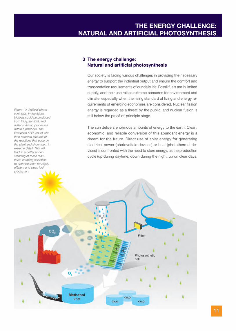
3 The energy challenge:Natural and artificial photosynthesis
Figure 10: Artificial photo-synthesis. In the future,biofuels could be producedfrom CO2, sunlight, andwater imitating processeswithin a plant cell. The European XFEL could taketime-resolved pictures ofthe reactions that occur inthe plant and show them inextreme detail. This willlead to a better under-standing of these reac-tions, enabling scientists to optimize them for highlyefficient and clean fuel production.

12
down on rainy ones; more during summer, less during winter) does
not match the consumption cycle. The storage of energy is currently
inefficient and expensive, and this limits the photovoltaic and
photothermal approach (in analogy to wind power).
In an effort to solve these problems, many scientists are trying to
optimize processes which would use sunlight energy to produce
storable and transportable fuels, inspired by the natural photosyn-
thetic process in plants, which uses sunlight energy. In plants, the
energy of the sun is used to split water into oxygen and hydrogen,
the latter of which is further split into a proton and an electron. Their
recombination into hydrogen provides the energy to power subse-
quent reactions that are necessary for the life and growth of plants.
Ideally, if one could mimic this process, one could use the hydrogen
released from water to react with carbon dioxide and produce
methanol or methane, which can be used as fuels (Figure 10).
In nature, the splitting of water molecules and the splitting of
hydrogen atoms into protons and electrons takes place in a complex
biomolecule, Photosystem II, containing a specific group of four
manganese atoms and one calcium atom.
In the laboratory, water can be split with moderate efficiency, but
only by employing some rare and heavy metals, such as platinum
or ruthenium, that are expensive and very polluting to the environ-
ment. It would be a tremendous breakthrough to understand the
intermediate steps of the chemistry of water-splitting in plants by
such cheap and abundant materials as manganese and calcium, or
by similar ones, and to reproduce and optimize the process to
improve its efficiency. The full process in plants and bacteria involves
the sequential absorption of light from the sun (four photons). Each
of the four photons absorbed induces a transformation in the
structure of the manganese-calcium complex and one step in the
reaction leading to the splitting of two water molecules. The fourth
photon brings the complex back to the starting configuration, ready
to start all over again.
Studies to elucidate the structures of the manganese calcium
complex in the different intermediate states of the process have
been hampered so far by the need to freeze the crystals to very low
temperatures to limit the effects of radiation damage. Here the FELs

13
have a significant and definite advantage, in the ability for work at
room temperature, in which the processes actually take place and
can be controlled using successive illuminations with optical laser
flashes that drive the complex through the different states of its
cycle.
The ability of FELs to follow the steps of such a chemical cycle like
in a slow-motion movie could help to better understand the efficient
water-splitting reaction in plants. The unique capabilities of the
European XFEL could have a major impact on this exciting pursuit
and provide the basis for the application of an optimized photo-
synthetic process on an industrial scale, to generate hydrogen and
liquid fuels from ingredients as cheap and abundant as sunlight,
water, carbon dioxide, and some light metals.
THE ENERGY CHALLENGE:NATURAL AND ARTIFICIAL PHOTOSYNTHESIS

14
4a Catalysis and catalytic reactionsThe previous discussion of photosynthesis provides an example of
catalysis in chemical reactions: the complex biomolecule containing
manganese and calcium is left unchanged by the reaction in which
the water molecules are split, but it is not a purely passive spectator.
It takes part in some intermediate steps of the process in a way that
makes it an indispensable ingredient for an efficient progress of the
reaction. The chemists call such a facilitator agent a catalyst, and
the process of facilitation catalysis.
Catalysts have been of fundamental importance for the chemical
industry for at least 100 years. In the early 20th century, Fritz Haber
and Carl Bosch developed catalysts to boost the efficiency of
ammonia production from nitrogen and hydrogen, starting the
synthetic fertilizer industry. Today, catalysts are also used to limit
the environmental damage of car exhaust gases (Figure 11) and
are fundamental in the petrochemical industry and in many other
applications, with a market of tens of billions of euro.
4 Materials and processes for advancedtechnologies
Figure 11: A car’s catalytic converter, which helps reduce pollution in the exhaust.The European XFEL could improve the understanding of the process of catalysis
and help make more efficient, less toxic catalysts. Credit: DESY

15
Despite their widespread use, the action of catalysts is very often
poorly understood. They have in many cases been developed by
trial and error, with what some people call “cooking recipes”. X-ray
FELs will give new impulses to systematic approaches based on the
possibility to shoot molecular movies of the catalyst’s action in the
various stages of a reaction. Understanding the molecular mecha-
nisms in detail will improve the development of better and more
environment-friendly catalysts, with important effects on a whole
range of important industrial processes.
4b Nano-magnetism and information technologyThe development and improvement of materials for the most modern
technologies also poses challenges that X-ray FELs can help to
solve. Progress in information technology, for example, results from
our ability to store information on smaller and smaller areas, and
also on the ability to read it and write it as rapidly as possible.
Present-day hard disk drives use layered magnetic materials in
which the tiny magnetic moment of each grain in the topmost layer
can be magnetized in the up or down direction to indicate a “0” or
a “1”, thus storing one bit of information. Writing usually takes place
by applying magnetic fields; reading occurs by measuring electrical
resistance that depends on the orientation of the magnetic moment
of the top layer (the so-called “giant magneto-resistance effect”, for
which the 2007 Nobel Prize in Physics was awarded to Albert Fert
and Peter Grünberg). In recent years it was discovered that a short
laser pulse with circularly polarized light, which has two possible
directions like a left-turning or right-turning screw, can also write in
a sample by changing the magnetic polarity, but more quickly. The
European XFEL is going to be equipped with a special device to pro-
duce circular polarized X-rays that will permit researchers to obtain
a map of the magnetization of a sample and of its evolution in time.
This will lead to a better understanding of ultrafast magnetization
erasure and re-writing by optical laser pulses and should contribute
to establishing the mechanism and approach the physical speed
limits of magnetic recording. Faster data recording is a central issue
for progress in information technology and for the development of
numerous applications and products in science and technology.
MATERIALS AND PROCESSESFOR ADVANCED TECHNOLOGIES

16
Even in the construction phase, the European XFEL project has
already stimulated the development of some high-technology
industrial sectors in the participating countries. To achieve the
demanding performance indispensable for the European XFEL, the
fabrication of many components requires all the sophisticated
know-how of the contributing research labs; on the other hand,
these components need to be produced in large series by industry.
For example, 800 accelerator resonant cavities, made of a metal
called niobium, are needed, plus some pre-series and spare ones.
The order was split between two industrial companies that equipped
themselves with the necessary infrastructure and were extensively
trained through the pre-series production, under the guidance of
experts from DESY and from the Italian research agency INFN. The
underlying TESLA technology, developed at DESY, is now emerging
as a standard for superconducting linear accelerators worldwide,
for applications not only in FELs but also in other research infra-
structures, such as spallation neutron sources and high-energy
physics colliders (Figure 12). The technology is foreseen for various
projects in the planning or in the pre-construction phase all over the
world, and the companies in question have acquired a solid ad-
vantageous position in a market niche that is bound to expand.
Another area in which the European XFEL has been stimulating a
considerable development effort is in detector devices. In order to
take complete advantage of the European XFEL’s high number of
5 The European XFEL construction and technologytransfer to industry
Figure 12: The accelerator technologyused in the European XFEL, based onthese specialized niobium cavities, has
already made an impact on thedevelopment of linear accelerators
worldwide. Credit: DESY

17
pulses per second, the detector (essentially a digital camera for
X-rays) must be able to record a high-quality image on a sensor,
transfer it to a temporary memory medium, clear the image from the
sensor, and be ready for the next one—all in an extremely short
period of time. The temporary memory storage has been designed
and prototyped with a capacity for several hundreds of images and
with the objective to reach a few thousands in the future. All this re-
quires great sophistication and expertise, so a number of European
laboratories were involved in the development of prototypes. Ad-
vanced electronic integrated circuits were designed and their fabri-
cation was given out to companies. Some features of the imaging
detectors with very high read-out speed will be of interest not only
for future high repetition rate FELs but also for research with synchro -
trons, and possibly in other areas, including commercial electronic
devices, where speed of data processing and of data storage and
recovery could open up new business opportunities.
All the information acquired during an experiment (hundreds of
thousands and sometimes millions of images) must be transferred
to memory. In addition, all images must be accompanied by
additional information (metadata) allowing identification of the
image (for example, to which experiment and sample it belongs
and the settings of the different pieces of apparatus). In this way,
the European XFEL contributes to the general area of current re-
search and development that goes under the name “data deluge”,
which increasingly affects not only the scientific enterprise in general
(one example: the experiments at CERN that led to the celebrated
discovery of the Higgs Boson) but also many aspects of business
and public services in our digital age. Richard Feynman estimated
in 1959 that the total information contained at the time in the major
libraries of the world (24 million books) amounted to 0.1 petabytes
of information (or 1014 bytes). In more modern terms, this amount
of information can be stored in about 10 000 high-quality DVDs;
nonetheless, the European XFEL is expected to produce an
equivalent amount of information within the first few months of
experiment time—and this rate will only increase! Some of the
solutions being developed for dealing efficiently with the data deluge
at the European XFEL and other large research infrastructures like
CERN are expected to be of use in other parts of society as well:
better weather forecasting, more accurate climate evolution simu-
lations, and global communication networks, where the amount of
data is growing inexorably, albeit at a slower pace.
THE EUROPEAN XFEL CONSTRUCTIONAND TECHNOLOGY TRANSFER TO INDUSTRY

18
CONCLUDING REMARKS
The European XFEL—a large and 3.4 km long research facility in the
Hamburg region constructed by 11 European countries and mostly
located in underground tunnels and halls—will be a major contri-
bution to consolidating Europe’s position at the forefront of basic
scientific research. It holds the promise of many exciting discoveries
for the benefit of society, a few examples of which we have pre-
sented in the preceding paragraphs.
The very construction of the facility poses scientific and techno-
logical challenges that are being addressed with the help of aca -
demia and industry, to the advantage of both.
In operation, the European XFEL will select the best and most inno-
vative experiments proposed by researchers from many different
disciplines, providing an opportunity to access a unique instrument.
Thus, this opportunity will also be available to young scientists at
the beginning of their careers, to those working in institutes or uni-
versities with modest funding, as long as their ideas are bright and
show promise to lead to a real advance in a field of science. With
6 instruments in the starting configuration and the possibility to up-
grade to up to 15, the European XFEL will be able to host more re-
searchers than any other X-ray FEL in the world. Located within a
cluster of prestigious institutions—such as DESY, CFEL, the Univer-
sity of Hamburg and the Max Planck Institutes, EMBL, CSSB, and
others—the European XFEL will maximize synergies and collabora-
tions to offer a unique and stimulating research environment.
The conclusion of the construction project in a few years will place
Europe at the top of the league of X-ray sources. It is expected that
the European XFEL will be yet another success story of the
European collaboration in science and technology.
6 Concluding remarks


EUROPEAN XFEL GMBHAlbert-Einstein-Ring 19 · 22761 Hamburg · Germany
www.xfel.eu