Embed Size (px)
Citation preview
Electron Capture Dissociation, Electron Detachment Dissociation,and Collision-Induced Dissociation of Polyamidoamine (PAMAM)Dendrimer Ions with Amino, Amidoethanol, and SodiumCarboxylate Surface Groups
Malgorzata A. Kaczorowska and Helen J. Cooper⁎School of Biosciences, University of Birmingham, Edgbaston, Birmingham, United Kingdom.
AbstractHere, we investigate the effect of the structure (generation) and nature of the surface groups ofdifferent polyamidoamine (PAMAM) dendrimers on electron-mediated dissociation, either electroncapture dissociation (ECD) or electron detachment dissociation (EDD), and compare thefragmentation with that observed in collision-induced dissociation (CID). ECD and EDD of thePAMAM dendrimers resulted in simple mass spectra, which are straightforward to interpret, whereasCID produced complex mass spectra. The results show that electron-mediated dissociation (ECDand EDD) of PAMAM dendrimers does not depend on the nature of the surface group but tends tooccur within the innermost generations. CID of the PAMAM dendrimers showed a strong dependenceon the nature of the surface group and occurred mostly in the outer generation. The resultsdemonstrate the potential utility of ECD and EDD as a tool for the structural analysis of PAMAMdendrimers.
Graphical AbstractInvestigation of the effect of the structure and nature of surface groups of different PAMAMdendrimers on electron-mediated dissociation (ECD, EDD) and collision-induced dissociation(CID).
© 2008 Elsevier Inc..⁎Address reprint requests to Dr. Helen J. Cooper, University of Birmingham, School of Biosciences, Edgbaston, Birmingham, B15 2TT,UK [email protected] document was posted here by permission of the publisher. At the time of deposit, it included all changes made during peer review,copyediting, and publishing. The U.S. National Library of Medicine is responsible for all links within the document and for incorporatingany publisher-supplied amendments or retractions issued subsequently. The published journal article, guaranteed to be such by Elsevier,is available for free, on ScienceDirect.Published online June 28, 2008
Sponsored document fromJournal of the American Society forMass Spectrometry
Published as: J Am Soc Mass Spectrom. 2008 September ; 19(9): 1312–1319.
Sponsored Docum
ent Sponsored D
ocument
Sponsored Docum
ent
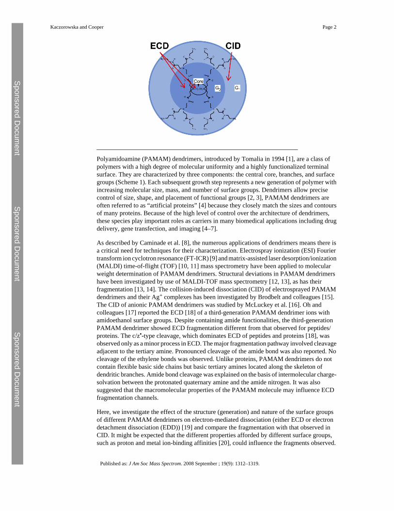
Polyamidoamine (PAMAM) dendrimers, introduced by Tomalia in 1994 [1], are a class ofpolymers with a high degree of molecular uniformity and a highly functionalized terminalsurface. They are characterized by three components: the central core, branches, and surfacegroups (Scheme 1). Each subsequent growth step represents a new generation of polymer withincreasing molecular size, mass, and number of surface groups. Dendrimers allow precisecontrol of size, shape, and placement of functional groups [2, 3], PAMAM dendrimers areoften referred to as “artificial proteins” [4] because they closely match the sizes and contoursof many proteins. Because of the high level of control over the architecture of dendrimers,these species play important roles as carriers in many biomedical applications including drugdelivery, gene transfection, and imaging [4–7].
As described by Caminade et al. [8], the numerous applications of dendrimers means there isa critical need for techniques for their characterization. Electrospray ionization (ESI) Fouriertransform ion cyclotron resonance (FT-ICR) [9] and matrix-assisted laser desorption/ionization(MALDI) time-of-flight (TOF) [10, 11] mass spectrometry have been applied to molecularweight determination of PAMAM dendrimers. Structural deviations in PAMAM dendrimershave been investigated by use of MALDI-TOF mass spectrometry [12, 13], as has theirfragmentation [13, 14]. The collision-induced dissociation (CID) of electrosprayed PAMAMdendrimers and their Ag+ complexes has been investigated by Brodbelt and colleagues [15].The CID of anionic PAMAM dendrimers was studied by McLuckey et al. [16]. Oh andcolleagues [17] reported the ECD [18] of a third-generation PAMAM dendrimer ions withamidoethanol surface groups. Despite containing amide functionalities, the third-generationPAMAM dendrimer showed ECD fragmentation different from that observed for peptides/proteins. The c/z•-type cleavage, which dominates ECD of peptides and proteins [18], wasobserved only as a minor process in ECD. The major fragmentation pathway involved cleavageadjacent to the tertiary amine. Pronounced cleavage of the amide bond was also reported. Nocleavage of the ethylene bonds was observed. Unlike proteins, PAMAM dendrimers do notcontain flexible basic side chains but basic tertiary amines located along the skeleton ofdendritic branches. Amide bond cleavage was explained on the basis of intermolecular charge-solvation between the protonated quaternary amine and the amide nitrogen. It was alsosuggested that the macromolecular properties of the PAMAM molecule may influence ECDfragmentation channels.
Here, we investigate the effect of the structure (generation) and nature of the surface groupsof different PAMAM dendrimers on electron-mediated dissociation (either ECD or electrondetachment dissociation (EDD)) [19] and compare the fragmentation with that observed inCID. It might be expected that the different properties afforded by different surface groups,such as proton and metal ion-binding affinities [20], could influence the fragments observed.
Kaczorowska and Cooper Page 2
Published as: J Am Soc Mass Spectrom. 2008 September ; 19(9): 1312–1319.
Sponsored Docum
ent Sponsored D
ocument
Sponsored Docum
ent
The results suggest that electron-mediated dissociation of PAMAM dendrimers does notdepend on the nature of surface groups, but is related to the structure of the polymer: irrespectiveof surface group, the dominant ECD fragmentation channel for these species is cleavage at thetertiary amine and pronounced amide bond cleavage. The majority of ECD fragmentationoccurred within the innermost generations. In contrast to ECD, collision-induced dissociationof the PAMAM dendrimers depended strongly on the nature of surface groups.
ExperimentalSample Preparation
Polyamidoamine (PAMAM) dendrimers were purchased from Sigma–Aldrich (Poole, Dorset,UK) and used without further purification. The PAMAM dendrimers with amino andamidoethanol surface groups were diluted to a concentration of 10 pmol/μL in solutions ofmethanol (FisherScientific, Loughborough, UK) to water (J. T. Baker, Middlesex, UK) toacetic acid (FisherScientific) (49:49:2, vol/vol). The 0.5-generation PAMAM dendrimer withsodium carboxylate surface groups was prepared (10 pmol/μL) in methanol:acetic acid (98:2)solution.
Mass SpectrometryMass spectrometry analysis was performed on a Thermo Finnigan LTQ FT mass spectrometer(Thermo Fisher Scientific, Bremen, Germany). Samples were injected by use of an AdvionBiosciences Triversa Nanomate electrospray source (Advion Biosciences, Ithaca, NY, USA).Data acquisition and analysis were conducted using the Xcalibur 2.0 (Thermo Fisher Scientific)software. Mass spectra were acquired at a resolution of 100,000 at m/z 400. Precursor ionswere selected and isolated for ECD in the linear ion trap before transfer to the ICR cell.Electrons were generated on the surface of an indirectly heated barium tungsten cylindricaldispenser cathode (5.1 mm diameter; Heat Wave Labs, Inc., Watsonville, CA, USA), situated154 mm from the cell, 1 mm off-axis. The current across the electrode was about 1.1 A. Ionswere irradiated with electrons for 70 ms. Each ECD scan consisted of five coadded microscans.CID experiments were performed in the front-end linear ion trap and the fragments transferredto the ICR cell for detection. Precursor ions were selected and isolated in the linear ion trap.CID experiments were performed with helium gas at a normalized collision energy of 35%.Each CID scan consisted of five coadded microscans. All tandem mass spectrometry (MS/MS)spectra were averaged over 30 scans and analyzed manually.
Results and DiscussionECD of PAMAM Dendrimers
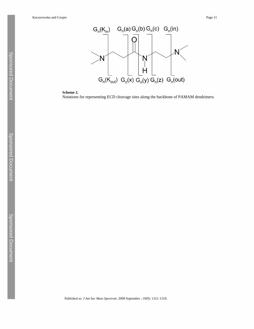
Assignment of ECD fragments ions of PAMAM dendrimers—ECD fragments wereassigned based on the nomenclature devised by Oh and colleagues [17] (see Scheme 2).Assignments are given in the form Gn(m), where subscript n refers to the generation in whichfragmentation takes place and m describes the type of fragmentation; a•/x, b/y, c/z•. Forexample, fragment G1(y) was derived from cleavage of the amide bond in generation 1.Gn(in) and Gn(out) notations concern fragmentation that takes place core-side of the tertiaryamines. K refers to cleavages surface-side of the tertiary amines.
Generation 2 PAMAM dendrimer with amidoethanol surface groups(PAMAMG2OH)—The second-generation PAMAM dendrimer with amidoethanol surfacegroups and ethylenediamine core is a symmetrical molecule that contains three tertiary aminebranches (generations 0, 1, and 2) and 16 neutral alcohol surface groups. Electrosprayionization of this PAMAM dendrimer leads to the formation of multiply protonated molecularions [M + 4H]4+ through [M + 7H]7+. The most abundant multiply protonated molecular ions
Kaczorowska and Cooper Page 3
Published as: J Am Soc Mass Spectrom. 2008 September ; 19(9): 1312–1319.
Sponsored Docum
ent Sponsored D
ocument
Sponsored Docum
ent
of the PAMAM dendrimer—[M + 4H]4+, [M + 5H]5+, and [M + 6H]6+ ions—were isolatedand subjected to ECD. Similar fragmentation behavior was observed for each precursor.
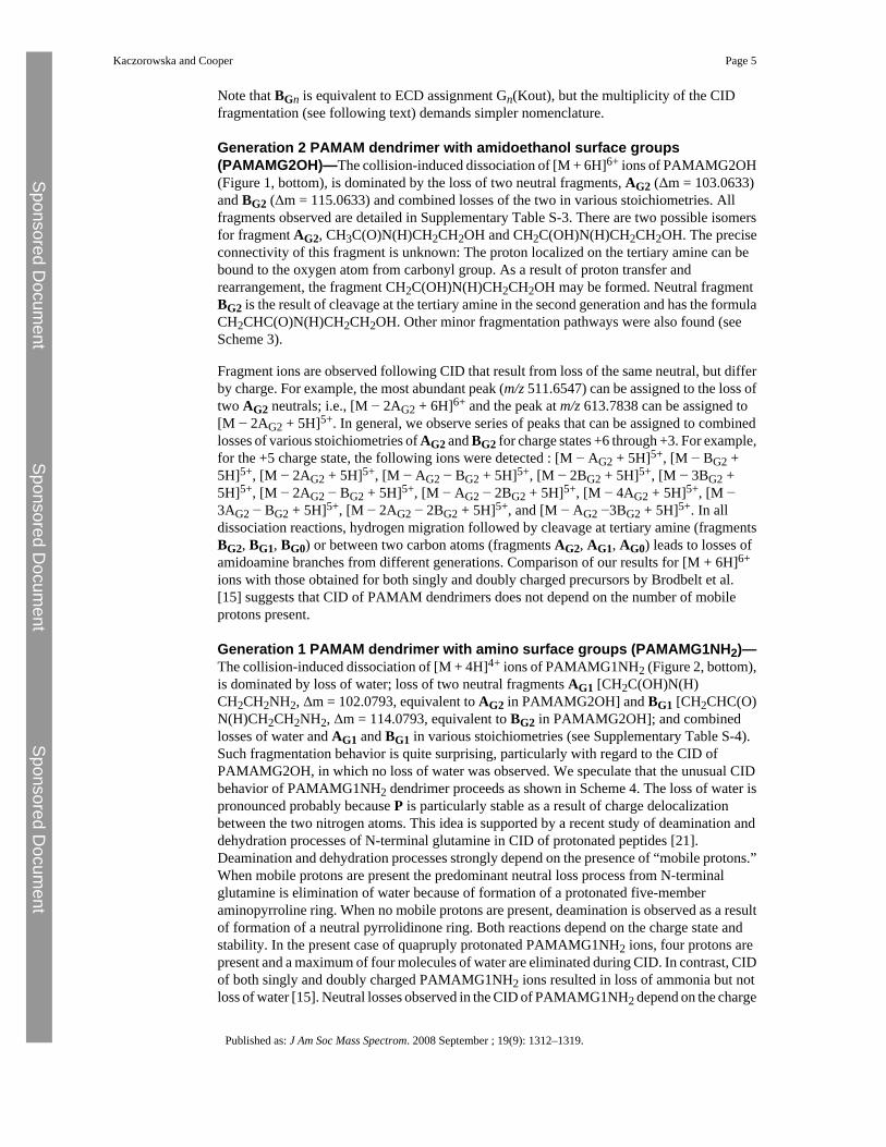
The ECD MS/MS spectrum of [M + 6H]6+ precursor ions is shown in Figure 1 (top). Allfragments detected are described in Supplementary Table S-1, which can be found in theelectronic version of this article. The most abundant peaks correspond to y fragments fromgeneration 1 and generation zero. No b(•) ions were observed. The mass spectrum shows peakscorresponding to triply charged fragments G1(in)3+, G0(in)3+ singly charged Gcore(in)+, andquadruply charged G1(in)4+. These four fragments result from cleavage at tertiary amines. Inaddition, G(out) fragments, which also result from cleavage at tertiary amines but with thecharge retained toward the surface of the dendrimer, were observed. These fragments alsoundergo secondary fragmentations, resulting in singly charged fragment ions [G1(out)G2(a)]+ at m/z 160.1205, [G0(out)G1(a)]+ at m/z 389.2624, and [G0(out)G1(out)]+ at m/z459.2915. The c/z• cleavages that are the most abundant in the ECD of peptides/proteins werefound here only as minor channels: G1(z)+ at m/z 274.1757 and G0(z)+ at m/z 732.4592.
As mentioned earlier, similar fragmentation patterns were seen for each charge state ofPAMAMG2OH. In each case, fragmentation within these cations occurred in the innermostgenerations, i.e., the core, G0 or G1. No fragmentation in the outer generation (G2) wasobserved.
Generation 1 PAMAM dendrimer with amino surface groups (PAMAMG1NH2)—Electrospray ionization of the generation 1 PAMAM dendrimer, containing ethylenediaminecore, two tertiary amine branches (generations 0 and 1), and eight primary amino surfacegroups, results in the formation of multiply protonated molecular ions [M + 2H]2+ through [M+ 4H]4+. The fragmentation for each charge state was very similar. Mass spectra shown herewere obtained from the most abundant charge state, i.e., [M + 4H]4+ ions. The ECD massspectrum of the [M + 4H]4+ ions of PAMAMG1NH2 is shown in Figure 2 (top) and thefragments observed are detailed in Supplementary Table S-2. The dominant fragmentsoriginate from the inner generation (G0). The peaks at m/z 592.9225 and 289.2342 can beassigned to G0(in)2+ and G0(y)+ fragments, respectively. The c/z• cleavages are found only asa minor channel. Other abundant fragment ions at m/z 388.3021 and 159.1366 are attributedto secondary fragmentation and can be assigned to [Gcore(out)G0(a)]+ and [G0(out)G1(a)]+,respectively. These fragments are presumably the result of cleavage of the amide bond followedby loss of CO, together with cleavage at the tertiary amine. Secondary fragmentation reactionsinvolving a cleavages were seen for both PAMAMG1NH2 and PAMAMG2OH.
Comparison of the ECD mass spectra obtained from PAMAMG1NH2 and PAMAMG2OHdendrimers leads to the conclusion that, despite the different chemical properties, the natureof the surface groups does not affect the ECD fragmentation behavior of these polymers. Inboth cases the mechanism of ECD is bound up with protonation of the tertiary amines and thepresence of amide functionalities in the polymer backbone and can be explained on the basisof a charge-solvation model (proton sharing) [17]. The generation number has a moresignificant influence on the electron capture dissociation. In both cases the ECD mass spectraare dominated by fragments that come from the inner generation(s). The most abundantfragments are of the same type for each dendrimer; Gn(y), Gn(in) and Gn(out), where n standsfor the inner generation(s) (n = 0, 1, in the case of PAMAMG2OH; n = 0 in the case ofPAMAMG1NH2).
CID of PAMAM DendrimersAssignment of CID fragments ions of PAMAM dendrimers—CID fragments wereassigned according to the nomenclature shown in Scheme 3. Assignments are given in formAGn, BGn, and CGn, where subscript Gn refers to the generation in which cleavage takes place.
Kaczorowska and Cooper Page 4
Published as: J Am Soc Mass Spectrom. 2008 September ; 19(9): 1312–1319.
Sponsored Docum
ent Sponsored D
ocument
Sponsored Docum
ent
Note that BGn is equivalent to ECD assignment Gn(Kout), but the multiplicity of the CIDfragmentation (see following text) demands simpler nomenclature.
Generation 2 PAMAM dendrimer with amidoethanol surface groups(PAMAMG2OH)—The collision-induced dissociation of [M + 6H]6+ ions of PAMAMG2OH(Figure 1, bottom), is dominated by the loss of two neutral fragments, AG2 (Δm = 103.0633)and BG2 (Δm = 115.0633) and combined losses of the two in various stoichiometries. Allfragments observed are detailed in Supplementary Table S-3. There are two possible isomersfor fragment AG2, CH3C(O)N(H)CH2CH2OH and CH2C(OH)N(H)CH2CH2OH. The preciseconnectivity of this fragment is unknown: The proton localized on the tertiary amine can bebound to the oxygen atom from carbonyl group. As a result of proton transfer andrearrangement, the fragment CH2C(OH)N(H)CH2CH2OH may be formed. Neutral fragmentBG2 is the result of cleavage at the tertiary amine in the second generation and has the formulaCH2CHC(O)N(H)CH2CH2OH. Other minor fragmentation pathways were also found (seeScheme 3).
Fragment ions are observed following CID that result from loss of the same neutral, but differby charge. For example, the most abundant peak (m/z 511.6547) can be assigned to the loss oftwo AG2 neutrals; i.e., [M − 2AG2 + 6H]6+ and the peak at m/z 613.7838 can be assigned to[M − 2AG2 + 5H]5+. In general, we observe series of peaks that can be assigned to combinedlosses of various stoichiometries of AG2 and BG2 for charge states +6 through +3. For example,for the +5 charge state, the following ions were detected : [M − AG2 + 5H]5+, [M − BG2 +5H]5+, [M − 2AG2 + 5H]5+, [M − AG2 − BG2 + 5H]5+, [M − 2BG2 + 5H]5+, [M − 3BG2 +5H]5+, [M − 2AG2 − BG2 + 5H]5+, [M − AG2 − 2BG2 + 5H]5+, [M − 4AG2 + 5H]5+, [M −3AG2 − BG2 + 5H]5+, [M − 2AG2 − 2BG2 + 5H]5+, and [M − AG2 −3BG2 + 5H]5+. In alldissociation reactions, hydrogen migration followed by cleavage at tertiary amine (fragmentsBG2, BG1, BG0) or between two carbon atoms (fragments AG2, AG1, AG0) leads to losses ofamidoamine branches from different generations. Comparison of our results for [M + 6H]6+
ions with those obtained for both singly and doubly charged precursors by Brodbelt et al.[15] suggests that CID of PAMAM dendrimers does not depend on the number of mobileprotons present.
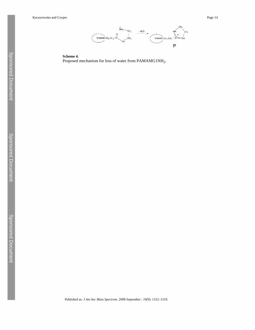
Generation 1 PAMAM dendrimer with amino surface groups (PAMAMG1NH2)—The collision-induced dissociation of [M + 4H]4+ ions of PAMAMG1NH2 (Figure 2, bottom),is dominated by loss of water; loss of two neutral fragments AG1 [CH2C(OH)N(H)CH2CH2NH2, Δm = 102.0793, equivalent to AG2 in PAMAMG2OH] and BG1 [CH2CHC(O)N(H)CH2CH2NH2, Δm = 114.0793, equivalent to BG2 in PAMAMG2OH]; and combinedlosses of water and AG1 and BG1 in various stoichiometries (see Supplementary Table S-4).Such fragmentation behavior is quite surprising, particularly with regard to the CID ofPAMAMG2OH, in which no loss of water was observed. We speculate that the unusual CIDbehavior of PAMAMG1NH2 dendrimer proceeds as shown in Scheme 4. The loss of water ispronounced probably because P is particularly stable as a result of charge delocalizationbetween the two nitrogen atoms. This idea is supported by a recent study of deamination anddehydration processes of N-terminal glutamine in CID of protonated peptides [21].Deamination and dehydration processes strongly depend on the presence of “mobile protons.”When mobile protons are present the predominant neutral loss process from N-terminalglutamine is elimination of water because of formation of a protonated five-memberaminopyrroline ring. When no mobile protons are present, deamination is observed as a resultof formation of a neutral pyrrolidinone ring. Both reactions depend on the charge state andstability. In the present case of quapruply protonated PAMAMG1NH2 ions, four protons arepresent and a maximum of four molecules of water are eliminated during CID. In contrast, CIDof both singly and doubly charged PAMAMG1NH2 ions resulted in loss of ammonia but notloss of water [15]. Neutral losses observed in the CID of PAMAMG1NH2 depend on the charge
Kaczorowska and Cooper Page 5
Published as: J Am Soc Mass Spectrom. 2008 September ; 19(9): 1312–1319.
Sponsored Docum
ent Sponsored D
ocument
Sponsored Docum
ent
state; for higher charge states, the dominant process is loss of water and, for lower charge states,elimination of NH3 is observed.
Unlike ECD, the results of the CID experiments performed for PAMAMG2OH andPAMAMG1NH2 suggest a strong dependence of the dissociation processes on the nature ofthe surface groups. For PAMAMG1NH2, the observed CID also depends on charge state. Forboth PAMAMG2OH and PAMAMG1NH2, dissociation occurs mainly in the outermostgeneration.
EDD and CID of Anionic Polyamidoamine (PAMAM) Dendrimer, Generation 0.5, with SodiumCarboxylate Surface Groups
The PAMAM dendrimer, generation 0.5, with eight sodium carboxylate surface groups andethylenediamine core (PAMAMG0.5COONa), forms multiply charged anions via negativeelectrospray. The dendrimer ions have mixtures of sodium and proton counterions becauseprotons present in the MeOH/NH4OH solution compete with sodium ions associated with thesurface groups [16]. Within each charge state, mixtures of counterions were found accordingto the formula [M − (n + m)Na + mH]n−, where n is charge state (n = 2–4), and m is the numberof protons replacing sodium in the surface groups. Figure 3 shows the EDD and CID massspectra obtained from [M −5Na + 2H]3− ions. The EDD mass spectrum (Figure 3, top) isdominated by peaks corresponding to the loss of one, two, and three T fragments [CH2CHC(O)O−] (Δm = 71.01333), which results from cleavage at an outer tertiary amine. Loss of Tfragments is also observed following CID (Figure 3, bottom). The only peak present in theEDD mass spectrum, but not present in the CID mass spectrum, occurs at m/z 497.7054 andcan be assigned to the [G1/2(in)]2− fragment (m/zcalc 497.7080). This fragment results fromcleavage at the tertiary amine, which was also a major fragmentation channel in the ECD ofPAMAM cations. No fragments corresponding to cleavage of the amide bond or c/z•-typecleavages were observed following EDD.
ConclusionsWe have investigated the electron-mediated dissociation (ECD and EDD) of three PAMAMdendrimers with the aim of determining the effect of the macromolecular properties (numberof generations) and the nature of the surface group. In all cases, fragmentation was dominatedby cleavage at the tertiary amines with some amide bond cleavages (ECD). The c/z•-typedissociation, prevalent in ECD of peptides and proteins, is observed only as a minor channel.The results suggest that ECD (and EDD) are independent of the nature of the surface group,but tend to occur within the innermost generations. In contrast, CID of the PAMAM dendrimerstends to occur in the outermost generation and is strongly dependent on the nature of the surfacegroup. In comparison with CID, ECD produces simple mass spectra that are straightforwardto interpret. The results demonstrate the potential utility of ECD as a tool for the structuralanalysis of PAMAM dendrimers.
Supplementary dataRefer to Web version on PubMed Central for supplementary material.
AcknowledgmentsWe gratefully acknowledge EPSRC and the Wellcome Trust (074131) for funding.
Kaczorowska and Cooper Page 6
Published as: J Am Soc Mass Spectrom. 2008 September ; 19(9): 1312–1319.
Sponsored Docum
ent Sponsored D
ocument
Sponsored Docum
ent
References1. Tomalia D. A., Kaplan D. A., Kruper W. J. Jr., Cheng R. C., Tomplinson I. A., Fazio M. J., Edwards
D. S. U.S. Patent 5 728 461, 1994.2. Tomalia D.A. Baker H. Dewald J. Hall M. Kallos G. Martin S. Roeck J. Ryder J. Smith P. A New
Class of Polymers: Starburst Dendritic Macromolecules. Polym. J 1985;17:117–132.3. Tomalia D.A. Esfand R. Dendrons, Dendrimers and Dendigrafts. Chem. Ind 1997;11:416–420.4. Esfand R. Tomalia D.A. Poly(amidoamine) (PAMAM) Dendrimers: From Biomimicry to Drug
Delivery and Biomedical Applications. Drug Disc. Today 2001;6:427–436.5. Boas U. Heegaard P.M.H. Dendrimers in Drug Research. Chem. Soc. Rev 2004;33:43–63. [PubMed:
14737508]6. Svenson S. Tomalia D.A. Commentary: Dendrimers in Biomedical Applications—Reflections on the
Field. Adv. Drug Del. Rev 2005;57:2106–2129.7. Tomalia D.A. Reyna L.A. Svenson S. Dendrimers as Multi-Purpose Nanodevices for Oncology Drug
Delivery and Diagnostic Imaging. Biochem. Soc. Trans 2007;35:61–67. [PubMed: 17233602]8. Caminade A.-M. Laurent R. Majoral J.-P. Characterization of Dendrimers. Adv. Drug Del. Rev
2005;57:2130–2146.9. Pasa-Tolic L. Anderson G.A. Smith R.D. Brothers H.M. Spindler R. Tomalia D.A. ESI FT-ICR Mass
Spectrometric Characterization of High Molecular Mass Starburst Dendrimers. Int. J. Mass Spectrom1997;165:405–418.
10. Muller R. Laschober C. Szymanski W.W. Allmaier G. Determination of Molecular Weight, ParticleSize, and Density of High Number Generation PAMAM Dendrimers Using MALDI-TOF-MS andnES-GEMMA. Macromolecules 2007;40:5599–5605.
11. Zhou L. Russell D.H. Zhao M. Crooks R.M. Characterization of PAMAM Dendrimers and TheirComplexes with Cu2+ by MALDI Mass Spectrometry. Macromolecules 2001;34:3567–3573.
12. Giordanengo R. Mazarin M. Wu J. Peng L. Charles L. Propagation of Structural Deviations ofPAMAM Fan-Shape Dendrimers (Generations 0–3) Characterized by MALDI and ElectrosprayMass Spectrometry. Int. J. Mass Spectrom 2007;266:62–75.
13. Peterson J. Allikmaa V. Subbi J. Pehk T. Lopp M. Structural Deviations in PAMAM Dendrimers: AMALDI-TOF-MS Analysis. Eur. Polym. J 2003;39:33–42.
14. Subbi J. Aguraiuja R. Tanner R. Allikmaa V. Lopp M. Fragmentation of PAMAM Dendrimers inMALDI. Eur. Polym. J 2005;41:2552–2558.
15. Mazzitelli C.L. Brodbelt J.S. Investigation of Silver-Binding to PAMAM Dendrimers by ESI TandemMass Spectrometry. J. Am. Soc. Mass Spectrom 2006;17:676–684. [PubMed: 16516486]
16. He M. McLuckey S.A. Tandem Mass Spectrometry of Half Generation PAMAM Dendrimer Anions.Rapid Commun. Mass Spectrom 2004;18:960–972. [PubMed: 15116423]
17. Lee S. Han S.Y. Lee T.G. Chung G. Lee D. Oh H.B. Observation of Pronounced b-Dot, y Cleavagesin the ECD Mass Spectrometry of PAMAM Dendrimer Ions with Amide Functionalities. J. Am. Soc.Mass Spectrom 2006;17:536–543. [PubMed: 16490359]
18. Zubarev R.A. Kelleher N.L. McLafferty F.W. ECD of Multiply Charged Protein Cations. J. Am.Chem. Soc 1998;120:3265–3266.
19. Budnik B.A. Haselmann K.F. Zubarev R.A. Electron Detachment Dissociation of Peptide Di-Anions:An Electron-Hole Recombination Phenomenon. Chem. Phys. Lett 2001;342:299–302.
20. Crooks, R.M.; Lemon, B.I.; Sun, L.; Yeung, L.K.; Zhao, M.Q. Dendrimer Encapsulated Metals andSemiconductors: Synthesis, Characterization, and Applications. In: Vogtle, F., editor. DendrimersIII: Design, Dimension, Function. Springer-Verlag; Berlin: 2001. p. 81-135.
21. Neta P. Pu Q.-L. Kilpatrick L. Yang X. Stein S.E. Dehydration versus Deamination of N-TerminalGlutamine in CID of Protonated Peptides. J. Am. Soc. Mass Spectrom 2006;18:27–36. [PubMed:17005415]
Kaczorowska and Cooper Page 7
Published as: J Am Soc Mass Spectrom. 2008 September ; 19(9): 1312–1319.
Sponsored Docum
ent Sponsored D
ocument
Sponsored Docum
ent
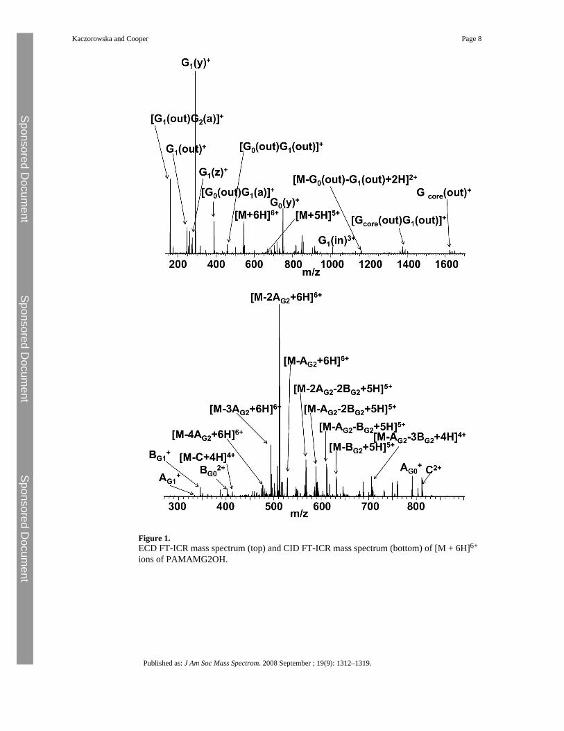
Figure 1.ECD FT-ICR mass spectrum (top) and CID FT-ICR mass spectrum (bottom) of [M + 6H]6+
ions of PAMAMG2OH.
Kaczorowska and Cooper Page 8
Published as: J Am Soc Mass Spectrom. 2008 September ; 19(9): 1312–1319.
Sponsored Docum
ent Sponsored D
ocument
Sponsored Docum
ent
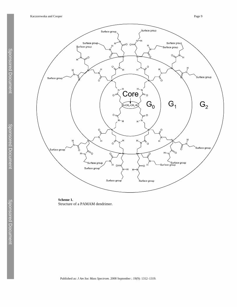
Scheme 1.Structure of a PAMAM dendrimer.
Kaczorowska and Cooper Page 9
Published as: J Am Soc Mass Spectrom. 2008 September ; 19(9): 1312–1319.
Sponsored Docum
ent Sponsored D
ocument
Sponsored Docum
ent
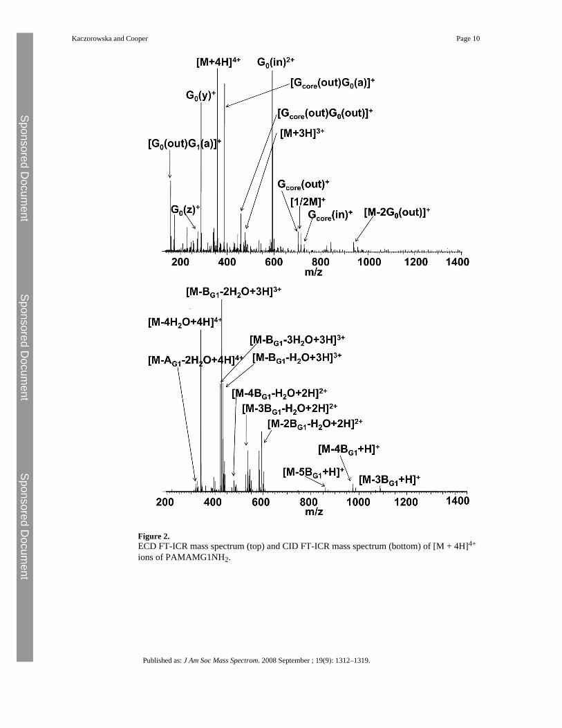
Figure 2.ECD FT-ICR mass spectrum (top) and CID FT-ICR mass spectrum (bottom) of [M + 4H]4+
ions of PAMAMG1NH2.
Kaczorowska and Cooper Page 10
Published as: J Am Soc Mass Spectrom. 2008 September ; 19(9): 1312–1319.
Sponsored Docum
ent Sponsored D
ocument
Sponsored Docum
ent
Scheme 2.Notations for representing ECD cleavage sites along the backbone of PAMAM dendrimers.
Kaczorowska and Cooper Page 11
Published as: J Am Soc Mass Spectrom. 2008 September ; 19(9): 1312–1319.
Sponsored Docum
ent Sponsored D
ocument
Sponsored Docum
ent
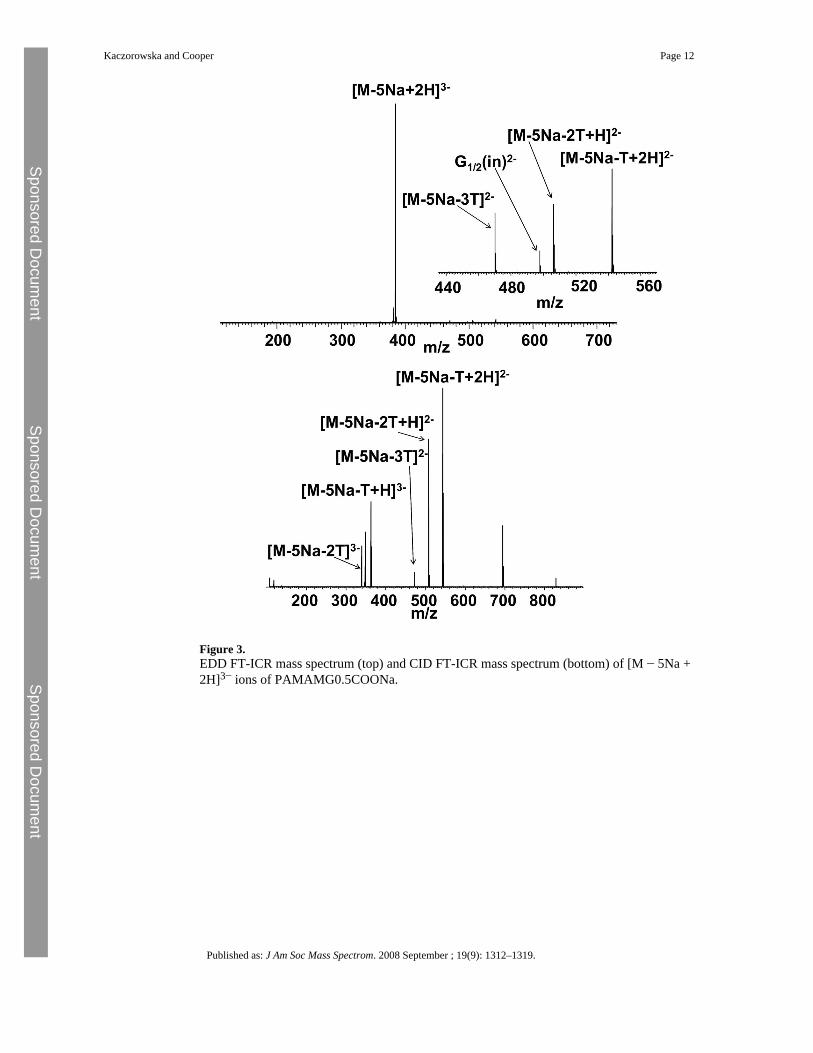
Figure 3.EDD FT-ICR mass spectrum (top) and CID FT-ICR mass spectrum (bottom) of [M − 5Na +2H]3− ions of PAMAMG0.5COONa.
Kaczorowska and Cooper Page 12
Published as: J Am Soc Mass Spectrom. 2008 September ; 19(9): 1312–1319.
Sponsored Docum
ent Sponsored D
ocument
Sponsored Docum
ent
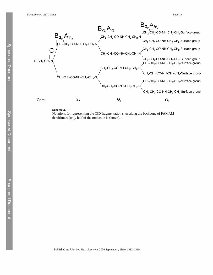
Scheme 3.Notations for representing the CID fragmentation sites along the backbone of PAMAMdendrimers (only half of the molecule is shown).
Kaczorowska and Cooper Page 13
Published as: J Am Soc Mass Spectrom. 2008 September ; 19(9): 1312–1319.
Sponsored Docum
ent Sponsored D
ocument
Sponsored Docum
ent
Scheme 4.Proposed mechanism for loss of water from PAMAMG1NH2.
Kaczorowska and Cooper Page 14
Published as: J Am Soc Mass Spectrom. 2008 September ; 19(9): 1312–1319.
Sponsored Docum
ent Sponsored D
ocument
Sponsored Docum
ent