Embed Size (px)
Citation preview
Subscriber access provided by University of Texas Libraries
The Journal of Physical Chemistry is published by the American Chemical Society. 1155Sixteenth Street N.W., Washington, DC 20036
Electron spin resonance spectra of 9-phenylanthraceneand 9,10-diphenylanthracene anion radicals
L. O. Wheeler, K. S. V. Santhanam, and Allen J. BardJ. Phys. Chem., 1967, 71 (7), 2223-2227• DOI: 10.1021/j100866a040 • Publication Date (Web): 01 May 2002
Downloaded from http://pubs.acs.org on February 19, 2009
More About This Article
The permalink http://dx.doi.org/10.1021/j100866a040 provides access to:
• Links to articles and content related to this article• Copyright permission to reproduce figures and/or text from this article
ELECTRON SPIN RESONANCE SPECTRA OF 9-PHENYLANTHRACENE 2223
Electron Spin Resonance Spectra of 9=Phenylanthracene
and 9,lO-Diphenylanthracene Anion Radicals
by L. 0. Wheeler, K. S . V. Santhanam, and Allen J. Bardl
Department of Chemistry, The Univeraity of Texas, Austin, Texas ‘78716 (Received December 60, 1966)
The electron spin resonance (esr) spectra of 9-phenylanthracene and 9, lo-diphenylanthra- cene anion radicals prepared by electroreduction in N,N-dimethylformamide and potassium metal reduction in dimethoxyethane are given. The assigned experimental coupling constants are shown to be in good agreement with those calculated using molecular orbital theory. A brief study of how different factors in radical ion preparation affect esr spec- trum resolution is described.
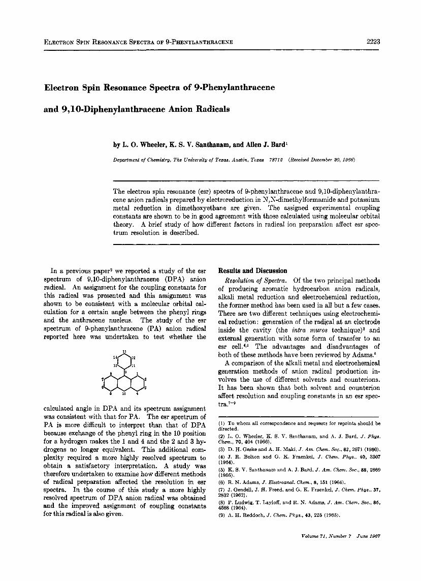
In a previous paper2 we reported a study of the esr spectrum of 9,lO-diphenylanthracene (DPA) anion radical. An assignment for the coupling constants for this radical was presented and this assignment was shown to be consistent with a molecular orbital cal- culation for a certain angle between the phenyl rings and the anthracene nucleus. The study of the esr spectrum of 9-phenylanthracene (PA) anion radical reported here was undertaken to test whether the
7
6 6 10 4
calculated angle in DPA and its spectrum assignment was consistent with that for PA. The esr spectrum of PA is more difficult to interpret than that of DPA because exchange of the phenyl ring in the 10 position for a hydrogen makes the 1 and 4 and the 2 and 3 hy- drogens no longer equivalent. This additional com- plexity required a more highly resolved spectrum to obtain a satisfactory interpretation. A study was therefore undertaken to examine how different methods of radical preparation affected the resolution in esr spectra. In the course of this study a more highly resolved spectrum of DPA anion radical was obtained and the improved assignment of coupling constants for this radical is also given.
Results and Discussion Resolution of Spectra. Of the two principal methods
of producing aromatic hydrocarbon anion radicals, alkali metal reduction and electrochemical reduction, the former method has been used in all but a few cases. There are two different techniques using electrochemi- cal reduction: generation of the radical at an electrode inside the cavity (the intra muros te~hnique)~ and external generation with some form of transfer to an esr celL4t6 The advantages and disadvantages of both of these methods have been reviewed by Adams?
A comparison of the alkali metal and electrochemical generation methods of anion radical production in- volves the use of different solvents and counterions. It has been shown that both solvent and counterion affect resolution and coupling constants in an esr spec- tra.7-D
(1) To whom all correspondence and requests for reprints should be directed. (2) L. 0. Wheeler, K. S. V. Santhanam, and A. J. Bard, J . Phys. Chem., 70, 404 (1966). (3) D. H. Geske and A. H. Maki, J . Am. Chem. SOC., 82,2671 (1960). (4) J. R. Bolton and G. K. Fraenkel, J . Chem. Phgs., 40, 3307 (1964). (5) K. 9. V. Santhanam and A. J. Bard, J . Am. Chem. Soc., 88, 2669 (1966). (6) R. N. Adams, J . Electroanal. Chem., 8 , 151 (1964). (7) J. Gendell, J. H. Freed, and G. K. Fraenkel, J . Chem. Phys., 37, 2832 (1962). (8) P. Ludwig, T. Layloff, and R. N. Adams, J . Am. Chem. Soc., 86, 4668 (1964). (9) A. H. Reddoch, J . Chem. Phys.. 43, 225 (1965).
Volume 71. Numbg 7 June 1987
2224 L. 0. WHEELER, K. S. V. SANTHANAM, AND A. J. BARD
T O VACUUM LINE
f7L 24/40
PLATINUM C 0 N T A C T
MERCURY POOL WORKING
ELECTRODE
1 3mm TUBIN6
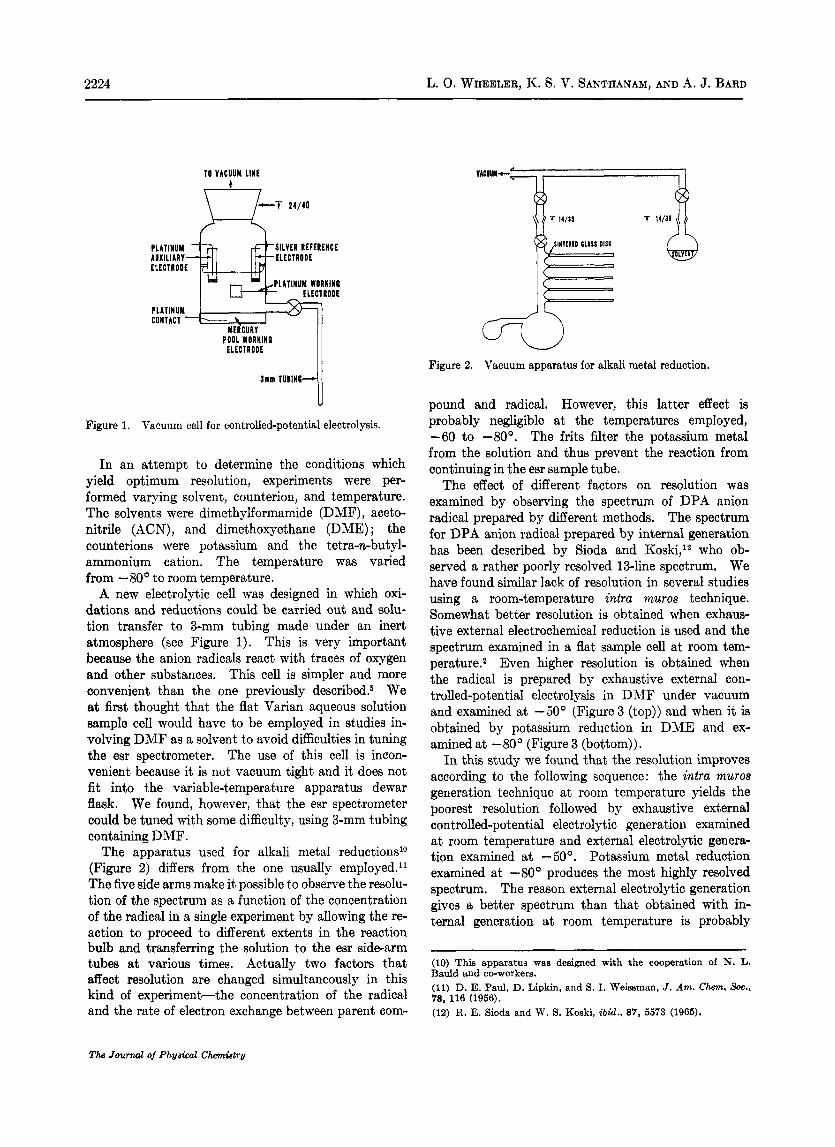
Figure 1. Vacuum cell for controlled-potential electrolysis.
In an attempt to determine the conditions which yield optimum resolution, experiments were per- formed varying solvent, counterion, and temperature. The solvents were dimethylformamide (DMF), aceto- nitrile (ACN), and dimethoxyethane (DME) ; the counterions were potassium and the tetra-n-butyl- ammonium cation. The temperature was varied from -80' to room temperature.
A new electrolytic cell was designed in which oxi- dations and reductions could be carried out and solu- tion transfer to 3-mm tubing made under an inert atmosphere (see Figure 1). This is very important because the anion radicals react with traces of oxygen and other substances. This cell is simpler and more convenient than the one previously de~cribed.~ We at first thought that the flat Varian aqueous solution sample cell would have to be employed in studies in- volving DMF as a solvent to avoid difficulties in tuning the esr spectrometer. The use of this cell is incon- venient because it is not vacuum tight and it does not fit into the variable-temperature apparatus dewar flask. We found, however, that the esr spectrometer could be tuned with some difficulty, using 3-mm tubing containing DMF.
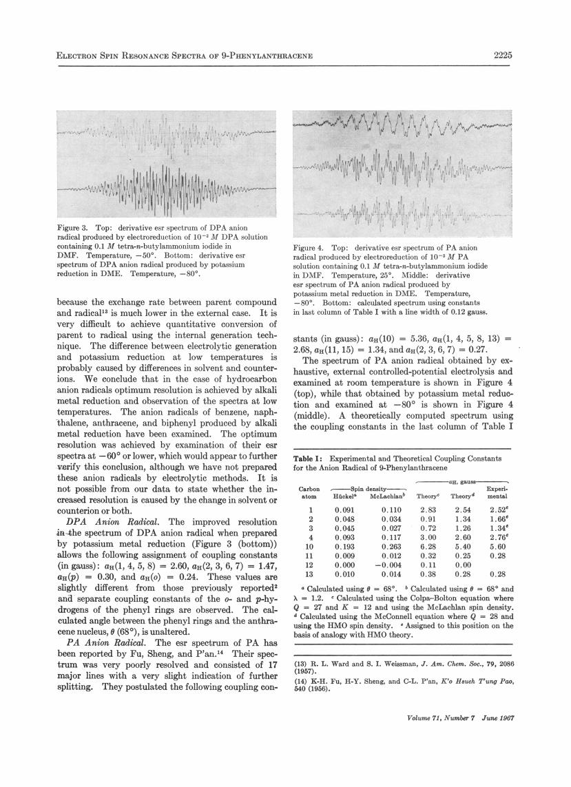
The apparatus used for alkali metal reductions1° (Figure 2) differs from the one usually employed." The five side arms make it possible to observe the resolu- tion of the spectrum as a function of the concentration of the radical in a single experiment by allowing the re- action to proceed to different extents in the reaction bulb and transferring the solution to the esr side-arm tubes at various times. Actually two factors that affect resolution are changed simultaneously in this kind of experiment-the concentration of the radical and the rate of electron exchange between parent com-
T 14/11
Figure 2. Vacuum apparatus for alkali metal reduction.
pound and radical. However, this latter effect is probably negligible at the temperatures employed, -60 to -80'. The frits filter the potassium metal from the solution and thus prevent the reaction from continuing in the esr sample tube.
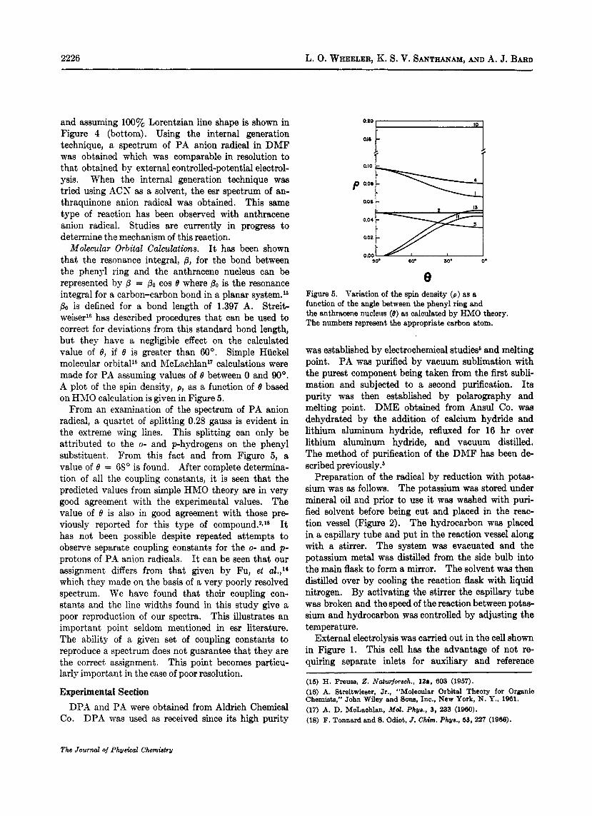
The effect of different factors on resolution was examined by observing the spectrum of DPA anion radical prepared by different methods. The spectrum for DPA anion radical prepared by internal generation has been described by Sioda and Koski,I2 who ob- served a rather poorly resolved 13-line spectrum. We have found similar lack of resolution in several studies using a room-temperature intra muros technique. Somewhat better resolution is obtained when exhaus- tive external electrochemical reduction is used and the spectrum examined in a flat sample cell at room tem- perature.2 Even higher resolution is obtained when the radical is prepared by exhaustive external con- trolled-potential electrolysis in DMF under vacuum and examined at -50' (Figure 3 (top)) and when it is obtained by potassium reduction in DME and ex- amined at -80' (Figure 3 (bottom)).
In this study we found that the resolution improves according to the following sequence: the intra muros generation technique at room temperature yields the poorest resolution followed by exhaustive external controlled-potential electrolytic generation examined at room temperature and external electrolytic genera- tion examined at -50'. Potassium metal reduction examined a t -80' produces the most highly resolved spectrum. The reason external electrolytic generation gives a better spectrum than that obtained with in- ternal generation at room temperature is probably
(10) This apparatus was designed with the cooperation of N. L. Bauld and cc-workers. (11) D. E. Paul, D. Lipkin, and S. I. Weisaman, J. Am. Chem. SOC., 78, 118 (1956). (12) R. E. Sioda and W. 8. Koski, {bid., 87, 5573 (1965).
The Journal of' Physiea2 Chemistry
ELECTRON SPIN RESONANCE SPECTRA OF %PHENYLANTHRACENE 2225
Figure 3. Top: derivative esr spectrum of DPA anion radical produced by electroreduction of 10-8 M UPA solution containing 0.1 M tetmwbutylammonium iodide in DMF. Temperature, -50'. Bottom: derivative esr spectrum of DPA anion radical produced by potassium reduction in DME. Temperature, -80".
because the exchange rate between parent compound and radical'* is much lower in the external case. It is very difficult to achieve quantitative conversion of parent to radical using the internal generation tech- nique. The difference between electrolytic generation and potassium reduction at low temperatures is probably caused by differences in solvent and counter- ions. We conclude that in the case of hydrocarbon anion radicals optimum resolution is achieved by alkali metal reduction and observation of the spectra at low temperatures. The anion radicals of benzene, naph- thalene, anthracene, and biphenyl produced by alkali metal reduction have been examined. The optimum resolution was achieved by examination of their esr spectra at --60° or lower, which would appear to further verify this conclusion, although we have not prepared these anion radicals by electrolytic methods. It is not poasible from our data to state whether the in- creased resolution is caused by the change in solvent or counterion or both.
DPA Anion Radical. The improved resolution in-the spectrum of DPA anion radical when prepared by potassium metal reduction (Figure 3 (bottom)) allows the following assignment of coupling eonstants (in gauss): Q X ( ~ , 4, 5, 8) = 2.60, a&, 3, 6, 7) = 1.47, a&) = 0.30, and QA(O) = 0.24. These values are slightly different from those previously reported' and separate coupling constants of the 0- and phy- drogens of the phenyl rings are observed. The cal- culated angle between the phenyl rings and the anthra- cene nucleus, 0 (6S0), is unaltered.
P A Anion Radical. The esr spectrum of PA has been reported by Fu, Sheng, and P'an." Their spec- trum was very poorly resolved and consisted of 17 major lines with a very slight indication of further splitting. They postulated the following coupling con-
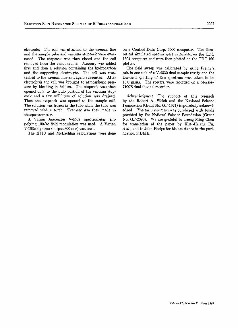
Figure 4. Top: derivative esr spectrum of PA anion radical produced by electroreduction of lo-' M PA solution containing 0.1 M tetra-n-butylammonium iodide in DMF. Temperature, 25'. Middle: derivative esr spectrum of PA anion radical produced by potgssiurn metal reduction in DME. Temperature, -80'. Bottom: calculated spectrum using eonstants in last column of Table I with a line width of 0.12 gausa.
stants (in gauss): ~ ~ ( 1 0 ) = 5.36, a& 4, 5, 8, 13) = 2 . 6 8 , ~ ~ ( 1 1 , 15) = 1.34, anda&3,6,7) = 0.27.
The spectrum of PA anion radical obtained by ex- haustive, external controlled-potential electrolysis and examined a t room temperature is shown in Figure 4 (top), while that obtained by potassium metal reduc- tion and examined at -80" is shown in Figure 4 (middle). A theoretically computed spectrum using the coupling constants in the last column of Table I
Table I: Ekperirnental and Theoretical Coupling Constants for the Anion Radical of %Phenylanthracene
C.rhbon atom
1 2 3 4 10 11 12 13
---Spin density- Hacksla Melachh.nb
0.091 0.110 0.048 0.034 0.045 0.027 0.093 0.117 0.193 0.263 0,009 0.012 0.000 -0.004 0.010 0.014
-E. mu- Expad.
T h e o e Thoolyg msoUl
2.83 2.54 2.52' 0.91 1.34 1.66' 0.72 1.26 1.34' 3.00 2.60 2.76' 6.28 5.40 5.60 0.32 0.25 0.28 0.11 0.00 0.38 0.28 0.28
a Calculated using 8 - 68". 6 Calculated using 0 = 68' and A = 1.2. Calculated using the Colpa-Bolton equation where Q = 27 and K = 12 and using the McLachlan spin denaity. d Calculated using the McConnell equation where Q = 28 and using the HMO spin density. Assigned to this position on the basis of analogy with HMO theory.
(13) R. L. Ward snd 8. I. Weissman, J . Am. C h . Em.. 7% 2088 (1967). ~. (14) K-H. Fu, H-Y. 8heng. and GL. P'sn. R'o H-h T'uno Pao. 640 (1066).
Vdum 71. Nu- 7 JUM 1067
2226 L. 0. WHEELER, K. S. V. SANTHANAM, AND A. J. BARD
and assuming 100% Lorentzian line shape is shown in Figure 4 (bottom). Using the internal generation technique, a spectrum of PA anion radical in DMF was obtained which was comparable in resolution to that obtained by external controlled-potential electrol- ysis. When the internal generation technique was tried using ACN as a solvent, the esr spectrum of an- thraquinone anion radical was obtained. This same type of reaction has been observed with anthracene anion radical. Studies are currently in progress to determine the mechanism of this reaction.
Molecular Orbital Calculations. It has been shown that the resonance integral, p, for the bond between the phenyl ring and the anthracene nucleus can be represented by P = PO cos e where PO is the resonance integral for a carbon-carbon bond in a planar system.16 PO is defined for a bond length of 1.397 A. Streit- weiserlB has described procedures that can be used to correct for deviations from this standard bond length, but they have a negligible effect on the calculated value of 8, if e is greater than 60’. Simple Huckel molecular orbital1* and McLachlan17 calculations were made for PA assuming values of 0 between 0 and 90’. A plot of the spin density, p, as a function of e based on HMO calculation is given in Figure 5.
From an examination of the spectrum of PA anion radical, a quartet of splitting 0.28 gauss is evident in the extreme wing lines. This splitting can only be attributed to the 0- and p-hydrogens on the phenyl substituent. From this fact and from Figure 5, a value of e = 68’ is found. After complete determina- tion of all the coupling constants, it is seen that the predicted values from simple HMO theory are in very good agreement with the experimental values. The value of e is also in good agreement with those pre- viously reported for this type of compound.**l* It has not been possible despite repeated attempts to observe separate coupling constants for the 0- and p- protons of P-4 anion radicals. It can be seen that our assignment differs from that given by Fu, et aZ.,14 which they made on the basis of a very poorly resolved spectrum. We have found that their coupling con- stants and the line widths found in this study give a poor reproduction of our spectra. This illustrates an important point seldom mentioned in esr literature. The ability of a given set of coupling constants to reproduce a spectrum does not guarantee that they are the correct assignment. This point becomes particu- larly important in the case of poor resolution.
Experimental Section DPA and PA were obtained from Aldrich Chemical
Co. DPA was used as received since its high purity
0.20 IO
O.I0 p 0.08
aoe I- I
O . O 4 ~ 0.02 0.00
90. 6Q 30‘ 0.
e Figure 5. Variation of the spin density ( p ) as a function of the angle between the phenyl ring and the anthracene nucleus (e) as calculated by HMO theory. The numbers represent the appropriate carbon atom.
was established by electrochemical studies6 and melting point. PA was purified by vacuum sublimation with the purest component being taken from the first subli- mation and subjected to a second purification. Its purity was then established by polarography and melting point. DME obtained from Ansul Co. was dehydrated by the addition of calcium hydride and lithium aluminum hydride, refluxed for 16 hr over lithium aluminum hydride, and vacuum distilled. The method of purification of the DMF has been de- scribed previously.6
Preparation of the radical by reduction with potas- sium was as follows. The potassium was stored under mineral oil and prior to use it was washed with puri- fied solvent before being cut and placed in the reac- tion vessel (Figure 2). The hydrocarbon was placed in a capillary tube and put in the reaction vessel along with a stirrer. The system was evacuated and the potassium metal was distilled from the side bulb into the main flask to form a mirror. The solvent was then distilled over by cooling the reaction flask with liquid nitrogen. By activating the stirrer the capillary tube was broken and the speed of the reaction between potas- sium and hydrocarbon Tvas controlled by adjusting the temperature.
External electrolysis was carried out in the cell shown in Figure 1. This cell has the advantage of not re- quiring separate inlets for auxiliary and reference
(16) H. Preuas, 2. Naturfotech., 12a, 603 (1967). (16) A. Streitwieeer, Jr., “Molecular Orbital Theory for Organic Chemists,” John Wiley and Sons, Inc., New York, N. Y., 1961. (17) A. D. McLachlan, Md. Phys., 3, 233 (1960). (18) F. Tonnard and S. Odiot, J . Chim. Phys., 63, 227 (1906).
Tb Journal of Physical Chemistry
ELECTRON SPIN RESONANCE SPECTRA OF 9-PHENYLANTHRACENE 2227
electrode. The cell was attached to the vacuum line and the sample tube and vacuum stopcock were evac- uated. The stopcock was then closed and the cell removed from the vacuum line. Mercury was added first and then a solution containing the hydrocarbon and the supporting electrolyte. The cell was reat- tached to the vacuum line and again evacuated. After electrolysis the cell was brought to atmospheric pres- sure by bleeding in helium. The stopcock was then opened only to the bulb portion of the vacuum stop- cock and a few milliliters of solution was drained. Then the stopcock was opened to the sample cell. The solution was frozen in the tube while the tube was removed with a torch. Transfer was then made to the spectrometer.
A Varian Associates V-4502 spectrometer em- polying 100-kc field modulation was used. A Varian V-153c klystron (output 300 mw) was used.
The HMO and McLachlan calculations were done
on a Control Data Corp. 6600 computer. The theo- retical simulated spectra were calculated on the CDC 1604 computer and were then plotted on the CDC 160 plotter.
The field sweep was calibrated by using Fremy’s salt in one side of a V-4532 dual sample cavity and the low-field splitting of this spectrum was taken to be 13.0 gauss. The spectra were recorded on a Moseley 7100B dual channel recorder.
Acknowledgment. The support of this research by the Robert A. Welch and the National Science Foundation (Grant No. GP-1921) is gratefully acknowl- edged. The esr instrument was purchased with funds provided by the National Science Foundation (Grant No. GP-2090). We are grateful to Tzeng-Ming Chen for translation of the paper by Kuei-Hsiang Fu, et al., and to John Phelps for his assistance in the puri- fication of DME.
Volume 71, N u m b 7 June 1867