Embed Size (px)
Citation preview
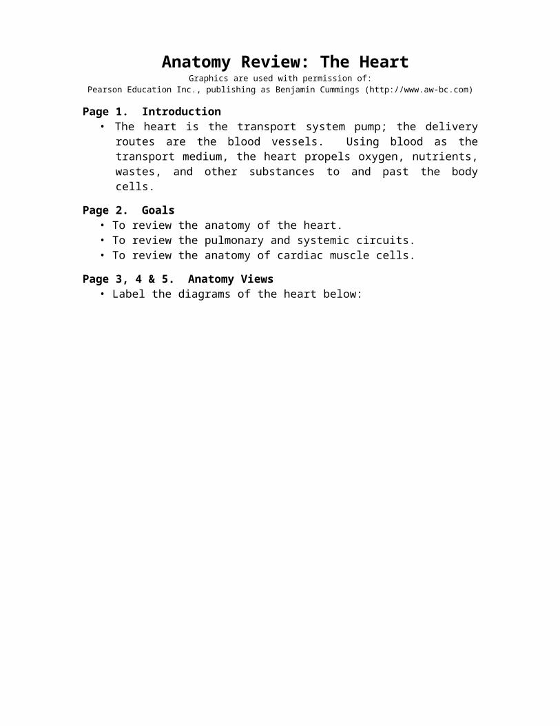
Anatomy Review: The Heart Graphics are used with permission of:
Pearson Education Inc., publishing as Benjamin Cummings (http://www.aw-bc.com)
Page 1. Introduction• The heart is the transport system pump; the delivery routes are the blood vessels.
Using blood as the transport medium, the heart propels oxygen, nutrients, wastes, and other substances to and past the body cells.
Page 2. Goals• To review the anatomy of the heart.• To review the pulmonary and systemic circuits.• To review the anatomy of cardiac muscle cells.
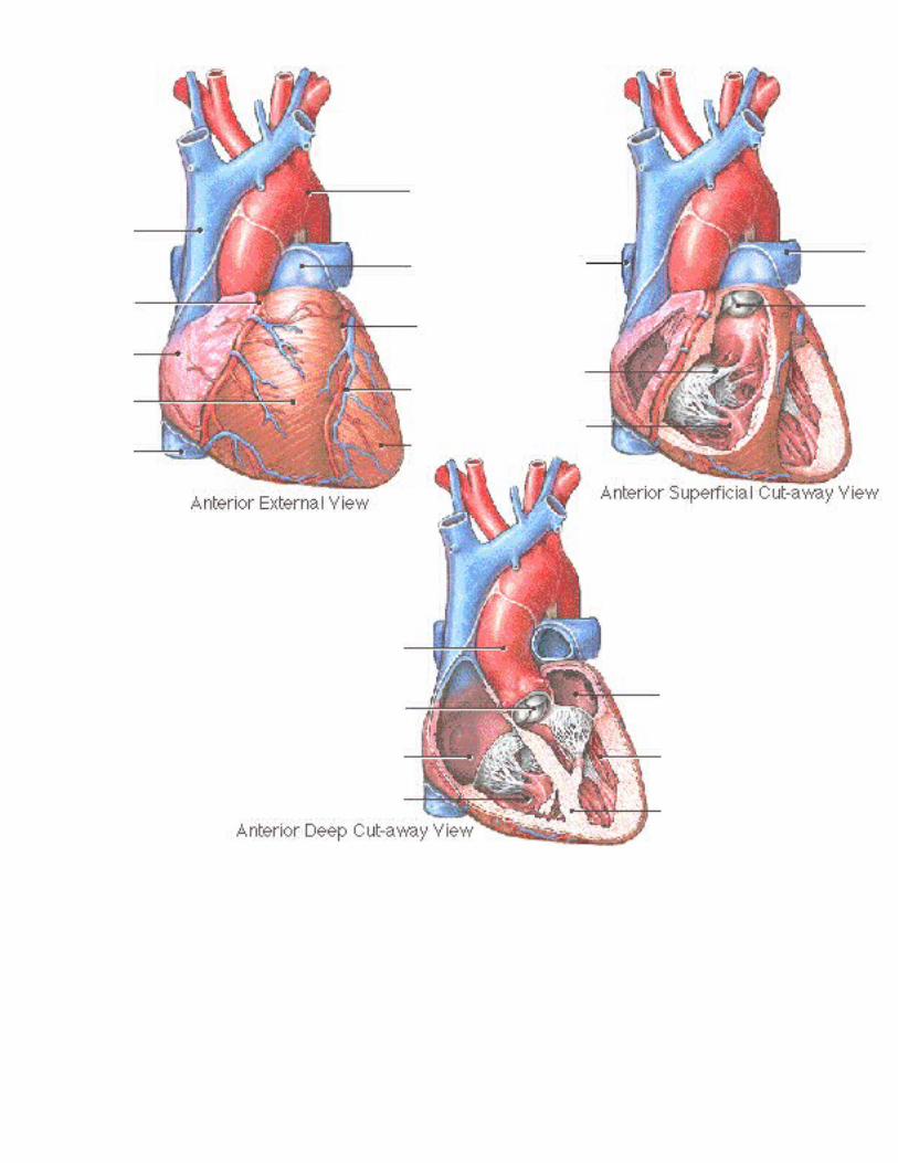
Page 3, 4 & 5. Anatomy Views• Label the diagrams of the heart below:
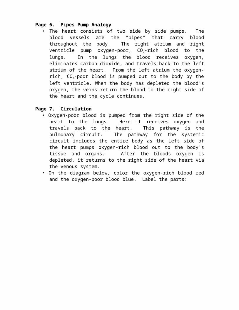
Page 6. Pipes-Pump Analogy• The heart consists of two side by side pumps. The blood vessels are the "pipes" that
carry blood throughout the body. The right atrium and right ventricle pump oxygen-poor, CO2-rich blood to the lungs. In the lungs the blood receives oxygen, eliminates carbon dioxide, and travels back to the left atrium of the heart. From the left atrium the oxygen-rich, CO2-poor blood is pumped out to the body by the left ventricle. When the body has depleted the blood's oxygen, the veins return the blood to the right side of the heart and the cycle continues.
Page 7. Circulation• Oxygen-poor blood is pumped from the right side of the heart to the lungs. Here it
receives oxygen and travels back to the heart. This pathway is the pulmonary circuit. The pathway for the systemic circuit includes the entire body as the left side of the heart pumps oxygen-rich blood out to the body's tissue and organs. After the bloods oxygen is depleted, it returns to the right side of the heart via the venous system.
• On the diagram below, color the oxygen-rich blood red and the oxygen-poor blood blue. Label the parts:
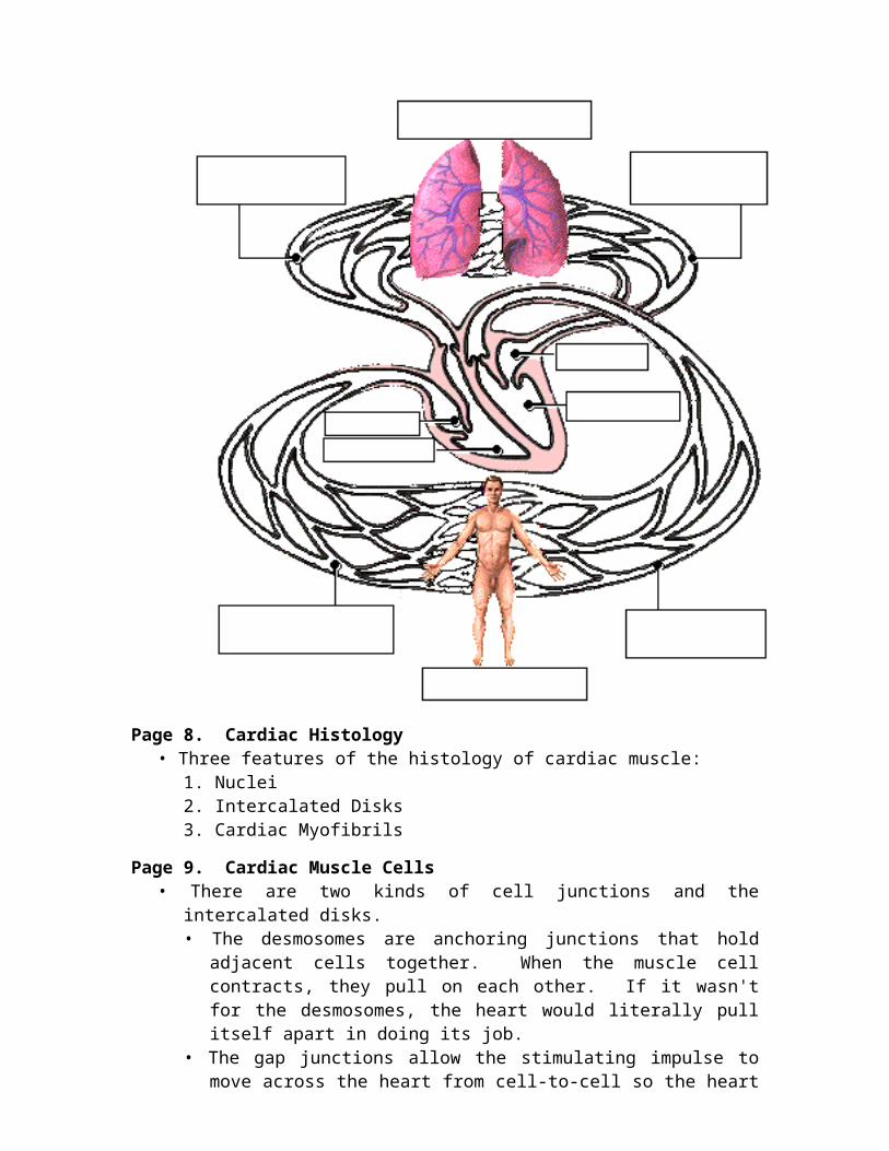
Page 8. Cardiac Histology• Three features of the histology of cardiac muscle:
1. Nuclei2. Intercalated Disks3. Cardiac Myofibrils
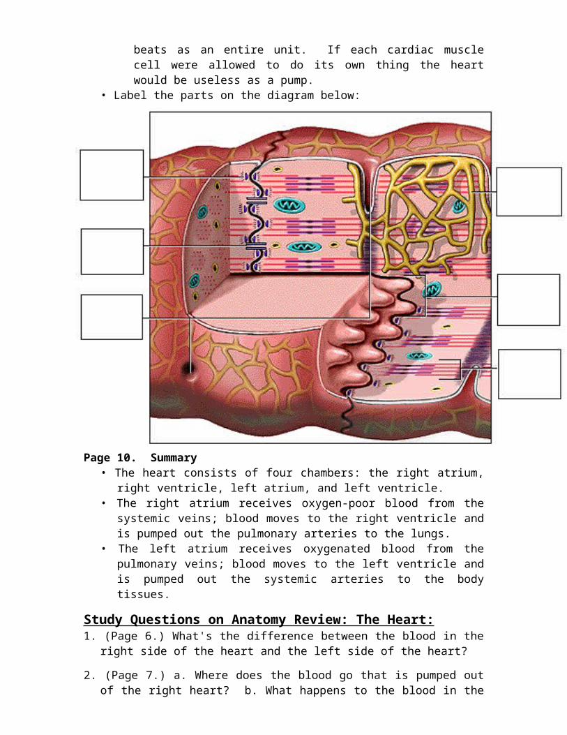
Page 9. Cardiac Muscle Cells• There are two kinds of cell junctions and the intercalated disks.
• The desmosomes are anchoring junctions that hold adjacent cells together. When the muscle cell contracts, they pull on each other. If it wasn't for the desmosomes, the heart would literally pull itself apart in doing its job.
• The gap junctions allow the stimulating impulse to move across the heart from cell-to-cell so the heart beats as an entire unit. If each cardiac muscle cell were allowed to do its own thing the heart would be useless as a pump.
• Label the parts on the diagram below:
Page 10. Summary• The heart consists of four chambers: the right atrium, right ventricle, left atrium, and
left ventricle.• The right atrium receives oxygen-poor blood from the systemic veins; blood moves
to the right ventricle and is pumped out the pulmonary arteries to the lungs.• The left atrium receives oxygenated blood from the pulmonary veins; blood moves
to the left ventricle and is pumped out the systemic arteries to the body tissues.
Study Questions on Anatomy Review: The Heart:
1. (Page 6.) What's the difference between the blood in the right side of the heart and the left side of the heart?
2. (Page 7.) a. Where does the blood go that is pumped out of the right heart? b. What happens to the blood in the lungs? c. Where does the blood go that is pumped out of the left heart?
3. (Page 7.) What is the pulmonary circuit and the systemic circuit?
4. (Page 8.) What three structural features are found on histological images of cardiac muscle?
5. (Page 9.) What are the names of the two types of cell junctions in cardiac muscle cells?
6. (Page 9.) What is the function of desmosomes?
7. (Page 9.) What is the function of gap junctions?
Intrinsic Conduction SystemGraphics are used with permission of:
Pearson Education Inc., publishing as Benjamin Cummings (http://www.aw-bc.com)
Page 1. Introduction• The intrinsic conduction system sets the basic rhythm of the beating heart. • It consists of autorhythmic cardiac cells that initiate and distribute impulses (action
potentials) throughout the heart.
Page 2. Goals• To identify the components of the intrinsic conduction system.• To recognize that the intrinsic conduction system coordinates heart activity by
determining the direction and speed of heart depolarization.• To relate heart electrical activity to an ECG wave tracing.
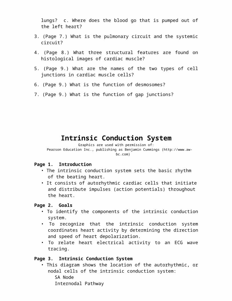
Page 3. Intrinsic Conduction System• This diagram shows the location of the autorhythmic, or nodal cells of the intrinsic
conduction system:SA NodeInternodal PathwayAV NodeAV BundleBundle BranchesPurkinje Fibers
• Label this diagram:
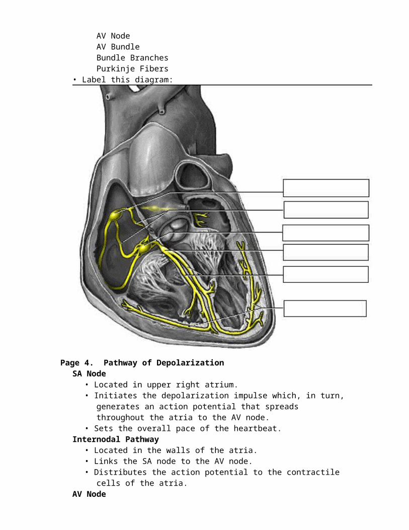
Page 4. Pathway of DepolarizationSA Node
• Located in upper right atrium.• Initiates the depolarization impulse which, in turn, generates an action potential
that spreads throughout the atria to the AV node.• Sets the overall pace of the heartbeat.
Internodal Pathway• Located in the walls of the atria.• Links the SA node to the AV node.• Distributes the action potential to the contractile cells of the atria.
AV Node• Located in the inferior interatrial septum.• The action potential is delayed here briefly, while the atria contract, before being
transmitted to the AV bundle.AV Bundle
• The only electrical connection between the atria and the ventricles.• Allows the action potential to move from the interatrial septum to the
interventricular septum, connecting the AV node to the Bundle Branches.Bundle Branches
• Convey the action potential down the interventricular septum.Purkinje Fibers
• Begin at the lower interventricular septum to the apex of the heart, then continue superiorly through the myocardium of the ventricles.
• The Purkinje fibers convey the action potential to the contractile cells of the ventricle.
• Action potentials, which spread from the autorhythmic cells of the intrinsic conduction system to the contractile cells are electrical events.
• Subsequent contraction of the contractile cells is a mechanical event that causes a heartbeat.
** Now is a good time to go to quiz question 1:• Click the Quiz button on the left side of the screen.• After answering question 1, click the Back to Topic button on the left side of the screen.• To get back to where you left off, click on the scrolling page list at the top of the screen and choose "5.
ECG Wave".
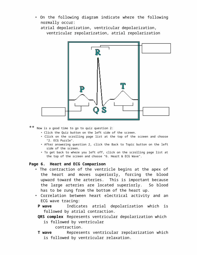
Page 5. ECG WaveECG Waves:
P Wave • Small upward wave.• Indicates atrial depolarization.
QRS Wave • Downward deflection, then a large upward peak, ending as a downward deflection.
• Represents ventricular depolarization.T Wave • Dome-shaped wave.
• Represents ventricular repolarization.• In a normal ECG tracing, atrial repolarization is hidden by the QRS complex.
• On the following diagram indicate where the following normally occur:atrial depolarization, ventricular depolarization, ventricular repolarization, atrial
repolarization
** Now is a good time to go to quiz question 2:• Click the Quiz button on the left side of the screen.• Click on the scrolling page list at the top of the screen and choose "2. ECG Puzzle".• After answering question 2, click the Back to Topic button on the left side of the screen.
• To get back to where you left off, click on the scrolling page list at the top of the screen and choose "6. Heart & ECG Wave".
Page 6. Heart and ECG Comparison• The contraction of the ventricle begins at the apex of the heart and moves
superiorly, forcing the blood upward toward the arteries. This is important because the large arteries are located superiorly. So blood has to be rung from the bottom of the heart up.
• Correlation between heart electrical activity and an ECG wave tracing:P wave Indicates atrial depolarization which is followed by atrial
contraction.QRS complex Represents ventricular depolarization which is followed by
ventricular contraction.T wave Represents ventricular repolarization which is followed by
ventricular relaxation.
Page 7. Summary• The intrinsic conduction system of the heart initiates depolarization impulses.• Action potentials spread throughout the heart, causing coordinated heart
contraction.• An ECG wave tracing records the electrical activity of the heart.
** Now is a good time to go to quiz questions 3 and 4:• Click the Quiz button on the left side of the screen.• Click on the scrolling page list at the top of the screen and choose "3a. Left Bundle branch Block".• Work through questions 3a, 3b, and 4.
Notes on Quiz Questions:Quiz Question #1. Conduction Pathway
• This question asks you to match the various autorhythmic cells of heart to their functions.
Quiz Question # 2. ECG Puzzle• This question asks you to piece together a normal ECG Tracing.
Quiz Question #3a & 3b. Create Left Bundle Branch Block• This question asks you to create a left bundle branch block and predict what
would happen to the ECG tracing. • If you have a difficult time understanding the correct answer, please note that
normally the left ventricle is depolarized when impulses move along the left bundle branch and to the Purkinje fibers. If the left bundle branch is blocked, ventricular depolarization takes longer because impulses in the left ventricle must travel from cell to cell. Because ventricular depolarization is taking longer, the QRS complex is wider.
Quiz Question #4. ECG for Tachycardia• This question allows you to chose the ECG Wave tracing that corresponds to
Tachycardia • With a normal heart rate of 75 beats per minute, one heartbeat takes 0.8 seconds.
(1 minute/75 beats) (60 seconds/1 minute) = 0.8 seconds• An abnormally fast heart rate, such as 120 beats per minutes, one heartbeat takes 0.5 seconds.
(1 minute/120 beats) (60 seconds/1 minute) = 0.5 seconds
Study Questions on the Intrinsic Conduction System:1. (Page 1.) What is the purpose of the intrinsic conduction system of the heart?
2. (Page 1.) What type of cells are present in the intrinsic conduction system of the heart?
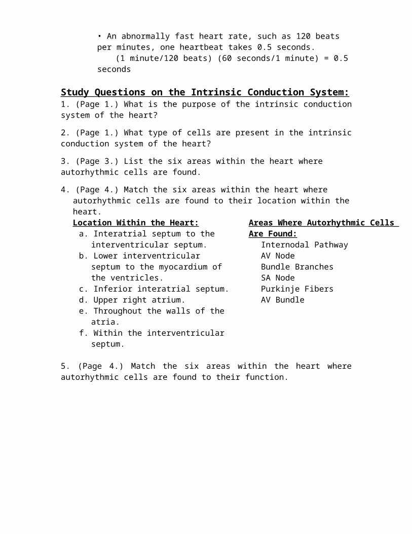
3. (Page 3.) List the six areas within the heart where autorhythmic cells are found.
4. (Page 4.) Match the six areas within the heart where autorhythmic cells are found to their location within the heart. Location Within the Heart:
a. Interatrial septum to the interventricular septum.
b. Lower interventricular septum to the myocardium of the ventricles.
c. Inferior interatrial septum.d. Upper right atrium.e. Throughout the walls of the atria.f. Within the interventricular septum.
Areas Where Autorhythmic Cells Are Found:Internodal PathwayAV NodeBundle BranchesSA Node Purkinje FibersAV Bundle
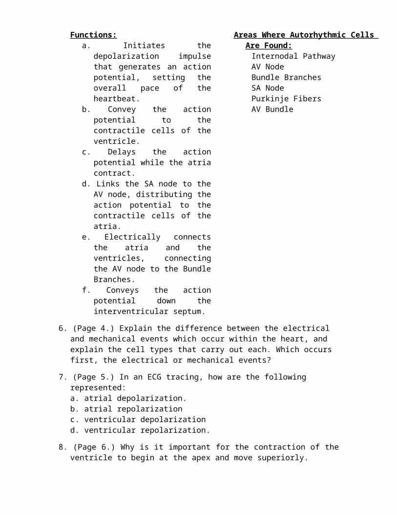
5. (Page 4.) Match the six areas within the heart where autorhythmic cells are found to their function.
Functions:a. Initiates the depolarization impulse
that generates an action potential, setting the overall pace of the heartbeat.
b. Convey the action potential to the contractile cells of the ventricle.
c. Delays the action potential while the atria contract.
d. Links the SA node to the AV node, distributing the action potential to the contractile cells of the atria.
e. Electrically connects the atria and the ventricles, connecting the AV node to the Bundle Branches.
f. Conveys the action potential down the interventricular septum.
Areas Where Autorhythmic Cells Are Found:
Internodal PathwayAV NodeBundle BranchesSA Node Purkinje FibersAV Bundle
6. (Page 4.) Explain the difference between the electrical and mechanical events which occur within the heart, and explain the cell types that carry out each. Which occurs first, the electrical or mechanical events?
7. (Page 5.) In an ECG tracing, how are the following represented: a. atrial depolarization.b. atrial repolarization c. ventricular depolarizationd. ventricular repolarization.
8. (Page 6.) Why is it important for the contraction of the ventricle to begin at the apex and move superiorly.
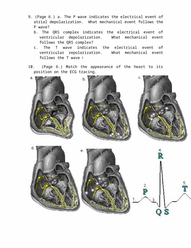
9. (Page 6.) a. The P wave indicates the electrical event of atrial depolarization. What mechanical event follows the P wave? b. The QRS complex indicates the electrical event of ventricular depolarization.
What mechanical event follows the QRS complex?c. The T wave indicates the electrical event of ventricular repolarization. What
mechanical event follows the T wave ?
10. (Page 6.) Match the appearance of the heart to its position on the ECG tracing.
Blood Pressure RegulationGraphics are used with permission of:
Pearson Education Inc., publishing as Benjamin Cummings (http://www.aw-bc.com)
Page 1. Introduction• There are two basic mechanisms for regulating blood pressure:
(1) short-term mechanisms, which regulate blood vessel diameter, heart rate and contractility
(2) long-term mechanisms, which regulate blood volume
Page 2. Goals• To compare and contrast the short-term mechanisms that respond
to rising blood pressure with the short-term mechanisms that respond to falling blood pressure.
• To understand the process of long-term regulation of low blood pressure.
• To describe the long-term and short-term effects of increased osmolarity on blood pressure.
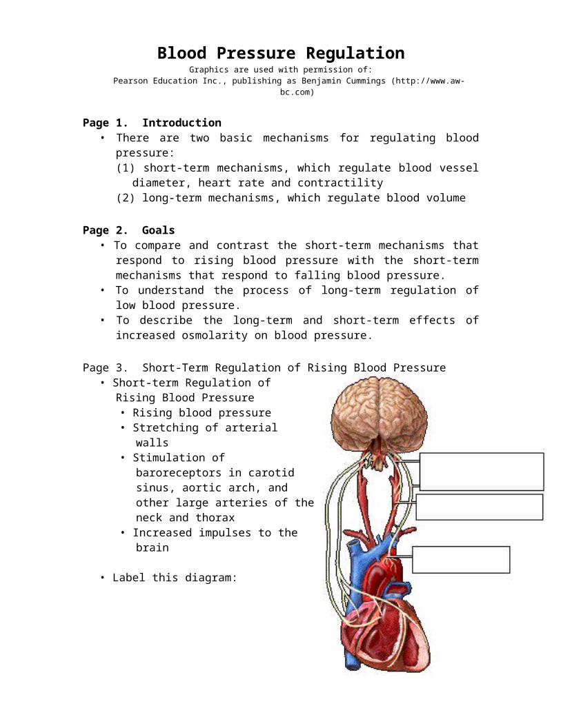
Page 3. Short-Term Regulation of Rising Blood Pressure• Short-term Regulation of Rising
Blood Pressure• Rising blood pressure• Stretching of arterial walls• Stimulation of baroreceptors in
carotid sinus, aortic arch, and other large arteries of the neck and thorax
• Increased impulses to the brain
• Label this diagram:
Page 4. Effect of Baroreceptors • Increased impulses to brain from baroreceptors• Increased parasympathetic activity and decreased sympathetic
activity• Reduction of heart rate and increase in arterial diameter• Lower blood pressure
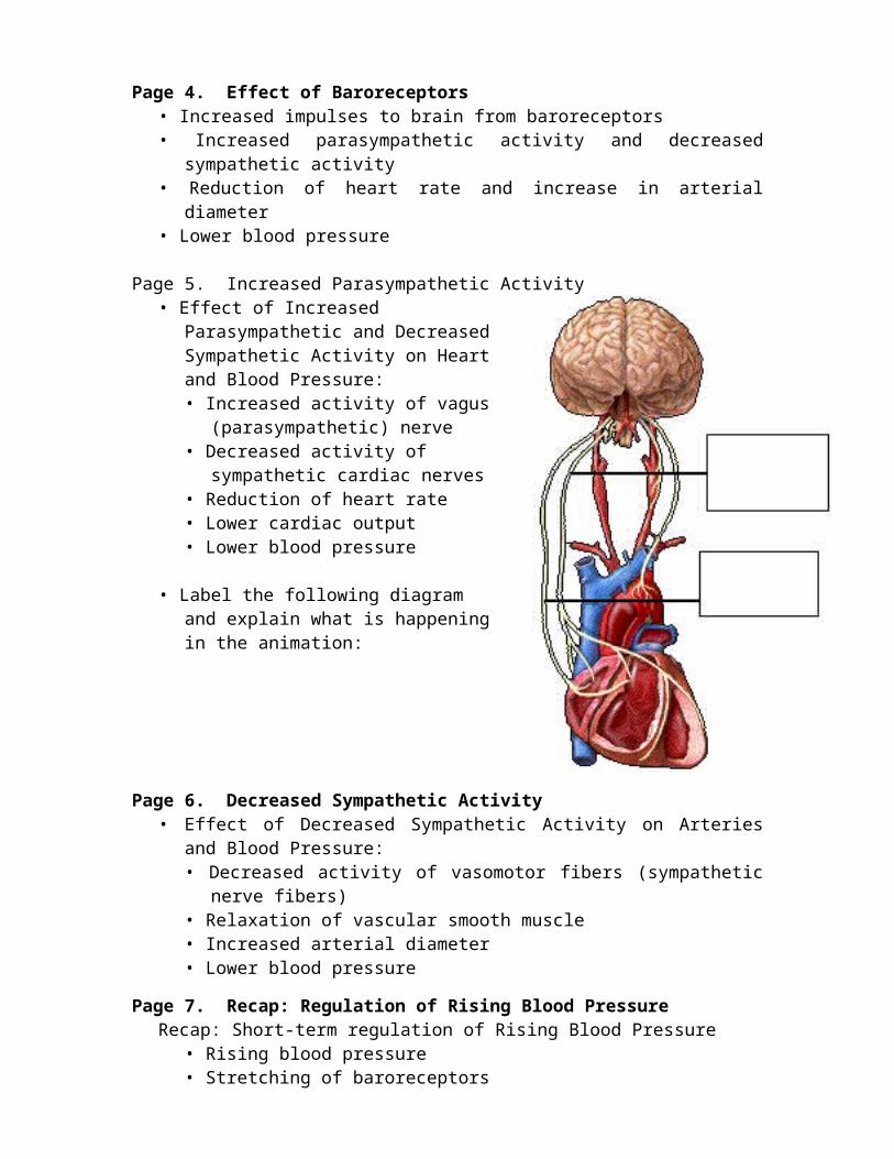
Page 5. Increased Parasympathetic Activity
• Effect of Increased Parasympathetic and Decreased Sympathetic Activity on Heart and Blood Pressure:• Increased activity of vagus
(parasympathetic) nerve• Decreased activity of sympathetic
cardiac nerves• Reduction of heart rate• Lower cardiac output• Lower blood pressure
• Label the following diagram and explain what is happening in the animation:
Page 6. Decreased Sympathetic Activity • Effect of Decreased Sympathetic Activity on Arteries and Blood
Pressure:• Decreased activity of vasomotor fibers (sympathetic nerve
fibers)• Relaxation of vascular smooth muscle• Increased arterial diameter• Lower blood pressure
Page 7. Recap: Regulation of Rising Blood Pressure Recap: Short-term regulation of Rising Blood Pressure
• Rising blood pressure• Stretching of baroreceptors• Increased impulses to the brain• Increased parasympathetic activity• Decreased sympathetic activity• Slowing of heart rate• Increased arterial pressure• Reduction of blood pressure
• Take notes on the diagram above as the animation proceeds.Page 8. Short-term Regulation of Falling Blood Pressure
• Short-term Regulation of Falling Blood Pressure:• Falling blood pressure• Baroreceptors inhibited• Decreased impulses to the brain
• Decreased parasympathetic activity, increased sympathetic activity
• Three effects:1. Heart: increased heart rate and increased contractility2. Vessels: increased vasoconstriction3. Adrenal gland: release of epinephrine and norepinephrine
which enhance heart rate, contractility, and vasoconstriction
• Increased blood pressure
Page 9. Sympathetic Activity on Heart and Blood Pressure• Effect of Increased Sympathetic Activity on Heart and Blood
Pressure:• Increased activity of sympathetic cardiac nerves• Decreased activity of vagus (parasympathetic) nerve• Increased heart rate and contractility• Higher cardiac output• Increased blood pressure
Page 10. Vasomotor Fibers• Effect of Increased Sympathetic Activity on Arteries and Blood
Pressure:• Increased activity of vasomotor fibers (sympathetic nerve
fibers)• Constriction of vascular smooth muscle• Decreased arterial diameter• Increased blood pressure
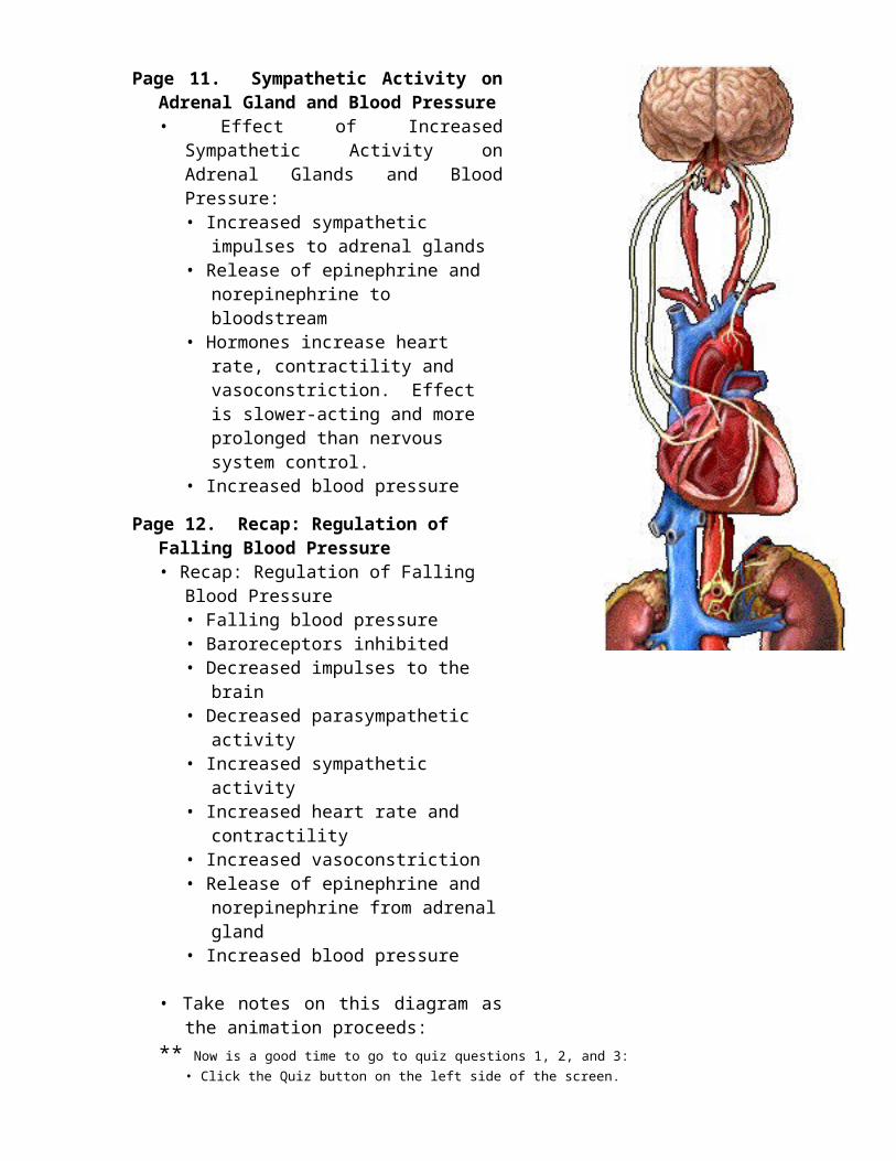
Page 11. Sympathetic Activity on Adrenal Gland and Blood Pressure• Effect of Increased Sympathetic
Activity on Adrenal Glands and Blood Pressure:• Increased sympathetic impulses
to adrenal glands• Release of epinephrine and
norepinephrine to bloodstream• Hormones increase heart rate,
contractility and vasoconstriction. Effect is slower-acting and more prolonged than nervous system control.
• Increased blood pressurePage 12. Recap: Regulation of Falling Blood
Pressure• Recap: Regulation of Falling Blood
Pressure• Falling blood pressure• Baroreceptors inhibited• Decreased impulses to the brain• Decreased parasympathetic
activity• Increased sympathetic activity• Increased heart rate and
contractility• Increased vasoconstriction• Release of epinephrine and
norepinephrine from adrenal gland
• Increased blood pressure
• Take notes on this diagram as the animation proceeds:
** Now is a good time to go to quiz questions 1, 2, and 3:• Click the Quiz button on the left side of the screen.• After answering question 3, click the Back to Topic button on the left side of the
screen.• To get back to where you left off, click on the scrolling page list at the top of the
screen and choose "13. Introduction: Long-Term Regulation of Low BP".
Page 13. Introduction: Long-Term Regulation of Low BP• Long-term regulation of blood pressure is primarily accomplished
by altering blood volume.• The loss of blood through hemorrhage, accident, or donating a
pint of blood will lower blood pressure and trigger processes to restore blood volume and therefore blood pressure back to normal.
• Long-term regulatory processes promote the conservation of body fluids via renal mechanisms and stimulate intake of water to normalize blood volume and blood pressures.
Page 14. Loss of Blood• When there is a loss of blood, blood pressure and blood volume
decrease.Page 15. Kidney Juxtaglomerular Cells
• Juxtaglomerular cells in the kidney monitor alterations in the blood pressure. If blood pressure falls too low, these specialized cells release the enzyme renin into the bloodstream.
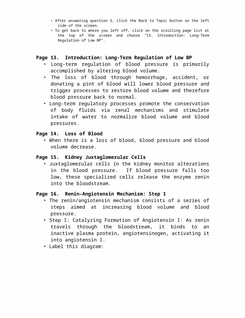
Page 16. Renin-Angiotensin Mechanism: Step 1• The renin/angiotensin mechanism consists of a series of steps
aimed at increasing blood volume and blood pressure.• Step 1: Catalyzing Formation of Angiotensin I: As renin travels
through the bloodstream, it binds to an inactive plasma protein, angiotensinogen, activating it into angiotensin I.
• Label this diagram:
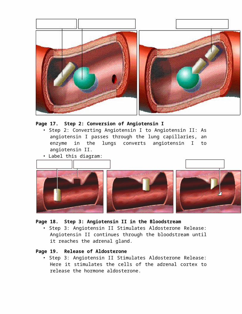
Page 17. Step 2: Conversion of Angiotensin I• Step 2: Converting Angiotensin I to Angiotensin II: As angiotensin I
passes through the lung capillaries, an enzyme in the lungs converts angiotensin I to angiotensin II.
• Label this diagram:
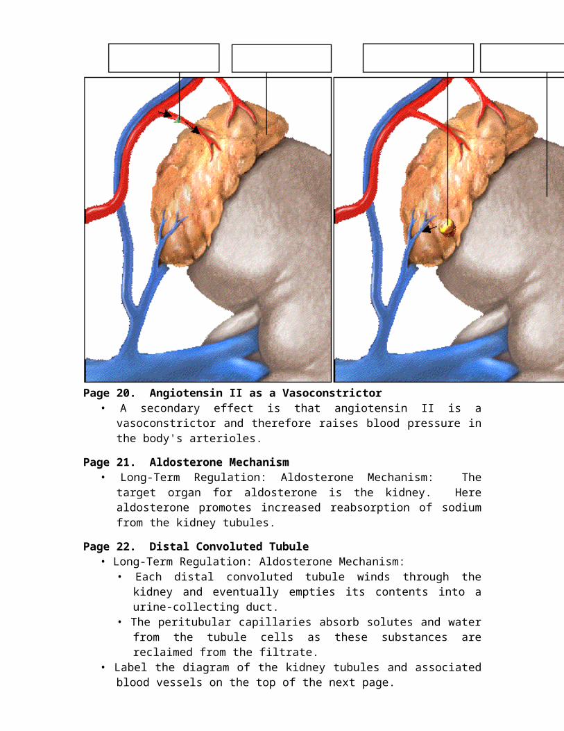
Page 18. Step 3: Angiotensin II in the Bloodstream• Step 3: Angiotensin II Stimulates Aldosterone Release:
Angiotensin II continues through the bloodstream until it reaches the adrenal gland.
Page 19. Release of Aldosterone• Step 3: Angiotensin II Stimulates Aldosterone Release: Here it
stimulates the cells of the adrenal cortex to release the hormone aldosterone.
Page 20. Angiotensin II as a Vasoconstrictor• A secondary effect is that angiotensin II is a vasoconstrictor and
therefore raises blood pressure in the body's arterioles.Page 21. Aldosterone Mechanism
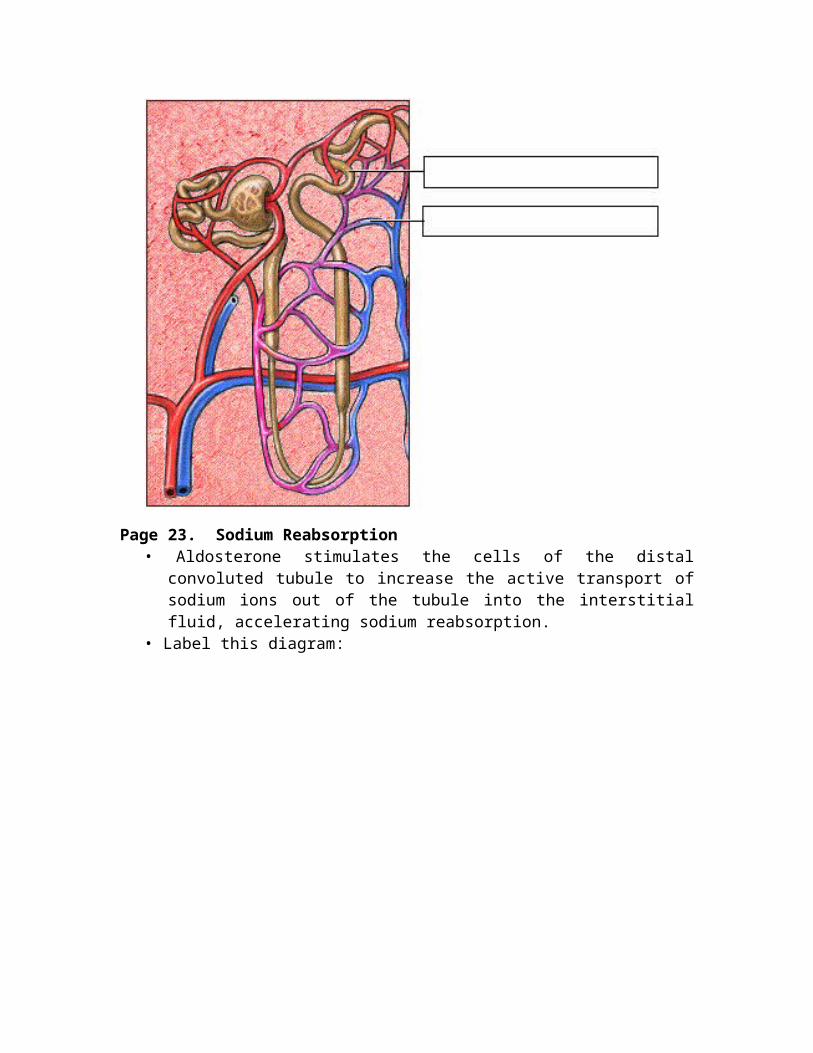
• Long-Term Regulation: Aldosterone Mechanism: The target organ for aldosterone is the kidney. Here aldosterone promotes increased reabsorption of sodium from the kidney tubules.
Page 22. Distal Convoluted Tubule• Long-Term Regulation: Aldosterone Mechanism:
• Each distal convoluted tubule winds through the kidney and eventually empties its contents into a urine-collecting duct.
• The peritubular capillaries absorb solutes and water from the tubule cells as these substances are reclaimed from the filtrate.
• Label the diagram of the kidney tubules and associated blood vessels on the top of the next page.
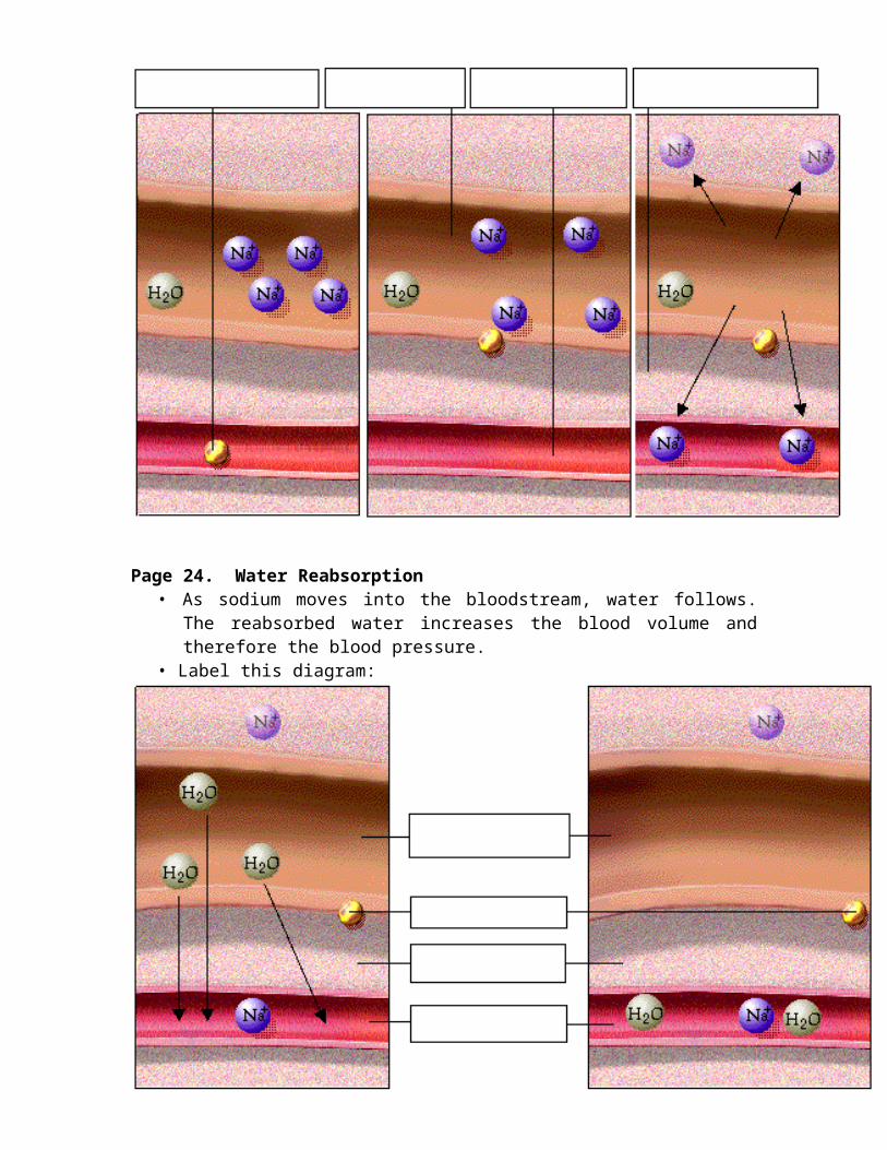
Page 23. Sodium Reabsorption• Aldosterone stimulates the cells of the distal convoluted tubule to
increase the active transport of sodium ions out of the tubule into the interstitial fluid, accelerating sodium reabsorption.
• Label this diagram:
Page 24. Water Reabsorption• As sodium moves into the bloodstream, water follows. The
reabsorbed water increases the blood volume and therefore the blood pressure.
• Label this diagram:
Page 25. Increase in Osmolarity• Dehydration due to sweating, diarrhea, or excessive urine flow will
cause an increase in osmolarity of the blood and a decrease in blood volume and blood pressure.
Page 26. Long-Term Effect of Osmolarity on BP• As increased osmolarity is detected there is both a short and long-
term effect. For the long-term effect, the hypothalamus sends a signal to the posterior pituitary to release antidiuretic hormone (ADH).
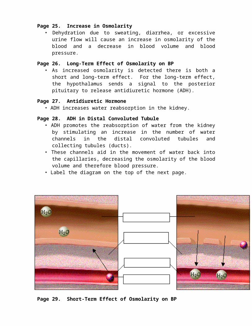
Page 27. Antidiuretic Hormone• ADH increases water reabsorption in the kidney.
Page 28. ADH in Distal Convoluted Tubule• ADH promotes the reabsorption of water from the kidney by
stimulating an increase in the number of water channels in the distal convoluted tubules and collecting tubules (ducts).
• These channels aid in the movement of water back into the capillaries, decreasing the osmolarity of the blood volume and therefore blood pressure.
• Label the diagram on the top of the next page.
Page 29. Short-Term Effect of Osmolarity on BP• A short-term effect of increased osmolarity is the excitation of the
thirst center in the hypothalamus. The thirst center stimulates the individual to drink more water and thus rehydrate the blood and extracellular fluid, restoring blood volume and therefore blood pressure.
Page 30. Other Chemicals That Influence BP
• There are many other chemicals which influence blood flow and blood vessel diameter. Most of them act by influencing blood vessel diameter.
Page 31. Summary• In the short-term, rising blood pressure stimulates increased
parasympathetic activity, which leads to reduced heart rate, vasodilation and lower blood pressure.
• Falling blood pressure stimulates increased sympathetic activity, which leads to increased heart rate, contractility, vasoconstriction, and blood pressure.
• Long-term blood pressure regulation involves renal regulation of blood volume via the renin-angiotensin mechanism and aldosterone mechanism.
• Increased blood osmolarity stimulates release of antidiuretic hormone (ADH), which promotes reabsorption of water, and excites the thirst center, resulting in increased blood volume and blood pressure.
** Now is a good time to go to quiz questions 4 and 5:• Click the Quiz button on the left side of the screen.• Click on the scrolling page list at the top of the screen and choose "4. Blood Volume
Chain Reaction". • Work through quiz questions 4-5.
Notes on Quiz Questions:Quiz Question #1: Identification
• This question asks you to label structures which are important in blood pressure regulation.
Quiz Question #2: High Blood Pressure• This question asks you to lower blood pressure by clicking on
the appropriate nerve.Quiz Question #3: Chemical Heart Stabilization
• This question asks you to identify the chemical that will increase blood pressure.
Quiz Question #4: Blood Volume Chain Reaction• This question asks you to list the proper sequence of events
that occurs when blood volume and blood pressure increases or decreases.
Quiz Question #5: Dehydration Chain Reaction• This question asks you to list the proper sequence of events
that occurs when dehydration increases or decreases.
Study Questions on Blood Pressure Regulation:1. (Page 3.) What are baroreceptors?2. (Page 3.) Where are the baroreceptors that sense blood pressure
located?
3. (Page 3.) What happens to baroreceptors when blood pressure is high?
4. (Page 4.) What happens to both parasympathetic activity and sympathetic activity when blood pressure is high?
5. (Page 4.) What is the effect of increased parasympathetic activity and decreased sympathetic activity on both heart rate and blood pressure?
6. (Page 5.) What is the name of the parasympathetic nerve that decreases heart rate?
7. (Page 5.) How does a decrease in heart rate decrease blood pressure?
8. (Page 6.) What is a vasomotor fiber?9. (Page 6.) What is the effect of high blood pressure on arteries?10. (Page 6.) How does vasodilation decrease blood pressure?11. (Page 8.) What happens to baroreceptors when the blood pressure
is low? What effect does that have on the brain?12. (Page 8.) What are the three effects of an increased sympathetic
activity and decreased parasympathetic activity?13. (Page 9.) How does an increase in heart rate increase blood
pressure?14. (Page 10.) What is the effect of low blood pressure on arteries?15. (Page 10.) How does vasoconstriction increase blood pressure?16. (Page 11.) What is the effect of sympathetic activity on the adrenal
gland?17. (Page 11.) Why are the effects of epinephrine and norepinephrine
from the adrenal gland slower-acting and more prolonged than nervous system control?
18. (Page 13.) When there is a loss of blood through hemorrhage, accident, or donating a pint of blood, what two long-term regulatory processes will restore blood volume and therefore blood pressure back to normal?
19. (Page 14.) What happens to blood volume and blood pressure when there is blood loss?
20. (Page 15.) If blood pressure falls too low, what do the juxtaglomerular cells of the kidney release into the bloodstream?
21. (Page 16.) Label the diagram on p. 16.22. (Page 16.) What is angiotensinogen?23. (Page 16.) How is angiotensinogen activated?24. (Page 17.) Label the diagram on p. 17.
25. (Page 17.) How is angiotensin I converted into Angiotensin II?26. (Pages 16-19) Place the following steps in the release of
aldosterone in order:a. An enzyme in the lungs converts angiotensin I to angiotensin II.b. Angiotensinogen is activated into angiotensin I.c. Angiotensin II stimulates the cells of the adrenal cortex to
release the hormone aldosterone.d. As renin travels through the bloodstream, it binds to an inactive
plasma protein, angiotensinogen.e. Angiotensin II continues through the bloodstream until it
reaches the adrenal gland. f. Angiotensin I passes through the lung capillaries.
27. (Page 19.) Label the diagram on p. 19.28. (Page 18,19) What happens when Angiotensin II reaches the
adrenal gland?29. (Page 20.) What are two effects of angiotensin II?30. (Page 21.) What is the target organ for aldosterone?31. (Page 21.) What is the effect of aldosterone?32. (Page 22.) What is "filtrate" and where is it located within the
kidneys? What is its relationship to the blood capillaries.33. (Page 22.) What is the process of reabsorption within the kidneys?34. (Page 23.) What happens when aldosterone binds to the cells of the
distal convoluted tubule?35. (Page 23.) Label the diagram on p. 23.36. (Page 24.) Label the diagram on p. 24.37. (Page 24.) How does aldosterone increase the blood volume and
blood pressure?38. (Page 25.) What effect does dehydration due to sweating, diarrhea,
or excessive urine flow have on osmolarity of the blood, blood volume, and blood pressure?
39. (Page 26.) An increased osmolarity of the blood causes the release of what hormone?
40. (Page 27.) What is the effect of ADH?41. (Page 28.) How does ADH increase water reabsorption in the
kidney?42. (Page 29.) What is the short-term effect of increased osmolarity of
the blood on blood pressure?43. (Summary) When blood volume and blood pressure are increased,
do the following increase or decrease?
a. Renin release from the kidney ________.b. Angiotensinogen into Angiotensin I _______.c. Angiotensin I into Angiotensin II _______.d. Aldosterone release from the adrenal gland _______.e. Sodium reabsorption from the filtrate into the blood _______.f. Water reabsorption _______.g. Blood volume and blood pressure _______.
44. (Summary) When blood volume and blood pressure are decreased, do the following increase or decrease?
a. Renin release from the kidney ________.b. Angiotensinogen into Angiotensin I _______.c. Angiotensin I into Angiotensin II _______.d. Aldosterone release from the adrenal gland _______.e. Sodium reabsorption from the filtrate into the blood _______.f. Water reabsorption _______.g. As a result blood volume and blood pressure _______.
45. (Summary) When there is an increase in dehydration, are the following increased or decreased?
a. Body water _______.b. Blood volume and blood pressure _______.c. Blood osmolarity _______.d. ADH release from the pituitary _______.e. Water permeability of the kidney tubules _______.f. Urine output and blood osmolarity _______.g. As a result, blood volume and blood pressure _______.