Embed Size (px)
Citation preview
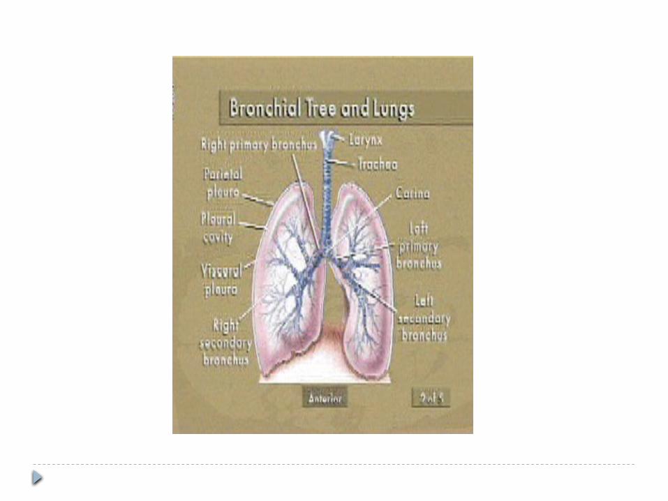
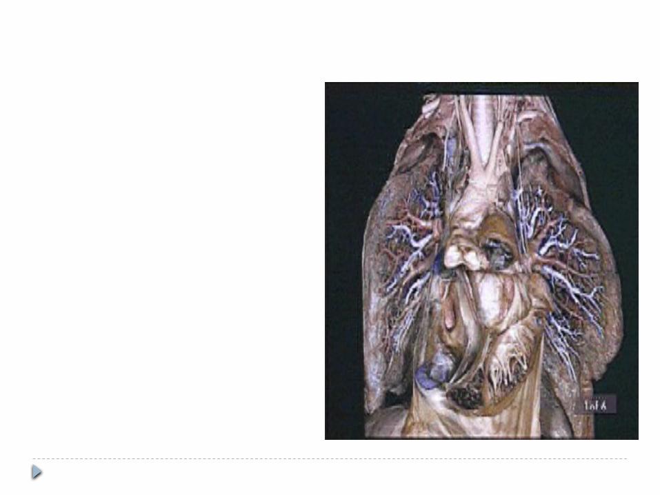
ANATOMY OF TRACHEOBRONCHIAL TREE
PRESENTED BY- Dr. CHITRA
MODERATOR-Dr.AJAY SOOD
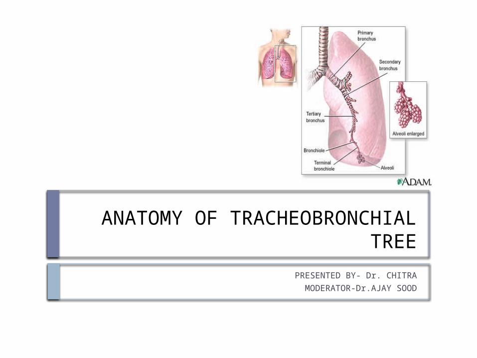
Respiratory System
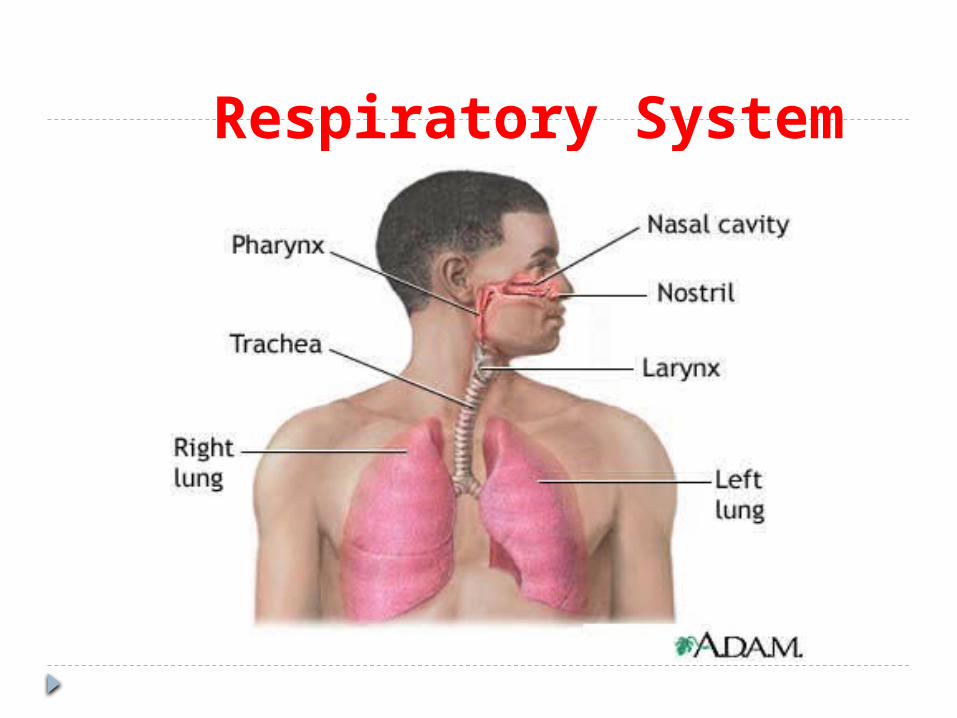
TRACHEAIt is a cartilaginous and membranous tube
EXTENT - from 6th cervical vertebra till the body of 5th thoracic vertebra
During expiration the bifurcation rises by one vertebral level and during inspiration may be lowered as far as 6th thoracic vertebra
LENGTH - 11cm DIAMETER - 2 to 2.5cm CHILDREN - smaller, deeper, more moveable
STRUCTURE CARTILAGES-16to20 in no.,each forms an
incomplete ring,which occupies anterior two third of circumference of trachea
Are placed horizontally above each other,separated by narrow intervals
4mm deep and 1mm thick Outer surface is flattened in vertical direction and
convex from inner side Highly elastic,but may calcify in later stages FIRST TRACHEAL CARTILAGE-broader,divided
connected to lower end of cricoid by cricotracheal ligament
LAST TRACHEAL CARTILAGE-thick and broad in midlle, lower border is prolonged to a triangular hook shaped process which curves downward and backward between two bronchi

FIBROUS MEMBRANE-cartilages are enclosed in an elastic fibrous membrane which consists of two layers,one passes over the outer surface the other one over the inner surface
At upper and lower margins they blend together to form a single membrane
MUSCULAR TISSUES-two layers of non –striated muscles longitudinal and transeverse
Longitudinal fibres are external,consist of few scattered bundles only
Transeverse fibers(trachealis muscle) are internal,extends between the end of cartilages
MUCUS MEMBRANE-continous above with larynx and below with bronchus
Consist of areolar and lymphoid tissue,basement membrane,supporting stratified epithelium ,surface layer of which is columnar and ciliated
Beneath basement membrane there is a layer of longitudinal elastic fibre
Submucus layer,composed of loose meshwork of connective tissue
VESSELS AND NERVES- ARTERIAL SUPPLY-Inferior thyroid arteries NERVE SUPPLY-vagus nerve,recurrent
nerve,sympathetic nerves
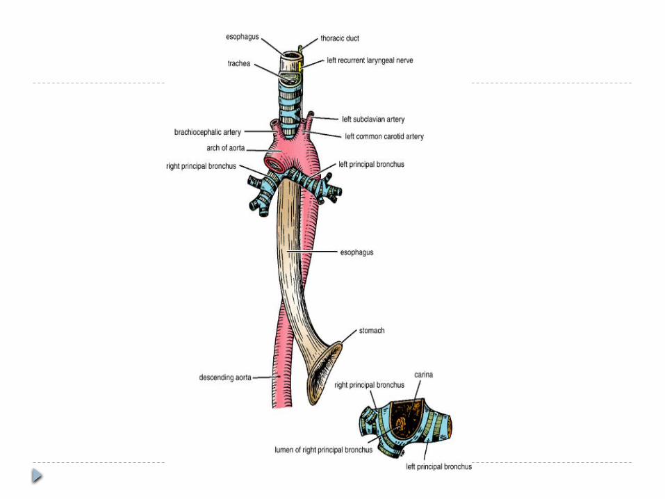
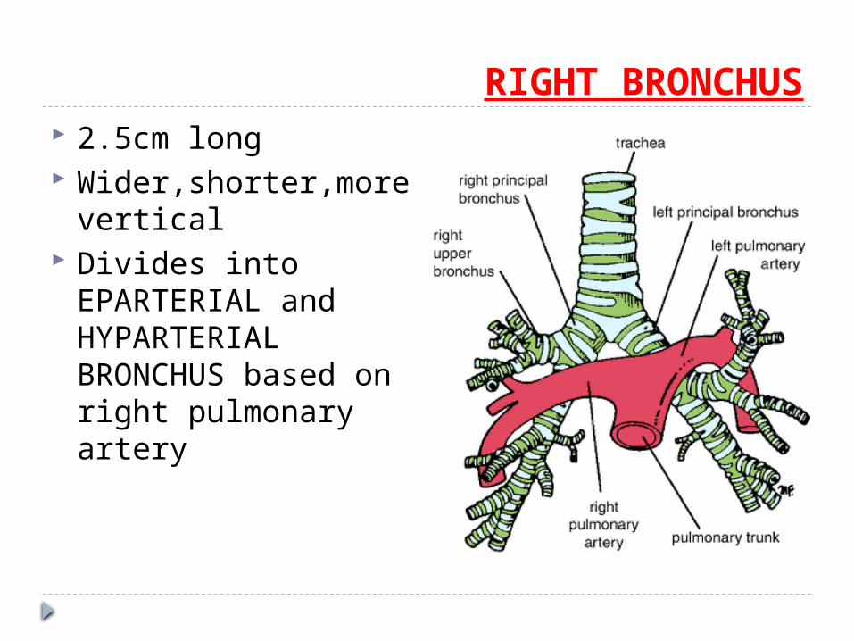
RIGHT BRONCHUS 2.5cm long Wider,shorter,more
vertical Divides into
EPARTERIAL and HYPARTERIAL BRONCHUS based on right pulmonary artery
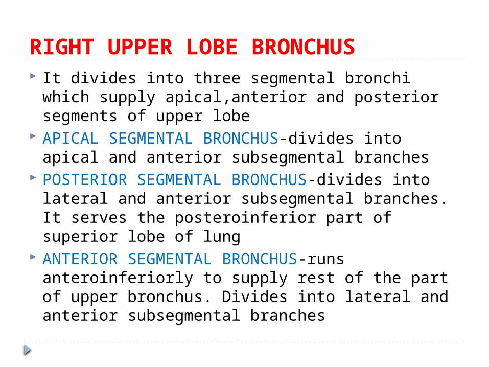
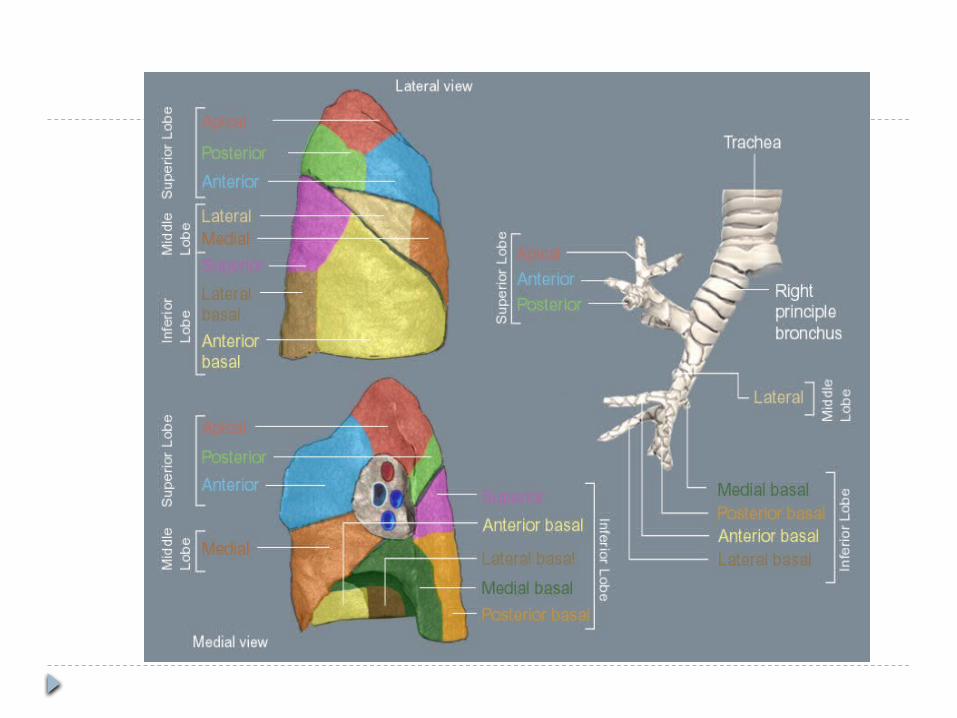
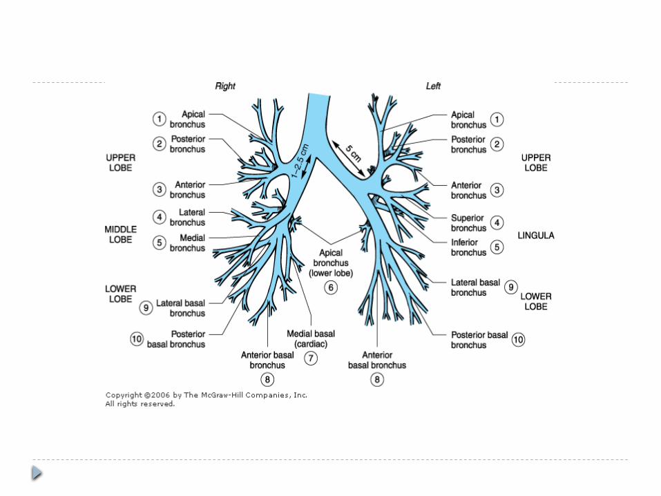
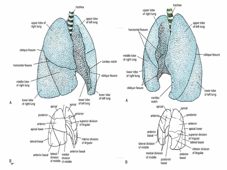
RIGHT UPPER LOBE BRONCHUS It divides into three segmental bronchi which
supply apical,anterior and posterior segments of upper lobe
APICAL SEGMENTAL BRONCHUS-divides into apical and anterior subsegmental branches
POSTERIOR SEGMENTAL BRONCHUS-divides into lateral and anterior subsegmental branches. It serves the posteroinferior part of superior lobe of lung
ANTERIOR SEGMENTAL BRONCHUS-runs anteroinferiorly to supply rest of the part of upper bronchus. Divides into lateral and anterior subsegmental branches

RIGHT MIDDLE LOBE BRONCHUS Divides into lateral and medial subsegments
RIGHT LOWER LOBE BRONCHUS Continuation of principal stem beyond the
origin of middle lobe bronchus Supplies 5 segments of the lung Apical segmental bronchus Medial basal segmental bronchus Anterior basal segmental bronchus Lateral and posterior basal segmental
bronchus
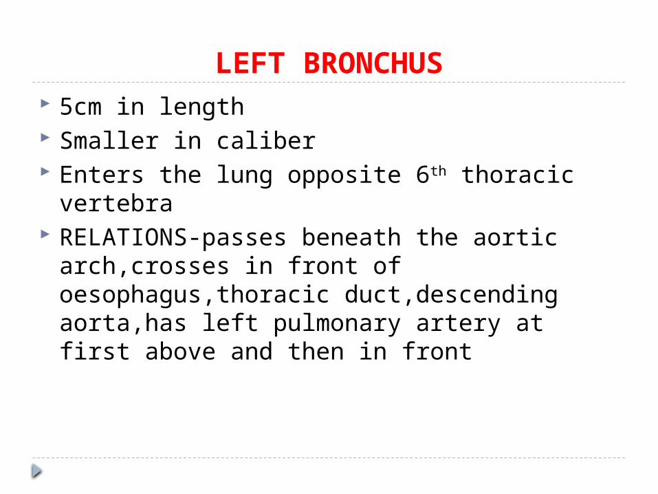
LEFT BRONCHUS 5cm in length Smaller in caliber Enters the lung opposite 6th thoracic vertebra RELATIONS-passes beneath the aortic
arch,crosses in front of oesophagus,thoracic duct,descending aorta,has left pulmonary artery at first above and then in front
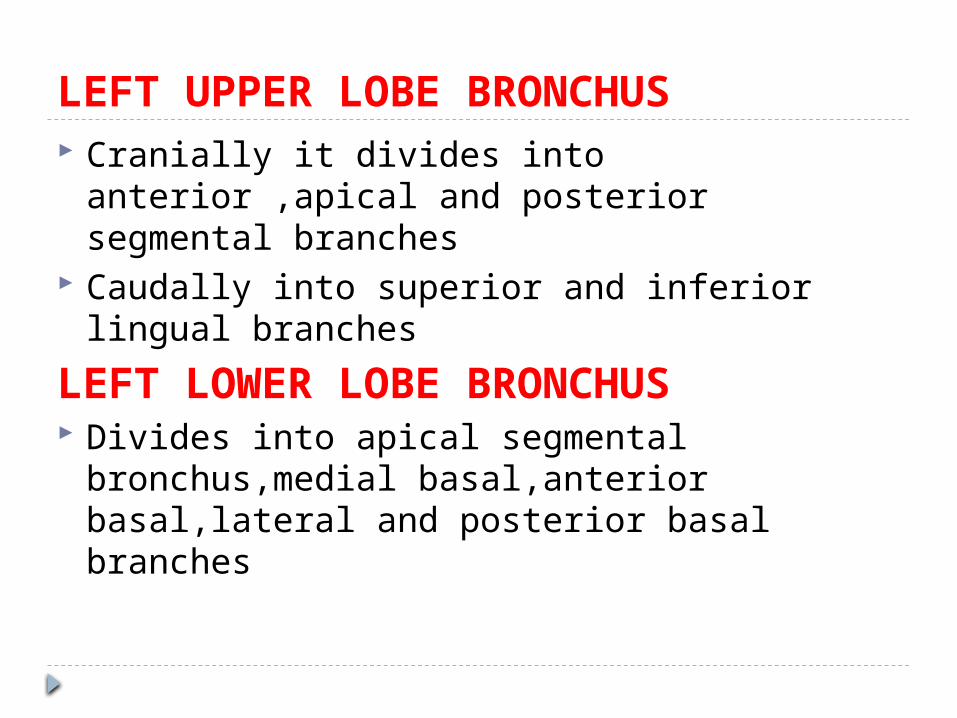
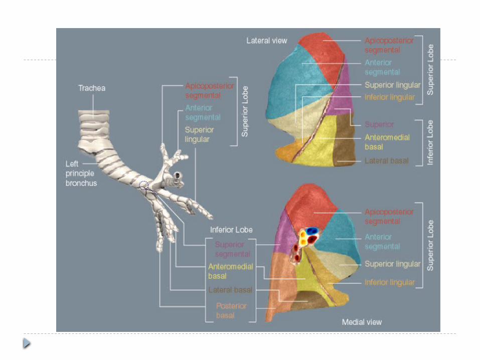
LEFT UPPER LOBE BRONCHUS Cranially it divides into anterior ,apical and
posterior segmental branches Caudally into superior and inferior lingual
branches
LEFT LOWER LOBE BRONCHUS Divides into apical segmental bronchus,medial
basal,anterior basal,lateral and posterior basal branches
-
BRONCHI
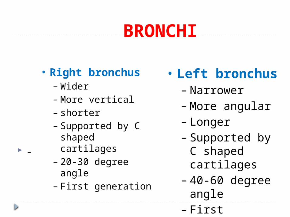
• Right bronchus–Wider–More vertical– shorter– Supported by C
shaped cartilages– 20-30 degree
angle– First generation
• Left bronchus– Narrower–More angular– Longer– Supported by C
shaped cartilages
– 40-60 degree angle
– First generation
CLINICAL SIGNIFICANCE Right main bronchus is more in line with
trachea Inhaled foreign bodies and gastric contents
enter right bronchial tree If patient is lying on his side,lateral
subsegments of anterior and posterior segments are more likely to get such material
If patient is supine,then apical segmental bronchus which arise from right or left lower lobe bronchus is the most common part of lung for the aspirated material to collect
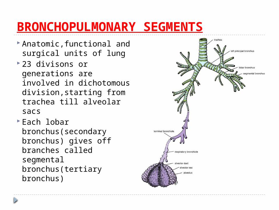
BRONCHOPULMONARY SEGMENTS Anatomic,functional and
surgical units of lung 23 divisons or
generations are involved in dichotomous division,starting from trachea till alveolar sacs
Each lobar bronchus(secondary bronchus) gives off branches called segmental bronchus(tertiary bronchus)
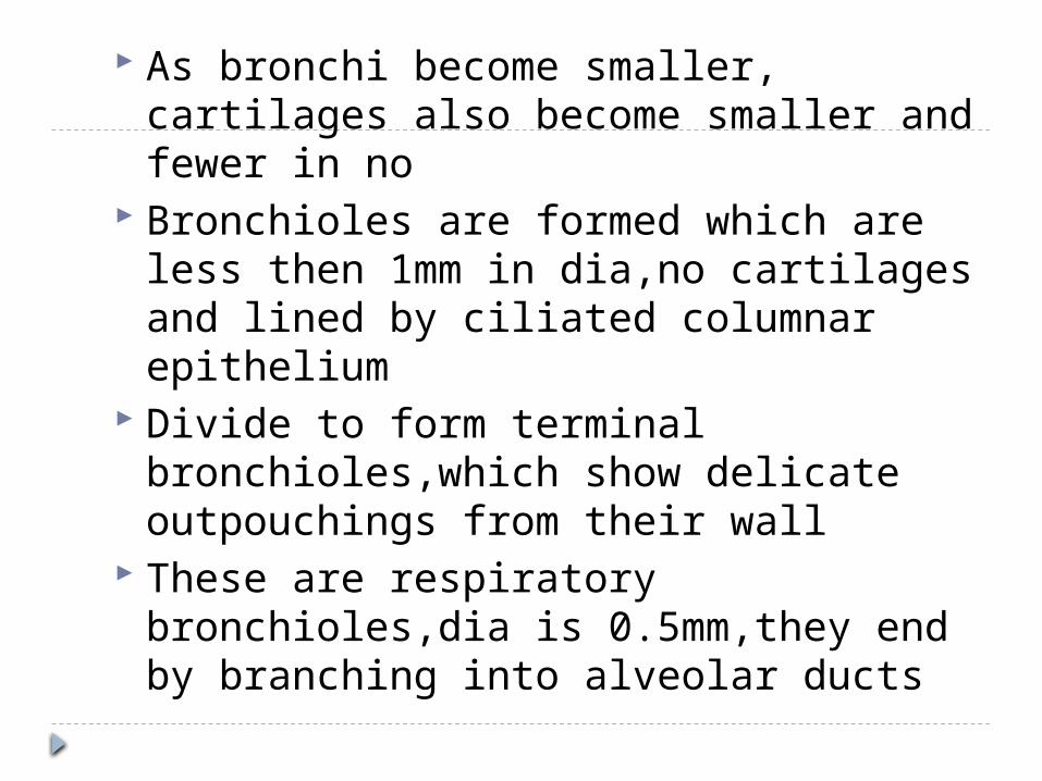
As bronchi become smaller, cartilages also become smaller and fewer in no
Bronchioles are formed which are less then 1mm in dia,no cartilages and lined by ciliated columnar epithelium
Divide to form terminal bronchioles,which show delicate outpouchings from their wall
These are respiratory bronchioles,dia is 0.5mm,they end by branching into alveolar ducts
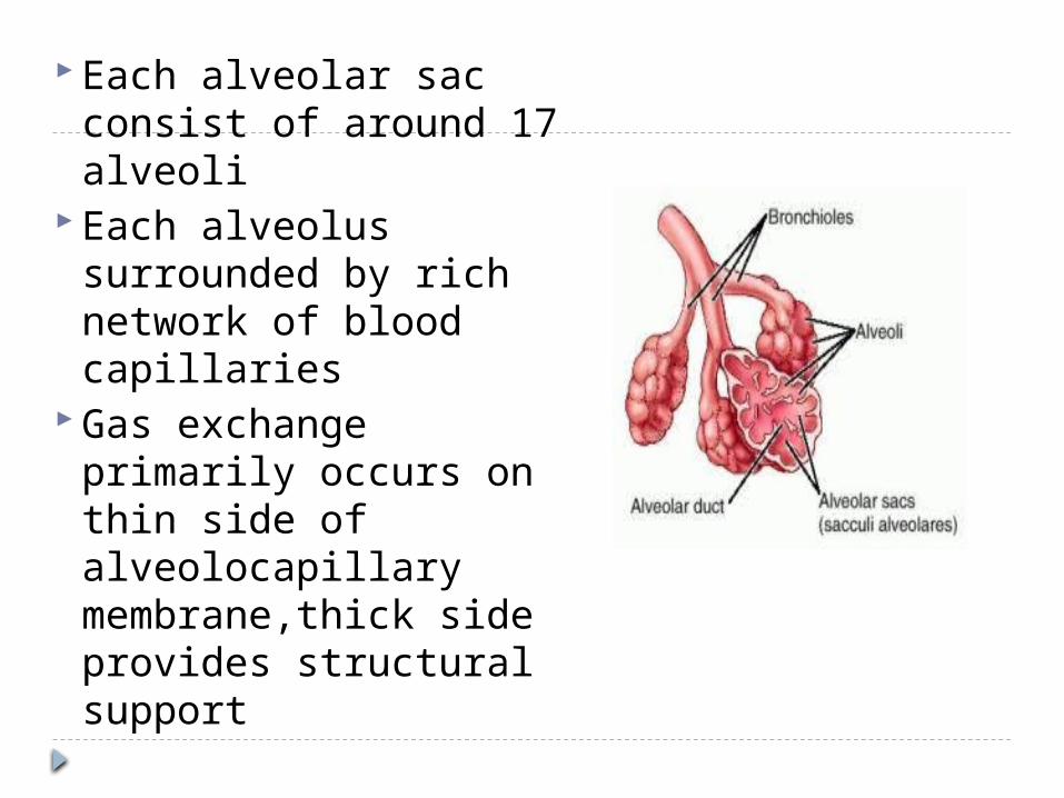
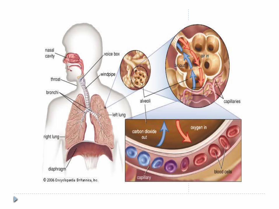
Each alveolar sac consist of around 17 alveoli
Each alveolus surrounded by rich network of blood capillaries
Gas exchange primarily occurs on thin side of alveolocapillary membrane,thick side provides structural support
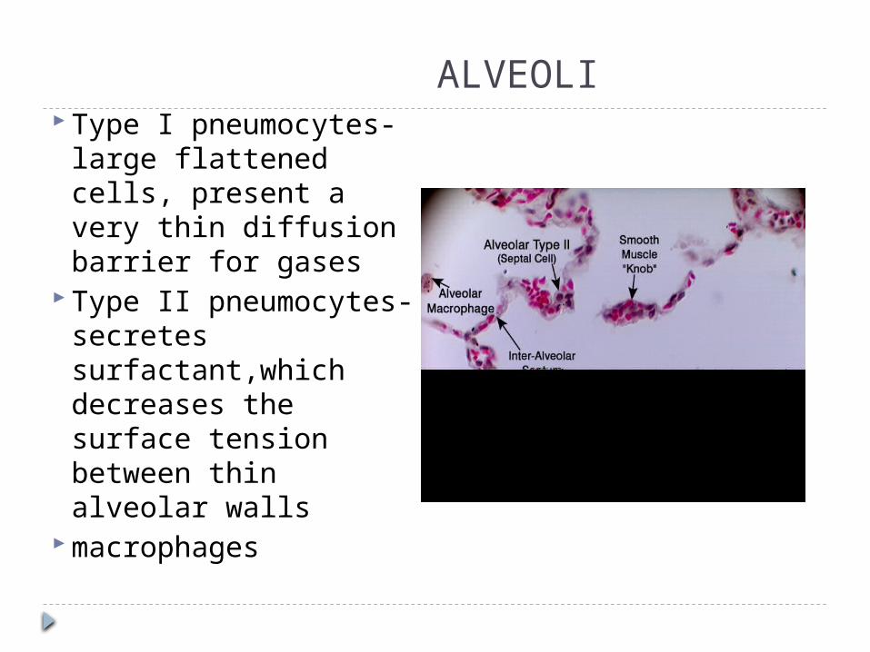
ALVEOLI Type I pneumocytes-
large flattened cells, present a very thin diffusion barrier for gases
Type II pneumocytes-secretes surfactant,which decreases the surface tension between thin alveolar walls
macrophages
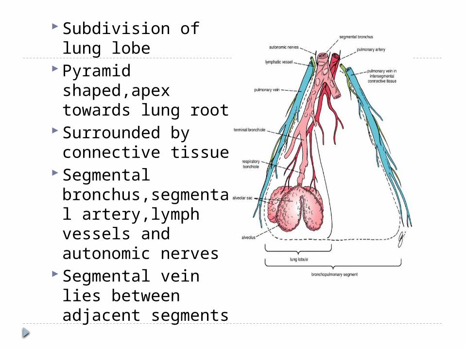
Subdivision of lung lobe
Pyramid shaped,apex towards lung root
Surrounded by connective tissue
Segmental bronchus,segmental artery,lymph vessels and autonomic nerves
Segmental vein lies between adjacent segments
BLOOD SUPPLY OF LUNGS By bronchial arteries which are
branches of descending aortaNERVE SUPPLY OF LUNGS Pulmonary plexus-efferent and
afferent autonomic nerve fibres Sympathetic efferent fibres produce
bronchodilation and vasoconstriction
Parasympathetic efferent produces bronchoconstriction,vasodilation,increase glandular secretions