Embed Size (px)
DESCRIPTION
Anatomy of the Hand and Wrist. Mnemonic for Learning Carpals. S he L ikes T o P lay. Lunate In the moonlight. Scaphoid A boat. Triquetrum The third T Bone. Pisiform Pea-shaped. Hamate A hambone With a hook. Trapezium: “It’s by the thumb”. Capitate. Trapezoid “Is by its side”. - PowerPoint PPT Presentation
Citation preview
Anatomy of the Hand and Wrist
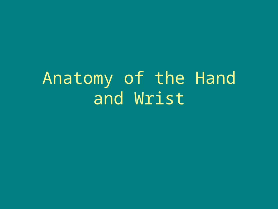
Mnemonicfor
LearningCarpals
She Likes To PlayLunateIn the moonlight
TriquetrumThe third T Bone
PisiformPea-shaped
Try To Catch Her
Trapezium:“It’s by the thumb”
Trapezoid“Is by its side”
Capitate
HamateA hamboneWith a hook
ScaphoidA boat
Click R Button for Slideshow
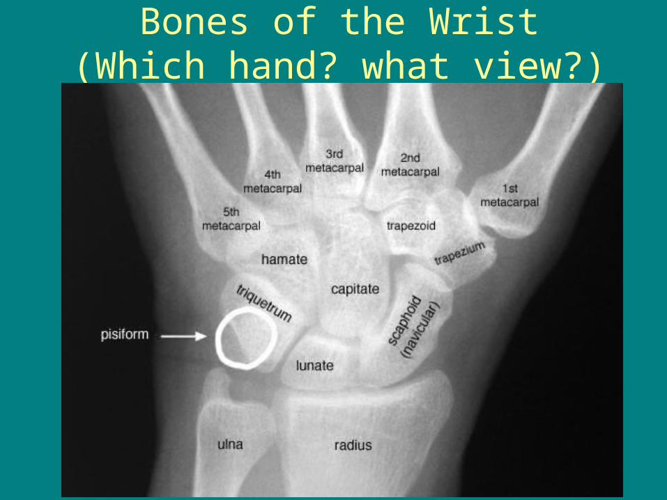
Bones of the Wrist (Which hand? what view?)
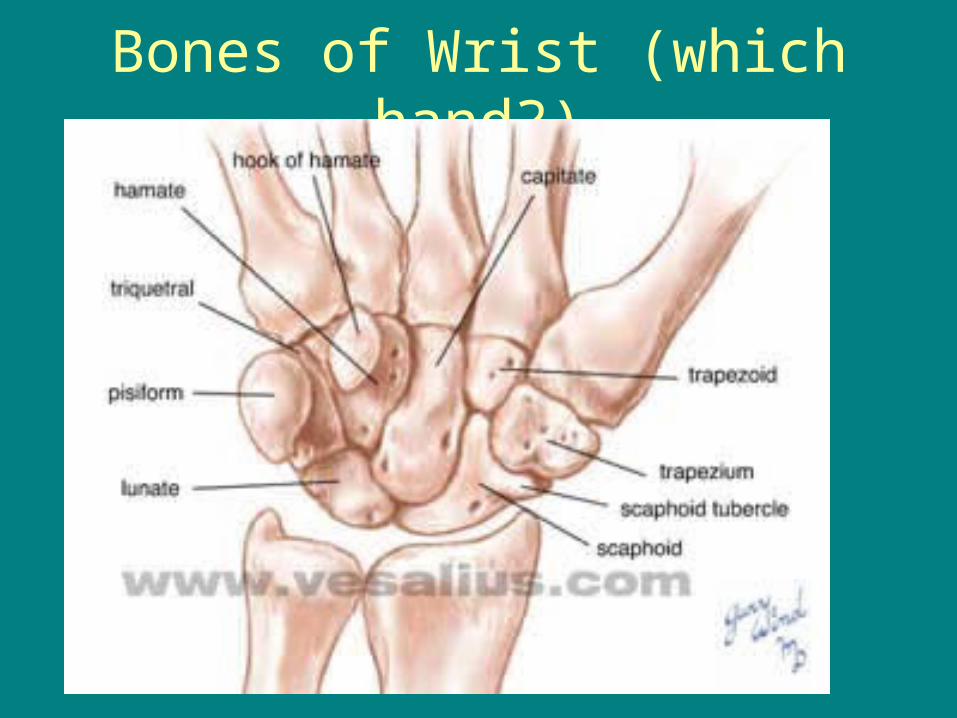
Bones of Wrist (which hand?)

Flexor Tendons
• The muscles that flex your wrist are on the palmer side.
• A group of the begin at the medial epicondyle of the humerus at the elbow
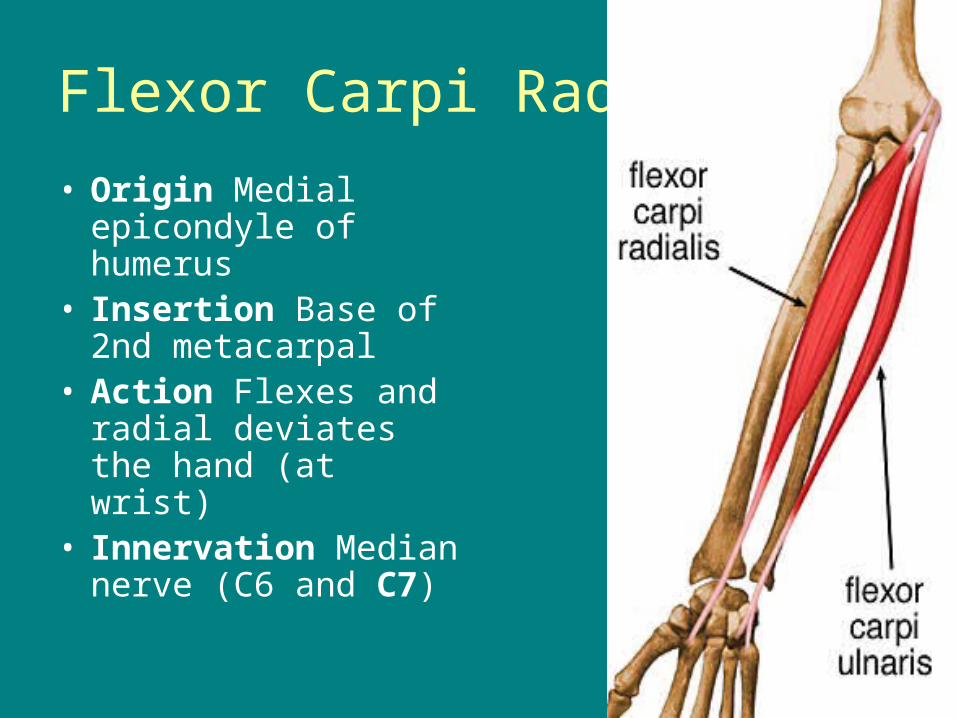
Flexor Carpi Radialis
• Origin Medial epicondyle of humerus
• Insertion Base of 2nd metacarpal
• Action Flexes and radial deviates the hand (at wrist)
• Innervation Median nerve (C6 and C7)
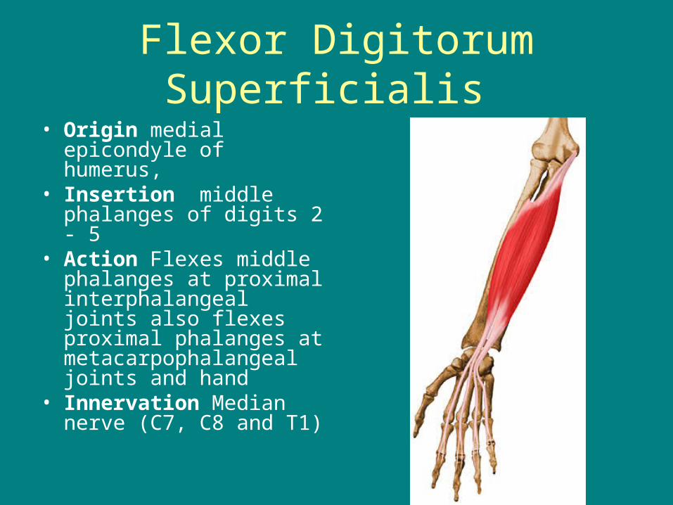
Flexor Digitorum Superficialis
• Origin medial epicondyle of humerus,
• Insertion middle phalanges of digits 2 - 5
• Action Flexes middle phalanges at proximal interphalangeal joints also flexes proximal phalanges at metacarpophalangeal joints and hand
• Innervation Median nerve (C7, C8 and T1)
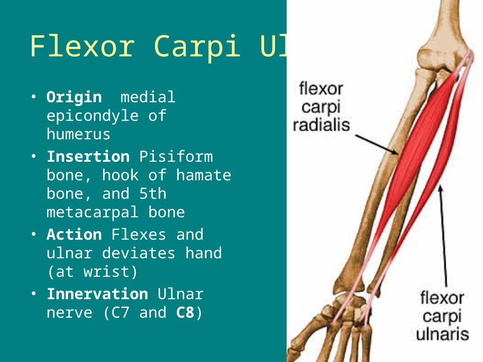
Flexor Carpi Ulnaris
• Origin medial epicondyle of humerus
• Insertion Pisiform bone, hook of hamate bone, and 5th metacarpal bone
• Action Flexes and ulnar deviates hand (at wrist)
• Innervation Ulnar nerve (C7 and C8)
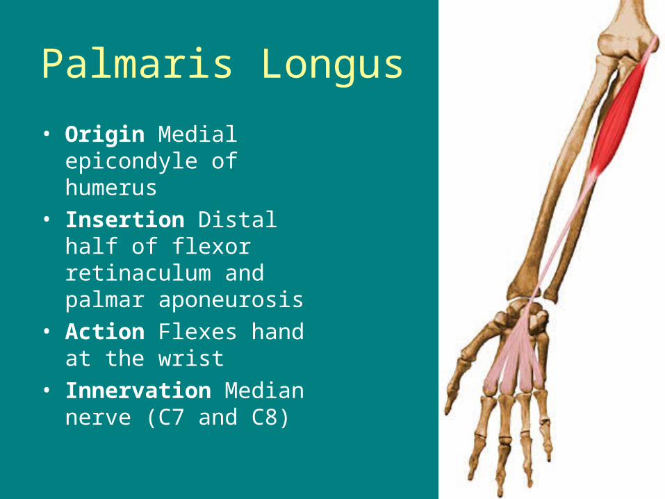
Palmaris Longus
• Origin Medial epicondyle of humerus
• Insertion Distal half of flexor retinaculum and palmar aponeurosis
• Action Flexes hand at the wrist
• Innervation Median nerve (C7 and C8)
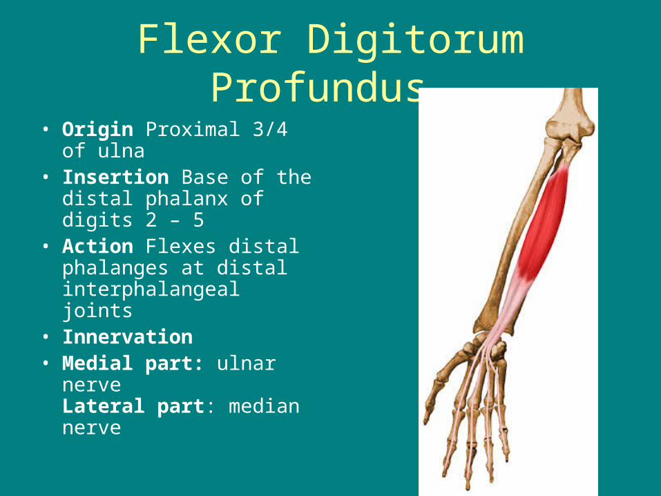
Flexor Digitorum Profundus
• Origin Proximal 3/4 of ulna
• Insertion Base of the distal phalanx of digits 2 – 5
• Action Flexes distal phalanges at distal interphalangeal joints
• Innervation • Medial part: ulnar nerve
Lateral part: median nerve
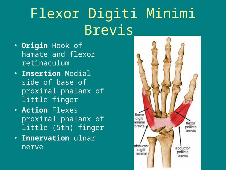
Flexor Digiti Minimi Brevis
• Origin Hook of hamate and flexor retinaculum
• Insertion Medial side of base of proximal phalanx of little finger
• Action Flexes proximal phalanx of little (5th) finger
• Innervation ulnar nerve
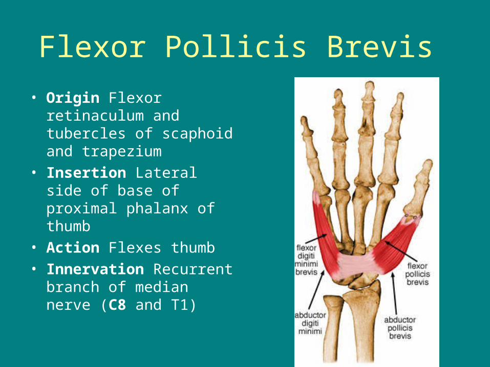
Flexor Pollicis Brevis
• Origin Flexor retinaculum and tubercles of scaphoid and trapezium
• Insertion Lateral side of base of proximal phalanx of thumb
• Action Flexes thumb • Innervation Recurrent
branch of median nerve (C8 and T1)

Flexor Pollicis Longus
• Origin Anterior surface of radius and adjacent interosseous membrane
• Insertion Base of distal phalanx of thumb
• Action Flexes phalanges of 1st digit (thumb)
• Innervation Anterior interosseous nerve from median nerve (C8 and T1)
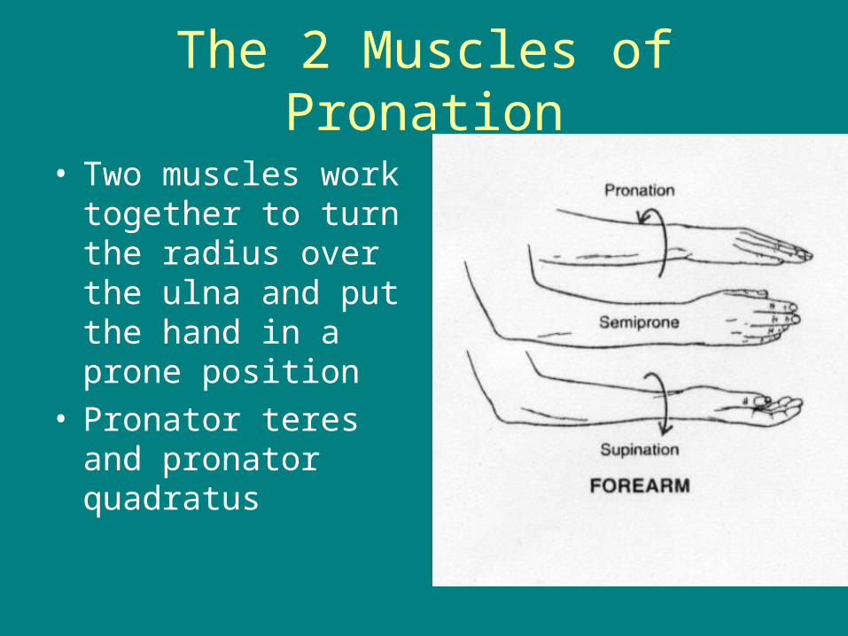
The 2 Muscles of Pronation
• Two muscles work together to turn the radius over the ulna and put the hand in a prone position
• Pronator teres and pronator quadratus

Pronator Quadratus
• Origin Distal 1/4 of anterior surface of ulna
• Insertion Distal 1/4 of anterior surface of radius
• Action Pronates forearm;
• Innervation median nerve
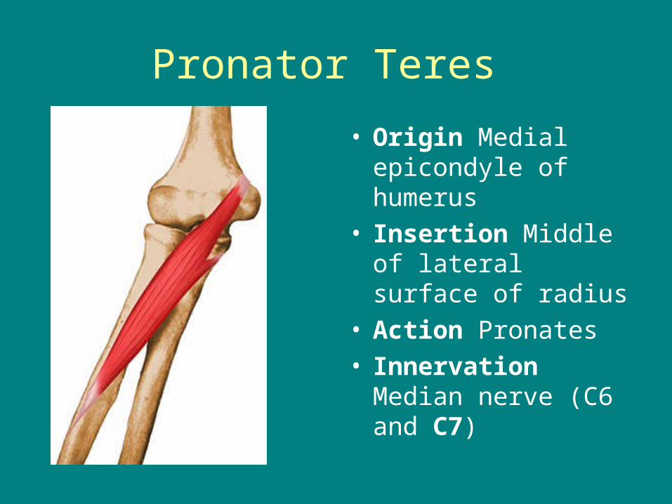
Pronator Teres
• Origin Medial epicondyle of humerus
• Insertion Middle of lateral surface of radius
• Action Pronates• Innervation Median
nerve (C6 and C7)

Abduction at Hand
• Abduction really only occurs at the thumb and little fingers
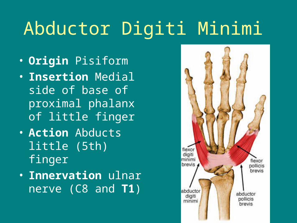
Abductor Digiti Minimi
• Origin Pisiform• Insertion Medial side
of base of proximal phalanx of little finger
• Action Abducts little (5th) finger
• Innervation ulnar nerve (C8 and T1)
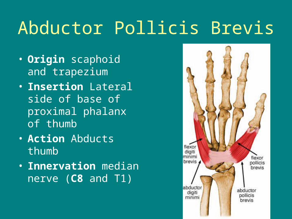
Abductor Pollicis Brevis
• Origin scaphoid and trapezium
• Insertion Lateral side of base of proximal phalanx of thumb
• Action Abducts thumb
• Innervation median nerve (C8 and T1)
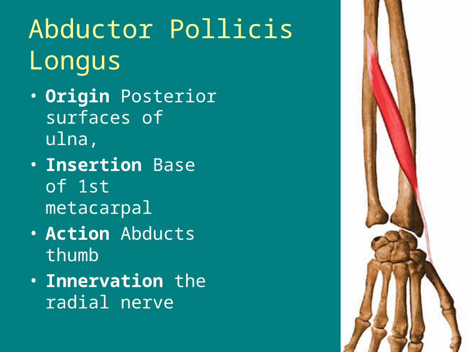
Abductor Pollicis Longus • Origin Posterior
surfaces of ulna, • Insertion Base of 1st
metacarpal • Action Abducts
thumb • Innervation the radial
nerve
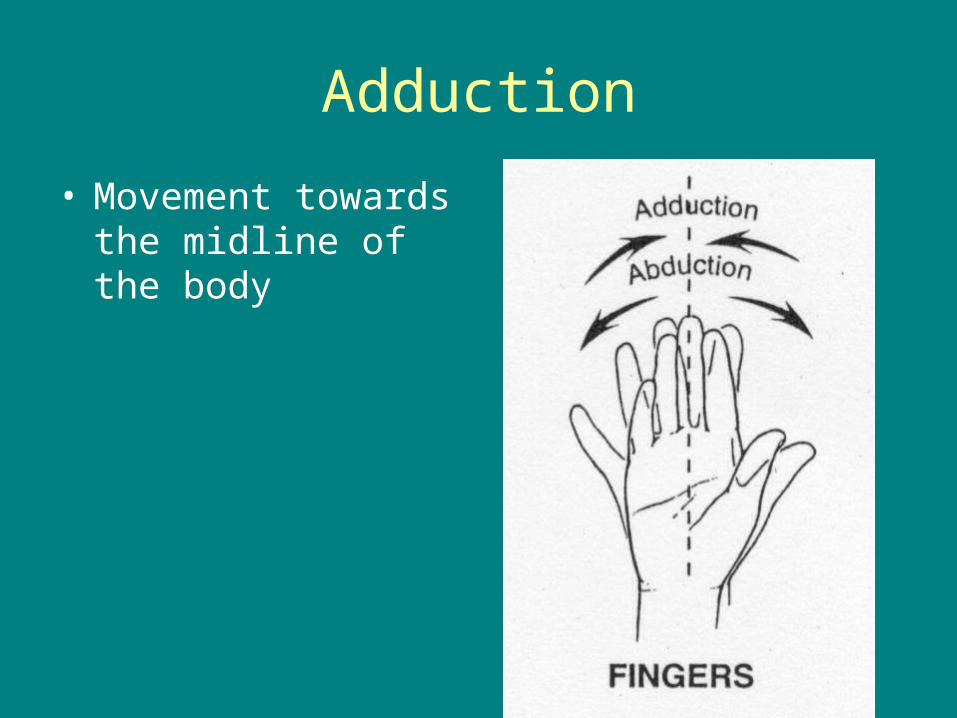
Adduction
• Movement towards the midline of the body

Adductor Pollicis
• Origin 2nd and 3rd metacarpals, capitate,
• Insertion Medial side of base of proximal phalanx of thumb
• Action Adducts thumb
• Innervation ulnar nerve

Wrist Extensors
• The extensors of the wrist are on the Dorsal side of the forearm
• A majority of the wrist extensors begin at the lateral epicondyle
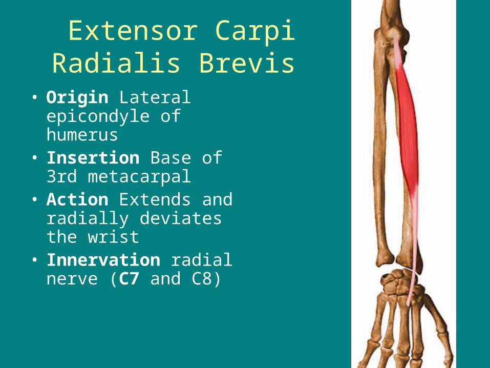
Extensor Carpi Radialis Brevis
• Origin Lateral epicondyle of humerus
• Insertion Base of 3rd metacarpal
• Action Extends and radially deviates the wrist
• Innervation radial nerve (C7 and C8)
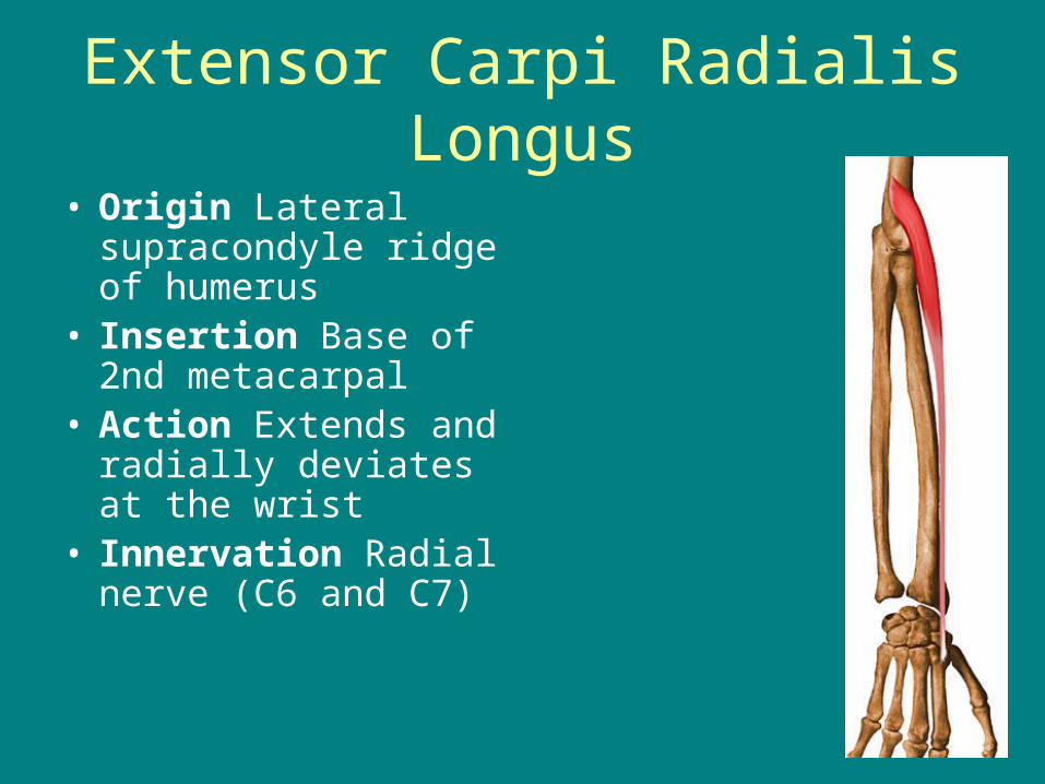
Extensor Carpi Radialis Longus
• Origin Lateral supracondyle ridge of humerus
• Insertion Base of 2nd metacarpal
• Action Extends and radially deviates at the wrist
• Innervation Radial nerve (C6 and C7)
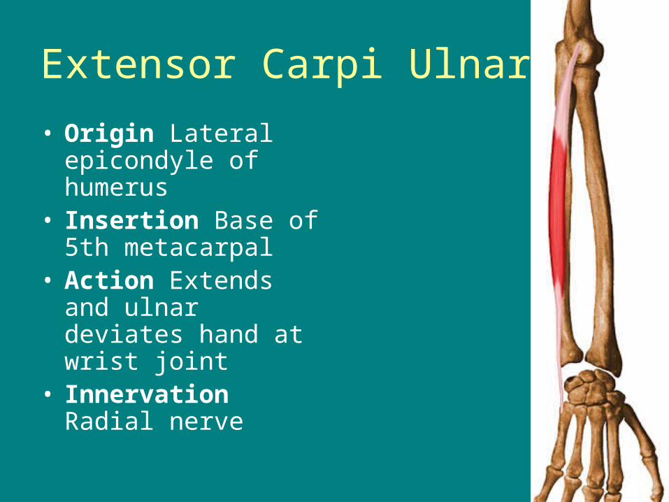
Extensor Carpi Ulnaris
• Origin Lateral epicondyle of humerus
• Insertion Base of 5th metacarpal
• Action Extends and ulnar deviates hand at wrist joint
• Innervation Radial nerve
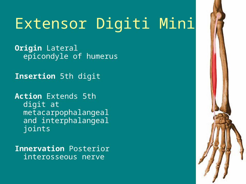
Extensor Digiti Minimi
Origin Lateral epicondyle of humerus
Insertion 5th digit
Action Extends 5th digit at metacarpophalangeal and interphalangeal joints
Innervation Posterior interosseous nerve

Extensor Digitorum
• Origin Lateral epicondyle of humerus
• Insertion Extensor expansions of medial four digits
• Action Extends the four digits and the wrist
• Innervation Posterior interosseous nerve
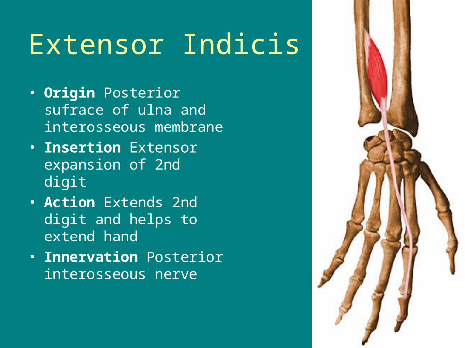
Extensor Indicis
• Origin Posterior sufrace of ulna and interosseous membrane
• Insertion Extensor expansion of 2nd digit
• Action Extends 2nd digit and helps to extend hand
• Innervation Posterior interosseous nerve

Extensor Pollicis Brevis
• Origin Posterior sufraces of radius and interosseous membrane
• Insertion Base of proximal phalanx of thumb
• Action Extends proximal phalanx of thumb at carpometacarpal joint
• Innervation Posterior interosseous nerve
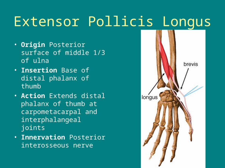
Extensor Pollicis Longus
• Origin Posterior surface of middle 1/3 of ulna
• Insertion Base of distal phalanx of thumb
• Action Extends distal phalanx of thumb at carpometacarpal and interphalangeal joints
• Innervation Posterior interosseous nerve
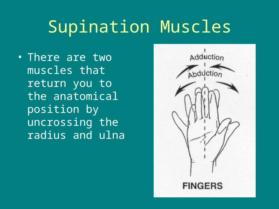
Supination Muscles
• There are two muscles that return you to the anatomical position by uncrossing the radius and ulna
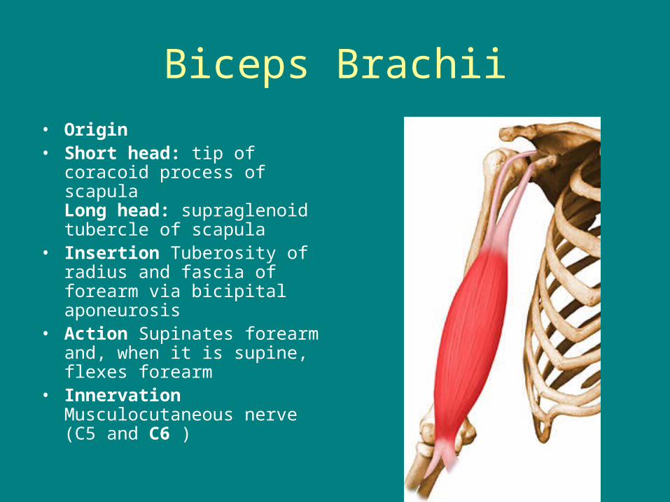
Biceps Brachii
• Origin • Short head: tip of coracoid
process of scapulaLong head: supraglenoid tubercle of scapula
• Insertion Tuberosity of radius and fascia of forearm via bicipital aponeurosis
• Action Supinates forearm and, when it is supine, flexes forearm
• Innervation Musculocutaneous nerve (C5 and C6 )

Supinator
• Origin Lateral epicondyle of humerus,
• Insertion Lateral, posterior and anterior surfaces of proximal 1/3 of radius
• Action Supinates forearm
• Innervation Deep branch of radial nerve (C5 and C6)
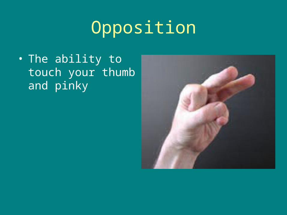
Opposition
• The ability to touch your thumb and pinky
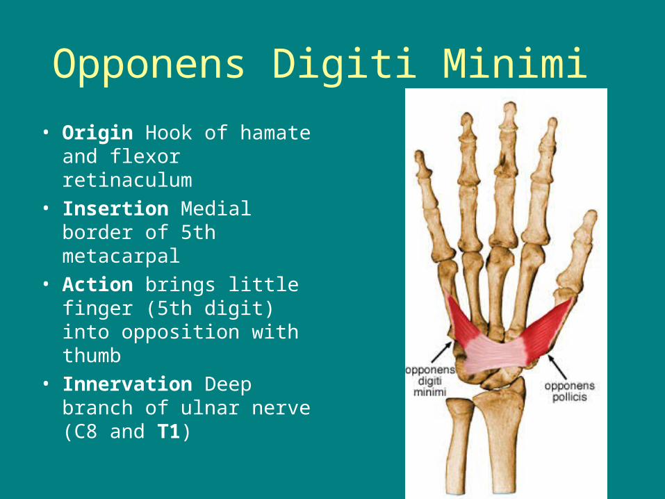
Opponens Digiti Minimi
• Origin Hook of hamate and flexor retinaculum
• Insertion Medial border of 5th metacarpal
• Action brings little finger (5th digit) into opposition with thumb
• Innervation Deep branch of ulnar nerve (C8 and T1)
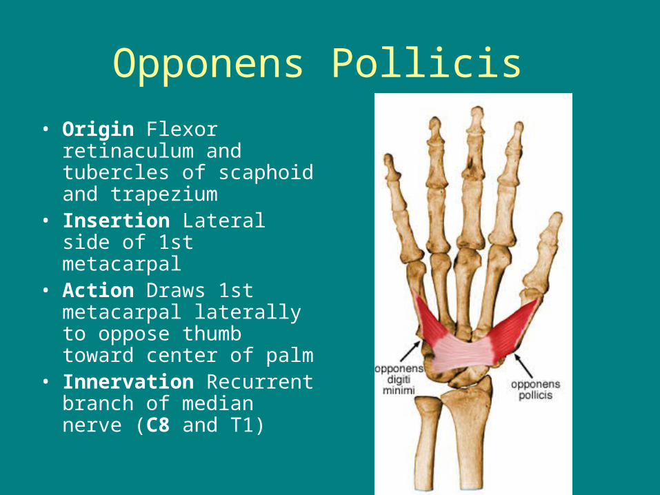
Opponens Pollicis
• Origin Flexor retinaculum and tubercles of scaphoid and trapezium
• Insertion Lateral side of 1st metacarpal
• Action Draws 1st metacarpal laterally to oppose thumb toward center of palm
• Innervation Recurrent branch of median nerve (C8 and T1)

Test
• Test Wednesday
Review packet will be given out Monday, I won’t be here
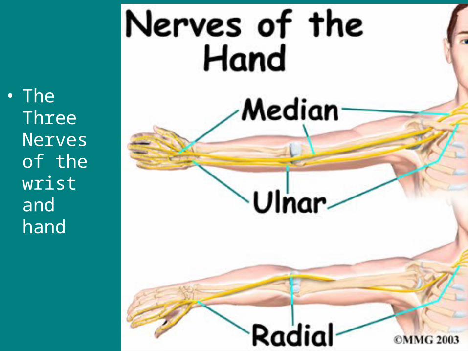
• The Three Nerves of the wrist and hand
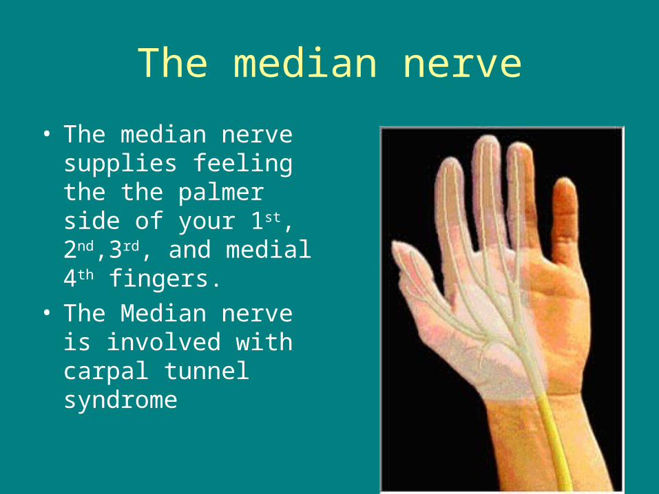
The median nerve
• The median nerve supplies feeling the the palmer side of your 1st, 2nd,3rd, and medial 4th fingers.
• The Median nerve is involved with carpal tunnel syndrome
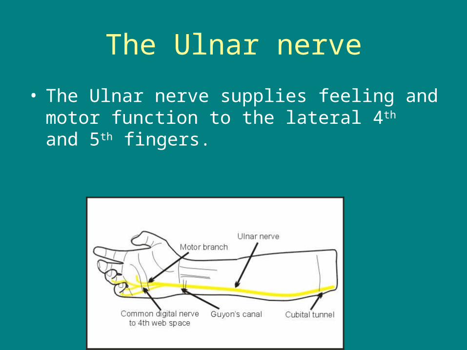
The Ulnar nerve
• The Ulnar nerve supplies feeling and motor function to the lateral 4th and 5th fingers.
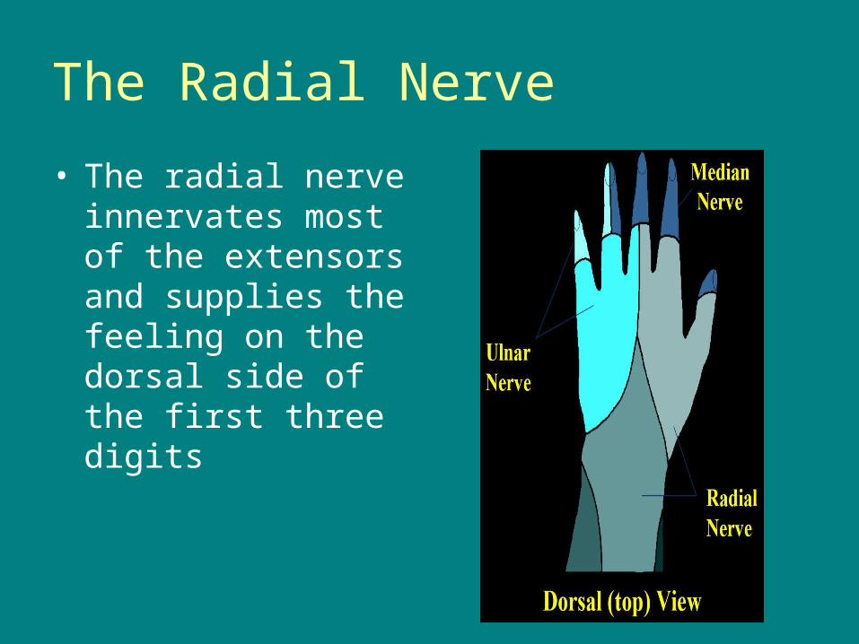
The Radial Nerve
• The radial nerve innervates most of the extensors and supplies the feeling on the dorsal side of the first three digits
Joint – the place where two bones come together (4 types)

Ball and socket joint – greatest range of motion allowing bones to swing in a circle
Example: shoulder or hip
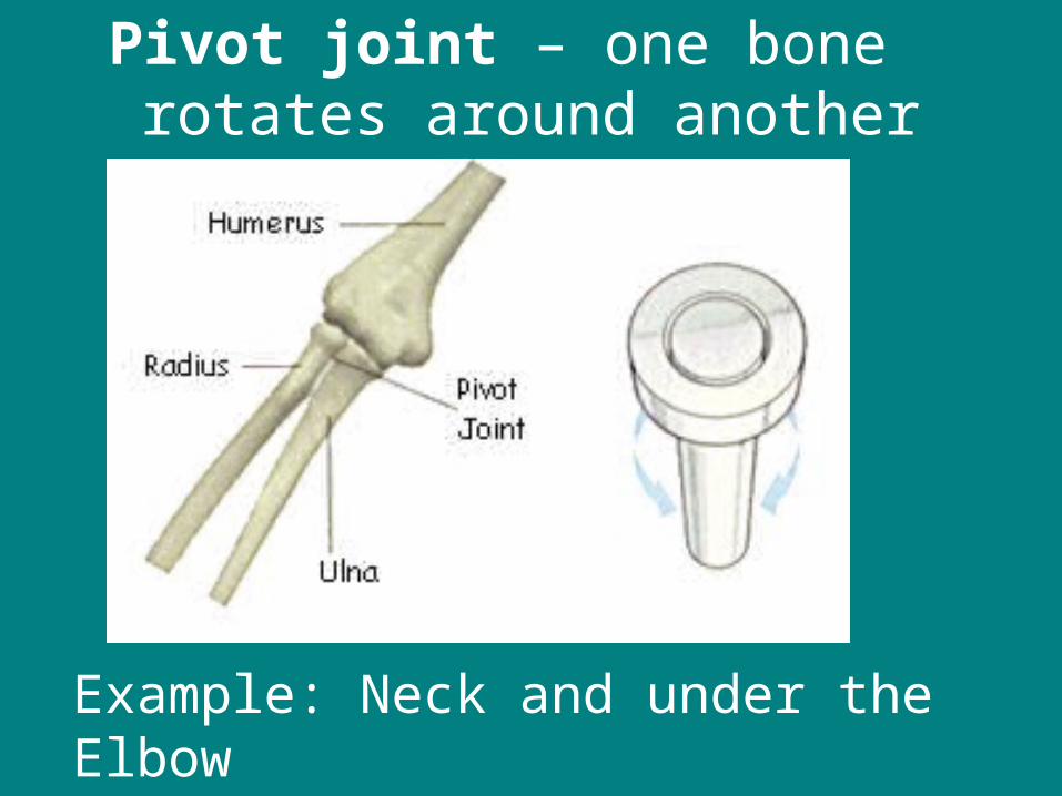
Pivot joint – one bone rotates around another
Example: Neck and under the Elbow

Hinge joint – bones bend like a hinge forward and backward
Example: Knee and Elbow

• Gliding joint – allows one bone to slide over another
Example: Wrist and Ankle
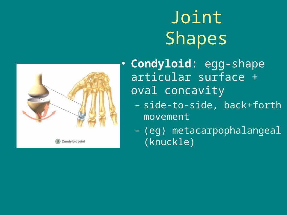
Joint Shapes
• Condyloid: egg-shape articular surface + oval concavity– side-to-side, back+forth
movement– (eg) metacarpophalangeal
(knuckle)
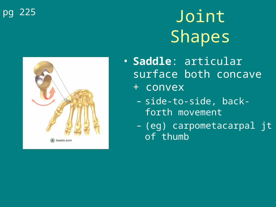
Joint Shapes
• Saddle: articular surface both concave + convex– side-to-side, back-forth
movement– (eg) carpometacarpal jt of
thumb
pg 225
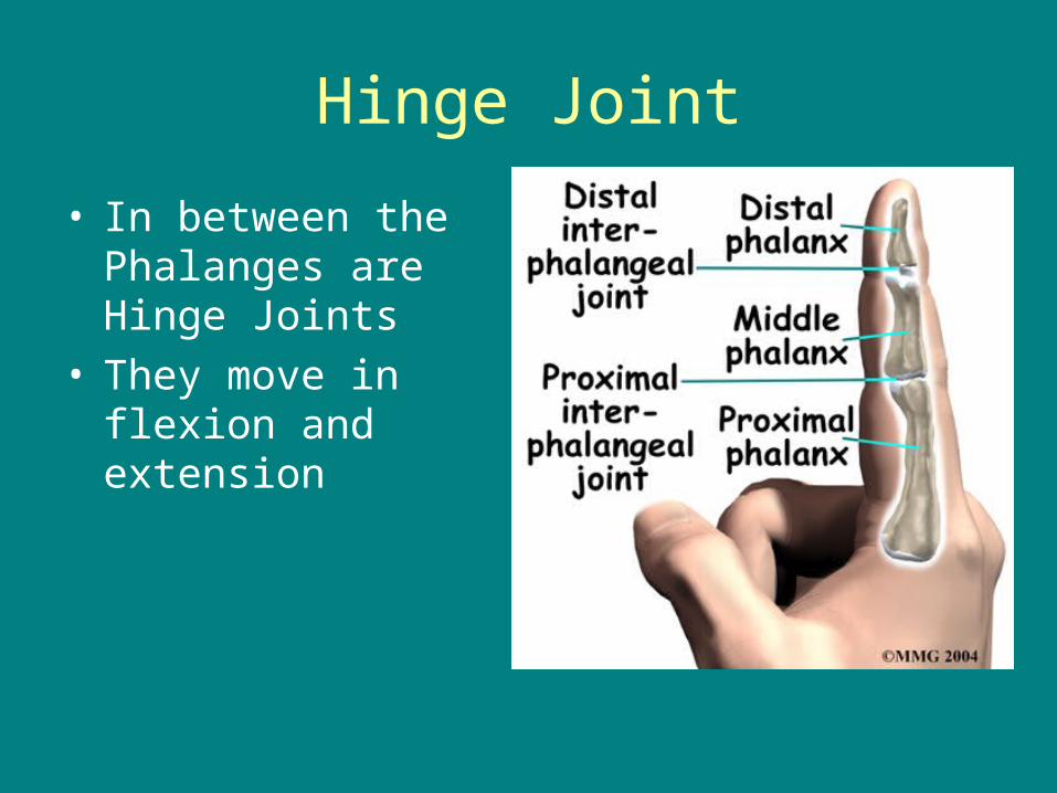
Hinge Joint
• In between the Phalanges are Hinge Joints
• They move in flexion and extension
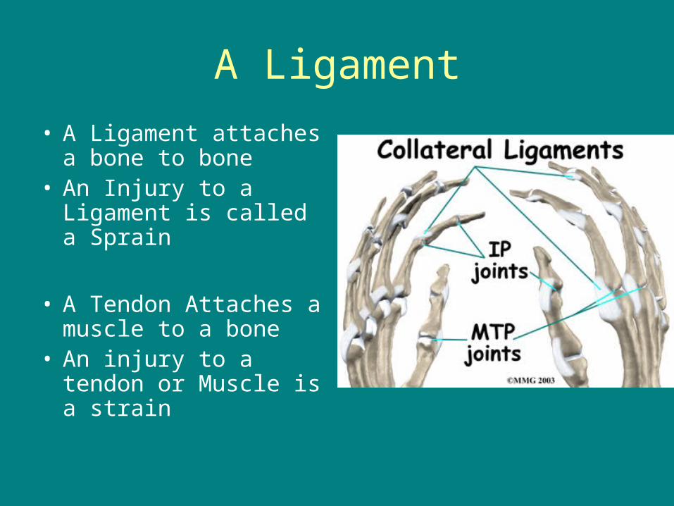
A Ligament
• A Ligament attaches a bone to bone
• An Injury to a Ligament is called a Sprain
• A Tendon Attaches a muscle to a bone
• An injury to a tendon or Muscle is a strain
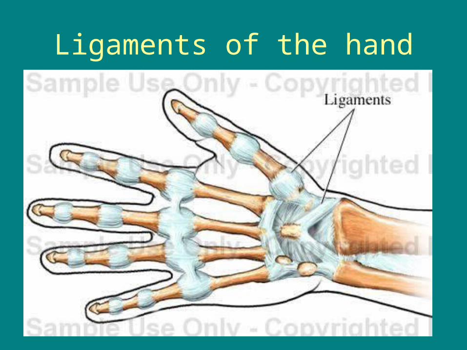
Ligaments of the hand
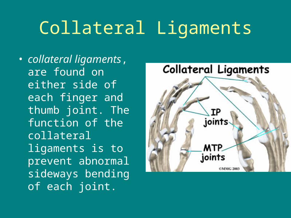
Collateral Ligaments
• collateral ligaments, are found on either side of each finger and thumb joint. The function of the collateral ligaments is to prevent abnormal sideways bending of each joint.
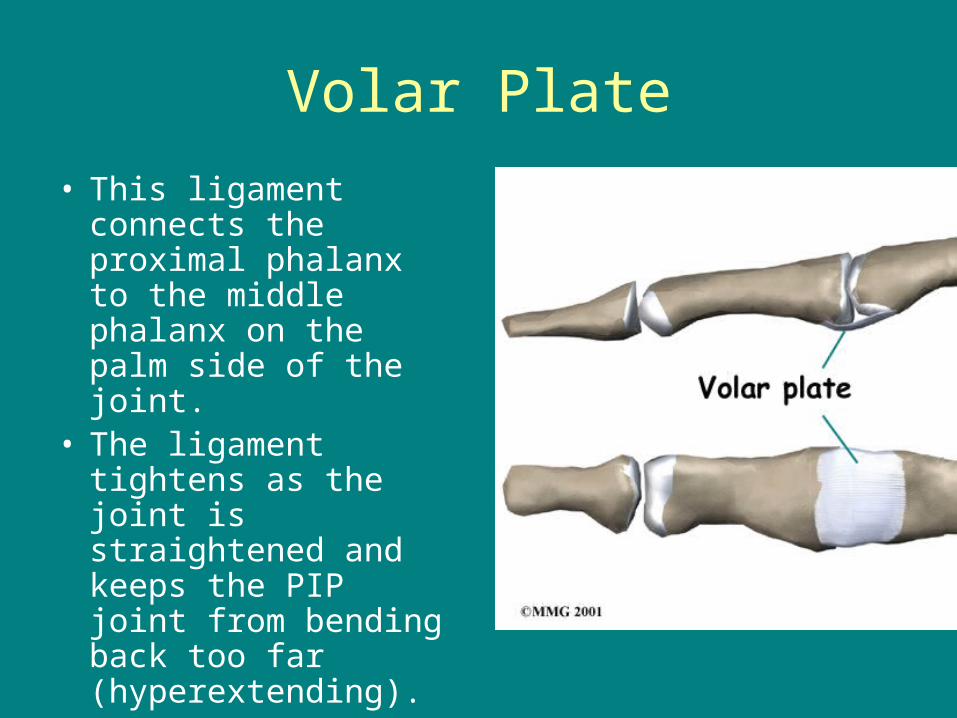
Volar Plate
• This ligament connects the proximal phalanx to the middle phalanx on the palm side of the joint.
• The ligament tightens as the joint is straightened and keeps the PIP joint from bending back too far (hyperextending).
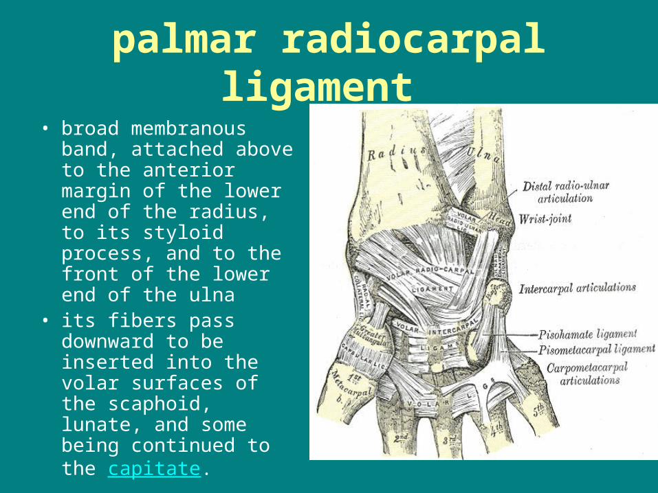
palmar radiocarpal ligament
• broad membranous band, attached above to the anterior margin of the lower end of the radius, to its styloid process, and to the front of the lower end of the ulna
• its fibers pass downward to be inserted into the volar surfaces of the scaphoid, lunate, and some being continued to the capitate.

dorsal radiocarpal ligament
• less thick and strong than the palmer ligament
• The ligament begins on the radius
• its fibers are directed downward and medially, and are fixed, below, to the dorsal surfaces of the scaphoid and lunate
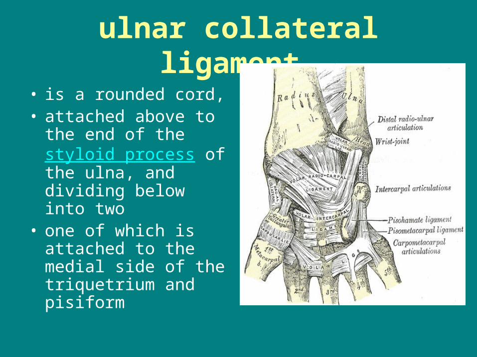
ulnar collateral ligament
• is a rounded cord,• attached above to the
end of the styloid process of the ulna, and dividing below into two
• one of which is attached to the medial side of the triquetrium and pisiform
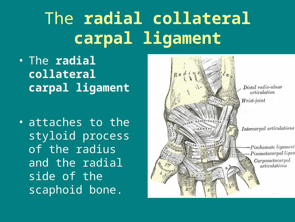
The radial collateral carpal ligament
• The radial collateral carpal ligament
• attaches to the styloid process of the radius and the radial side of the scaphoid bone.
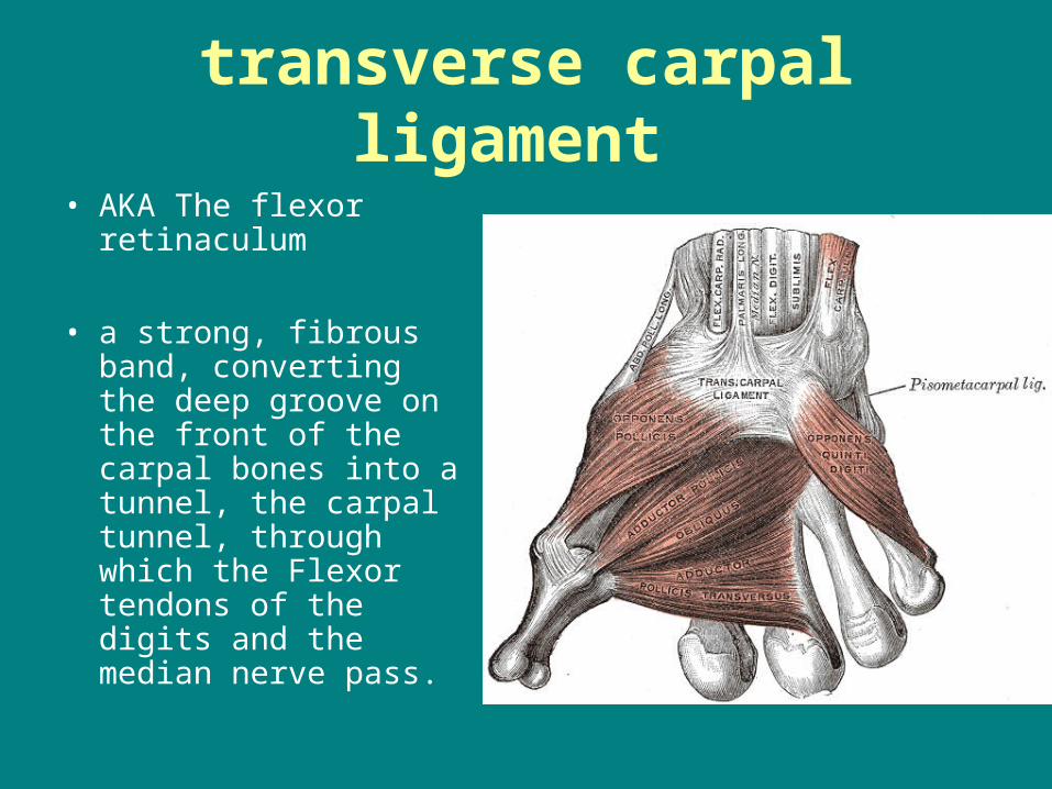
transverse carpal ligament
• AKA The flexor retinaculum
• a strong, fibrous band, converting the deep groove on the front of the carpal bones into a tunnel, the carpal tunnel, through which the Flexor tendons of the digits and the median nerve pass.

Hypothenar eminence
• is the body of muscle on the palm of the human hand just beneath the 5th phalange
• Abductor digiti minimi & Flexor digiti minimi Opponens digiti minimi
• “OAF”
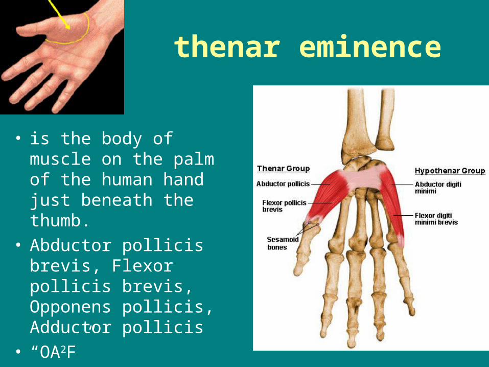
thenar eminence
• is the body of muscle on the palm of the human hand just beneath the thumb.
• Abductor pollicis brevis, Flexor pollicis brevis, Opponens pollicis, Adductor pollicis
• “OA2F”
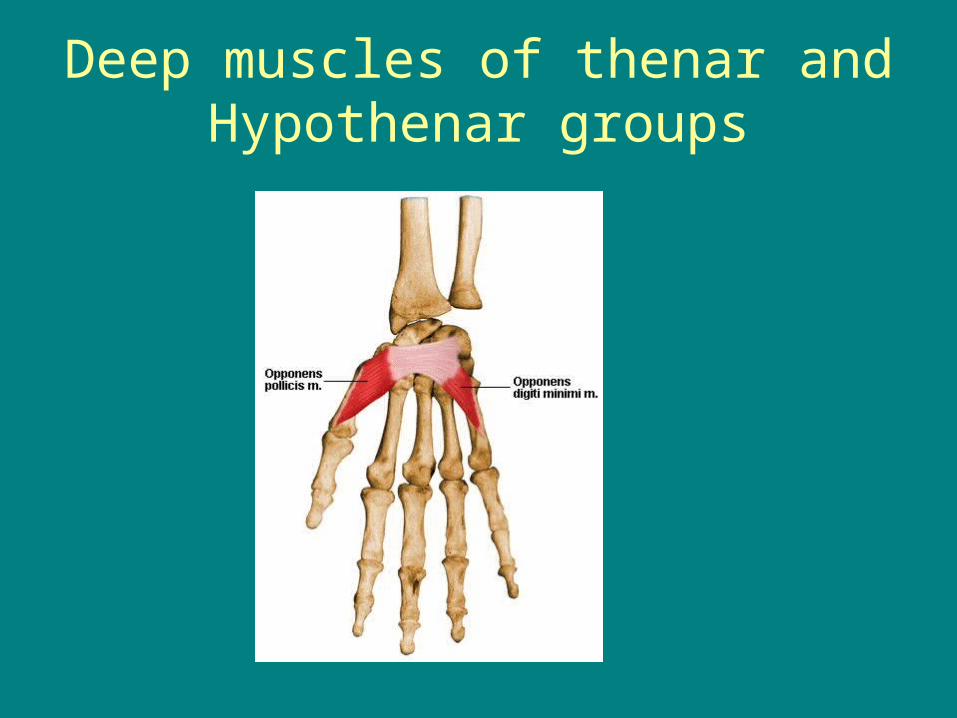
Deep muscles of thenar and Hypothenar groups