Embed Size (px)
Citation preview
Anatomy of the Abdomenin Computed Tomography
Michael C. Ficorelli, RT
Lesson Description
To explain the various exams pertaining to the abominal cavity using computed tomography, incorporating cross sectional anatomy from images
Lesson Description
• To be able to identify anatomy of the abdominal cavity. Understand the clinical indications for exams of the abdomen. To understand the methods of patient scanning, positioning, and protocols. To understand indications for contrast.
CT of the Abdomen
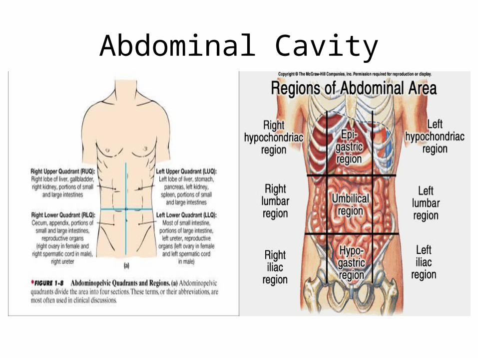
Abdominal Cavity• Region between the diaphragm and the sacral promontory (L-5 / S-1)
– Divided into 4 quadrants or 9 regions• Contains:
– Liver– Gallbladder– Biliary System– Pancreas– Spleen– Adrenal Glands– Kidneys– Ureters– Stomach– Intestines– Vascular Structures
Abdominal Cavity
Peritoneum
• Thin serous membrane which lines the walls of the abdominal cavity– 2 Layers separated by fluid used for lubrication
• Parietal Peritoneum – lines abdominal wall• Visceral Peritoneum – lines organs
– In males, cavity is closed. In females, cavity extends outward via uterine tubes, uterus and vagina
– Folds extend between organs allowing them to hold position and enclose the vessels and nerves between them• Mesentery – encloses intestines• Omentum – Attached to stomach
– Greater omentum – greater curvature to spleen– Lesser omentum – lesser curvature to duodenum
Peritoneum
• Contains:– Stomach, 1st part of duodenum, jejunum, ileum,
cecum, appendix, transverse colon, sigmoid, upper 1/3 or rectum, liver, spleen and pancreas• In women – fallopian tubes and ovaries
Peritoneum
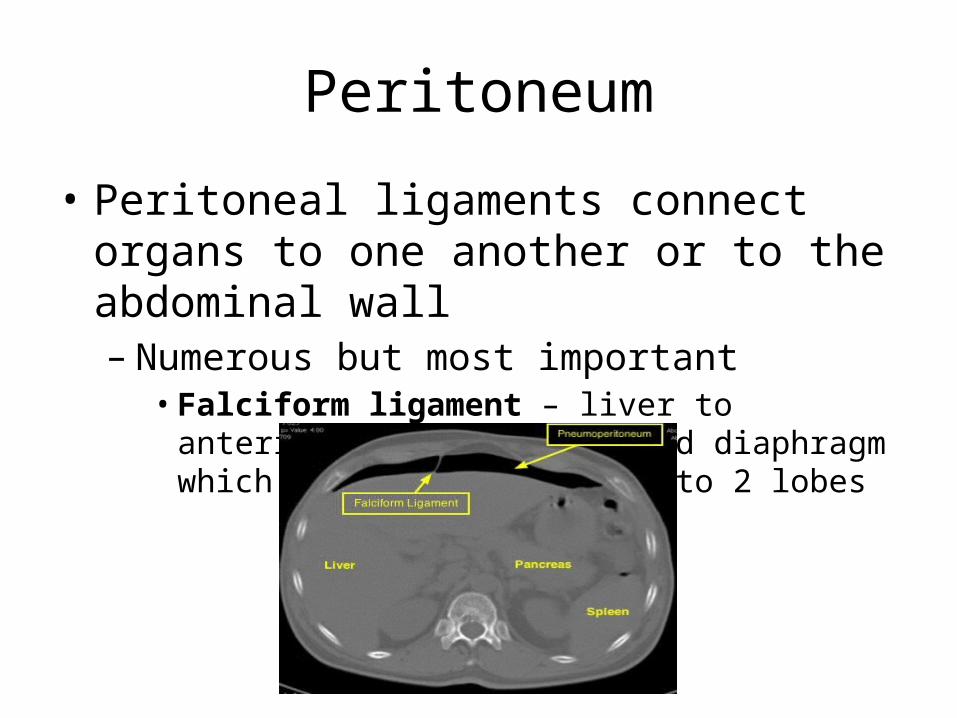
• Peritoneal ligaments connect organs to one another or to the abdominal wall– Numerous but most important• Falciform ligament – liver to anterior abdominal wall
and diaphragm which divides the liver into 2 lobes
Retroperitoneum
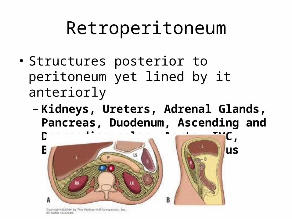
• Structures posterior to peritoneum yet lined by it anteriorly– Kidneys, Ureters, Adrenal Glands, Pancreas,
Duodenum, Ascending and Descending colon, Aorta, IVC, Bladder, Prostate, and Uterus
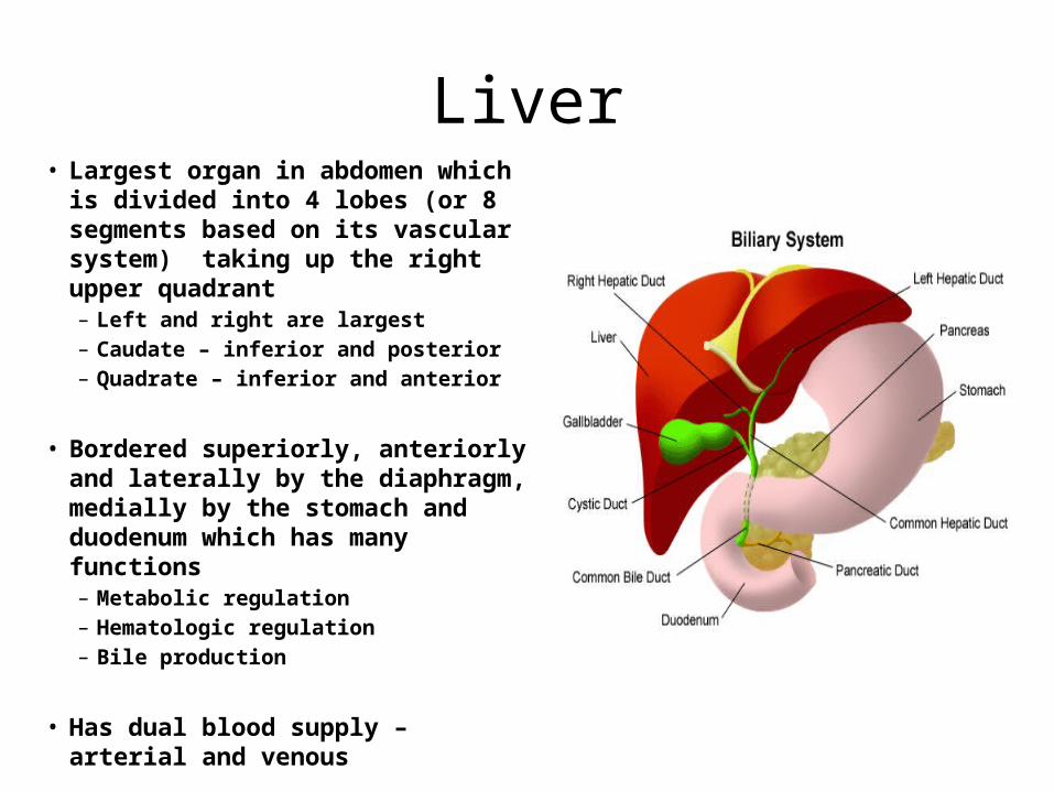
Liver• Largest organ in abdomen which is
divided into 4 lobes (or 8 segments based on its vascular system) taking up the right upper quadrant– Left and right are largest– Caudate – inferior and posterior– Quadrate – inferior and anterior
• Bordered superiorly, anteriorly and laterally by the diaphragm, medially by the stomach and duodenum which has many functions– Metabolic regulation– Hematologic regulation– Bile production
• Has dual blood supply – arterial and venous
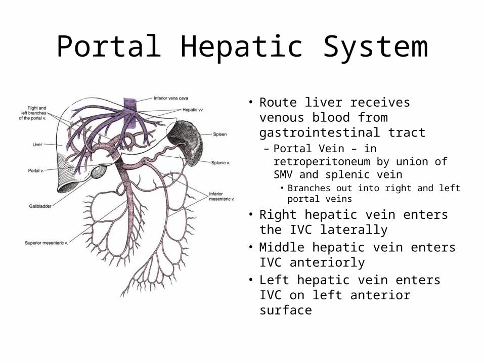
Portal Hepatic System
• Route liver receives venous blood from gastrointestinal tract– Portal Vein – in retroperitoneum
by union of SMV and splenic vein• Branches out into right and left
portal veins
• Right hepatic vein enters the IVC laterally
• Middle hepatic vein enters IVC anteriorly
• Left hepatic vein enters IVC on left anterior surface
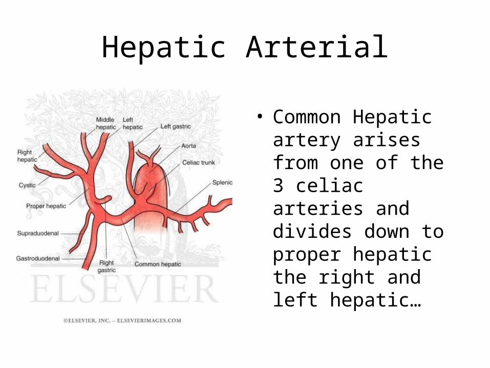
Hepatic Arterial
• Common Hepatic artery arises from one of the 3 celiac arteries and divides down to proper hepatic the right and left hepatic…
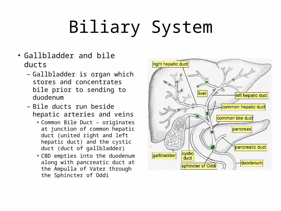
Biliary System
• Gallbladder and bile ducts– Gallbladder is organ which
stores and concentrates bile prior to sending to duodenum
– Bile ducts run beside hepatic arteries and veins• Common Bile Duct – originates
at junction of common hepatic duct (united right and left hepatic duct) and the cystic duct (duct of gallbladder)
• CBD empties into the duodenum along with pancreatic duct at the Ampulla of Vater through the Sphincter of Oddi
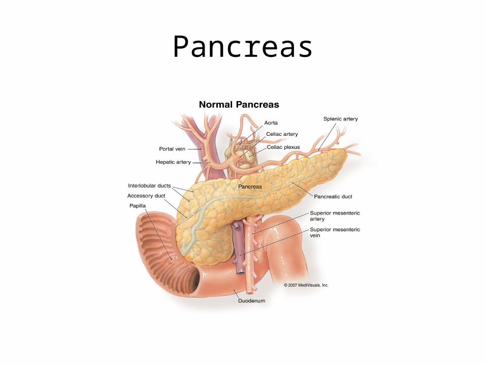
Pancreas• Located in retroperitoneum• Long, narrow organ lying posterior to the stomach and extends
transversely at an oblique angle between the duodenum and splenic hilum– Head – broad and flat; lies nestled in the curve of the duodenum around
L – 2 / L – 3– Body – largest anterior portion– Tail – extends to the left anterior pararenal space
• Functions in endochrine system by producing insulin and glucagon– Also produces digestive enzymes
• Main pancreatic duct carries products into the duodenum through the ampulla of vater
Pancreas
Spleen and Adrenal Glands
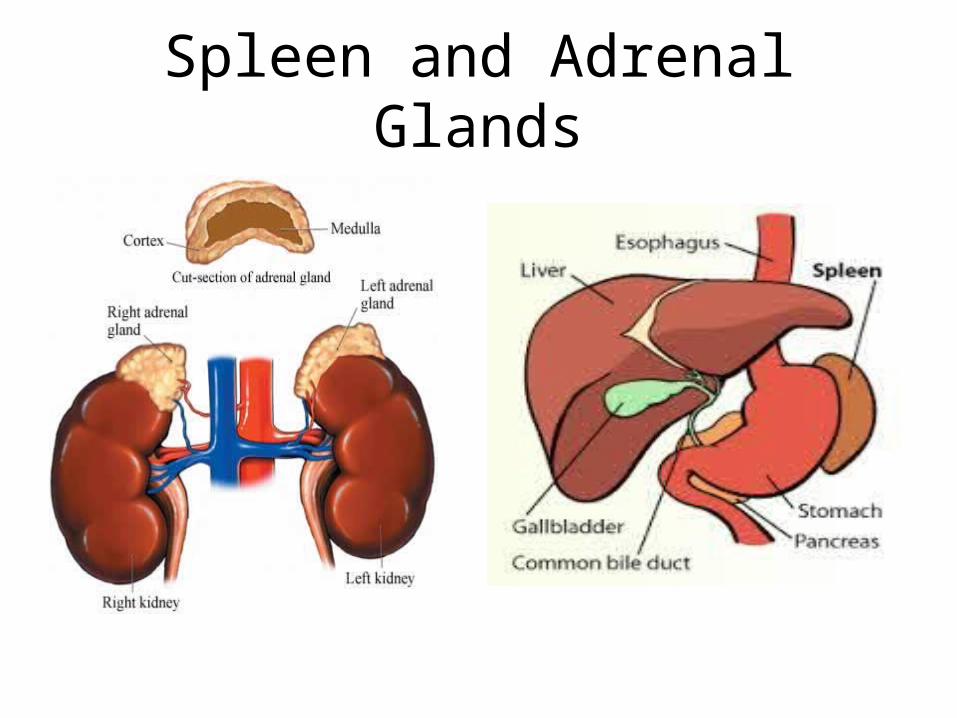
• Spleen – in peritoneum, posterior to the stomach; largest lymph organ which functions to produce white blood cells, remove abnormal blood cells, and aid in immunity
• Adrenal Glands – located superior to each kidney– Right is located just posterior to IVC, medial to the posterior
part of the right hepatic lobe – lower and more medial than the left
– Left is triangular shaped• Both produce more than 2 dozen hormones including corticosteroids
Spleen and Adrenal Glands
Kidneys
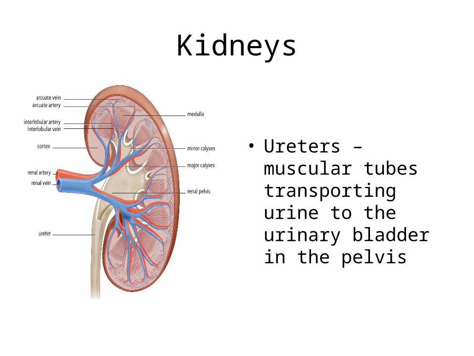
• Bean shaped organs lying obliquely on the posterior abdominal wall on either side of the vertebral column, contains medial hilum where renal arteries and veins enter– Gerota’s (Renal) Fascia – protective layer anchoring the kidneys to the
surrounding structures– Renal Cortex – outer third containing the nephrons (functional unit of
kidney)– Renal Medulla – consists of renal pyramids which contain the loops of
Henle (beginning of collecting system)– Renal Pelvis - kidney has 7-14 minor calyces which merge into 2 – 3
major calyces…they merge together to form renal pelvis• Largest point of collecting system which is continuous with the ureter
• Filters blood of impurities and begins transportation out
Kidneys
• Ureters – muscular tubes transporting urine to the urinary bladder in the pelvis
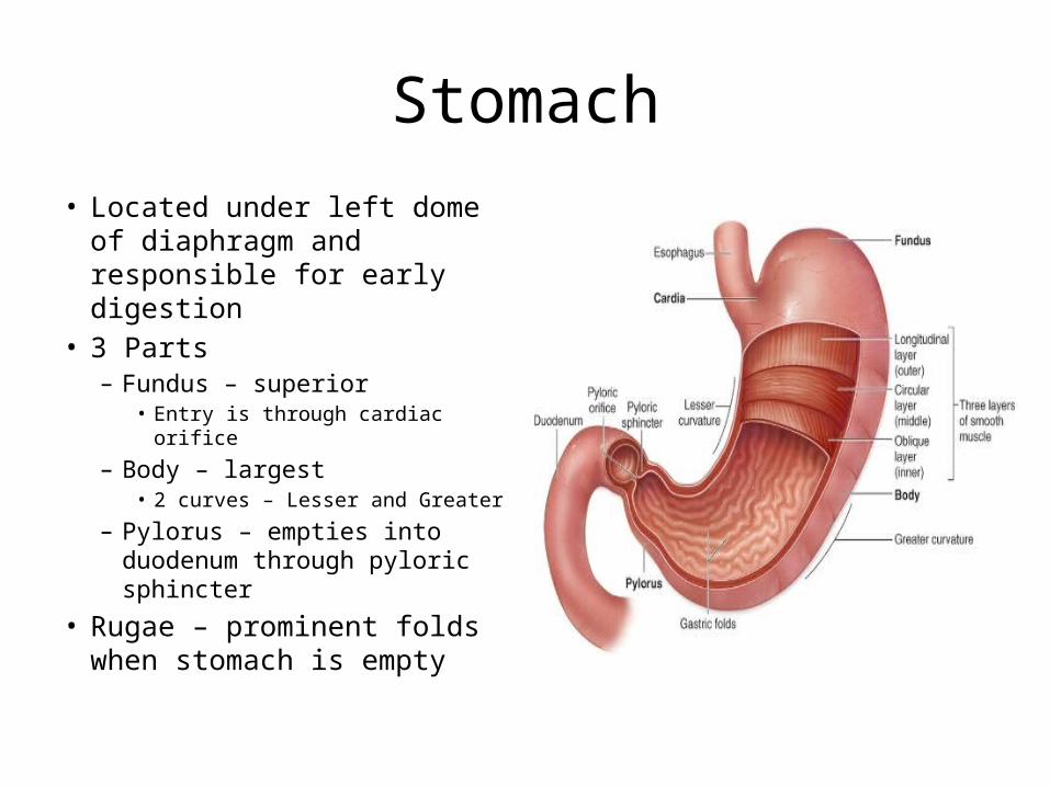
Stomach• Located under left dome of
diaphragm and responsible for early digestion
• 3 Parts– Fundus – superior
• Entry is through cardiac orifice
– Body – largest• 2 curves – Lesser and Greater
– Pylorus – empties into duodenum through pyloric sphincter
• Rugae – prominent folds when stomach is empty

Small Intestine
• Loops of bowel approx. 6 – 7 meters in length
• 3 portions– Duodenum – short, begins
following pylorus and curves around head of pancreas• Bulb – 1st 2 inches
– Jejunum – 2.5 meters long, bulk of digestion occurs here
– Ileum – Lower 3/5, longest portion• Ileocecal Valve – termination
of small bowel
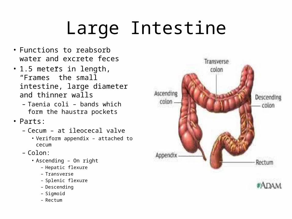
Large Intestine• Functions to reabsorb water and
excrete feces• 1.5 meters in length, “Frames” the
small intestine, large diameter and thinner walls– Taenia coli – bands which form the
haustra pockets
• Parts:– Cecum – at ileocecal valve
• Veriform appendix – attached to cecum
– Colon:• Ascending – On right
– Hepatic flexure– Transverse– Splenic flexure– Descending– Sigmoid– Rectum
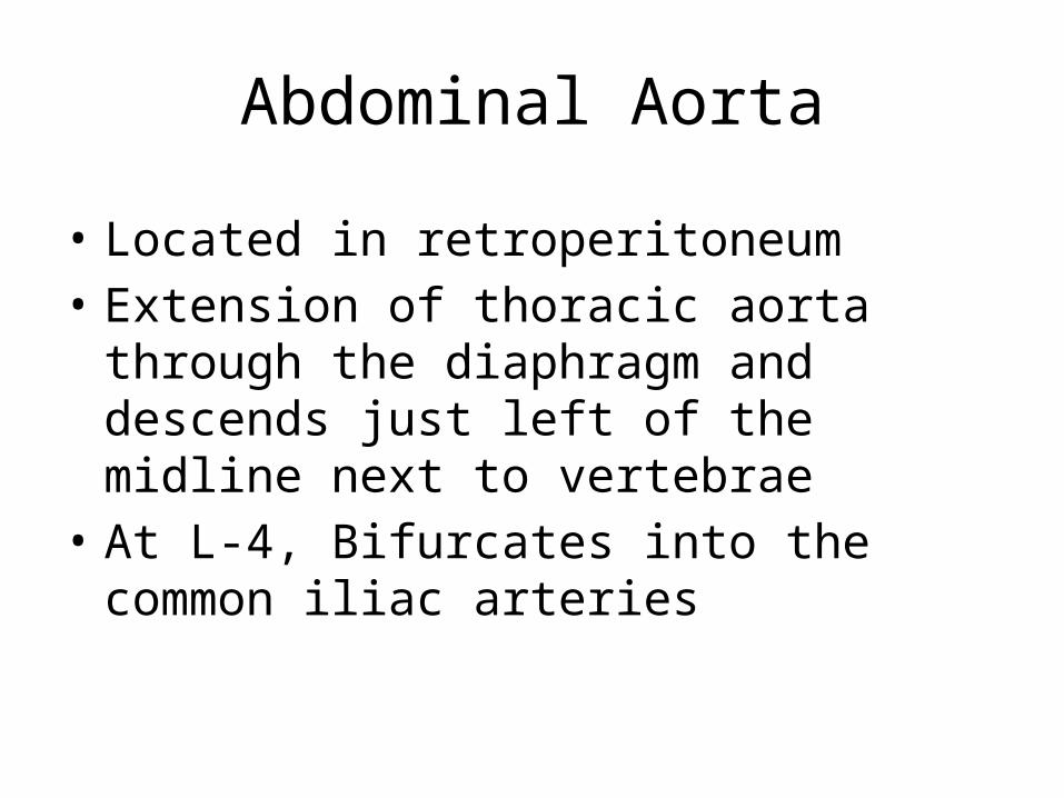
Abdominal Aorta
• Located in retroperitoneum• Extension of thoracic aorta through the
diaphragm and descends just left of the midline next to vertebrae
• At L-4, Bifurcates into the common iliac arteries
Abdominal Aorta
• Branches in order descending…– Inferior Phrenic – Paired– Celiac Trunk – divides into left gastric, common
hepatic and splenic arteries– Superior Mesenteric Artery (SMA) – Level L-1 – feeds
pancreas and both small and large intestines– Suprarenal– Renal – L-1 / L-2– Gonadal– Inferior Mesenteric Artery (IMA) – Level L-3/4
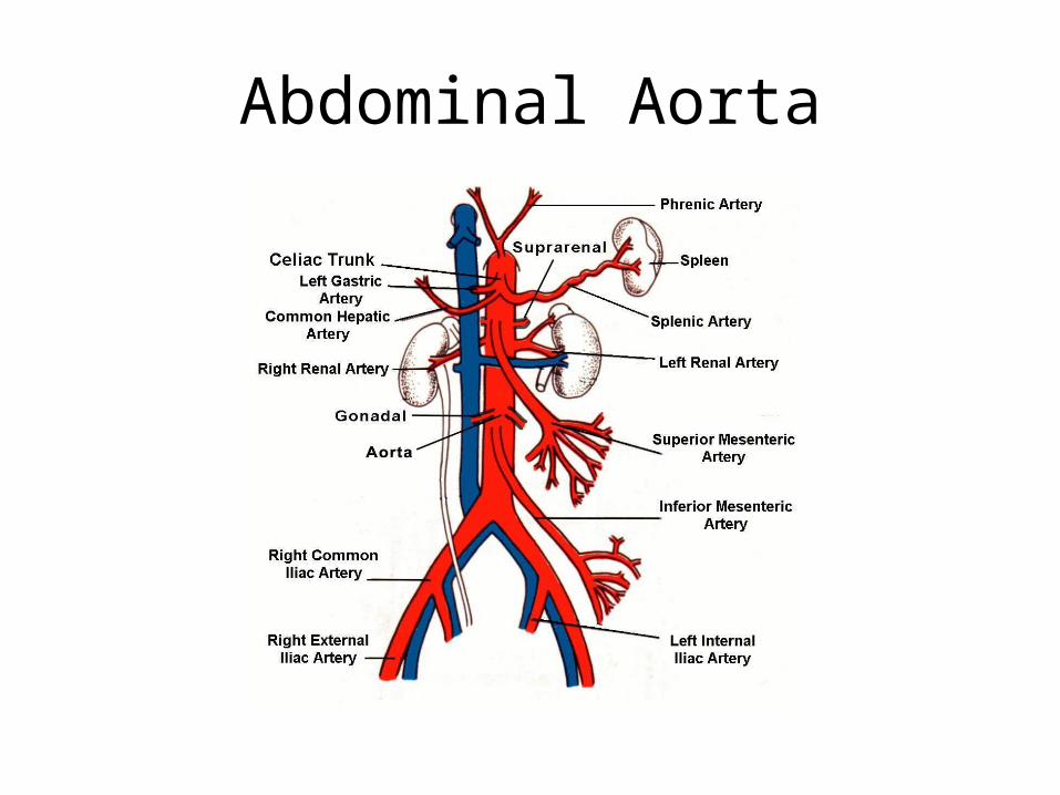
Abdominal Aorta
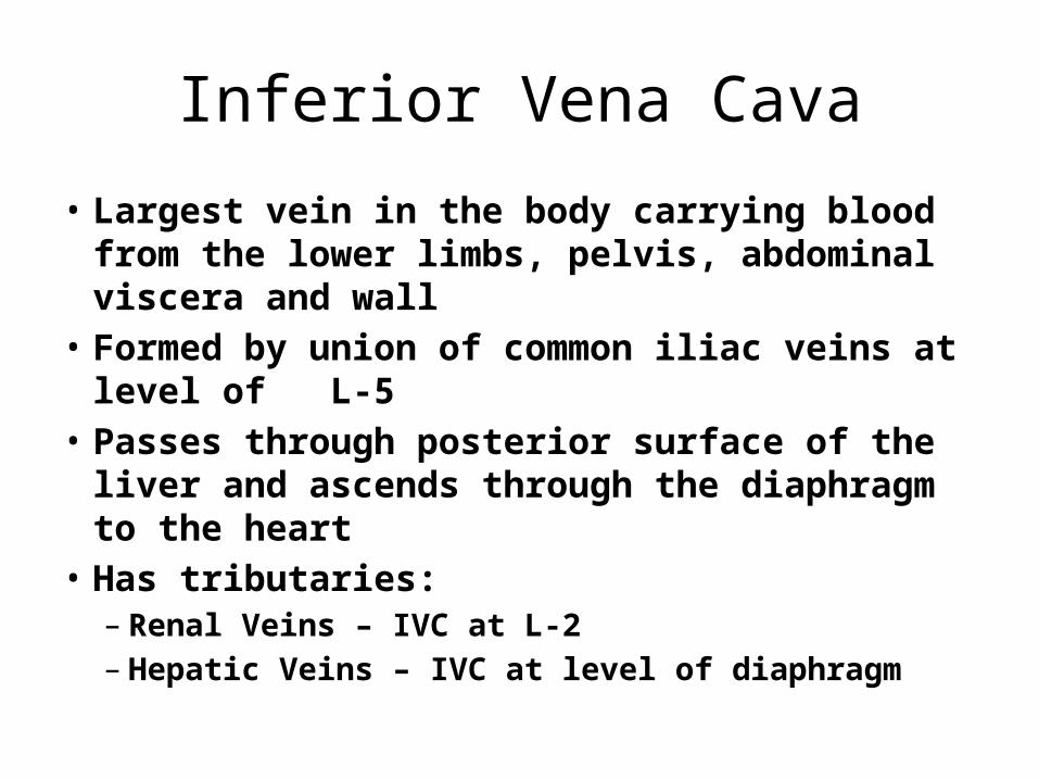
Inferior Vena Cava
• Largest vein in the body carrying blood from the lower limbs, pelvis, abdominal viscera and wall
• Formed by union of common iliac veins at level of L-5
• Passes through posterior surface of the liver and ascends through the diaphragm to the heart
• Has tributaries:– Renal Veins – IVC at L-2– Hepatic Veins – IVC at level of diaphragm
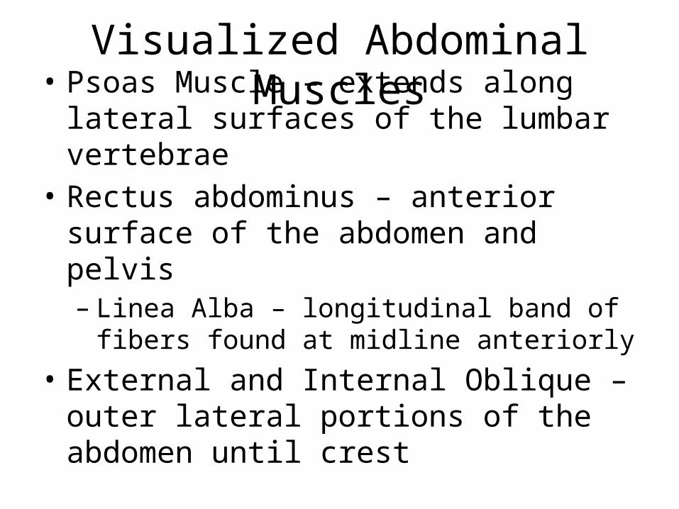
Visualized Abdominal Muscles
• Psoas Muscle – extends along lateral surfaces of the lumbar vertebrae
• Rectus abdominus – anterior surface of the abdomen and pelvis– Linea Alba – longitudinal band of fibers found at
midline anteriorly• External and Internal Oblique – outer lateral
portions of the abdomen until crest
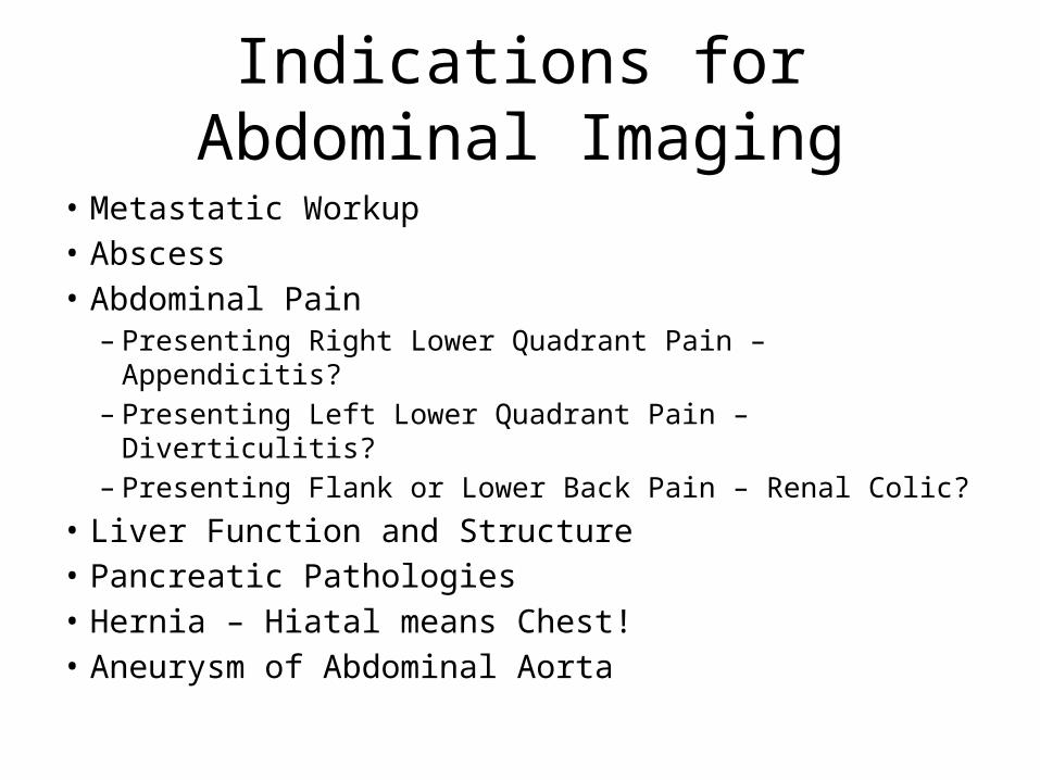
Indications for Abdominal Imaging
• Metastatic Workup• Abscess• Abdominal Pain– Presenting Right Lower Quadrant Pain – Appendicitis?– Presenting Left Lower Quadrant Pain – Diverticulitis?– Presenting Flank or Lower Back Pain – Renal Colic?
• Liver Function and Structure• Pancreatic Pathologies• Hernia – Hiatal means Chest!• Aneurysm of Abdominal Aorta
Patient Preparation
• If exam is done with I.V. contrast patient must be NPO 4-6 hours
• Lab work for I.V. contrast exams• Oral contrast can be given the night before and the
morning of the exam, or can be given 2 hours before the exam
• Oral contrast can be given at a constant rate of 900ml over a 45 minute period, 200ml is given at the time of the exam to distend the stomach
• Air contrast can also be used to give a negative (black) appearance
• Lung nodules• Cancer• Vascular disease• Effusion and infiltration• Trauma• Pulmonary Parenchymal diseases• Hilar Masses
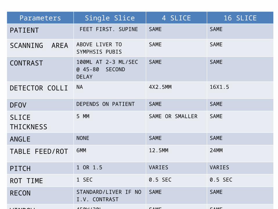
Parameters Single Slice 4 SLICE 16 SLICE
PATIENT FEET FIRST. SUPINE SAME SAME
SCANNING AREA ABOVE LIVER TO SYMPHSIS PUBIS
SAME SAME
CONTRAST 100ML AT 2-3 ML/SEC @ 45-80 SECOND DELAY
SAME SAME
DETECTOR COLLI NA 4X2.5MM 16X1.5
DFOV DEPENDS ON PATIENT SAME SAME
SLICE THICKNESS 5 MM SAME OR SMALLER SAME
ANGLE NONE SAME SAME
TABLE FEED/ROT 6MM 12.5MM 24MM
PITCH 1 OR 1.5 VARIES VARIES
ROT TIME 1 SEC 0.5 SEC 0.5 SEC
RECON STANDARD/LIVER IF NO I.V. CONTRAST
SAME SAME
WINDOW 450W/30L SAME SAME
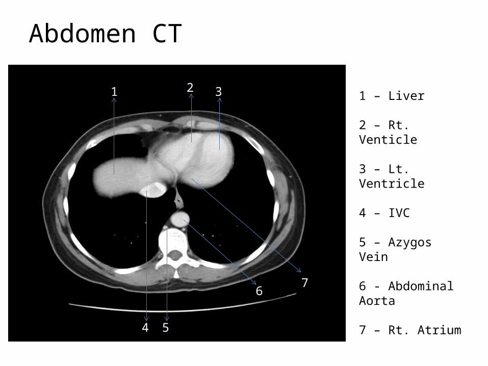
Abdomen CT
1 2 3
4
6
1 – Liver
2 – Rt. Venticle
3 – Lt. Ventricle
4 – IVC
5 – Azygos Vein
6 - Abdominal Aorta
7 – Rt. Atrium
5
7
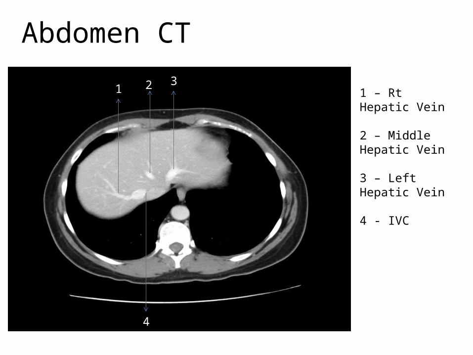
Abdomen CT
1 2 3
4
1 – Rt Hepatic Vein
2 – Middle Hepatic Vein
3 – Left Hepatic Vein
4 - IVC
Abdomen CT
12
3
4 5
6
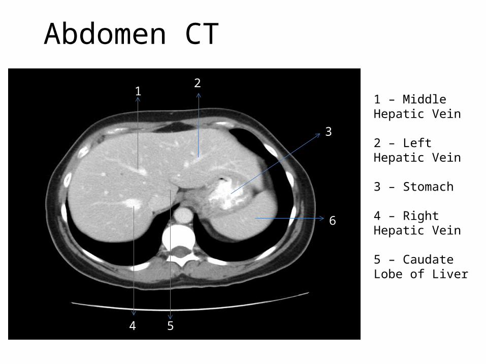
1 – Middle Hepatic Vein
2 – Left Hepatic Vein
3 – Stomach
4 – Right Hepatic Vein
5 – Caudate Lobe of Liver
Abdomen CT
1 2 3 4
5
6
7
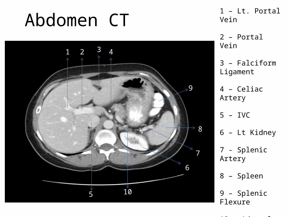
1 – Lt. Portal Vein
2 – Portal Vein
3 – Falciform Ligament
4 – Celiac Artery
5 – IVC
6 – Lt Kidney
7 - Splenic Artery
8 – Spleen
9 – Splenic Flexure
10 – Adrenal Gland
8
9
10
Abdomen CT
1
2
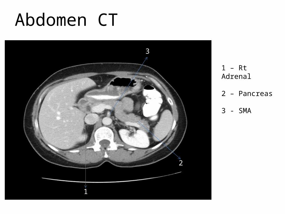
1 – Rt Adrenal
2 – Pancreas
3 - SMA
3
Abdomen CT
1 2 3
4
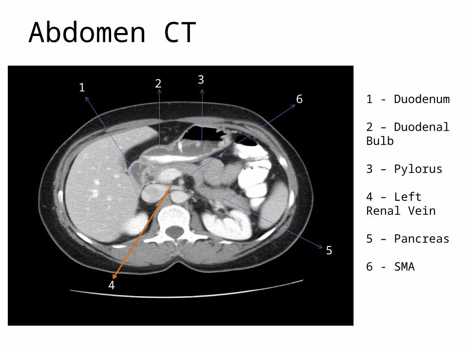
1 - Duodenum
2 – Duodenal Bulb
3 – Pylorus
4 – Left Renal Vein
5 – Pancreas
6 - SMA5
6
Abdomen CT
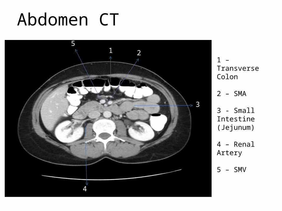
1 2
3
1 – Transverse Colon
2 – SMA
3 - Small Intestine (Jejunum)
4 – Renal Artery
5 – SMV
4
5
Abdomen CT
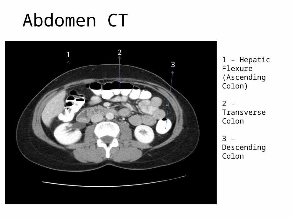
1 2
31 – Hepatic Flexure (Ascending Colon)
2 – Transverse Colon
3 – Descending Colon
Abdomen CT
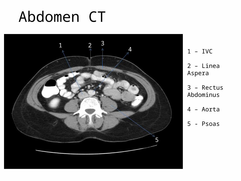
1 2 34
5
1 – IVC
2 – Linea Aspera
3 – Rectus Abdominus
4 – Aorta
5 - Psoas
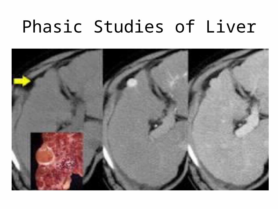
Phasic Studies of Liver
A three phase CT scan, usually of the liver, requires an injection of contrast medium which helps outline the vessels of your body by giving the x-rays something to be absorbed by besides blood which has a very low absorption rate. The phases are: 1. Scan during injection: Arterial Phase, this will highlight lesions in or around the artery leading into the liver. 2. Scan during injection or shortly after: Portal Venous Phase, this will show lesions in or around the portal vein. 3. Delayed scan after injection: Equilibrium Phase, this will allow the soft tissue to absorb the contrast and may highlight changes in tissue.
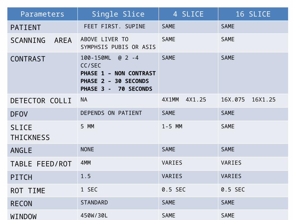
Parameters Single Slice 4 SLICE 16 SLICE
PATIENT FEET FIRST. SUPINE SAME SAME
SCANNING AREA ABOVE LIVER TO SYMPHSIS PUBIS OR ASIS
SAME SAME
CONTRAST 100-150ML @ 2 -4 CC/SECPHASE 1 – NON CONTRASTPHASE 2 – 30 SECONDSPHASE 3 - 70 SECONDS
SAME SAME
DETECTOR COLLI NA 4X1MM 4X1.25 16X.075 16X1.25
DFOV DEPENDS ON PATIENT SAME SAME
SLICE THICKNESS 5 MM 1-5 MM SAME
ANGLE NONE SAME SAME
TABLE FEED/ROT 4MM VARIES VARIES
PITCH 1.5 VARIES VARIES
ROT TIME 1 SEC 0.5 SEC 0.5 SEC
RECON STANDARD SAME SAME
WINDOW 450W/30L SAME SAME
Phasic Studies of Liver
CT Urography
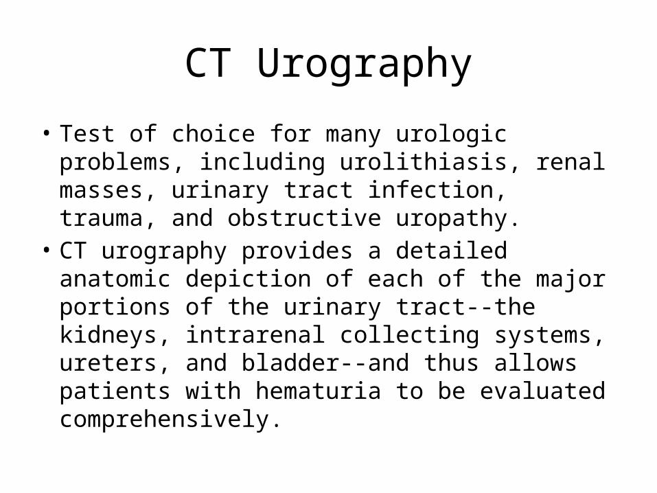
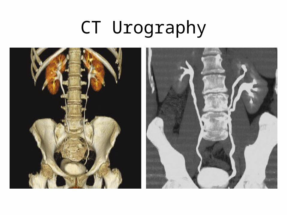
• Test of choice for many urologic problems, including urolithiasis, renal masses, urinary tract infection, trauma, and obstructive uropathy.
• CT urography provides a detailed anatomic depiction of each of the major portions of the urinary tract--the kidneys, intrarenal collecting systems, ureters, and bladder--and thus allows patients with hematuria to be evaluated comprehensively.
CT Urography