Embed Size (px)
Citation preview
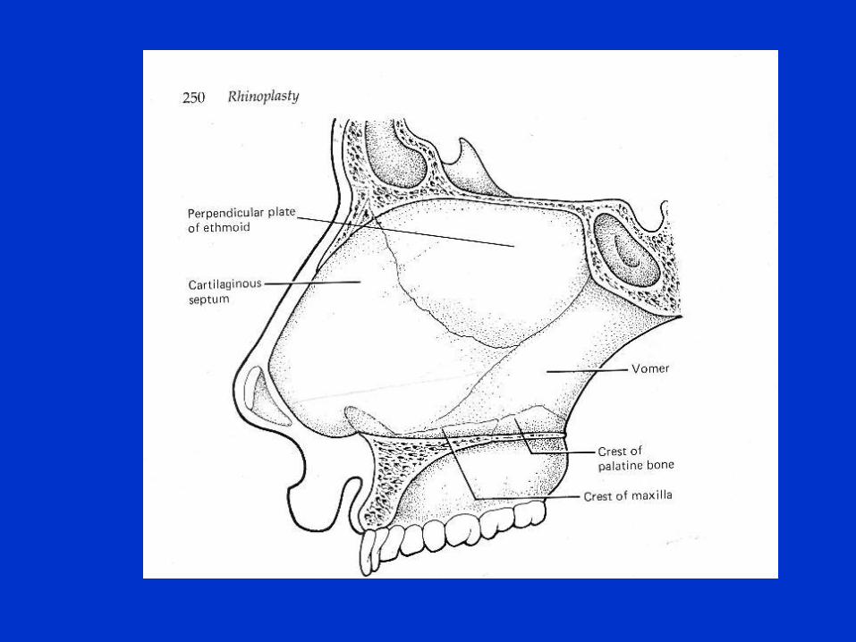
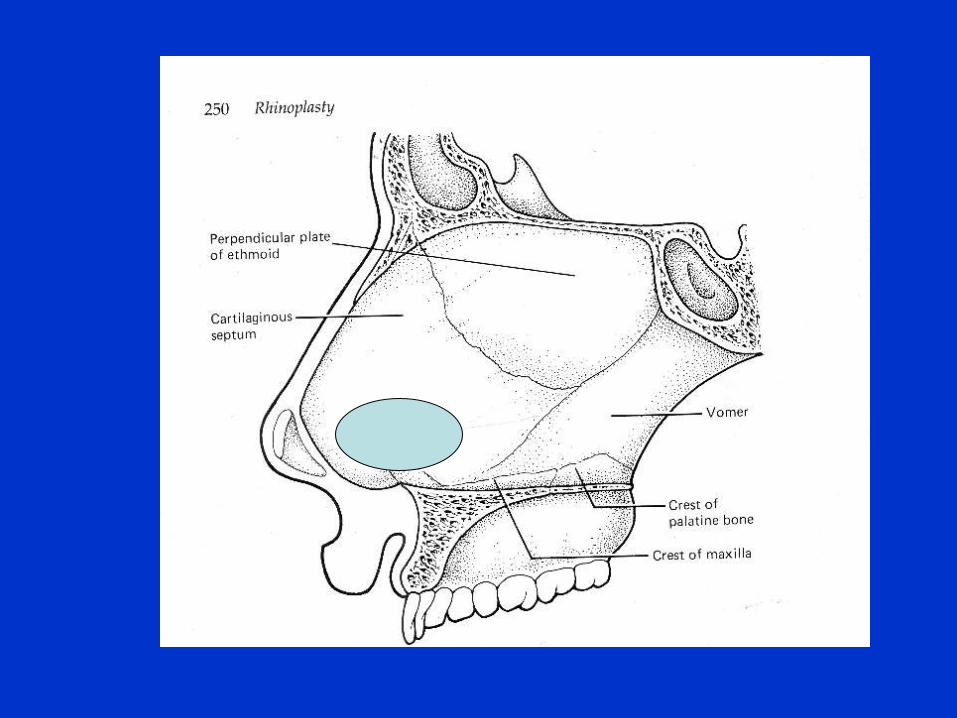
Anatomy of nose and
paranasal sinuses
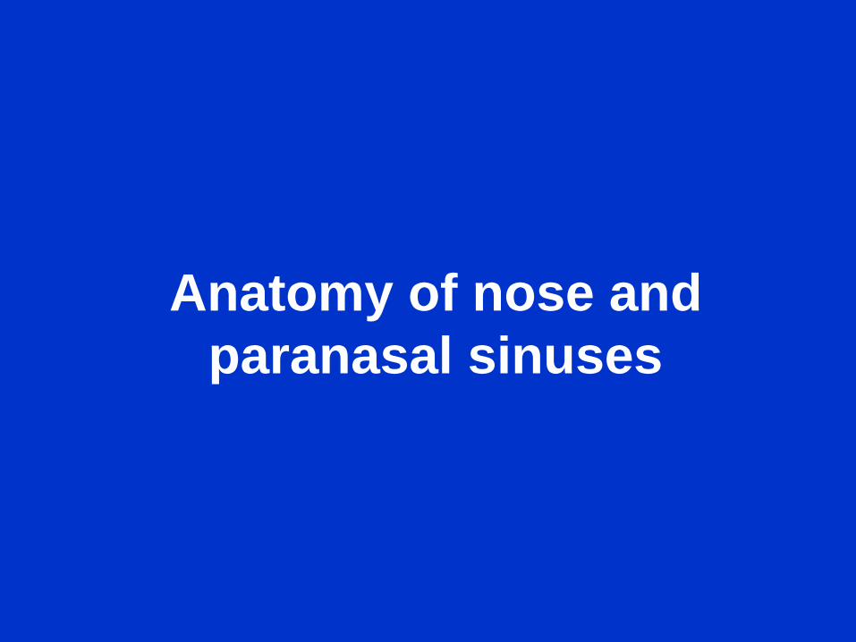
Nasal vestibule
• Is part of external nose and is lined by skin
and contains hair called vibrissae
• It is limited above and behind by curved
ridge, the limen nasi
Nasal fossae
• The right and left nasal fossae are
separated by nasal septum
• The nasal fossa includes only that part
which is lined by mucous membrane
• Each nasal fossa communicates with
paranasal sinuses and nasopharynx

Nasal fossa floor
• Palatine process of maxilla and horizontal
parts of palatine bone
Nasal fossa roof
• Nasal process of frontal bone ,cribriform
plate of ethmoid ,and body of sphenoid
bone
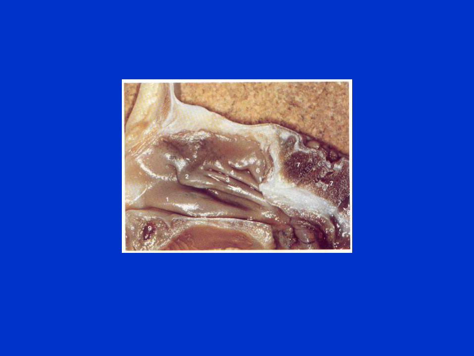
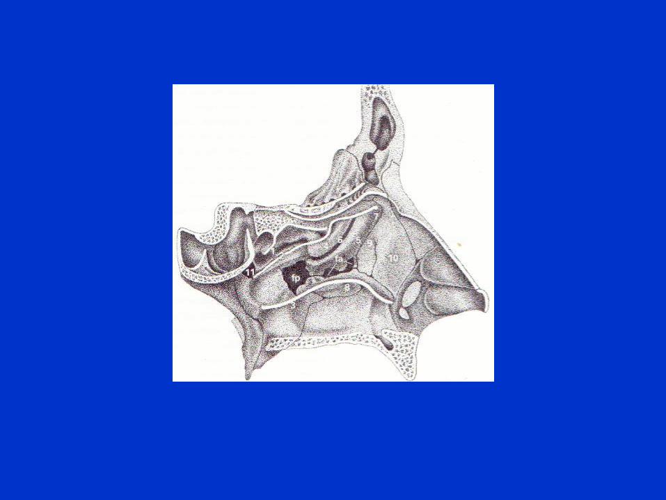
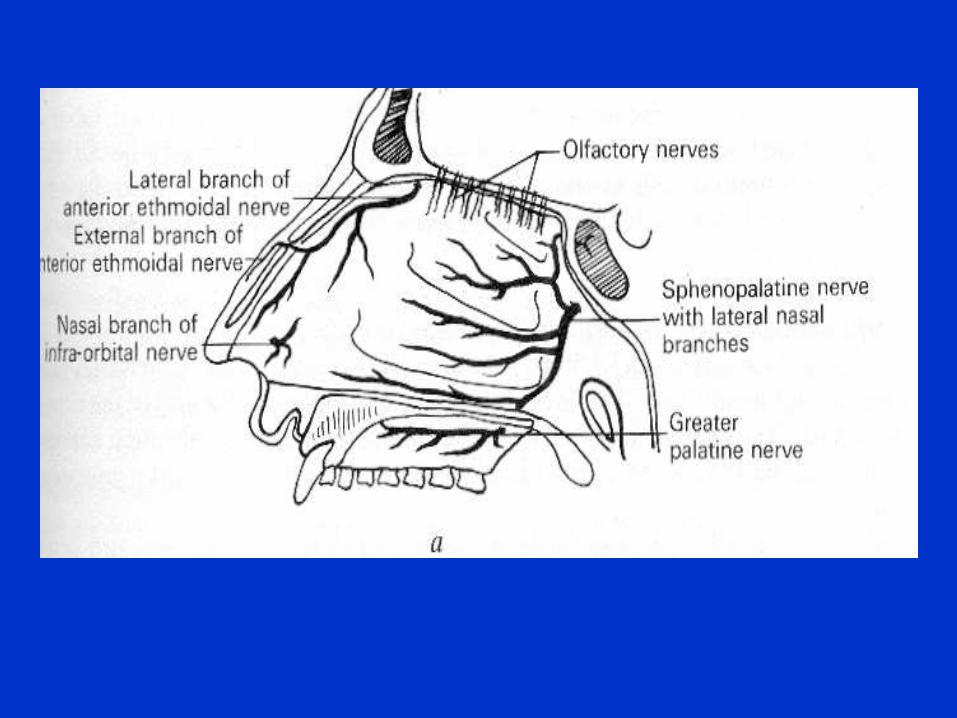
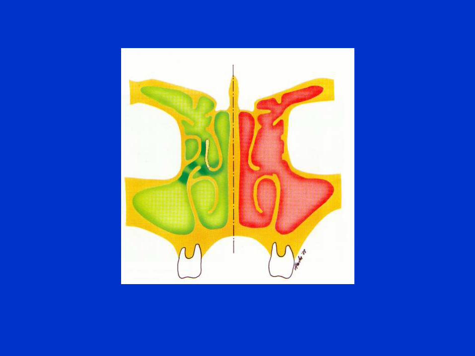
Lateral wall
• Medial walls of maxilla bone
• Lateral mass of ethmoid and lacrimal bone
• Ascending process of maxilla
• Perpendicular part of palatine bone and
medial pterygoid plate


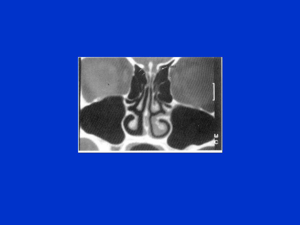
Paranasal sinuses
• These are air spaces within certain bones
of the skull.There are four on each side.
• Maxillary sinus
• Ethmoid sinuses
• Frontal sinus
• Sphenoid sinus
Maxillary sinus
• Is pyramidal in shape.It occupies the body of the maxilla.
• The base lies medially .
• The apex in the zygomatic portion of the maxilla.
• It is the largest of the sinuses.
• Average capacity is 15ml in adults
• Medial wall is the party wall between the sinus and nasal fossa
Mxillary sinus cont.
• Apex may extend into zygomatic process of
maxilla
• Roof is the thin floor of orbit .It is grooved by
infraorbital nerve
• Floor is formed by alveolar process and hard
palate. In children it lies at or above the level of
floor of nasal fossa.In adults it lies one cm.
below the nasal fossa floor. The roots of many
teeth may be related to floor.
Maxillary sinus cont.
• Posterior wall is pierced by dental canals
which transmit the posterior superior
dental vessels and nerves to molar teeth.
• Anterior wall separates the sinus from skin
of the cheek. It contains the anterior
superior dental vessels and nerves and
the foramen for the infraorbital nerve.
Ethmoid sinuses
• 7-15 in number
• Divided into anterior and posterior group
• Anterior cranial fossa lies above, the orbit
is lateral to these.
• Lacrimal sac is also lateral
• Optic nerve is closely related to posterior
group

Frontal sinus
• An upward extension of anterior ethmoid
cells.
• Present in frontal bone
• Average capacity is 7 ml. Opens into
middle meatus


![University of Massachusetts Amherstvis- · HfHfHfH;H^HfHfH;HfHfH^H;HfHfHfH;HfH^HfH;HfHfHfH^H;HfHfHfH;HfH^HfH;HfHfHfH H$7 ~ H O3V= 02X 6 ep]^9 :WXf9 v0>b2Xf74= 027]`::](https://img.pdfslide.us/doc/110x75/5fd2b18a79477a759921650c/university-of-massachusetts-amherstvis-hfhfhfhhhfhfhhfhfhhhfhfhfhhfhhfhhfhfhfhhhfhfhfhhfhhfhhfhfhfh.jpg)