Embed Size (px)
Citation preview
20-1
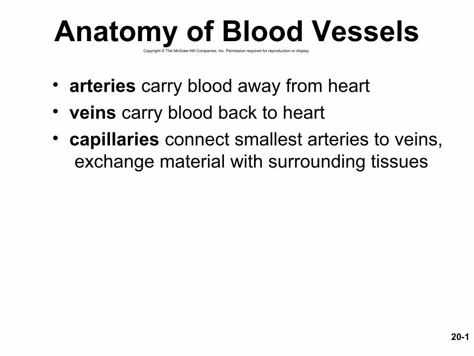
Anatomy of Blood Vessels
• arteries carry blood away from heart• veins carry blood back to heart• capillaries connect smallest arteries to veins,
exchange material with surrounding tissues
Copyright © The McGraw-Hill Companies, Inc. Permission required for reproduction or display.
20-3
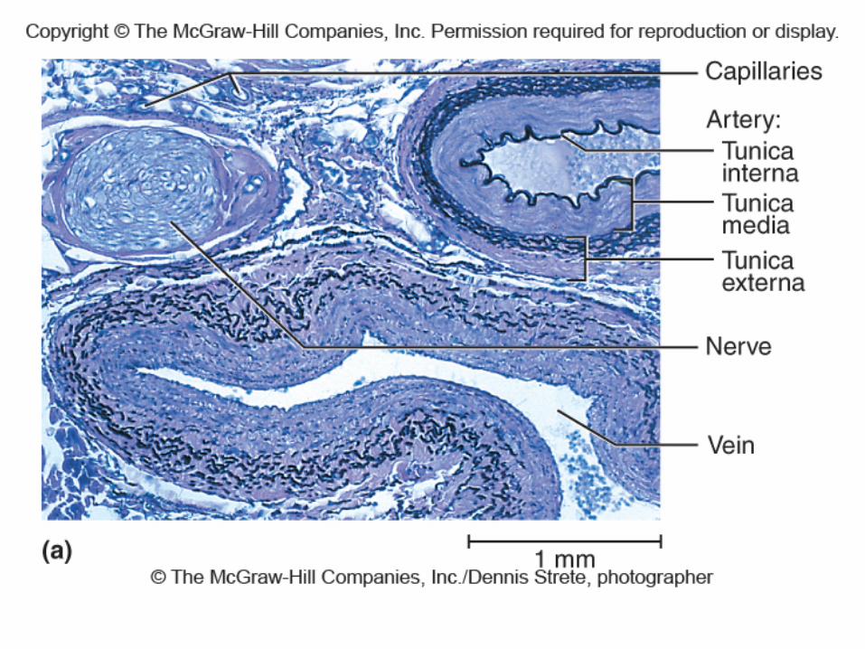
Vessel Wall• tunica interna
– lines the blood vessel and is exposed to blood– endothelium – simple squamous epithelium overlying a
basement membrane and a sparse layer of loose connective tissue
• acts as a selectively permeable barrier
• secrete chemicals that stimulate dilation or constriction of the vessel
• normally repels blood cells and platelets that may adhere to it and form a clot
• when tissue around vessel is inflamed, the endothelial cells produce cell-adhesion molecules that induce leukocytes to adhere to the surface
– causes leukocytes to congregate in tissues where their defensive actions are needed
20-4
Vessel Wall
• tunica media
– middle layer
– consists of smooth muscle, collagen, and elastic tissue
– strengthens vessel and prevents blood pressure from rupturing them
– changes in diameter of the blood vessel brought about by smooth muscle
20-5
Vessel Wall
• tunica externa
– outermost layer
– consists of loose connective tissue that often merges with that of neighboring blood vessels, nerves, or other organs
– anchors the vessel and provides passage for small nerves, lymphatic vessels

20-7
Arteries• conducting arteries
• biggest arteries• aorta, common carotid, subclavian, pulmonary trunk, and
common iliac arteries
• expand during systole, recoil during diastole which lessens fluctuations in blood pressure
• contain additional layers of elastic tissue
• distributing arteries• distributes blood to specific organs• brachial, femoral, renal, and splenic arteries• smooth muscle layers constitute three-fourths of wall thickness

20-8
Aneurysm• aneurysm - weak point in an artery or the heart
wall– forms a thin-walled, bulging sac that pulsates with each
heartbeat and may rupture at any time– most common sites: abdominal aorta, renal arteries,
and arterial circle at the base of the brain
– can cause pain by putting pressure on other structures
– can rupture causing hemorrhage
– result from congenital weakness of the blood vessels or result of trauma or bacterial infections such as syphilis
• most common cause is atherosclerosis and hypertension

20-9
Arteries and Metarterioles
• resistance (small) arteries– arterioles – smallest arteries
• control amount of blood to various organs
• metarterioles – short vessels that link arterioles to capillaries– muscle cells form a precapillary sphincter about
entrance to capillary• constriction of these sphincters reduces or shuts off blood
flow through their respective capillaries
• diverts blood to other tissues
20-10
Capillaries• capillaries - site where nutrients, wastes, and
hormones pass between the blood and tissue fluid through the walls of the vessels (exchange vessels)– composed of endothelium and basal lamina
20-11
Three Types of Capillaries• continuous capillaries - occur in most tissues
– endothelial cells have tight junctions forming a continuous tube with intercellular clefts
• allow passage of solutes such as glucose– pericytes wrap around the capillaries and contain the same
contractile protein as muscle • contract and regulate blood flow
• fenestrated capillaries - kidneys, small intestine– organs that require rapid absorption or filtration – endothelial cells riddled with holes called filtration pores
(fenestrations)• allows passage of only small molecules
• sinusoids (discontinuous capillaries) - liver, bone marrow, spleen– irregular blood-filled spaces with large fenestrations– allow proteins (albumin), clotting factors, and new blood cells to
enter the circulation

20-12
Continuous Capillary
Figure 20.5
Copyright © The McGraw-Hill Companies, Inc. Permission required for reproduction or display.
Pericyte
Erythrocyte
Basallamina
Intercellularcleft
Pinocytoticvesicle
Endothelialcell
Tightjunction
20-13
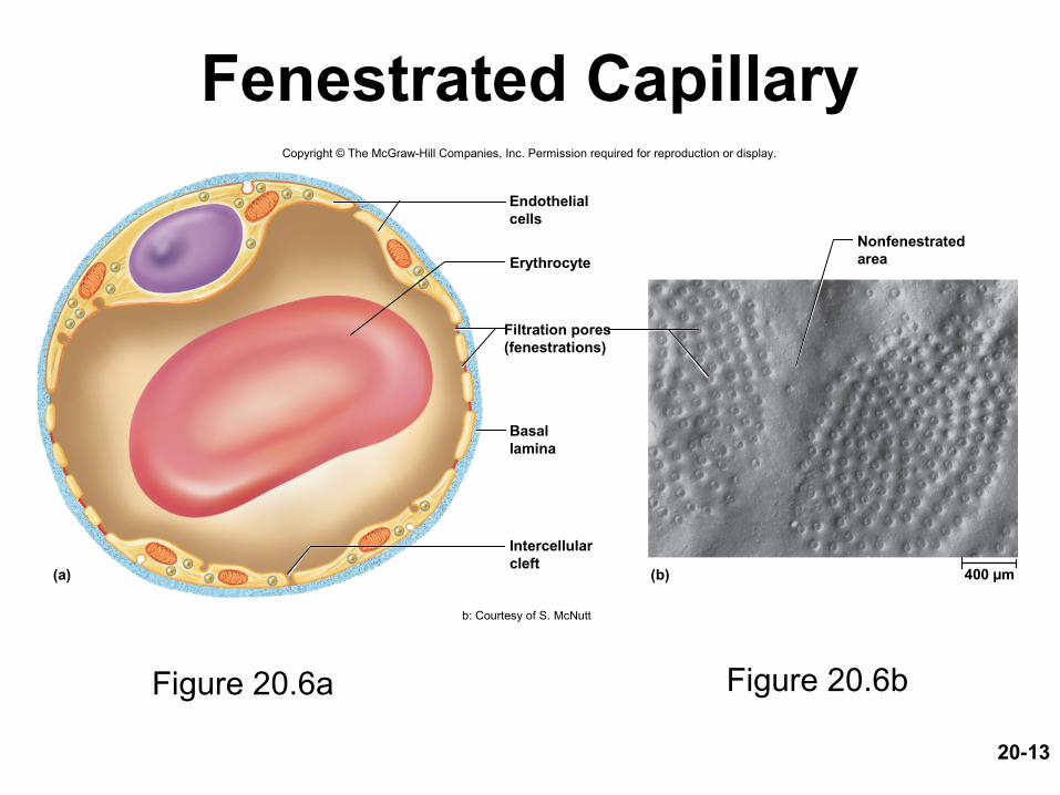
Fenestrated Capillary
Figure 20.6a Figure 20.6b
Copyright © The McGraw-Hill Companies, Inc. Permission required for reproduction or display.
(a) (b)
Erythrocyte
Endothelialcells
Filtration pores(fenestrations)
Basallamina
Intercellularcleft
Nonfenestratedarea
400 µm
b: Courtesy of S. McNutt
20-14
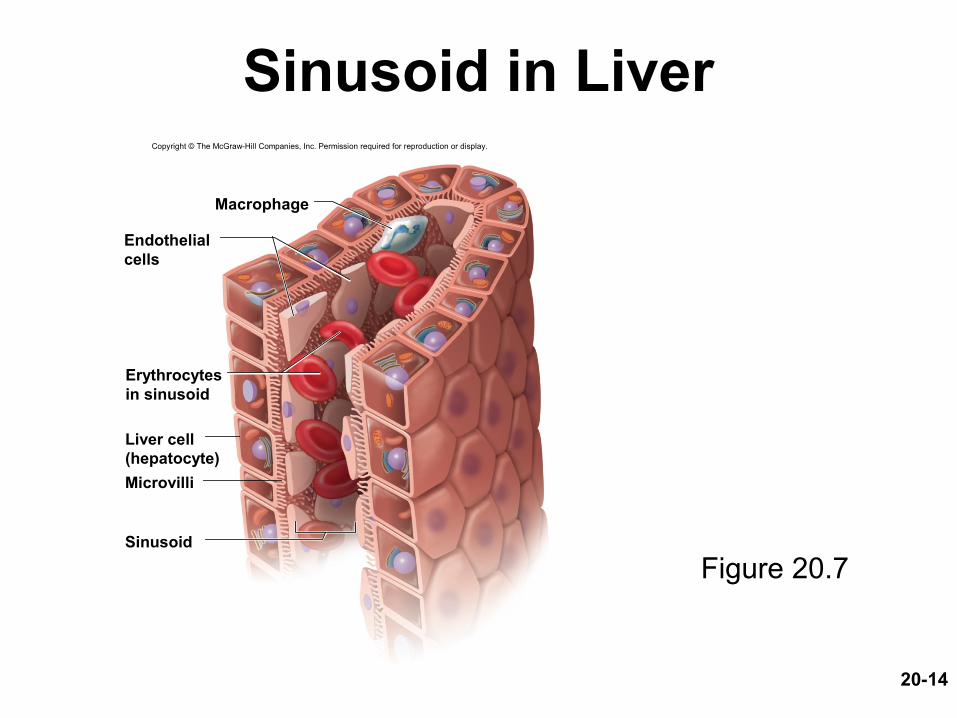
Sinusoid in Liver
Figure 20.7
Copyright © The McGraw-Hill Companies, Inc. Permission required for reproduction or display.
Macrophage
Sinusoid
Microvilli
Endothelialcells
Erythrocytesin sinusoid
Liver cell(hepatocyte)
20-15
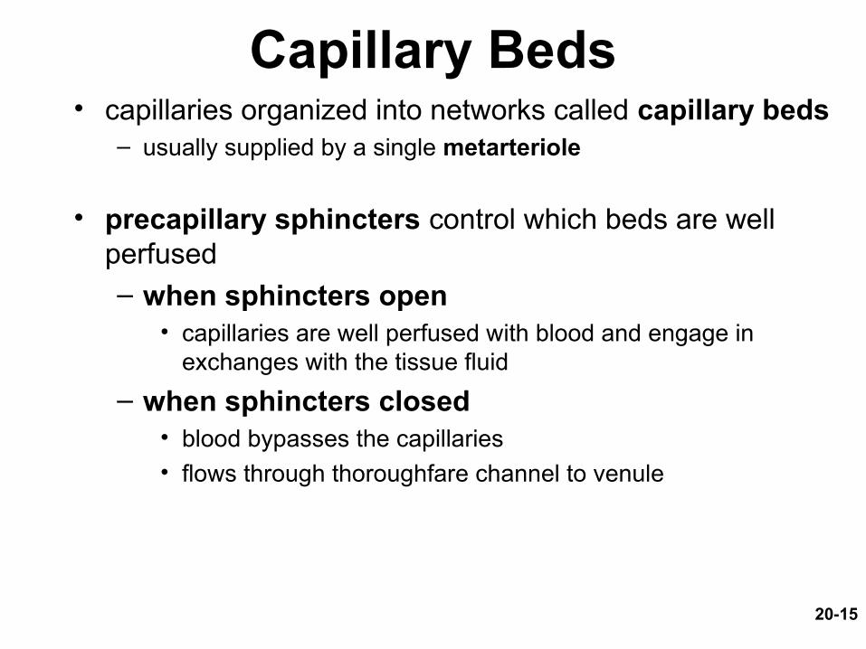
Capillary Beds• capillaries organized into networks called capillary beds
– usually supplied by a single metarteriole
• precapillary sphincters control which beds are well perfused
– when sphincters open• capillaries are well perfused with blood and engage in
exchanges with the tissue fluid
– when sphincters closed • blood bypasses the capillaries• flows through thoroughfare channel to venule
20-16
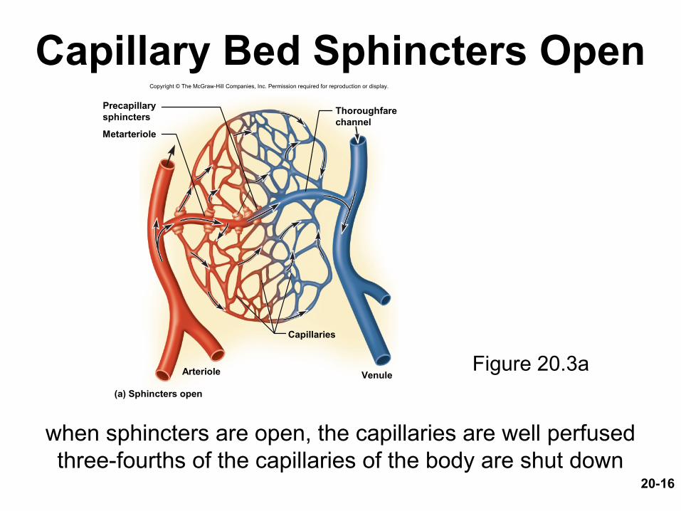
Capillary Bed Sphincters Open
Figure 20.3a
when sphincters are open, the capillaries are well perfusedthree-fourths of the capillaries of the body are shut down
Copyright © The McGraw-Hill Companies, Inc. Permission required for reproduction or display.
Capillaries
Metarteriole
Arteriole
Precapillarysphincters
Thoroughfarechannel
Venule
(a) Sphincters open
20-17
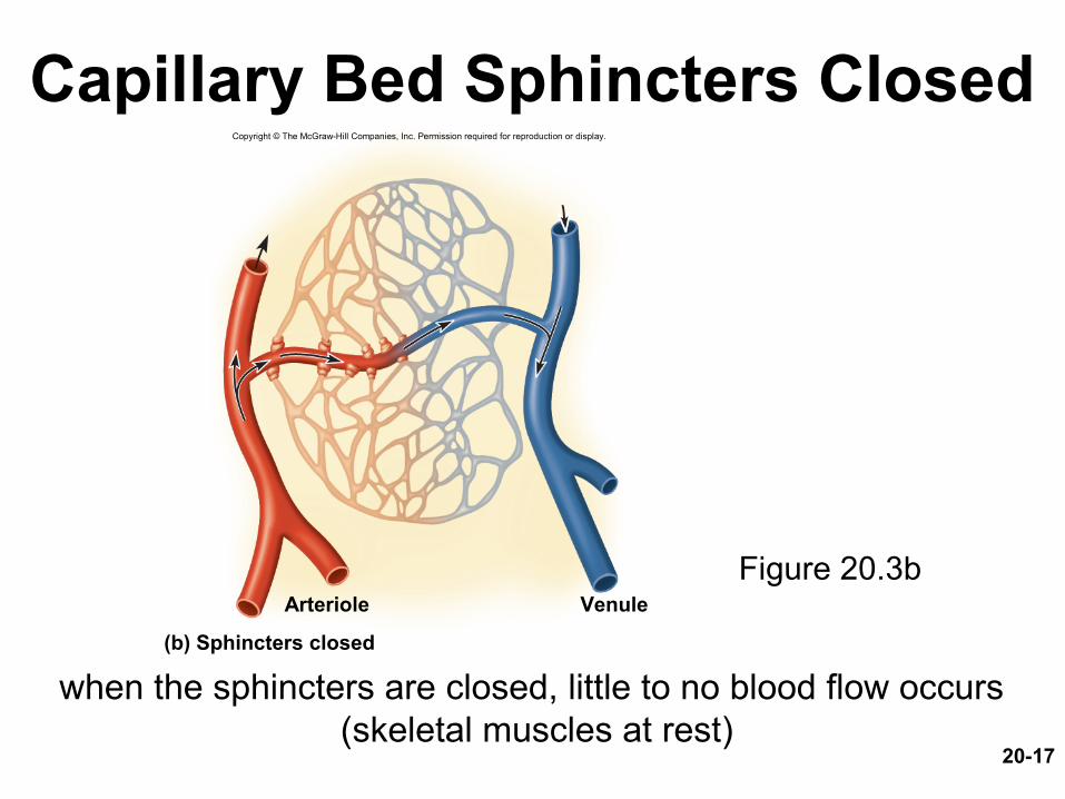
Capillary Bed Sphincters Closed
Figure 20.3b
when the sphincters are closed, little to no blood flow occurs
(skeletal muscles at rest)
VenuleArteriole
Copyright © The McGraw-Hill Companies, Inc. Permission required for reproduction or display.
(b) Sphincters closed
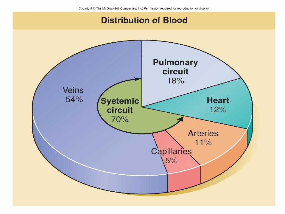
Veins (Capacitance Vessels)• greater capacity for blood containment than arteries• thinner walls, flaccid, less muscular and elastic tissue • collapse when empty, expand easily • have steady blood flow
• merge to form larger veins
• subjected to relatively low blood pressure– remains 10 mm Hg with little fluctuation
20-20
Blood Flow Pathway• postcapillary venules – smallest veins
– even more porous than capillaries so also exchange fluid with surrounding tissues
• muscular venules – up to 1 mm in diameter
• medium veins – up to 10 mm in diameter– tunica interna forms venous valves– skeletal muscle pump propels venous blood back toward the
heart
20-21
Blood Flow Pathway• venous sinuses
– veins with especially thin walls, large lumens, and no smooth muscle
– Example: coronary sinus of the heart– not capable of vasomotion
• large veins – larger than 10 mm– venae cavae, pulmonary veins, internal jugular veins,
and renal veins
20-22
Varicose Veins• blood pools in the lower legs in people who stand
for long periods stretching the veins– cusps of the valves pull apart in enlarged superficial
veins further weakening vessels– blood backflows and further distends the vessels, their
walls grow weak and develop into varicose veins
• hereditary weakness, obesity, and pregnancy also promote problems
• hemorrhoids are varicose veins of the anal canal
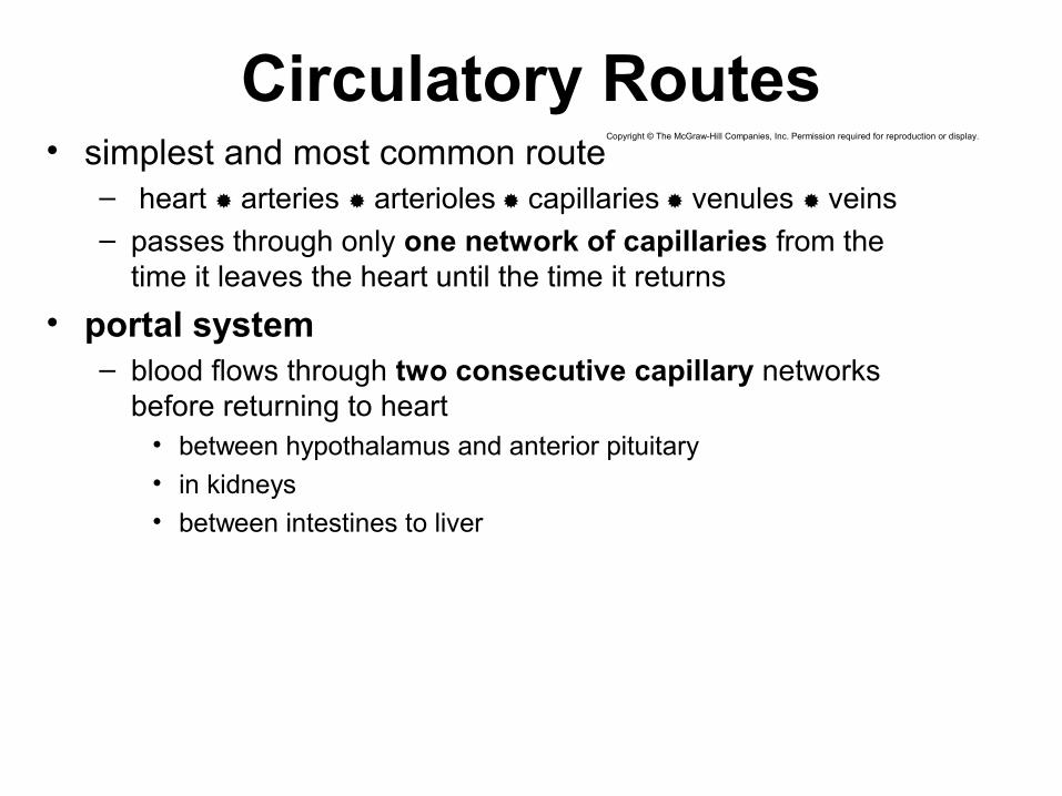
Circulatory Routes• simplest and most common route
– heart arteries arterioles capillaries venules veins– passes through only one network of capillaries from the
time it leaves the heart until the time it returns
• portal system– blood flows through two consecutive capillary networks
before returning to heart• between hypothalamus and anterior pituitary
• in kidneys• between intestines to liver
Copyright © The McGraw-Hill Companies, Inc. Permission required for reproduction or display.
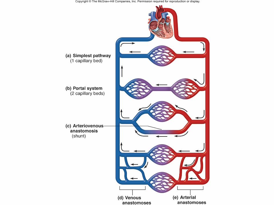
Anastomoses• anastomosis – the point where two blood vessels merge
• arteriovenous anastomosis (shunt)– artery flows directly into vein bypassing capillaries
• venous anastomosis– most common– one vein empties directly into another– reason vein blockage less serious
than an arterial blockage
• arterial anastomosis– two arteries merge
– provides collateral (alternative) routes of blood supply to a tissue– coronary circulation and around joints
20-26
Blood Pressure• blood pressure (bp) – the force that blood exerts against a vessel wall
• measured at brachial artery of arm using sphygmomanometer
• two pressures are recorded:– systolic pressure: peak arterial BP taken during ventricular contraction
(ventricular systole)– diastolic pressure: minimum arterial BP taken during ventricular
relaxation (diastole) between heart beats
• normal value, young adult: 120/75 mm Hg
20-27
Abnormalities of Blood Pressure
• hypertension – high blood pressure– chronic is resting BP > 140/90– consequences
• can weaken small arteries and cause aneurysms
• hypotension – chronic low resting BP– caused by blood loss, dehydration, anemia
20-28
Blood Pressure• one of the body’s chief mechanisms in preventing
excessive blood pressure is the ability of the arteries to stretch and recoil during the cardiac cycle
• importance of arterial elasticity– expansion and recoil maintains steady flow of blood throughout
cardiac cycle, smoothes out pressure fluctuations and decreases stress on small arteries
• BP rises with age – arteries less distensible and absorb less systolic force
• BP determined by cardiac output, blood volume and peripheral resistance
20-30
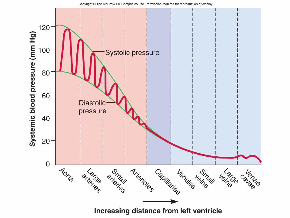
Flow at Different Points• from aorta to capillaries, blood velocity (speed)
decreases for three reasons:– greater distance, more friction to reduce speed– smaller radii of arterioles and capillaries offers more
resistance– farther from heart, the number of vessels and their total
cross-sectional area becomes greater and greater
• from capillaries to vena cava, flow increases again– decreased resistance going from capillaries to veins
– large amount of blood forced into smaller channels
– never regains velocity of large arteries
20-31
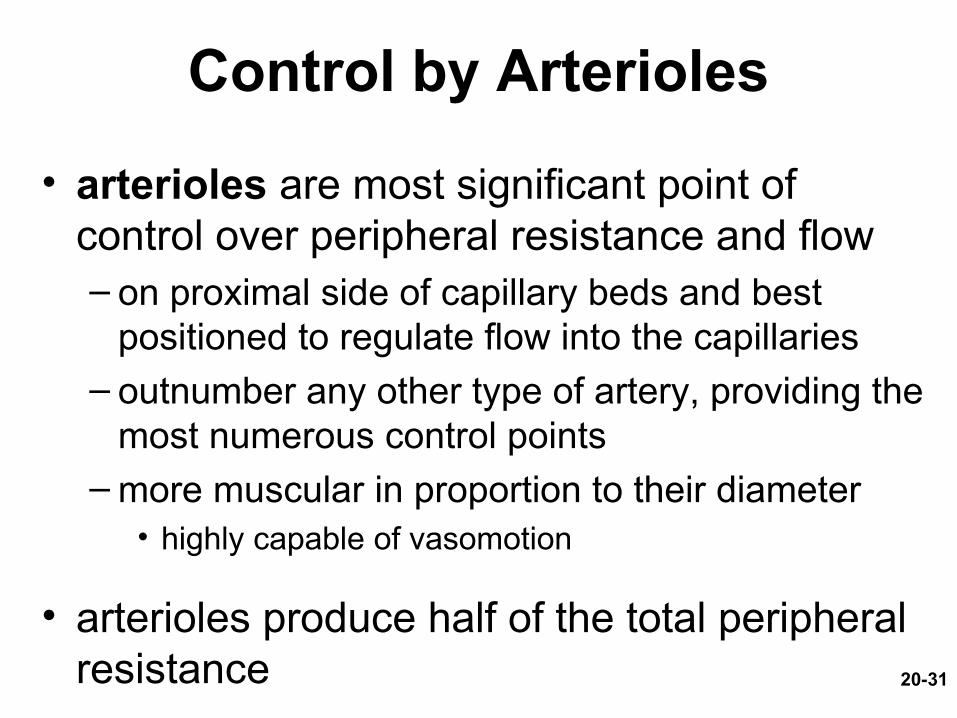
Control by Arterioles
• arterioles are most significant point of control over peripheral resistance and flow– on proximal side of capillary beds and best
positioned to regulate flow into the capillaries– outnumber any other type of artery, providing the
most numerous control points– more muscular in proportion to their diameter
• highly capable of vasomotion
• arterioles produce half of the total peripheral resistance
20-32
Regulation of BP and Flow
• vasomotion is a quick and powerful way of altering blood pressure and flow
• three ways of controlling vasomotion:– local control– neural control– hormonal control
20-33
Local Control of BP and Flow• autoregulation – the ability of tissues to regulate their own blood
supply
• vasoactive chemicals - substances secreted by platelets, endothelial cells, and perivascular tissue stimulate vasomotion
• angiogenesis - growth of new blood vessels– occurs in regrowth of uterine lining, around coronary artery obstructions, in
exercised muscle, and malignant tumors– controlled by growth factors

20-34
Neural Control of Blood Vessels• vessels under remote control by the central and autonomic
nervous systems
• vasomotor center of medulla oblongata exerts sympathetic control over blood vessels throughout the body– stimulates most vessels to constrict, but dilates vessels in skeletal
and cardiac muscle to meet demands of exercise• precapillary sphincters respond only to local and hormonal control
due to lack of innervation
– vasomotor center is the integrating center for three autonomic reflexes
• Baroreflexes – carotid sinuses• Chemoreflexes – aoritic and catotid bodies• medullary ischemic reflex – medulla oblongata
20-35
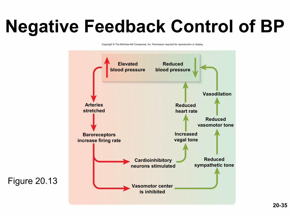
Negative Feedback Control of BP
Figure 20.13
Copyright © The McGraw-Hill Companies, Inc. Permission required for reproduction or display.
Vasodilation
Elevatedblood pressure
Reducedblood pressure
Reducedheart rate
Arteriesstretched
Baroreceptorsincrease firing rate
Reducedvasomotor tone
Reducedsympathetic tone
Increasedvagal tone
Cardioinhibitoryneurons stimulated
Vasomotor centeris inhibited
20-36
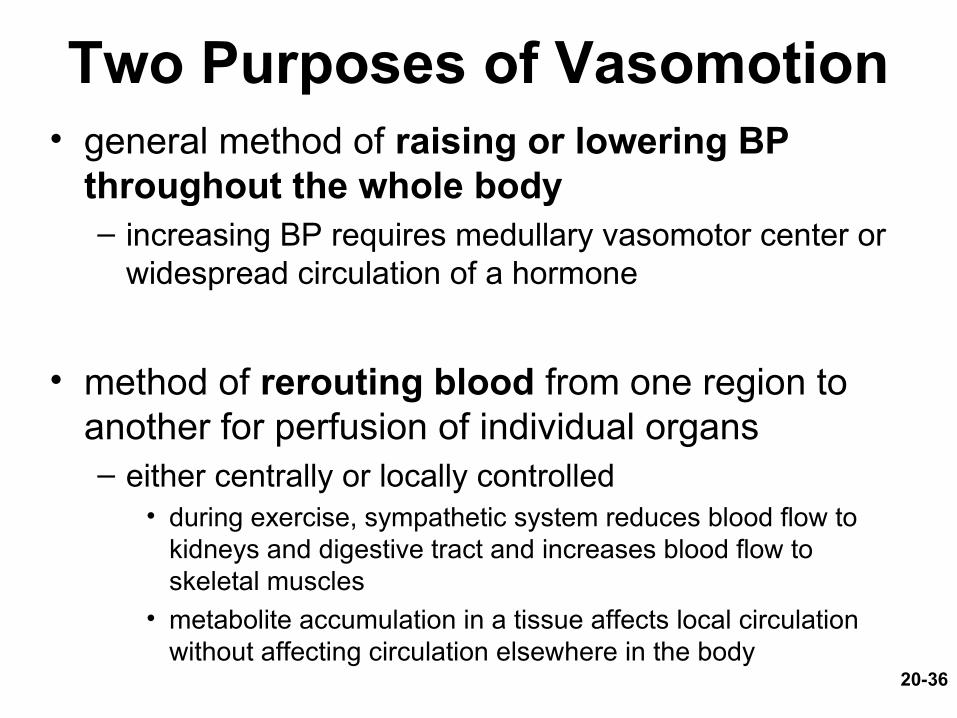
Two Purposes of Vasomotion• general method of raising or lowering BP
throughout the whole body– increasing BP requires medullary vasomotor center or
widespread circulation of a hormone
• method of rerouting blood from one region to another for perfusion of individual organs– either centrally or locally controlled
• during exercise, sympathetic system reduces blood flow to kidneys and digestive tract and increases blood flow to skeletal muscles
• metabolite accumulation in a tissue affects local circulation without affecting circulation elsewhere in the body
20-37
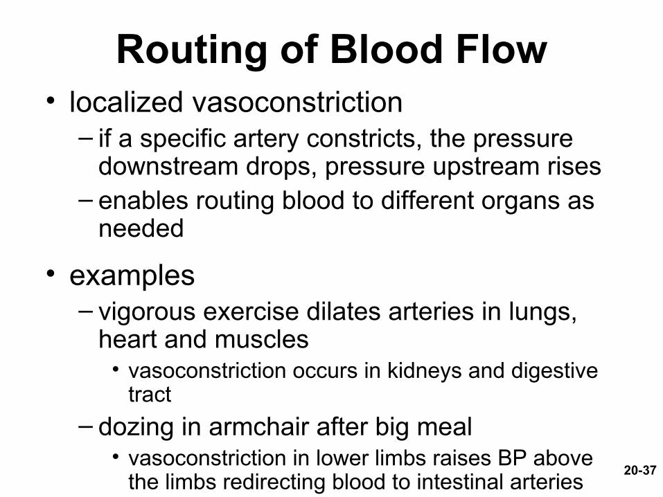
Routing of Blood Flow• localized vasoconstriction
– if a specific artery constricts, the pressure downstream drops, pressure upstream rises
– enables routing blood to different organs as needed
• examples– vigorous exercise dilates arteries in lungs,
heart and muscles• vasoconstriction occurs in kidneys and digestive
tract
– dozing in armchair after big meal• vasoconstriction in lower limbs raises BP above
the limbs redirecting blood to intestinal arteries
20-38
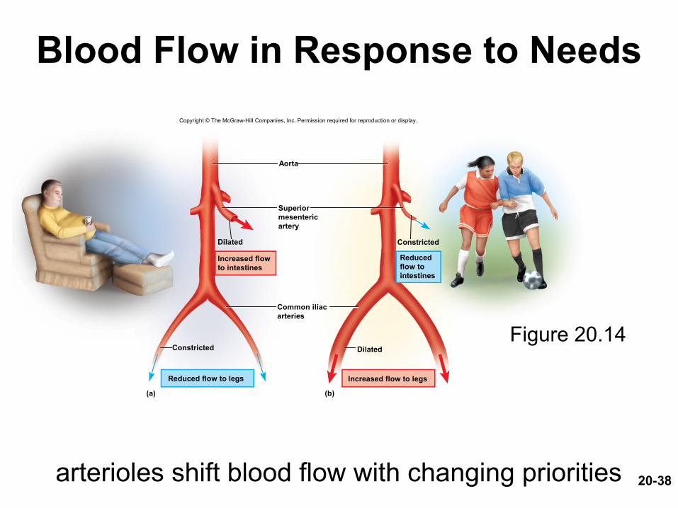
Blood Flow in Response to Needs
arterioles shift blood flow with changing priorities
Copyright © The McGraw-Hill Companies, Inc. Permission required for reproduction or display.
Constricted
Dilated
Aorta
(a) (b)
Constricted
Dilated
Reduced flow to legs
Superiormesentericartery
Increased flowto intestines
Common iliacarteries
Reducedflow tointestines
Increased flow to legs
Figure 20.14
20-39
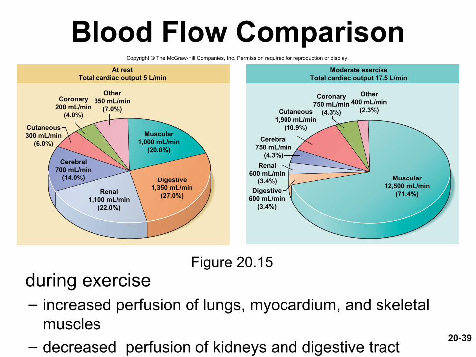
Blood Flow Comparison
during exercise– increased perfusion of lungs, myocardium, and skeletal
muscles – decreased perfusion of kidneys and digestive tract
Figure 20.15
Copyright © The McGraw-Hill Companies, Inc. Permission required for reproduction or display.
At restTotal cardiac output 5 L/min
Other350 mL/min
(7.0%)
Coronary200 mL/min
(4.0%)
Cutaneous300 mL/min
(6.0%)
Muscular1,000 mL/min
(20.0%)
Digestive1,350 mL/min
(27.0%)
Cerebral700 mL/min
(14.0%)
Renal1,100 mL/min
(22.0%)
Moderate exerciseTotal cardiac output 17.5 L/min
Other400 mL/min
(2.3%)
Coronary750 mL/min
(4.3%)Cutaneous1,900 mL/min
(10.9%)
Cerebral750 mL/min
(4.3%)
Renal600 mL/min
(3.4%)
Digestive600 mL/min
(3.4%)
Muscular12,500 mL/min
(71.4%)
20-40
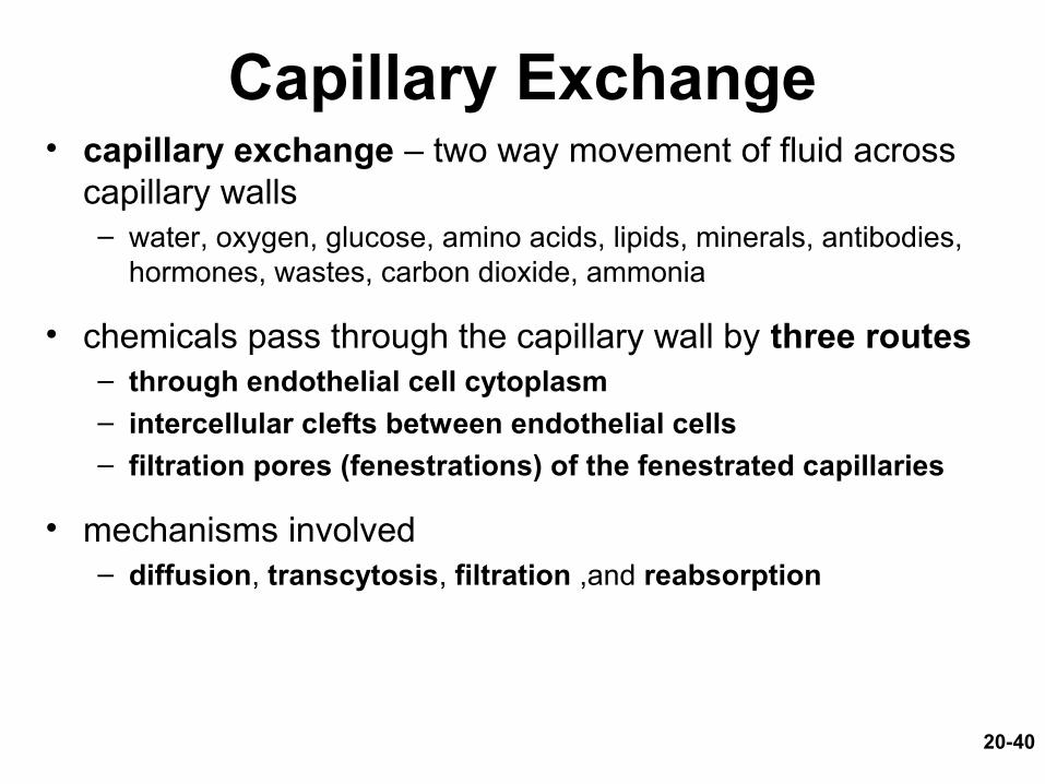
Capillary Exchange• capillary exchange – two way movement of fluid across
capillary walls– water, oxygen, glucose, amino acids, lipids, minerals, antibodies,
hormones, wastes, carbon dioxide, ammonia
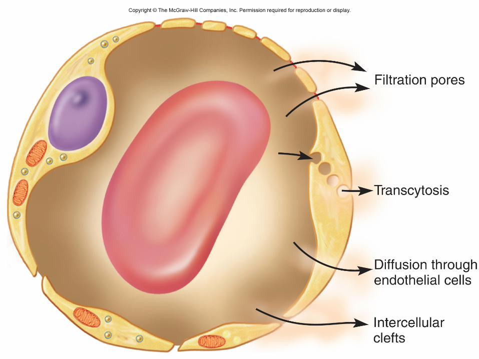
• chemicals pass through the capillary wall by three routes– through endothelial cell cytoplasm– intercellular clefts between endothelial cells– filtration pores (fenestrations) of the fenestrated capillaries
• mechanisms involved– diffusion, transcytosis, filtration ,and reabsorption
20-41
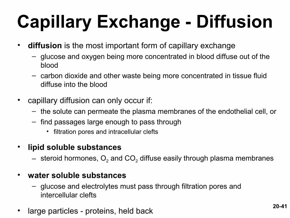
Capillary Exchange - Diffusion• diffusion is the most important form of capillary exchange
– glucose and oxygen being more concentrated in blood diffuse out of the blood
– carbon dioxide and other waste being more concentrated in tissue fluid diffuse into the blood
• capillary diffusion can only occur if:– the solute can permeate the plasma membranes of the endothelial cell, or– find passages large enough to pass through
• filtration pores and intracellular clefts
• lipid soluble substances– steroid hormones, O2 and CO2 diffuse easily through plasma membranes
• water soluble substances– glucose and electrolytes must pass through filtration pores and
intercellular clefts
• large particles - proteins, held back

Capillary Exchange - Transcytosis• endothelial cells pick up material on one side of the plasma
membrane by pinocytosis or receptor-mediated endocytosis, transport vesicles across cell, and discharge material on other side by exocytosis
• important for fatty acids, albumin and some hormones (insulin)
20-44
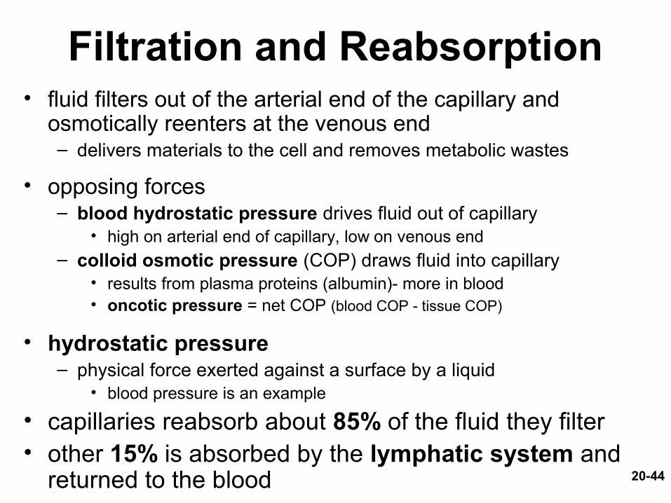
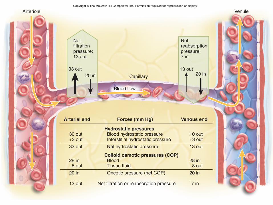
Filtration and Reabsorption• fluid filters out of the arterial end of the capillary and
osmotically reenters at the venous end– delivers materials to the cell and removes metabolic wastes
• opposing forces– blood hydrostatic pressure drives fluid out of capillary
• high on arterial end of capillary, low on venous end– colloid osmotic pressure (COP) draws fluid into capillary
• results from plasma proteins (albumin)- more in blood• oncotic pressure = net COP (blood COP - tissue COP)
• hydrostatic pressure– physical force exerted against a surface by a liquid
• blood pressure is an example
• capillaries reabsorb about 85% of the fluid they filter• other 15% is absorbed by the lymphatic system and
returned to the blood
20-46
Variations in Capillary Activity• capillaries usually reabsorb most of the fluid they
filter – exception:– kidney capillaries in glomeruli do not reabsorb– alveolar capillaries in lung absorb completely to keep
fluid out of air spaces
• capillary activity varies from moment to moment– collapsed in resting tissue, reabsorption predominates
since BP is low– metabolically active tissue has increase in capillary flow
and BP • increase in muscular bulk by 25% due to accumulation of fluid
20-47
Edema
• edema – the accumulation of excess fluid in a tissue– occurs when fluid filters into a tissue faster than it is
absorbed
• three primary causes– increased capillary filtration
• kidney failure, histamine release, old age, poor venous return
– reduced capillary absorption• hypoproteinemia, liver disease, dietary protein deficiency
– obstructed lymphatic drainage• surgical removal of lymph nodes

20-48
Consequences of Edema
• tissue necrosis– oxygen delivery and waste removal impaired
• pulmonary edema– suffocation threat
• cerebral edema– headaches, nausea, seizures, and coma
• severe edema or circulatory shock– excess fluid in tissue spaces causes low blood volume
and low blood pressure

20-49
Mechanisms of Venous Return• venous return – the flow of blood back to the heart
– pressure gradient • blood pressure is the most important force in venous return• 7-13 mm Hg venous pressure towards heart• venules (12-18 mm Hg) to central venous pressure – point where the
venae cavae enter the heart (~5 mm Hg)
– gravity drains blood from head and neck
– skeletal muscle pump in the limbs• contracting muscle squeezed out of the compressed part of the vein
– thoracic (respiratory) pump• inhalation - thoracic cavity expands and thoracic pressure decreases,
abdominal pressure increases forcing blood upward– central venous pressure fluctuates
• 2mm Hg- inhalation, 6mm Hg-exhalation
• blood flows faster with inhalation
– cardiac suction of expanding atrial space

20-50
Skeletal Muscle Pump
Figure 20.19 a-b
Copyright © The McGraw-Hill Companies, Inc. Permission required for reproduction or display.
To heart
Valve open
Valve closed
(a) Contracted skeletal muscles (b) Relaxed skeletal muscles
Venousblood
20-51
Venous Return and Physical Activity• exercise increases venous return in many ways:
– heart beats faster, harder increasing CO and BP– vessels of skeletal muscles, lungs, and heart dilate and
increase flow– increased respiratory rate, increased action of thoracic
pump– increased skeletal muscle pump
• venous pooling occurs with inactivity– venous pressure not enough force blood upward– with prolonged standing, CO may be low enough to
cause dizziness• prevented by tensing leg muscles, activate skeletal muscle
pump
20-52
Special Circulatory Routes- Brain• total blood flow to the brain fluctuates less than that of any
other organ (700 mL/min)– seconds of deprivation causes loss of consciousness – 4-5 minutes causes irreversible brain damage– blood flow can be shifted from one active brain region to another
20-53
TIAs and CVAs• transient ischemic attacks (TIAs ) – brief episodes of
cerebral ischemia– caused by spasms of diseased cerebral arteries– dizziness, loss of vision, weakness, paralysis, headache or
aphasia– lasts from a moment to a few hours– often early warning of impending stroke
• stroke - cerebral vascular accident (CVA)– sudden death of brain tissue caused by ischemia
• atherosclerosis, thrombosis, ruptured aneurysm
– effects range from unnoticeable to fatal• blindness, paralysis, loss of sensation, loss of speech common
– recovery depends on surrounding neurons, collateral circulation
20-54
Special Circulatory Routes Skeletal Muscle
• highly variable flow depending on state of exertion• at rest:
– arterioles constrict
– most capillary beds shut down
– total flow about 1L/min
• during exercise:– arterioles dilate in response to epinephrine and sympathetic
nerves
– precapillary sphincters dilate due to muscle metabolites like lactic acid, CO2
– blood flow can increase 20 fold
• muscular contraction impedes flow– isometric contraction causes fatigue faster than intermittent
isotonic contractions
20-55
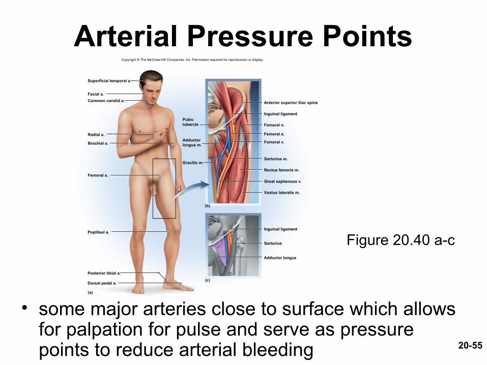
Arterial Pressure Points
• some major arteries close to surface which allows for palpation for pulse and serve as pressure points to reduce arterial bleeding
Figure 20.40 a-c
Superficial temporal a.
Facial a.
Common carotid a.
Radial a.
Brachial a.
Femoral a.
Popliteal a.
Dorsal pedal a.
Posterior tibial a.
Inguinal ligament
Anterior superior iliac spine
Inguinal ligament
Femoral n.
Femoral a.
Sartorius m.
Rectus femoris m.
(b)
(c)
(a)
Sartorius
Adductor longus
Gracilis m.
Pubictubercle
Adductorlongus m.
Femoral v.
Great saphenous v.
Vastus lateralis m.
Copyright © The McGraw-Hill Companies, Inc. Permission required for reproduction or display.
20-56
Hypertension• hypertension – most common cardiovascular disease
affecting about 30% of Americans over 50
• “the silent killer”– major cause of heart failure, stroke, and kidney failure
• damages heart by increasing afterload– myocardium enlarges until overstretched and inefficient
• renal arterioles thicken in response to stress– drop in renal BP leads to salt retention (aldosterone) and worsens
the overall hypertension
• primary hypertension– obesity, sedentary behavior, diet, nicotine
• secondary hypertension – secondary to other disease – kidney disease, hyperthyroidism
20-57
Blood Pressure Drugs• Beta blocker– Inhibit beta adrenergic receptors
• Block effects of epinephrine• Resting – heart reduces output lowering blood pressure
• Calcium channel blocker– Reduce intracellular calcium in heart muscle and smooth muscle in
blood vessels• Reduced cardiac output and dilation of blood vessels