Embed Size (px)
Citation preview

Anatomy of Articulation and ResonationCSDI 4037/5037
1

Overview
• Bones in the facial skeleton
• Muscles of the mastication
• Cavities of the vocal tract
2

Bones in the Facial Skeleton
• Mandible• Maxillae• Zygomatic bone• Nasal bone• Palatine bone and nasal conchae• Lacrimal bone• Vomer
3

Mandible
• The mandible forms lower portion of the jaw.
• Anterior view:
• Alveolar margin (border) – Superior aspect of the bone that contains the sockets (alveoli) for the teeth.
• Mental protuberance – raised, triangular shaped area where two halves of the bone have fused
Alveolar margin
Mental protuberance
4

Mandible
• Mental tubercle – raised bases of the mental protuberance
• Mental symphysis – slight, vertical ridge above the protuberance that was formed by the fusion of the right and left halves of the mandible
• Mental foramen –hole on the anterior aspect of the bone below the second premolar.
Mental tubercle
Mental symphysis
Mental foramen
5

Mandible
• Lateral view • Ramus – vertical portion of the
bone
• Body – horseshoe-shaped portion of the bone
• Angle- region of the bone where ramus and body join
• Condylar process – posterior extension of the ramus
• Coronoid process –sharply angled anterior extension of the ramus
6

Mandible
• Lateral view• Mandibular condyle –
rounded head of the condylar process; articulates with the mandibular process of the temporal bone
• Mandibular notch - concavity located between the condylar and coronoid process.
Mandibular notch
Mandibular condyle
7

Mandible
• Inner surface of the mandible
• Mylohyoid line• The mandibular foramen
is the conduit for the inferiror alveolar nerve of V trigeminal, providing sensory innervation for the teeth and gums.
Mandibular notch
Mandibular condyle
8

Maxilla Bone• The maxilla bone is the facial
bone that is positioned between the orbits, nose, and mouth. It forms upper portion of the jaw.
• Anterior view:• Alveolar process – inferior
extension that contains the sockets for the teeth.
• Infraorbital foramen – prominent hole located in the inferior to the orbit.
• Frontal process – narrow extension that projects toward the frontal bone
• Zygomatic process – extension that project toward the zygomatic bone.
9
Alveolar process
Frontal process
Infraorbital foramen
Zygomatic process

Maxilla
• Inferior orbital fissure – narrow, elongated opening that runs diagonally along the floor of the orbit, serves as a passageway for the maxillary nerve and infraorbital vessels
10
Inferior orbital fissure

Maxilla
• Inferior view • Incisive foramen – depression found posterior to the incisors; forms a passageway for nasopalatine vessels
• Palatine process – thin plate that forms part of the roof of the mouth (or hard palate)
• Two palatine processes of the maxilla articulate at the intermaxillary suture
11

Zygomatic bone
• The zygomatic is the facial bone that forms the cheek bone
• Frontal process – narrow extension that projects toward the frontal bone
• Temporal process – narrow extension that joins with the zygomatic process of the temporal
bone to form the zygomatic arch.• Maxillary process – narrow
extension that medially toward the maxilla.
12

Nasal bones• The nasal bones are
small, making up the superior nasal surface.
• They form the bridge of the nose.
• The nasal bones articulates with the frontal bones superiorly, the maxilla laterally, and the perpendicular plate of the ethmoid bone and the nasal septal cartilage.13

Lateral View
14

Palatine bone
• Together, the two L-shaped palatines form the posterior floor of the nasal cavity.
• Horizontal plate: portion of the bone that articulates with the palatine process of the maxilla bone; forms the posterior portion of the hard palate.
• Perpendicular plate: superior projection; forms a portion of the nasal cavity lateral wall.
15

Palatine bone
• Greater palatine foramen: largest opening along the lateral edge of horizontal plate; serves as a passageway for greater palatine nerve
• Lesser palatine foramina: two or three small openings located posterior to the greater palatine foramen; serve as passageways for branches of the lesser palatine nerve.
16
Greater palatine foramen
Lesser

Inferior nasal conchae
• They are small, scroll-like bones located on the lateral surface of the nasal cavity.
• They articulate with the maxilla, palatine, and ethmoid bone
• The middle and superior nasal conchae processes of the ethmoid bone
17

Vomer
• The vomer is an unpaired, midline bone making up the inferior and posterior nasal septum
• Joins with the perpendicular plate of the ethmoid bone to form the bony septum that divides nasal cavity into right and left halves.
18

Vomer
• It articulates with the sphenoid rostrum and perpendicular plate of the ethmoid bone in the posterior –superior, and with the maxillae and palatine bones on the inferior margin.
19

Lacrimal bones
• The small lacrimal bones are almost completely hidden in the intact skull.
• They articulate with the maxillae, frontal bone, nasal bone, and inferior conchae.
• Lacrimal fossa: depression at the junction of the lacrimal and maxilla bones that holds the lacrimal sac; tears formed by the sac drain through a duct into the nasal cavity.
20
Lacrimal fossa

Ethmoid bone• Irregular shaped cranial bone
found at the top of the nasal cavity; bony projections from ethmoid extend into the nasal cavity, orbits and cranium.
• Orbital plate: portion of the bone that forms the medial side of the orbit.
• Perpendicular plate: very thin vertical plate that extends down into the nasal cavity and joins the vomer bone below.
21

Ethmoid bone
• Cribriform plate: thin horizontal plate that articulates with the frontal bone; numerous holes in the plate form passageways for olfactory nerves through the cranium.
• Crista galli: a thin vertical plate that extends up from the cribriform plate
22

Lateral View
23

Muscles of Mastication
• The process of chewing food, or mastication, requires the movement of mandible.
• The muscles of mastication include– The mandibular elevators– Muscles of protrusion– The mandibular depressors

Muscles of Mastication• Mandibular elevators
• Masseter• Temporalis• Medial pterygoid
• Mandible protruders• Lateral pterygoid
• Mandibular depressors• Digastricus• Mylohyoid muscle• Geniohyoid bone
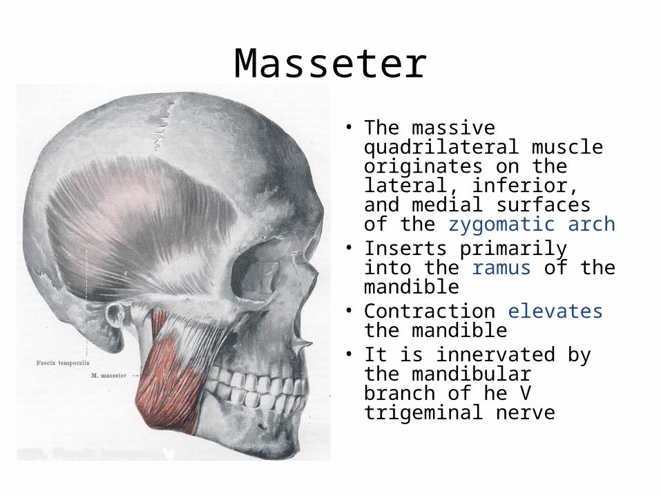
Masseter• The massive quadrilateral
muscle originates on the lateral, inferior, and medial surfaces of the zygomatic arch
• Inserts primarily into the ramus of the mandible
• Contraction elevates the mandible
• It is innervated by the mandibular branch of he V trigeminal nerve
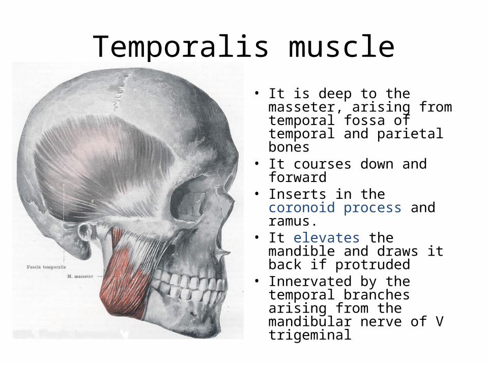
Temporalis muscle• It is deep to the masseter,
arising from temporal fossa of temporal and parietal bones
• It courses down and forward
• Inserts in the coronoid process and ramus.
• It elevates the mandible and draws it back if protruded
• Innervated by the temporal branches arising from the mandibular nerve of V trigeminal

Medial pterygoid muscle• It originates from the
medial pterygoid plate and fossa lateral to it.
• It courses down and back to insert into the mandibular ramus
• It elevates the mandible, acting in conjunction with the masseter; individually, they move mandible side to side.
• It is innervated by the mandibular division of the V trigeminal nerve.

Lateral pterygoid muscle• One head of the lateral
pterygoid arises from the lateral pterygoid plate; another head attaches to the greater wing of the sphenoid.
• It courses back to insert into the lower inner margin of the condyloid process.
• Its contraction protrude the mandible, and it works in contrast with the mandibular elevators.
• It is innervated by the mandibular branch of the V trigeminal nerve.

Digastricus• The digastricus anterior
originates on the inner surface of the mandible at the digastricus fossa
• The digastricus posterior originates on the mastoid process of the temporal bone.
• If the hyoid bone is fixed, contraction of the anterior component will result in depression of the mandible

Digastricus
• The anterior belly is innervated by the mandibular branch of the V trigeminal nerve.
• The posterior belly is supplied by the digastric branch of the VII facial nerve.

Mylohyoid muscle• The fanlike muscle courses
from the mylohyoid line of the mandible to the hyoid bone inferiorly.
• The mylohyoid depresses the mandible
• It is innervated by the alveolar nerve, arising from the V trigeminal nerve, mandibular branch.

Geniohyoid muscle
• It originates at the mental spines of the mandible and projects parallel to the anterior digastricus and inserts into the corpus hyoid.
• Contarction depresses the mandible
• Innervation is by means of the XII hypoglossal nerve.

Lips
• Covered externally by skin and internally by the mucous membrane
• Has four layers of tissues namely cutaneous, muscular, glandular, and mucous
• The skin terminates as a well-defined line called the cupids bow
34

Lips
• The vermilion zone is the transitional area between the skin and the mucous membrane
• The vertical groove is called philtrum while the slight projection at the end of the philtrum on the upper lip is the tubercle
• The lower lips are more mobile and contribute for the bilabial and labiodental sounds
35

Palate
• Soft Palate (velum): – attached to the posterior free border of the
palatine bones– This attachment is via the palatal aponeurosis– Its muscle fibers allow it to be lowered, elevated
or tensed. At rest, it is directed posteriorly and hangs like a curtain into the oropharynx
36