Embed Size (px)
Citation preview

ANATOMY
ANATOMY AND
MORPHOLOGY OF STEM
(PRIMARY)

ANATOMY OF STEM
(Primary)
Learning outcomes
Explain stem and bud parts, types,
and structural types
Explain the epidermis, ground tissue,
and vascular tissues in stem

STEM
MORPHOLOGY
3
MORPHOLOGY-STEM
AND BUDS
1. STEM
a. Stem Parts
i. Twig Surface Parts
Bud- an immature shoot.
Internode- a section or region of
stem between nodes.
Leaf scar - a mark indicating
former place of attachment of
petiole or leaf base.
4


MORPHOLOGY-STEM
Lenticel- a pore in the bark, as
breathing pores for gas exchange.
Node- region of stem from which
a leaf, leaves, or branches arise.
Prickle- a sharp-pointed out-
growth from the epidermis or
cortex of any organ.
Stipular scar - a mark indicating
former place of attachment of
stipule.
6

MORPHOLOGY-STEM
7

MORPHOLOGY-STEM
Terminal bud scale scar rings -
several marks in a ring indicating
former places of attachment of
bud scales.
Vascular bundle or trace scar -
a mark indicating former place of
attachment within the leaf scar of
the vascular bundle or trace.
8

MORPHOLOGY - STEM
Bark –tissues of plant outside
wood or xylem.
Pith – centermost tissue of stem,
usually soft. Not always visible
in older wood.
Wood – xylem consisting of vessels
and /or tracheids, fibers and
parenchyma cells.
ii. Major stem parts
9

MORPHOLOGY-STEM
Stem Types
(Classification based on
direction of growth
10

11
Arborescent – treelike in
appearance and size.

12
Climbing- growing upward by
means of tendrils, petioles or
adventitious root.

13
Decumbent – reclining or lying
on the ground with tips
ascending.

ANATOMY OF STEM (Primary)
⚫ Introduction
⚫ The stem, the part of the primary body, derived from the shoot apical meristem.
⚫ Meristems form the new cells of a plant and the apical meristem occur at the tip of the shoot and provides primary growth.
ANATOMY OF STEM (Primary)
⚫ The shoot meristem produces the cells that will form the major tissues of new stem and leaf:
⚫ i. Protoderm, epidermal cells that are still meristematic and in the early stages of differentiation.
⚫ ii. Ground meristem, young cells of pith and cortex.
⚫ iii. Provascular tissues, young cells of xylem and phloem. Fig. 1 and 2.

ANATOMY OF STEM (Primary)
Fig. 1
Apical meristem
of shoot.

ANATOMY OF STEM (Primary)
Fig. 2.
Stem near
meristem
1.& 2.Ground
meristem
3.Provascular
4.Protoderm

ANATOMY OF STEM (Primary)
⚫ Epidermis
⚫ Outermost surface, a layer of epidermal cell.
⚫ Prevent the lost of water, barrier against invasion by bacteria, fungi, and small insects.
⚫ Outer wall with cutin (fatty substance, impermeable to water), become a layer, cuticle.
⚫ A layer of wax may be present outside the cuticle. Fig. 3.

ANATOMY OF STEM (Primary)
Fig. 3.
Epidermis with
cuticle.

ANATOMY OF STEM (Primary)
⚫ Ground Tissue
⚫ Interior to the epidermis, simple or
complex.
⚫ Photosynthetic parenchyma and
sometimes collenchyma in simple cortex.
⚫ Complex cortex, presence of laticifers,
resin duct, calcium oxalate.
⚫ Fig. 4 to 7.

ANATOMY OF STEM (Primary)
Fig. 4
Helianthus,
Eudicot.
(sunflower)
Fig. 5. Zea mays. Monocot (corn)
Ground tissue

ANATOMY OF STEM (Primary)
Fig. 6. Laticifer,
Nerium oleander.
Fig. 7. Calcium oxalate,
Mangifera indica (Mango)
Ground tissue

ANATOMY OF STEM (Primary)
⚫ Vascular Tissues
⚫ Two types: Xylem and Phloem.
⚫ Xylem: Tracheary elements plus some parenchyma and sometimes sclerenc-hyma.
⚫ Two types of tracheary elements:
⚫ Tracheids and vessel members.
⚫ Fig. 8 to 10.

ANATOMY OF STEM (Primary)
Fig. 8
Tracheary
element with
secondary wall.
1. Parenchyma
2. Annular
3. Scalariform
4. Reticulate
5. Pitted

ANATOMY OF STEM (Primary)
Fig. 9. Tracheary element in the stem
of Mangifera indica (mango) x 1,500.
Reticulate
Annular
ANATOMY OF STEM (Primary)
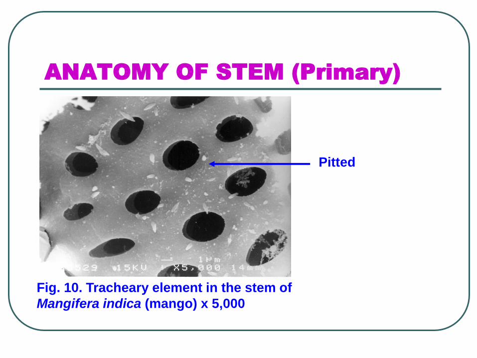
Fig. 10. Tracheary element in the stem of
Mangifera indica (mango) x 5,000
Pitted

ANATOMY OF STEM (Primary)
⚫ Phloem: sieve elements plus some
parenchyma and often some
sclerenchyma.
⚫ Two types of sieve elements:
⚫ Sieve cells and sieve tube members.

ANATOMY OF STEM (Primary)
⚫ Vascular bundles (V.B.)
⚫ Xylem and phloem occur together as vascular bundles.
⚫ Located just interior to the cortex.
⚫ In monocots, V.B. are distributed as a complex network throughout the inner part of the stem, between the bundles are parenchyma cells.

ANATOMY OF STEM (Primary)
⚫ In eudicots, V.B. are arranged in one ring
surrounding the pith, a region of
parenchyma similar to the cortex.
⚫ Vascular bundles are collateral when the
phloem is on the outside and the xylem
towards the inside. In some species the
bundles are bicollateral, xylem between
outer and inner phloem.
⚫ Fig. 11 and 12.

ANATOMY OF STEM (Primary)
Fig. 11. Collateral vascular
bundle.
ANATOMY OF STEM (Primary)
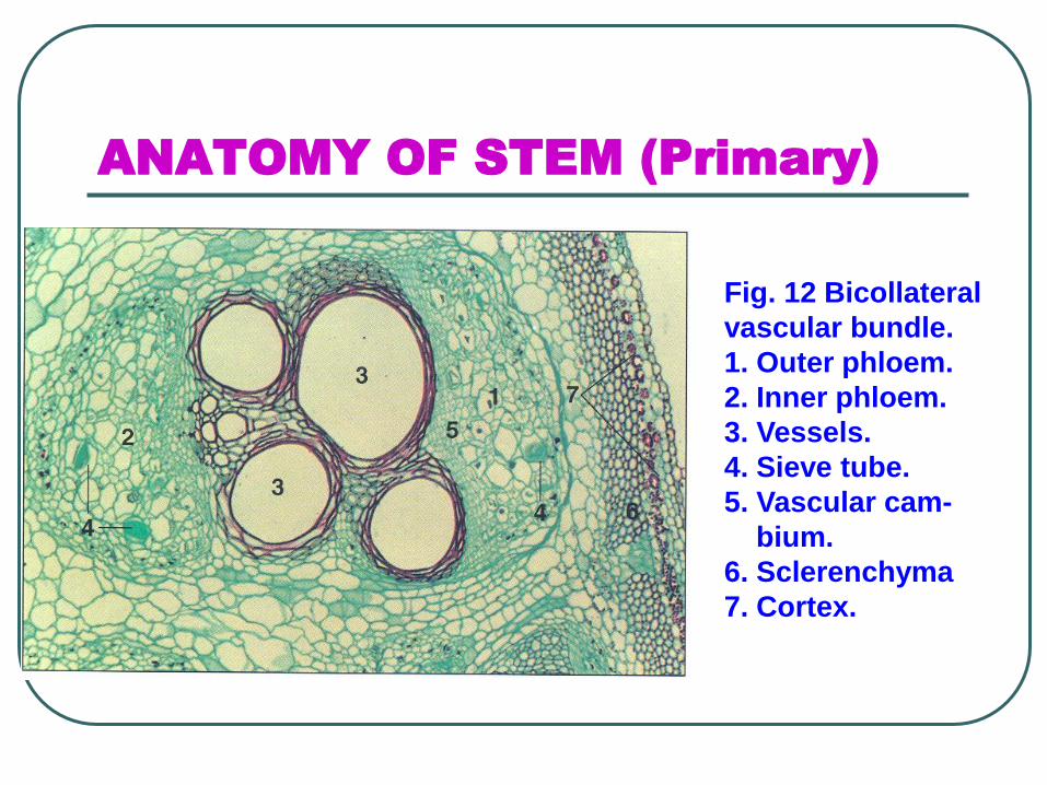
Fig. 12 Bicollateral
vascular bundle.
1. Outer phloem.
2. Inner phloem.
3. Vessels.
4. Sieve tube.
5. Vascular cam-
bium.
6. Sclerenchyma
7. Cortex.