Embed Size (px)
Citation preview

Semmelweis University Department of Anatomy, Histology and Embryology
Faculty of Medicine 1st year
ANATOMY HANDBOOK
Dr. Andrea D. Székely Associate Professor
Course Director of the English Language Program
Dr. Ágoston Szél Full Professor
Head of Department Rector of the Semmelweis University
Anatomy, Histology and Embryology for EM students
TEACHING DEPARTMENT: SEMMELWEIS UNIVERSITY Department of Anatomy, Histology and Embryology Budapest, Tűzoltó utca 58. H-1094 Budapest
LEARNING OBJECTIVES
Aims of the lectures in anatomy: Presentation of the important and/or complicated chapters such as
introductory chapters, thorax, pelvis, hand, foot, skull, heart, chapters of the visceral organs, central nervous
system, organs of special senses, topographical anatomy.
Aims of the lectures in cell biology and histology: Presentation of the cell, basic principles in cell
biology (mitosis, cytoskeleton, cellular motility), detailed presentation of the basic tissues (epithelial,
connective, muscle and nervous). Completing the gross anatomy with the detailed presentation of the fine
structure of the organs, including the ultrastructural details as well as the molecular arrangement. Important
chapters: basic tissues, viscera, central nervous system.
Aims of the lectures in embryology: Presentation of the early development from the differentiation of
the germ cells to the formation of the human embryo (general embryology). Presentation of the
development of the organs and functional systems parallel with the gross anatomical and histological
lectures including the frequently occurring malformations.
Aims of the practical sessions in the dissecting room: Based on the weekly programs the students
study the preparations (bones, joints, muscles, viscera, brain) and dissect (parts of or an entire cadaver). They
are aided by the lab instructors. Bones, joints, muscles and peripheral nervous system will be taught primarily
in the dissecting room.
Aims of the practical sessions in the histology room: Facilitate the understanding of the basic tissues
(epithelial, connective, muscle and nervous) and the fine structure of the organs through the observation and
interpretation of histological specimens.
Discussion of the more complicated chapters of the embryology is presented on small group
discussions connected to the practical sessions in the dissecting room.
The knowledge of the students will be checked by mid-term tests.
Lectures: first semester: 3x 45 min; second semester: 3x 45 min; third semester: 3x 45 min; fourth
semester: 1x 45 min.
Topics of the lectures:
First semester: Gross anatomy of the bones, joints and muscles, basic cytology, basic histology, basic
embryology, development of the skull, spine and limbs.
Second semester: Heart and vessels, lymphatic organs, viscera and body cavities; integrated gross
anatomy, cytology, histology and embryology.
Third semester: Central and peripheral nervous system, organs of special senses, endocrine organs;
integrated gross anatomy, cytology, histology and embryology.
Fourth semester: Topographical anatomy of the head, neck and body cavities (thorax, abdomen,
pelvis), cross sectional anatomy.
Practical course
First semester: 6x 45 min; second semester: 6x 45 min;
third semester: 4x 45 min; fourth semester: 2x 45 min;
First semester: Gross anatomy of the bones, joints and muscles, basic cytology, basic histology, basic
embryology, development of the skull, spine and limbs.
Second semester: Heart and vessels, lymphatic organs, viscera, topography of body cavities; integrated
gross anatomy, cytology, histology and embryology. Topographical anatomy of the body cavities.
Third semester: Central and peripheral nervous system, organs of special senses, endocrine organs;
integrated gross anatomy, cytology, histology and embryology. Topographical anatomy of the dorsal regions
of limbs and the trunk, including spinal cord.
Fourth semester: Topographical anatomy of the head, neck and body cavities (thorax, abdomen,
pelvis), cross sectional anatomy. Review of the subjects taught and studied during the four semesters.
Type of exams: first semester: semifinal; second semester: semifinal; third semester: semifinal; fourth
semester: final exam from the subjects of the four semesters..
ECTS credits: four semesters together: 27 (first semester: 8; second semester: 9; third semester: 7;
fourth semester: 3)
EM I ANNOUNCEMENTS
Evaluation is made using a five-grade scale (1-5).
Signing of the lecture book: active participation in lectures, dissection room and histology lab sessions
is obligatory. Students should attend at least 75% of the scheduled hours to gain a signature proving the
validity of the semester. Absences are therefore limited in 25%.
Midterm examinations: During the semester, both practical and theoretical knowledge will regularly
be evaluated. The midterm tests are obligatory and cannot be done at a different time, neither can they be
retaken. The anatomy and histology mid-terms may be oral or written exams. Anatomy mid-terms include
both identification of several structures on the specimen and theoretical questions related to the subject.
Histology midterms include the identification of a certain number of structures in slides, as well as,
theoretical questions related to general embryology. The results of all tests will appear on the personal
achievement cards.
A semester practical mark is calculated from the midterm marks together with the personal
achievement mark given by the group instructor. This practical mark will be counted into the semifinal
examination and will be written on the personal achievment cards.
Semifinal examinations are composed of the following parts:
1. written pretest,
2. oral examination - practical and theoretical questions in Macroscopy and Histology, including the
identification/description prosections and two histological specimen. Old curriculum students will
have an extra question concerning cell biology.
Notebooks should be used regularly in histology lab sessions in order to prepare schematic drawings of
the histological specimens. Students will be asked to present their histology notebooks, containing the
drawings, during the semifinal exams.
N.B. – In case, neither the first nor the repeated takes of a semifinal exam have been successful and so
the exam has to be postponed to the following, exam period (i.e. ’CV’ exam), only those students will be
allowed to go for a CV exam, parallel to continuing their studies whose average score of the midterm tests) is
equal, or higher than 2.00.
RULES AND REGULATIONS IN THE DISSECTING ROOM IT IS STRICTLY FORBIDDEN to eat, drink, smoke, to chow gums, or to use music devices or phones. Bags and coats should be left in the lockers before entering the dissecting room. The lockers will have to be locked using your padlocks. Please, remember to keep your valuables always on you, or lock them in the lockers since the department takes no responsibility for lost items.
Students are expected to be prepared for the practical work. Everybody is supposed to behave in the
dissecting room conforming to the spirit of the site. Loud speech, out-of-place jokes and any kind of
behaviour, disregarding the dignity of human corpses, should strictly be avoided.
The dissection room is a hazard area. Fire and work safety regulations should be maintained. Cleanliness
and order should be kept. Students should take care of the furniture and equipment of the dissecting room.
Do not sit on the dissection tables or stand on the tripod stools to avoid accidents.
Working in the dissection room involves the use of sharp and pointed tools, injuries should be reported to
the lab instructor. The technical personnel will provide first aid when necessary.
The white lab coats should be worn while in the dissection room, but should be removed before leaving the
dissection room area. The purpose of wearing the lab coats is to protect one’s clothing from contacting the
cadaver specimen. Furthermore we advise you to wear closed toed shoes and clothing covering the legs. In
the end of the class, lab coats should be emptied and left in order on the coat hangers. The department is not
responsible for valuables left in the dissecting room.
Only the members of the study group can participate in the sessions, visitors may be present only with prior
permission by the lab instructor. Students can leave the sessions only with the approval of the lab instructor.
Photos, or videos can only be made of line drawings with the agreement and in the presence of the lab
instructor.
Specimen preparations should be wrapped and labeled. Dissection materials of other groups or individuals
should not be handled. Dissected cadaver pieces should be discarded in a designated container and discarded
blades have to be collected separately in a specifically labeled container.
Dissecting rooms are closed between 6:00 PM to 8:00 AM and over the weekends. Students may not stay in
the dissecting room without the supervision of one of the assistants of the department. In the absence of an
instructor, the technical personnel should ask the students to leave the dissecting room.
SMOKING IS STRICTLY FORBIDDEN
ON THE DEPARTMENTAL PREMISES,
INCLUDING THE GARDEN AND THE YARD!
EM I.
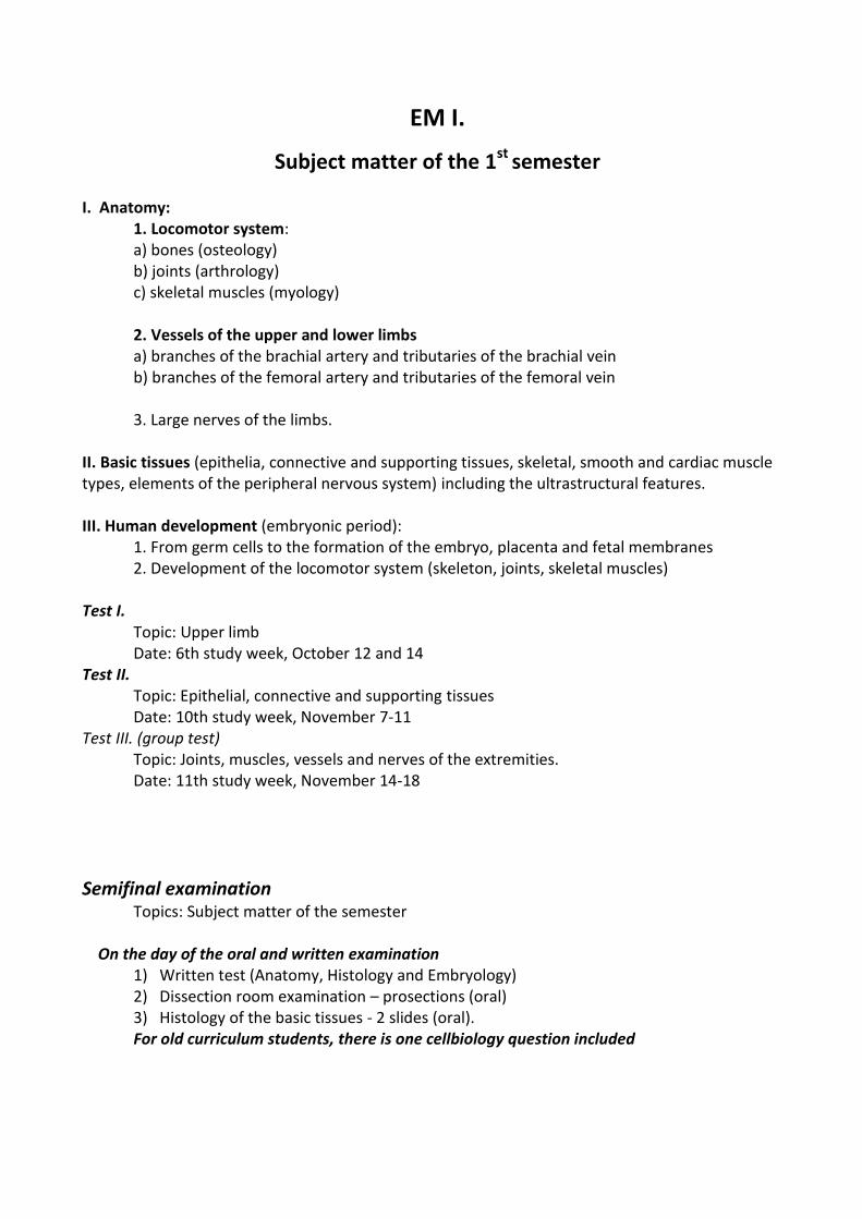
Subject matter of the 1st semester I. Anatomy: 1. Locomotor system: a) bones (osteology) b) joints (arthrology) c) skeletal muscles (myology) 2. Vessels of the upper and lower limbs a) branches of the brachial artery and tributaries of the brachial vein b) branches of the femoral artery and tributaries of the femoral vein 3. Large nerves of the limbs. II. Basic tissues (epithelia, connective and supporting tissues, skeletal, smooth and cardiac muscle types, elements of the peripheral nervous system) including the ultrastructural features. III. Human development (embryonic period): 1. From germ cells to the formation of the embryo, placenta and fetal membranes 2. Development of the locomotor system (skeleton, joints, skeletal muscles) Test I. Topic: Upper limb Date: 6th study week, October 12 and 14 Test II. Topic: Epithelial, connective and supporting tissues Date: 10th study week, November 7-11 Test III. (group test) Topic: Joints, muscles, vessels and nerves of the extremities. Date: 11th study week, November 14-18
Semifinal examination Topics: Subject matter of the semester On the day of the oral and written examination
1) Written test (Anatomy, Histology and Embryology) 2) Dissection room examination – prosections (oral) 3) Histology of the basic tissues - 2 slides (oral). For old curriculum students, there is one cellbiology question included
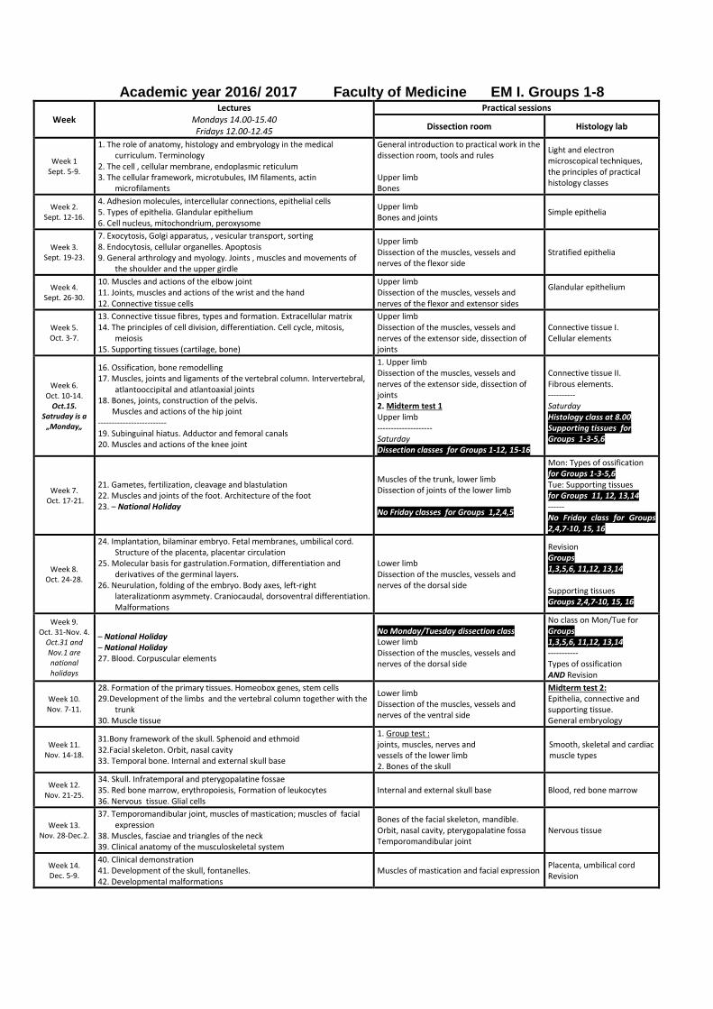
Academic year 2016/ 2017 Faculty of Medicine EM I. Groups 1-8
Week Lectures
Mondays 14.00-15.40 Fridays 12.00-12.45
Practical sessions
Dissection room Histology lab
Week 1 Sept. 5-9.
1. The role of anatomy, histology and embryology in the medical curriculum. Terminology
2. The cell , cellular membrane, endoplasmic reticulum 3. The cellular framework, microtubules, IM filaments, actin
microfilaments
General introduction to practical work in the dissection room, tools and rules Upper limb Bones
Light and electron microscopical techniques, the principles of practical histology classes
Week 2. Sept. 12-16.
4. Adhesion molecules, intercellular connections, epithelial cells 5. Types of epithelia. Glandular epithelium 6. Cell nucleus, mitochondrium, peroxysome
Upper limb Bones and joints
Simple epithelia
Week 3. Sept. 19-23.
7. Exocytosis, Golgi apparatus, , vesicular transport, sorting 8. Endocytosis, cellular organelles. Apoptosis 9. General arthrology and myology. Joints , muscles and movements of
the shoulder and the upper girdle
Upper limb Dissection of the muscles, vessels and nerves of the flexor side
Stratified epithelia
Week 4. Sept. 26-30.
10. Muscles and actions of the elbow joint 11. Joints, muscles and actions of the wrist and the hand 12. Connective tissue cells
Upper limb Dissection of the muscles, vessels and nerves of the flexor and extensor sides
Glandular epithelium
Week 5. Oct. 3-7.
13. Connective tissue fibres, types and formation. Extracellular matrix 14. The principles of cell division, differentiation. Cell cycle, mitosis,
meiosis 15. Supporting tissues (cartilage, bone)
Upper limb Dissection of the muscles, vessels and nerves of the extensor side, dissection of joints
Connective tissue I. Cellular elements
Week 6. Oct. 10-14.
Oct.15. Satruday is a „Monday„
16. Ossification, bone remodelling 17. Muscles, joints and ligaments of the vertebral column. Intervertebral,
atlantooccipital and atlantoaxial joints 18. Bones, joints, construction of the pelvis. Muscles and actions of the hip joint ------------------------- 19. Subinguinal hiatus. Adductor and femoral canals 20. Muscles and actions of the knee joint
1. Upper limb Dissection of the muscles, vessels and nerves of the extensor side, dissection of joints 2. Midterm test 1 Upper limb -------------------- Saturday Dissection classes for Groups 1-12, 15-16
Connective tissue II. Fibrous elements. ---------- Saturday Histology class at 8.00 Supporting tissues for Groups 1-3-5,6
Week 7. Oct. 17-21.
21. Gametes, fertilization, cleavage and blastulation 22. Muscles and joints of the foot. Architecture of the foot 23. – National Holiday
Muscles of the trunk, lower limb Dissection of joints of the lower limb No Friday classes for Groups 1,2,4,5
Mon: Types of ossification for Groups 1-3-5,6 Tue: Supporting tissues for Groups 11, 12, 13,14 ------ No Friday class for Groups 2,4,7-10, 15, 16
Week 8. Oct. 24-28.
24. Implantation, bilaminar embryo. Fetal membranes, umbilical cord. Structure of the placenta, placentar circulation
25. Molecular basis for gastrulation.Formation, differentiation and derivatives of the germinal layers.
26. Neurulation, folding of the embryo. Body axes, left-right lateralizationm asymmety. Craniocaudal, dorsoventral differentiation. Malformations
Lower limb Dissection of the muscles, vessels and nerves of the dorsal side
Revision Groups 1,3,5,6, 11,12, 13,14 Supporting tissues Groups 2,4,7-10, 15, 16
Week 9. Oct. 31-Nov. 4.
Oct.31 and Nov.1 are national holidays
– National Holiday – National Holiday 27. Blood. Corpuscular elements
No Monday/Tuesday dissection class Lower limb Dissection of the muscles, vessels and nerves of the dorsal side
No class on Mon/Tue for Groups 1,3,5,6, 11,12, 13,14 ----------- Types of ossification AND Revision
Week 10. Nov. 7-11.
28. Formation of the primary tissues. Homeobox genes, stem cells 29.Development of the limbs and the vertebral column together with the
trunk 30. Muscle tissue
Lower limb Dissection of the muscles, vessels and nerves of the ventral side
Midterm test 2: Epithelia, connective and supporting tissue. General embryology
Week 11. Nov. 14-18.
31.Bony framework of the skull. Sphenoid and ethmoid 32.Facial skeleton. Orbit, nasal cavity 33. Temporal bone. Internal and external skull base
1. Group test : joints, muscles, nerves and vessels of the lower limb 2. Bones of the skull
Smooth, skeletal and cardiac muscle types
Week 12. Nov. 21-25.
34. Skull. Infratemporal and pterygopalatine fossae 35. Red bone marrow, erythropoiesis, Formation of leukocytes 36. Nervous tissue. Glial cells
Internal and external skull base Blood, red bone marrow
Week 13. Nov. 28-Dec.2.
37. Temporomandibular joint, muscles of mastication; muscles of facial expression
38. Muscles, fasciae and triangles of the neck 39. Clinical anatomy of the musculoskeletal system
Bones of the facial skeleton, mandible. Orbit, nasal cavity, pterygopalatine fossa Temporomandibular joint
Nervous tissue
Week 14. Dec. 5-9.
40. Clinical demonstration 41. Development of the skull, fontanelles. 42. Developmental malformations
Muscles of mastication and facial expression Placenta, umbilical cord Revision
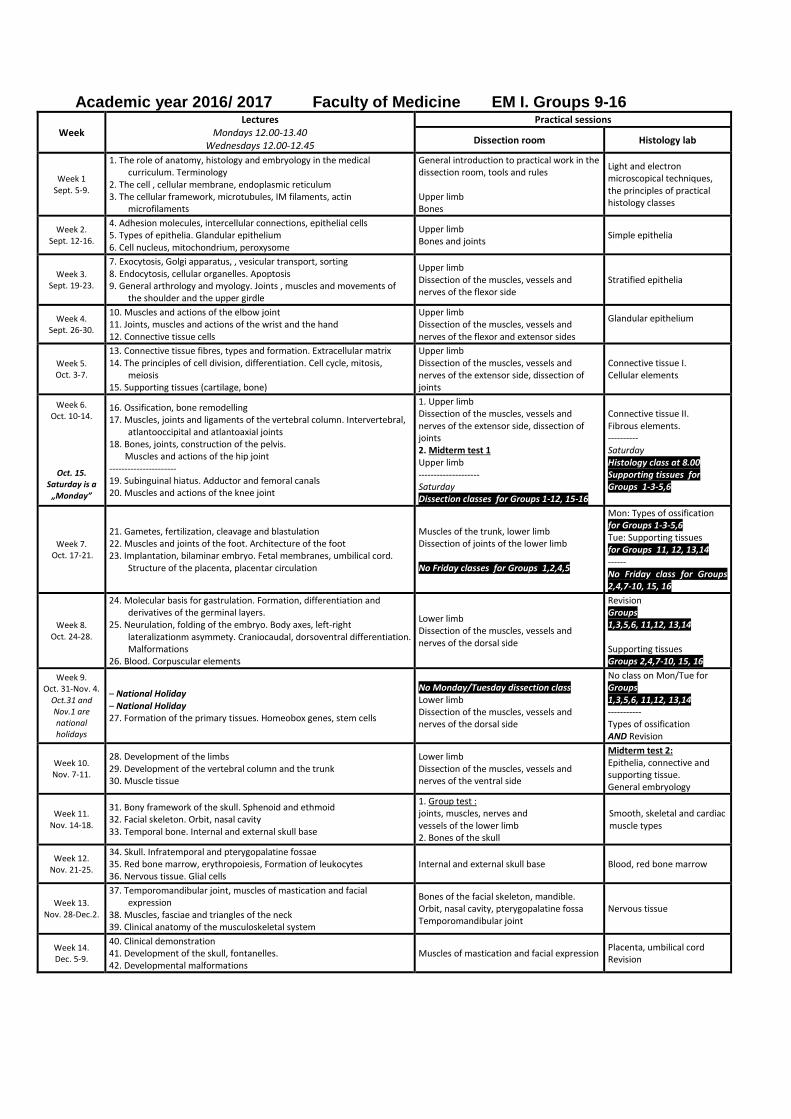
Academic year 2016/ 2017 Faculty of Medicine EM I. Groups 9-16
Week Lectures
Mondays 12.00-13.40 Wednesdays 12.00-12.45
Practical sessions
Dissection room Histology lab
Week 1 Sept. 5-9.
1. The role of anatomy, histology and embryology in the medical curriculum. Terminology
2. The cell , cellular membrane, endoplasmic reticulum 3. The cellular framework, microtubules, IM filaments, actin
microfilaments
General introduction to practical work in the dissection room, tools and rules Upper limb Bones
Light and electron microscopical techniques, the principles of practical histology classes
Week 2. Sept. 12-16.
4. Adhesion molecules, intercellular connections, epithelial cells 5. Types of epithelia. Glandular epithelium 6. Cell nucleus, mitochondrium, peroxysome
Upper limb Bones and joints
Simple epithelia
Week 3. Sept. 19-23.
7. Exocytosis, Golgi apparatus, , vesicular transport, sorting 8. Endocytosis, cellular organelles. Apoptosis 9. General arthrology and myology. Joints , muscles and movements of
the shoulder and the upper girdle
Upper limb Dissection of the muscles, vessels and nerves of the flexor side
Stratified epithelia
Week 4. Sept. 26-30.
10. Muscles and actions of the elbow joint 11. Joints, muscles and actions of the wrist and the hand 12. Connective tissue cells
Upper limb Dissection of the muscles, vessels and nerves of the flexor and extensor sides
Glandular epithelium
Week 5. Oct. 3-7.
13. Connective tissue fibres, types and formation. Extracellular matrix 14. The principles of cell division, differentiation. Cell cycle, mitosis,
meiosis 15. Supporting tissues (cartilage, bone)
Upper limb Dissection of the muscles, vessels and nerves of the extensor side, dissection of joints
Connective tissue I. Cellular elements
Week 6. Oct. 10-14.
Oct. 15. Saturday is a „Monday”
16. Ossification, bone remodelling 17. Muscles, joints and ligaments of the vertebral column. Intervertebral,
atlantooccipital and atlantoaxial joints 18. Bones, joints, construction of the pelvis. Muscles and actions of the hip joint ---------------------- 19. Subinguinal hiatus. Adductor and femoral canals 20. Muscles and actions of the knee joint
1. Upper limb Dissection of the muscles, vessels and nerves of the extensor side, dissection of joints 2. Midterm test 1 Upper limb -------------------- Saturday Dissection classes for Groups 1-12, 15-16
Connective tissue II. Fibrous elements. ---------- Saturday Histology class at 8.00 Supporting tissues for Groups 1-3-5,6
Week 7. Oct. 17-21.
21. Gametes, fertilization, cleavage and blastulation 22. Muscles and joints of the foot. Architecture of the foot 23. Implantation, bilaminar embryo. Fetal membranes, umbilical cord.
Structure of the placenta, placentar circulation
Muscles of the trunk, lower limb Dissection of joints of the lower limb No Friday classes for Groups 1,2,4,5
Mon: Types of ossification for Groups 1-3-5,6 Tue: Supporting tissues for Groups 11, 12, 13,14 ------ No Friday class for Groups 2,4,7-10, 15, 16
Week 8. Oct. 24-28.
24. Molecular basis for gastrulation. Formation, differentiation and derivatives of the germinal layers.
25. Neurulation, folding of the embryo. Body axes, left-right lateralizationm asymmety. Craniocaudal, dorsoventral differentiation. Malformations
26. Blood. Corpuscular elements
Lower limb Dissection of the muscles, vessels and nerves of the dorsal side
Revision Groups 1,3,5,6, 11,12, 13,14 Supporting tissues Groups 2,4,7-10, 15, 16
Week 9. Oct. 31-Nov. 4.
Oct.31 and Nov.1 are national holidays
– National Holiday – National Holiday 27. Formation of the primary tissues. Homeobox genes, stem cells
No Monday/Tuesday dissection class Lower limb Dissection of the muscles, vessels and nerves of the dorsal side
No class on Mon/Tue for Groups 1,3,5,6, 11,12, 13,14 ----------- Types of ossification AND Revision
Week 10. Nov. 7-11.
28. Development of the limbs 29. Development of the vertebral column and the trunk 30. Muscle tissue
Lower limb Dissection of the muscles, vessels and nerves of the ventral side
Midterm test 2: Epithelia, connective and supporting tissue. General embryology
Week 11. Nov. 14-18.
31. Bony framework of the skull. Sphenoid and ethmoid 32. Facial skeleton. Orbit, nasal cavity 33. Temporal bone. Internal and external skull base
1. Group test : joints, muscles, nerves and vessels of the lower limb 2. Bones of the skull
Smooth, skeletal and cardiac muscle types
Week 12. Nov. 21-25.
34. Skull. Infratemporal and pterygopalatine fossae 35. Red bone marrow, erythropoiesis, Formation of leukocytes 36. Nervous tissue. Glial cells
Internal and external skull base Blood, red bone marrow
Week 13. Nov. 28-Dec.2.
37. Temporomandibular joint, muscles of mastication and facial expression
38. Muscles, fasciae and triangles of the neck 39. Clinical anatomy of the musculoskeletal system
Bones of the facial skeleton, mandible. Orbit, nasal cavity, pterygopalatine fossa Temporomandibular joint
Nervous tissue
Week 14. Dec. 5-9.
40. Clinical demonstration 41. Development of the skull, fontanelles. 42. Developmental malformations
Muscles of mastication and facial expression Placenta, umbilical cord Revision
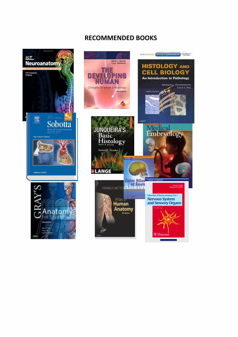
RECOMMENDED BOOKS
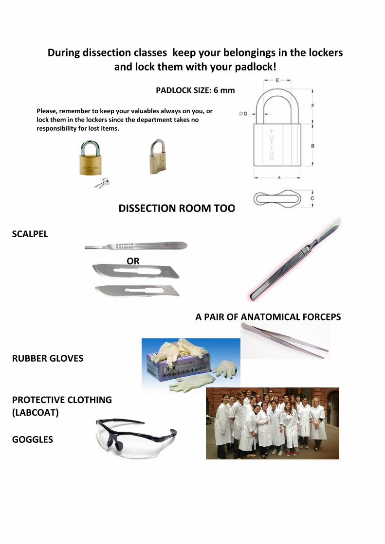
During dissection classes keep your belongings in the lockers and lock them with your padlock!
PADLOCK SIZE: 6 mm
Please, remember to keep your valuables always on you, or lock them in the lockers since the department takes no responsibility for lost items.
DISSECTION ROOM TOOLS
SCALPEL
OR
A PAIR OF ANATOMICAL FORCEPS
RUBBER GLOVES
PROTECTIVE CLOTHING (LABCOAT)
GOGGLES
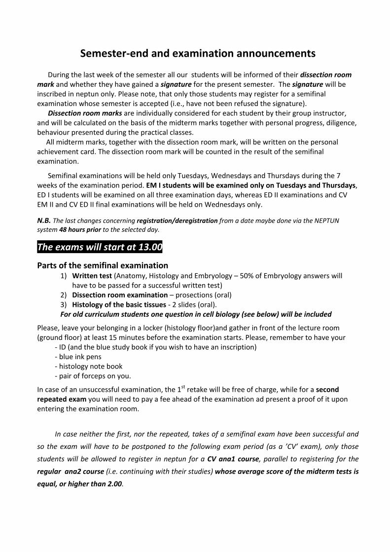
Semester-end and examination announcements
During the last week of the semester all our students will be informed of their dissection room mark and whether they have gained a signature for the present semester. The signature will be inscribed in neptun only. Please note, that only those students may register for a semifinal examination whose semester is accepted (i.e., have not been refused the signature). Dissection room marks are individually considered for each student by their group instructor, and will be calculated on the basis of the midterm marks together with personal progress, diligence, behaviour presented during the practical classes. All midterm marks, together with the dissection room mark, will be written on the personal achievement card. The dissection room mark will be counted in the result of the semifinal examination.
Semifinal examinations will be held only Tuesdays, Wednesdays and Thursdays during the 7 weeks of the examination period. EM I students will be examined only on Tuesdays and Thursdays, ED I students will be examined on all three examination days, whereas ED II examinations and CV EM II and CV ED II final examinations will be held on Wednesdays only.
N.B. The last changes concerning registration/deregistration from a date maybe done via the NEPTUN system 48 hours prior to the selected day.
The exams will start at 13.00
Parts of the semifinal examination 1) Written test (Anatomy, Histology and Embryology – 50% of Embryology answers will
have to be passed for a successful written test) 2) Dissection room examination – prosections (oral) 3) Histology of the basic tissues - 2 slides (oral). For old curriculum students one question in cell biology (see below) will be included
Please, leave your belonging in a locker (histology floor)and gather in front of the lecture room (ground floor) at least 15 minutes before the examination starts. Please, remember to have your
- ID (and the blue study book if you wish to have an inscription) - blue ink pens - histology note book - pair of forceps on you.
In case of an unsuccessful examination, the 1st retake will be free of charge, while for a second repeated exam you will need to pay a fee ahead of the examination ad present a proof of it upon entering the examination room.
In case neither the first, nor the repeated, takes of a semifinal exam have been successful and
so the exam will have to be postponed to the following exam period (as a ’CV’ exam), only those
students will be allowed to register in neptun for a CV ana1 course, parallel to registering for the
regular ana2 course (i.e. continuing with their studies) whose average score of the midterm tests is
equal, or higher than 2.00.
TOPICS OF THE SEMIFINAL EXAM
Academic year 2016/2017 First semester
EM I.
Histology (for both 8 and 9 credit courses)
Description of 2 histological slides with the help of a microscope
Further theoretical issues related to the inspected slide Concept of basic tissues Definition and classification of epithelial tissue Simple epithelia Stratified epithelia Membrane specializations of epithelia Glandular epithelia Cells of connective tissue Ground substance and fibres of connective tissue Types of connective tissue Umbilical cord and placenta Blood and the formed elements of blood Histology of the bone marrow, maturation of erythrocytes and platelets
Differentiation of granulocytes, lymphocytes and monocytes Histology of cartilage Histology of the bone tissue Intramembranous ossification Endochondral ossification Growth and remodeling of bone Smooth muscle and myoepithelial cells Skeletal muscle tissue Cardiac muscle tissue Histology of the peripheral nervous system (sensory and autonomic ganglia) Supporting cells in the peripheral nervous system Nerve fibers, myelin sheath Motor end-plate
Anatomy (for both 8 and 9 credit courses)
General osteology, classification of bones Bones, spaces and connections of the skull, external and internal skull bases Neurocranium, components and cavities (anterior, middle and posterior cranial fossae) Viscerocranium, components and cavities (walls and connections of the nasal cavity, orbit, oral cavity, pterygopalatine and infratemporal fossae) Bones of the axial and appendicular skeleton Vertebrae, ribs, sternum Bones of the girdles and limbs General arthrology Fibrous and cartilaginous joints Components of the synovial joints Classification of synovial joints; movements and mechanisms Structure of the vertebral column, the gross anatomy of the muscles acting upon it Movements and muscles of the head (atlantooccipital and atlantoaxial joints)
Joints of the shoulder girdle, the gross anatomy of the muscles acting upon them The shoulder joint, the gross anatomy of the muscles acting upon it The elbow joint, the gross anatomy of the muscles acting upon it Structure and movements of the wrist (radiocarpal) joint, the gross anatomy of the muscles acting upon it Metacarpophalangeal and interphalangeal joints, the gross anatomy of the muscles concerned with the movements Carpometacarpal, metacarpophalangeal and interphalangeal joints of the thumb, the gross anatomy of the muscles concerned with the movements The hip joint and the gross anatomy of the muscles concerned with the movements The knee joint and the gross anatomy of the muscles concerned with the movements The ankle joint together with the gross anatomy of the muscles acting upon it The subtalar and talocalcaneonavicular joints, the muscles acting upon them The temporomandibular joint and the gross anatomy of the muscles acting on it Architecture and classification of bones Structure and actions of somatic muscles Osteofibrous structure of the thoracic cage (bones, joints, ligaments, movements) Muscles and movements of the thorax Muscles of the back and nape (occipital region) The axilla, the quadrangular and triangular spaces The cubital fossa Muscles and cross section of the arm Muscles and cross section of the forearm Osteofibrous spaces and muscle compartments of the hand, tendinous sheaths Composition of the pelvis (bones, ligaments and membranes) Muscles of the buttock, the posterior abdominal wall and the pelvis (external and internal muscles of the hip) Osteofibrous compartments, muscles and cross section of the thigh Popliteal fossa Subinguinal hiatus, vascular and muscular compartments; adductor canal, femoral canal Osteofibrous compartments, muscles and the cross section of the leg Structure of the foot, arches of the foot Osteofibrous compartments of the foot, tendinous sheaths Muscles of mastication Superficial muscles of the neck and the muscle triangles Deep muscles of the neck and the laminae of the cervical fascia Muscles of facial expression
Embryology (for both 8 and 9 credit courses)Spermatogenesis, spermiogenesis Oogenesis Fertilization, cleavage of the zygote Blastocyst formation; the bilaminar embryonic disc Implantation Formation of the intraembryonic mesoderm; the notochord Neurulation (neural tube and neural crest)
Differentiation of the intraembryonic mesoderm; formation and derivatives of the somites Derivatives of the intermediate mesoderm Lateral plate mesoderm and its derivatives Folding of the embryo Development of the primitive cardiovascular system, the fetal circulation The structure and function of the placenta Development of the fetal membranes (chorion and amnion) and the umbilical cord Periods of embryonic / fetal life Twin formation Development of the limbs Development of the vertebral column Development of the skull Development of the skeletal muscular system ------ ------ ------- -------- ------ ------- ----- -------
CELL BIOLOGY TOPIC LIST ONLY FOR OLD CURRICULUM (9 credit course) STUDENTS General histological procedures (fixation, dehydration, embedding, cutting, rehydration, staining etc.) Different staining methods (chemical staining, impregnation, enzyme histochemistry, immunohistochemistry) Light and electron microscopy of cells or tissues The general structure of eukaryotic cells Biological membranes: structure, membrane proteins, membrane domains, glycocalyx Functions of cell membrane, membrane transport Structure and functions of cell nucleus. Chromosomes, karyogram, sex chromosomes, sex chromatin Structure, types and role of RNA, transcription Structure and function of ribosomes, translation Structure and function of the rough and smooth endoplasmic reticula Structure, function of the Golgi apparatus, protein sorting Protein secretion, exocytosis Vesicular transport Cytoskeleton in general Microtubules: structure, formation and degradation, regulating factors, associated motorproteins Structure and function of the centrosome (cytocenter), function of microtubules Structure and role of kinocilia and the primary (sensory) cilium Structure, regulation and associated proteins of icrofilaments. Functions of microfilaments, significance in cell trafficking processes Intermedier filaments: structure, function, associated proteins, appearance in different cell types Adhesion molecules (cell-cell, cell-ECM adhesion) Cell adhesion structures
The general concept of epithelial tissue, cell polarity, membrane domains, structures that increase the surface Structure and functionof the membrana basalis as well as of the lamina basalis Mitotic cell division Cell cycle phases and regulation Phagocytosis. Intracellular digestion Lyosomes, synthesis of lysosomal enzymes, their transport pathways, functions, lysosomal diseases. Pinocytosis. Endosome. Transcytosis. Apoptosis, autophagy and necrosis. Differentiation, stem cells Meiotic cell division Morphology, function and localization of mitochondria Mitochondrial genome, endosymbiotic theory Peroxisome