Embed Size (px)
Citation preview

ANATOMY ANDPHYSIOLOGY
5ANATOMY AND PHYSIOLOGY
As a beauty therapist it is important that you have a good understanding of anatomy and physiology, as many of yourtreatments aim to improve the particular functioning of systems of the body. For example a facial massage will improveblood and lymph circulation locally, as you massage the skin’s surface, increase cellular renewal as you improvenutrition to the living cells, and remove dead skin cells. The result is healthier looking skin.
ANAANATOMY AND PHYSIOLOGY KNOWLEDGE REQUIREMENTSTOMY AND PHYSIOLOGY KNOWLEDGE REQUIREMENTS
It is necessary for you to know and understand anatomy and physiology as relevant to each beauty therapy chapter.This may be assessed through oral questioning, written test or assignment. To guide you in your studies the essentialanatomy and physiology you need to know and understand for each chapter has been identified with a ✓ symbol.Look for the to remind you to check back here for your essential anatomy and physiology knowledge!
The beauty therapy chapters with an essential anatomy and physiology knowledge requirement are:
6. Improve and maintain facial skin condition
8. Remove hair using waxing techniques
9. Provide manicure treatment
10. Provide pedicure treatment
11. Provide make-up treatment
13. Provide nail art service
15. Assist with spa treatments
12. Extend and maintain nails
Anatomy and physiology knowledge and understanding is located in this chapter, but it can also be found within eachof the beauty therapy chapter where essential anatomy and physiology knowledge are identified as above.
Skin structure and function ✓ ✓ ✓ ✓ ✓ ✓ ✓ ✓
Factors affecting skin condition ✓ ✓ ✓ ✓ ✓ ✓
Structure of hair and types of hair growth ✓
Hair growth cycle ✓
ANATOMY AND PHYSIOLOGY ✓ ESSENTIAL KNOWLEDGE FOR CHAPTER
Chapter 6 8 9 10 11 12 13 15

Anatomy and Physiology 89
Nail structure and function ✓ ✓ ✓ ✓
Nail growth ✓ ✓ ✓ ✓
Muscle groups in parts of the body, ✓ ✓ ✓position, structure and function
Muscle tone ✓
Bones in parts of the body, position, ✓ ✓ ✓structure and function
Composition and function of blood ✓ ✓ ✓and lymph
Blood flow and pulse rate ✓ ✓ ✓
Central nervous system and ✓autonomic system
THE SKINTHE SKIN
TTHEHE SSTRUCTURETRUCTURE ANDAND FFUNCTIONUNCTION OFOF SSKINKIN ANDAND THETHE SSURROUNDINGURROUNDING TTISSUESISSUES
The skin varies in appearance according to our race, sex and age. It also alters from season to season and from yearto year, and reflects our general health, lifestyle and diet.
At puberty the chemical substances (hormones) that control many of our bodies’ activities become very active.Amongst other effects, this activity causes the skin to become more oily, and often blemishes appear on the skin’ssurface. Seven out of ten teenagers find that their skin becomes blemished with blackheads, inflamed angry spotsand even scars at this time: a skin disorder called acne vulgaris.
During the twenties the skin should look its best; any hormonal imbalance that occurred at puberty should by nowhave stabilised. As we grow older, the skin ages too. In our late twenties and early thirties we will see fine linesappearing on the skin’s surface, especially around the eyes where the skin is thinner and the skin gradually becomesdrier.
At around the age of 40, hormone activity in the body becomes slower and the skin begins to lose its strength andelasticity. The skin becomes increasingly drier, and lines and wrinkles appear on the surface. In the late fifties brownpatches of discoloured skin (lentigines) may appear: these are commonly seen at the temple region of the face andon the backs of the hands and are caused by ultra-violet light damage.
Fortunately help is at hand to care for the skin: there is an ever-increasing number of skin-care products from a vastand highly profitable cosmetics industry, and there are the skill and expertise of the qualified beauty therapist.
If it is your intention to become a qualified beauty professional, you need to learn about skin: its construction, itsfunction, and how and why it is changed by both internal and external influences.
CCELLSELLS
The human body consists of many trillions of microscopic cells. Each cell contains a chemical substance calledprotoplasm, which contains various specialised structures whose activities are essential to our health. If cells areunable to function properly, a disorder results.
Surrounding the cell is the cell membrane: this forms a boundary between the cell contents and their environment.The membrane has a porous surface which permits food to enter and waste materials to leave.

Anatomy and Physiology 91
Name of tissue Examples General functions
Tissues, may be grouped to form the larger functional and structural units we know as organs, such as the heart.
FUNCTIONS OF THE SKINFUNCTIONS OF THE SKIN
The human skin is an organ–the largest of the body. It provides a tough, flexiblecovering, with many different important functions. The skin has many functions.The main functions are listed below.
PPROTECTIONROTECTION
The skin protects the body from potentially harmful substances and conditions.
The outer surface is bactericidal, helping to prevent the multiplication ofharmful micro-organisms. It also prevents the absorption of many substances(unless the surface is broken), because of the construction of the cells on itsouter surface, which form a chemical and physical barrier.
The skin cushions the underlying structures from physical injury.
The skin provides a waterproof coating. Its natural oil, sebum, prevents theskin from losing vital water, and thus prevents skin dehydration.
The skin contains a pigment called melanin. This absorbs harmful rays of ultra-violet light.
HHEAEATT RREGULAEGULATIONTION
Humans maintain a normal body temperature of 36.8–37°C. Body temperature
is controlled in part by heat loss through the skin and by sweating. If thetemperature of the body is increased by 0.25–0.5°C, the sweat glands secrete sweatto the skin’s surface. The body is cooled by the loss of heat used to evaporate thesweat from the skin’s surface. If the body becomes too warm there is an increase inblood flow into the blood capillaries in the skin. The blood capillaries widen (dilate)and heat is lost from the skin.
EEXCRETIONXCRETION
Small amounts of certain waste products, such as urea, water and salt, are removed from the body in sweat byexcretion through the surface of the skin.
Nervous Neurones Forms a communication system between differentparts of the body
nucleusspindle-shapedcell
cells separatedfrom each other
Although the skin is structured to avoid penetration of harmfulsubstances by absorption, certainchemicals can be absorbed throughthe skin. Always protect the skinwhen using potentially harmfulsubstances, and wear gloves whenusing harsh chemical cleaningagents.
HEA
LTH
AN
D S
AFE
TY
++
Although the skin has a waterproofproperty it allows approximately500ml of water to be lost from thetissues through evaporation everyday,
TIP
�
If the external temperature becomeslow, blood flow nearer the skin’ssurface is decreased and the bloodcapillaries narrow (constrict),preventing heat loss and conservingheat.
HEA
LTH
AN
D S
AFE
TY
++

96 Beauty Therapy: The Foundations
The rThe reticular layereticular layer
The dermis contains a network of protein fibres called the reticular layer. Thesefibres allow the skin to expand, to contract, and to perform intricate, supplemovements.
This network is composed of two sorts of protein fibre: yellow elastin fibres andwhite collagen fibres. Elastin fibres give the skin its elasticity, and collagen fibres giveit its strength. The fibres are produced by specialised cells called fibroblasts, and areheld in a gel called the ground substance.
While this network is strong, the skin will appear youthful and firm. As the fibresharden and fragment, however, the network begins to collapse, losing its elasticity.The skin then begins to show visible signs of ageing.
A major cause of damage to this network is unprotected exposure of the skin toultra-violet light and to weather. Sometimes, too, the skin loses its elasticity becauseof a sudden increase in body weight, for example at puberty or pregnancy. Thisresults in the appearance of stretch marks, streaks of thin skin that is a differentcolour from the surrounding skin: on a white skin they appear as thin reddish streaks;on a black skin they appear slightly lighter than the surrounding skin. The lostelasticity cannot be restored.
NerNerve endingsve endings
The dermis contains different types of sensory nerve endings, which registertouch, pressure, pain and temperature. These send messages to the central
nervous system and the brain, informing us about the outside world and what ishappening on the skin’s surface. The appearance of each of these nerve endings isquite varied.
Appropriate external massagemovements can be used to increasethe blood supply within the dermis,bringing extra nutrients and oxygento the skin and to the underlyingmuscle. At the same time, thelymphatic circulation is increased,improving the removal of wasteproducts that may haveaccumulated.
TIP
�
When sunbathing, always protectthe skin with an appropriateprotective sun-screen product, andalways use an emollient after-sunpreparation to minimise thecumulative effects of prematureageing, by rehydrating and soothingthe skin.HEA
LTH
AN
D S
AFE
TY
++
Sensory nerve endings are mostnumerous in sensitive parts of theskin, such as the fingertips and thelips.
TIP
�
pressure receptor touch receptor pain receptor heat receptor cold receptor
Growth and repairGrowth and repair The body’s blood system of arteries and veins continually brings blood to the capillarynetworks in the skin and takes it away again. The blood carries the nutrients and oxygen essential for the skin’s health,maintenance and growth, and takes away waste products.
Sensory nerves
Anatomy and Physiology 97
DefenDefencece Within the dermis are the structures responsible for protecting theskin from harmful foreign bodies and irritants.
One set of cells, the mast cells, burst when stimulated during inflammation orallergic reactions, and release a chemical substance called histamine. This causesthe blood vessels nearby to enlarge, thereby bringing more blood to the site of theirritation to limit skin damage and begin repair.
In the blood, and also in the lymph and the connective tissue, are another groupof cells: the macrophages or ‘big eaters’. These destroy micro-organisms andengulf dead cells and other unwanted particles. When necessary, they travel to anarea where they are needed, for example the site of an infection. They form a rolein the immune system that protects the body from disease-causing micro-organisms.
Waste productsWaste products Lymph vessels in the skin carry a fluid called lymph, a straw-coloured fluid similar incomposition to blood plasma. Plasma is the liquid part of the blood that disperses from the blood capillaries intothe tissue spaces. Lymph is composed of water, lymphocytes (a type of white blood cell that plays a key role in theimmune system), oxygen, nutrients, hormones, salts and waste products. The waste products are eliminated andusable protein is recycled for further use by the body.
Control of functioningControl of functioning Hormones are chemical messengers transportedin the blood. They control the activity of many organs in the body, including thecells and glands in the skin. These include melanosomes, which produce skinpigment, and the sweat glands and sebaceous glands.
Hormone imbalance at different times of our life may disturb the normalfunctioning of these cells and structures, causing various skin disorders.
Skin appendagesSkin appendages
Within the dermis are structures called skin appendages. These include:
sweat glands;
sebaceous glands;
hair follicles, which produce hair;
nails.
Sweat glandsSweat glands Sweat glands or sudoriferous glands are composed ofepithelial tissue, which extends from the epidermis into the dermis. These glandsare found all over the body, but are particularly abundant on the palms of thehands and the soles of the feet. Their function is to regulate body temperaturethrough the evaporation of sweat from the surface of the skin. Fluid loss andcontrol of body temperature are important to prevent the body overheating,especially in hot, humid climates. For this reason, perhaps, sweat glands are largerand more abundant in black skins than white skins.
There are two types of sweat glands: eccrine glands and apocrine glands. Eccrine
glands are simple sweat-producing glands, found over most of the body,appearing as tiny tubes (ducts). The eccrine glands are responsive to heat. These
When the surface has been broken,the skin at the site of the injury isreplaced but may leave a scar. Thisinitially appears red, due to theincreased blood supply to the area,required while the skin heals.When healed, the redness will fade.H
EALT
H A
ND
SA
FET
Y
++
We each have approximately two tofive million sweat glands.
TIP
�
Excessive sweating, which canoccur through exposure to hightemperatures or during illness, canlead to skin ehydration–insufficientwater content. Fluid intake mustbe increased to rebalance the bodyfluids.
HEA
LTH
AN
D S
AFE
TY
++
Pores allow the absorption of somefacial cosmetics into the skin. Manyfacial treatments are therefore aimedat cleansing the pores, some with aparticularly deep cleansing action, aswith cosmetic cleansers and facialmasks.
The pores may become enlargeddue to congestion caused by dirt,dead skin cells and cosmetics. Theapplication of an astringent skin-care preparation creates a tighteningeffect upon the skin’s surface, slightlyreducing the size of the pores.
TIP
�
Anatomy and Physiology 99
Acid mantle Acid mantle Sweat and sebum combine on the skin’s surface, creating an acidfilm. This is known as the acid mantle, and discourages the growth of bacteria andfungi.
Acidity and alkalinity are measured by a number called the pH. An acidic solution hasa pH of 0–7; a neutral solution has a pH of 7; and an alkaline solution has a pH of7–14. The acid mantle of the skin has a pH of 5.5–5.6.
THE HAIRTHE HAIR
TTHEHE SSTRUCTURETRUCTURE ANDAND FFUNCTIONUNCTION OFOF HHAIRAIR ANDAND THETHE
SSURROUNDINGURROUNDING TTISSUESISSUES
A hair is a long, slender structure which grows out of, and is part of, the skin. Eachhair is made up of dead skin cells, which contain the protein called keratin.
Hairs cover the whole body, except for the palms of the hands, the soles of thefeet, the lips, and parts of the sex organs.
Hair has many functions:
scalp hair insulates the head against cold, protects it from the sun, andcushions it against bumps;
eyebrows cushion the browbone from bumps, and prevent sweat fromrunning into the eyes;
eyelashes help to prevent foreign particles entering the eyes;
nostril hair traps dust particles inhaled with the air;
ear hair helps to protect the ear canal;
body hair helps to provide an insulating cover (though this function is almostobsolete in humans), has a valuable sensory function, and is linked with thesecretion of sebum onto the surface of the skin.
Hair also plays a role in social communication.
The strThe structuructure of haire of hair
Most hairs are made up of three layers of different types of epithelial cells: themedulla, the cortex and the cuticle.
The medulla is the central core of the hair. The cells of the medulla contain softkeratin, and sometimes some pigment granules. The medulla only exists inmedium to coarser hair–there is usually no medulla in thinner hair.
The cortex is the thickest layer of the hair, and is made up of several layers ofclosely packed, elongated cells. These contain pigment granules and hard keratin.
Cosmetic moisturisers mimic sebumin providing an oily covering for theskin’s surface to reduce moisture lossT
IP
�
Because the skin has an acid pH, ifalkaline products are used on it theacid mantle will be disturbed. Itwill take several hours for thisprotective film to be restored; duringthis time, the skin will be irritatedand sensitive
HEA
LTH
AN
D S
AFE
TY
++
There are approximately 100,000hairs on the scalp.
TIP
�
A strand of hair is stronger than anequivalent strand of nylon or copper.
TIP
�
Sebaceous glands are not present onthe surface of the lips. For thisreason the lips should be protectedwith a lip emollient preparation toprevent them from becoming dryand chapped.
HEA
LTH
AN
D S
AFE
TY
++

Anatomy and Physiology 101
Each hair grows out of a tube-like indentation in the epidermis,the hair follicle. The walls of the follicle are a continuation ofthe epidermal layer of the skin.
The arrector pili muscle is attached at an angle to the base ofthe follicle. Cold, aggression or fright stimulates this muscle tocontract, pulling the follicle and the hair upright.
The sebaceous gland is attached to the upper part of thefollicle; from it, a duct enters directly into the hair follicle. Thegland produces an oily substance, sebum, which is secretedinto the follicle. Sebum waterproofs, lubricates and softens thehair and the surface of the skin; it also protects the skin againstbacterial and fungal infections. The contraction of the arrectorpili muscle aids the secretion of sebum.
The dermal papilla, a connective tissue sheath, is surrounded by a hair bulb. It has an excellent blood supply,necessary for the growth of the hair. It is not itself part of the follicle, but a separate tiny organ which serves thefollicle.
The bulb is the expanded base of the hair root. A gap at the base leads to a cavity inside, which houses thepapilla. The bulb contains in its lower part the dividing cells that create the hair. The hair continues to develop asit passes through the regions of the upper bulb and the root.
The matrix is the name given to the lower part of the bulb, which comprises actively dividing cells from whichthe hair is formed.
The hair follicle The hair follicle The hair follicle extends into the dermis, and is made upof three sheaths: the inner epithelial root sheath, the outer epithelial root sheathand the surrounding connective-tissue sheath.
The inner epithelial root sheath grows from the bottom of the follicle at thepapilla; both the hair and the inner root sheath grow upwards together. The innersurface of this sheath is covered with cuticle cells, in the same way as the outersurface of the hair: these cells lock together, anchoring the hair firmly in place.The inner root sheath ceases to grow when level with the sebaceous gland.
The outer epithelial root sheath forms the follicle wall. This does not grow up with the hair, but is stationary.It is a continuation of the growing layer of the epidermis of the skin.
The connective-tissue sheath surrounds both the follicle and the sebaceous gland, providing both a sensorysupply and a blood supply. The connective-tissue sheath includes, and is a continuation of, the papilla.
medulla
cortex hair shaft
cuticle
artery blood supplyto hair papillavein
matrix
dividing cells
dermal papilla
When hairs break off due toincorrect waxing technique, they willbreak at the level at which they arelocked into the follicle by the cells ofthe inner root sheath.
TIP
�
Hair shapes Curly Wavy Straight
Flat ribbon-like Less oval Round
The hair bulb

Anatomy and Physiology 105
THE NAILSTHE NAILS
TTHEHE SSTRUCTURETRUCTURE ANDAND FFUNCTIONUNCTION OFOF THETHE NNAILAIL
Nails grow from the ends of the fingers and toes and serve as a form ofprotection. They also help when picking up small objects. Dark streaks causedby pigmentation are common on the nail plate of black-skinned clients. Thesetend to increase with age.
The nail plateThe nail plate
The nail plate is composed of compact translucent layers of keratinisedepidermal cells: it is this that makes up the main body of the nail. The layersof cells are packed very closely together, with fat but very little moisture.
The nail gradually grows forward over the nail bed, until finally it becomes thefree edge. The underside of the nail plate is grooved by longitudinal ridges andfurrows, which help to keep it in place.
In normal health the plate curves in two directions:
transversely–from side to side across the nail;
longitudinally–from the base of the nail to the free edge.
There are no blood vessels or nerves in the nail plate: this is why the nails, like hair, can be cut without pain orbleeding. The pink colour of the nail plate derives from the blood vessels that pass beneath it–the nail bed.
Function: To protect the living nail bed of the fingers and toes.
The frThe free edgeee edge
The free edge is the part of the nail that extends beyond the fingertip; this is thepart that is filed. It appears white as there is no nail bed underneath.
Function: To protect the fingertip and the hyponychium (see page 106).
The matrixThe matrix
The matrix, sometimes called the nail root, is the growing area of the nail. It is formed by the division of cells in thisarea, called mitosis, which is part of the stratum germinativum layer of the epidermis. It lies under the eponychium(see page 106), at the base of the nail, nearest to the body. The process of keratinisation takes place in the epidermalcells of the matrix, forming the hardened tissue of the nail plate.
Function: To produce new nail cells.
The nail bedThe nail bed
The nail bed is the portion of skin upon which the nail plate rests. It has a pattern of grooves and furrowscorresponding to those found on the underside of the nail plate; these interlock, keeping the nail in place, but separateat the end of the nail to form the free edge. The nail bed is liberally supplied with blood vessels, which provide thenourishment necessary for continued growth; and sensory nerves, for protection.
Function: To supply nourishment and protection.
free edge
hyponychium
nail plate
nail wall
nail bed
perionychium
lunula
cuticle
matrix
nail groove
eponychium
nail mantle
The structure of the nail
nail plate
The nail plate
Fingernails grow more quickly thantoenails. Fingernails grow about0.1mm each day (4cm per year),and grow faster in summer than inwinter.
TIP
�

110 Beauty Therapy: The Foundations
The nervous system therefore co-ordinates the activities of the body by respondingto stimuli received by sense organs, including the nose, tongue, eyes, ears and skin.
THE MUSCULAR SYSTEMTHE MUSCULAR SYSTEM
Muscles are responsible for the movement of body parts. Each is made up of abundle of elastic fibres bound together in a sheath, the fascia. Muscular tissuecontracts (shortens) and produces movement. Muscles never completely relax–there are always a few contracted fibresin every muscle. These make the muscles slightly tense and this tension is called muscle tone.
The sympathetic division isassociated with stress.The parasympathetic system isassociated with peace.
TIP
�
Muscle
Muscle tissue has the following properties:
it has the ability to contract;
it is extensible (when the extensor muscle in a joint contracts the corresponding flexor muscle will be stretchedor lengthened);
it is elastic–following contraction or extension it returns to its original length;
it is responsive–it contracts in response to nerve stimulation.
A muscle is usually anchored by a strong tendon to one bone: the point of attachment is known as the muscle’sorigin. The muscle is likewise joined to a second bone: the attachment in this case is called the muscle’sinsertion. It is this second bone that is moved: the muscle contracts, pulling the two bones towards each other.(A different muscle, on the other side of the bone, has the contrary effect.) Not all muscles attach to bones,however: some insert into an adjacent muscle, or into the skin itself. The muscles with which we are concernedhere are those of the face, the neck and the shoulders.
FFACIALACIAL MMUSCLESUSCLES
Many of the muscles located in the face are very small and are attached to (‘insert into’) another small muscle or thefacial skin. When the muscles contract, they pull the facial skin in a particular way; this creates facial expressions.
With age, the facial expressions that we make every day produce lines on the skin–frown lines. The amount of tension,or tone, also decreases with age. When performing facial massage, the aim is to improve the general tone of thefacial muscles.
124 Beauty Therapy: The Foundations
the occipital branch supplies the back of the head and the scalp;
the temporal branch supplies the sides of the face, the head, the scalp and the skin;
the facial branch supplies the muscles and tissues of the face.
These arteries also divide repeatedly, successive vessels becoming smaller andsmaller until they form tiny blood capillaries. These vessels are just one cell thick,allowing substances carried in the blood to pass through them into the tissue fluid
which bathes and nourishes the cells of the various body tissues.
The blood capillaries begin to join up again, forming first small vessels called venules,then larger vessels called veins. These return the blood to the heart.
Veins are less elastic than arteries, and are closer to the skin’s surface. Along theircourse are valves, which prevent the backflow of blood.
The main veins are the external and internal jugular veins. The internal jugular vein
and its main branch, the facial vein, carry blood from the face and head. The external jugular vein carries bloodfrom the scalp and has two branches: the occipital branch and the temporal branch. The jugular veins join to enterthe subclavian vein, which lies above the clavicle.
Blood returns to the heart, which pumps it to the lungs, where the red blood cells take on fresh oxygen, and wherecarbon dioxide is expelled from the blood. The blood returns to the heart, and begins its next journey round the body.
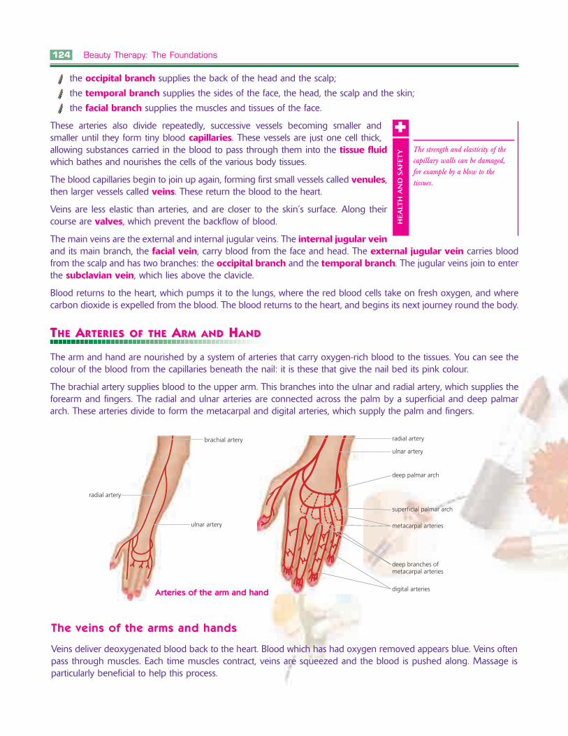
TTHEHE AARRTERIESTERIES OFOF THETHE AARMRM ANDAND HHANDAND
The arm and hand are nourished by a system of arteries that carry oxygen-rich blood to the tissues. You can see thecolour of the blood from the capillaries beneath the nail: it is these that give the nail bed its pink colour.
The brachial artery supplies blood to the upper arm. This branches into the ulnar and radial artery, which supplies theforearm and fingers. The radial and ulnar arteries are connected across the palm by a superficial and deep palmararch. These arteries divide to form the metacarpal and digital arteries, which supply the palm and fingers.
The strength and elasticity of thecapillary walls can be damaged,for example by a blow to thetissues.
HEA
LTH
AN
D S
AFE
TY
++
radial artery
brachial artery
ulnar artery
radial artery
ulnar artery
deep palmar arch
superficial palmar arch
metacarpal arteries
deep branches ofmetacarpal arteries
digital arteriesArteries of the arm and hand
The veins of the arThe veins of the arms and handsms and hands
Veins deliver deoxygenated blood back to the heart. Blood which has had oxygen removed appears blue. Veins oftenpass through muscles. Each time muscles contract, veins are squeezed and the blood is pushed along. Massage isparticularly beneficial to help this process.

Anatomy and Physiology 127
These principal lymphatic vessels then empty their contents into a vein at the base of the neck, which in turn emptiesinto the vena cava. The lymph is mixed into the venous blood as it is returned to the heart.
LLYMPHYMPH NNODESODES
Lymph nodes or glands are tiny oval structures which filter the lymph, extracting poisons, pus and bacteria, and thusdefending the body against infection by destroying harmful organisms. Lymphocytes, found in the lymph glands,are special cells which produce antibodies which enable us to resist invasion by micro-organisms.
When performing massage, the hands should be used to apply pressure to direct the lymph towards the nearestlymph node: this encourages the speedy removal of waste products. Various groups of lymph nodes drain the lymphof the head and neck.
LLymph nodes of the headymph nodes of the head
The buccal group drains the eyelids, the nose and the skin of the face.
The mandibular group drains the chin, the lips, the nose and the cheeks.
The mastoid group drains the skin of the ear and the temple area.
The occipital group drains the back of the scalp and the upper neck.
The submental group drains the chin and the lower lip.
The parotid group drains the nose, eyelids and ears.
LLymph nodes of the neckymph nodes of the neck
The superficial cervical group drains the back of the head and the neck.
The lower deep cervical group drains the back area of the scalp and theneck.
LLymph nodes of the chest and arymph nodes of the chest and armsms
The nodes of the armpit area drain various regions of the arms and chest.
occipital nodes
post-auricular nodes(mastoid)
superficial cervical nodes
deep cervical nodes
parotid nodes
buccal nodes
submental nodes
submandibular nodes
Lymph nodes of the head and neck
Learn and remember these names ofthe main regions of the head andneck. Not only will this assist you inrecalling the names and locations ofthe bones, it will also help yougreatly with the names and locationsof muscles, arteries, veins, nerves andlymph nodes.
TIP
�
128 Beauty Therapy: The Foundations
ASSESSMENT OF KNOWLEDGE AND UNDERSTASSESSMENT OF KNOWLEDGE AND UNDERSTANDINGANDING
You have now learnt about the related anatomy and physiology for the beauty therapy chapters with an essentialknowledge requirement.
To test your level of knowledge, answer the following short questions. These will prepare you for your summative(final) assessment.
Skin structure and functionSkin structure and function
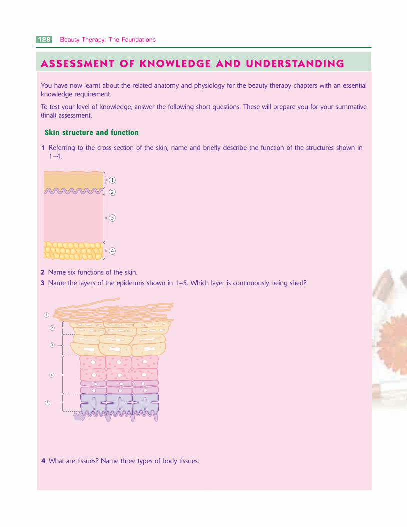
1 Referring to the cross section of the skin, name and briefly describe the function of the structures shown in1–4.
1
2
3
4
2 Name six functions of the skin.
3 Name the layers of the epidermis shown in 1–5. Which layer is continuously being shed?
1
2
3
4
5
4 What are tissues? Name three types of body tissues.
130 Beauty Therapy: The Foundations
2 What is the soft downy hair on the face called?
3 What is coarse pigmented hair called?
4 What is the name of the protein found in skin cells?
5 What is the function of the dermal papilla in relation to the hair and the hair growth cycle?
Hair growHair growth cycleth cycle
1 What are the different stages of the hair growth cycle called?
2 What happens to the hair at each stage?
3 What relevance has the hair growth cycle for a wax depilation treatment?
4 What is the difference in the time between the hair growth cycle anagen to telogen for scalp hair and eyebrowhair?
Nail structure and functionNail structure and function
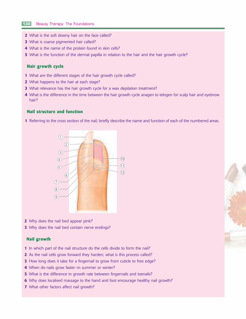
1 Referring to the cross section of the nail, briefly describe the name and function of each of the numbered areas.
6
1
5
4
2
3
7
8
9
10
11
12
2 Why does the nail bed appear pink?
3 Why does the nail bed contain nerve endings?
Nail growNail growthth
1 In which part of the nail structure do the cells divide to form the nail?
2 As the nail cells grow forward they harden; what is this process called?
3 How long does it take for a fingernail to grow from cuticle to free edge?
4 When do nails grow faster–in summer or winter?
5 What is the difference in growth rate between fingernails and toenails?
6 Why does localised massage to the hand and foot encourage healthy nail growth?
7 What other factors affect nail growth?
Anatomy and Physiology 131
Muscle groups in parts of the body, position structure and functionMuscle groups in parts of the body, position structure and function
1 What happens when muscles contract?
2 What structure attaches a muscle to a bone?
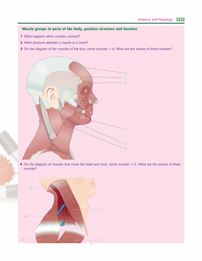
3 On the diagram of the muscles of the face, name muscles 1–6. What are the actions of these muscles?
6
1
5
4
2
3
5
4
3
1
2
4 On the diagram of muscles that move the head and neck, name muscles 1–5. What are the actions of thesemuscles?
134 Beauty Therapy: The Foundations
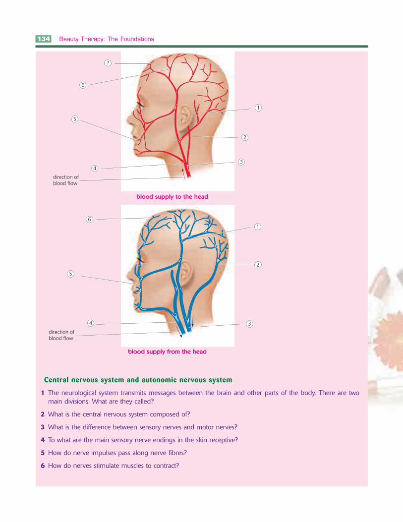
7
6
5
4
1
2
3
1
2
3
6
5
4
direction ofblood flow
direction ofblood flow
blood supply to the head
blood supply from the head
Central nervous system and autonomic nervous systemCentral nervous system and autonomic nervous system
1 The neurological system transmits messages between the brain and other parts of the body. There are twomain divisions. What are they called?
2 What is the central nervous system composed of?
3 What is the difference between sensory nerves and motor nerves?
4 To what are the main sensory nerve endings in the skin receptive?
5 How do nerve impulses pass along nerve fibres?
6 How do nerves stimulate muscles to contract?

Anatomy and Physiology 135
7 What is meant by the autonomic nervous system?
8 How many pairs of cranial nerves emerge from the brain?
9 Those of concern to the beauty therapist when performing facial massage are the 5th, 7th and 11th cranialnerves. What is the function of the: ■ 5th, known as trigeminal nerve■ 7th, known as the facial nerve■ 11th, known as the accessory nerve?
10 Name the main branches of the 7th cranial facial nerve, items 1–5 in the diagram.
1
2
3
4
5