Embed Size (px)
Citation preview

Handbook of Clinical Neurology, Vol. 102 (3rd series)Neuro-ophthalmologyC. Kennard and R.J. Leigh, Editors# 2011 Elsevier B.V. All rights reserved
Chapter 1
Anatomy and physiology of the afferent visual system
SASHANK PRASAD1* AND STEVEN L. GALETTA2
1Division of Neuro-ophthalmology, Department of Neurology, Brigham and Women�s Hospital, Harvard Medical School,Boston, MA, USA
2Neuro-ophthalmology Division, Department of Neurology, Hospital of the University of Pennsylvania, Philadelphia, PA, USA
INTRODUCTION
Visual processing poses an enormous computationalchallenge for the brain, which has evolved highlyorganized and efficient neural systems to meet thesedemands. In primates, approximately 55% of the cortexis specialized for visual processing (compared to 3% forauditory processing and 11% for somatosensory pro-cessing) (Felleman and Van Essen, 1991). Over the pastseveral decades there has been an explosion in scientificunderstanding of these complex pathways and net-works. Detailed knowledge of the anatomy of the visualsystem, in combination with skilled examination, allowsprecise localization of neuropathological processes.Moreover, these principles guide effective diagnosisand management of neuro-ophthalmic disorders.
The visual pathways perform the function of receiv-ing, relaying, and ultimately processing visual informa-tion. These structures include the eye, optic nerves,chiasm, tracts, lateral geniculate nucleus (LGN) of thethalamus, radiations, striate cortex, and extrastriateassociation cortices. Form follows function, and struc-tural relationships often directly determine the under-lying mechanisms of visual processing. The goal ofthis chapter is to describe in detail the anatomy andphysiology of vision.
EYE
The eye is the primary sensory organ for vision, respon-sible for collecting light, focusing it, and encoding thefirst neural signals of the visual pathway. To reach theretina, light must pass through the ocular media, consist-ing of the tear film, cornea, anterior chamber, lens, andthe posterior-chamber vitreous (Fig. 1.1). The corneal epi-thelium and stroma are transparent to permit passage of
*Correspondence to: Sashank Prasad, MD, Department of Neurolo
Hospital, Harvard Medical School, Boston, MA, USA. Tel: (617)
light without distortion (Maurice, 1970). The tear–airinterface and cornea contribute more to the focusingof light than the lens does; unlike the lens, however, thefocusing power of the cornea is fixed. The ciliary mus-cles dynamically adjust the shape of the lens in orderto focus light optimally from varying distances uponthe retina (accommodation). The total amount of lightreaching the retina is controlled by regulation of thepupil aperture. Ultimately, the visual image becomesprojected upside-down and backwards on to the retina(Fishman, 1973).
The majority of the blood supply to structures of theeye arrives via the ophthalmic artery, which is the firstbranch of the internal carotid artery (Hayreh, 2006).The ophthalmic artery enters the orbit via the opticcanal, traveling beside the optic nerve. It then gives riseto two groups of vessels: those supplying the globe(including the central retinal artery, the muscularartery, the anterior ciliary arteries, and the long andshort posterior ciliary arteries) and those supplyingother orbital structures (including the lacrimal artery,supraorbital artery, ethmoidal arteries, frontal artery,and nasal artery). There are minor contributions fromcollateral vessels that originate in the external carotidarteries (via the infraorbital artery and the orbitalbranch of the middle meningeal artery). The patternsof blood supply to the eye and orbit can be quite com-plex, with tremendous variation between individuals(Meyer, 1887; Hayreh, 2006).
RETINA
When light reaches the retina, its energy is converted byretinal photoreceptors into an electrochemical signalthat is then relayed by neurons. To arrive at the
gy, Division of Neuro-Ophthalmology, Brigham and Women’s
732-7432, Fax: (617) 732-6083, E-mail: [email protected]

Posterior chamber
RetinaSuperior rectus muscle
ChoroidFovea
Macula
ScleraVorticose vein
Optic nerve
Central retinal vein
Central retinal artery
Retinal arteries and veins
Medial rectus muscleInferior rectus muscle
Inferior oblique muscle
Conjunctiva
Lens nucleus
Lens cortex
Iris
Cornea
Anterior chamber
Pupil
Canal of Schlemm
Ciliary zonules
Ciliary muscle
Optic nerve sheath
Optic disc
Fig. 1.1. The eye. Light passes through the anterior chamber, the lens, and the posterior chamber, and is then focused
upside-down and backwards upon the retina.
4 S. PRASAD AND S.L. GALETTA
photoreceptors, light must first pass through transpar-ent inner layers of the neurosensory retina, comprisedof the nerve fiber layer, ganglion cells, amacrine cells,and bipolar cells (Fig. 1.2). Immediately outside thephotoreceptor layer is the retinal pigment epithelium
250 μm
TemporalParafovea
Foveola
Fovea centralis
Fig. 1.2. Structures of the neurosensory retina. Top, high-resolutio
Bottom, schematic depiction of retinal layers. (Adapted and rep
www.webvision.med.utah.edu.)
(RPE). The RPE provides structural and metabolicsupport for the photoreceptors, primarily through thevital function of vitamin A metabolism (Wald, 1933).In addition, the RPE absorbs any intraocular light thathas passed through the photoreceptor layer; this reduces
Inner limiting membrane/nerve fiber layerGanglion cell layer
Inner nuclear layerOuter nuclear layerPhotoreceptor layer
Retinal pigment epitheliumChoriocapillarisChoroid
Inner limiting membrane/nerve fiber layerGanglion cell layer
Inner nuclear layer
NasalParafovea
Outer nuclear layerPhotoreceptor layer
Retinal pigment epitheliumChoriocapillarisChoroid
Inner limiting membrane
Outer limiting membrane
ConesRods
Ganglion cell layer
Amacrine cells (Inner nuclear layer)
Bipolar cells (Inner nuclear layer)Horizontal cells (Inner nuclear layer)
Mueller cells
Retinal pigment epithelium
Nerve fiber layer
n optical coherence tomography. Middle, histological section.
rinted with permission from Jaffe and Caprioli (2004), and

F T
backscatter of light and maintains high-fidelity acuity.In contrast, some animals prioritize night vision at theexpense of spatial acuity, and in these species the outer-most layer of the eye (known as the tapetum) is highlyreflective and provides photoreceptors with an additionalopportunity to absorb light. (The reflective tapetum isalso the reason why a cat’s eyes, for example, seem toglow at night.)
Photoreceptors use a highly efficient mechanism toconvert a photon of light into an electrochemical neuralsignal (Fig. 1.3). They contain photopigment, consistingof a membrane protein known as opsin and achromophore molecule called 11-cis-retinal. When lightreaches the photopigment, it causes the chromophore’sconformation to change from 11-cis-retinal to all-trans-retinal. All-trans-retinal detaches from the opsinmolecule because of a low binding affinity. Theunbound opsin molecule then activates another mem-brane complex, the G-protein transducin, by replacinga guanosine diphosphate (GDP) molecule with guano-sine triphosphate (GTP) (Palczewski et al., 2000).Activated transducin subsequently stimulates the effec-tor enzyme phosphodiesterase (PDE), which hydrolyzesthe cytoplasmic second messenger cyclic guanosinemonophosphate (cGMP). Reduction of cytosolic cGMPlevels causes membrane sodium channels to close,reducing the inward sodium current and finally causingthe cell to become hyperpolarized. As a consequence,the photoreceptor cell reduces its neurotransmitter
ANATOMY AND PHYSIOLOGY O
11-cis-retinal
GDP
A
β γ
α
transducin
op
sin
all-trans-retinal
BGTP
β γα
Na+
Na+
cG
PD
E
op
sin
Fig. 1.3. The photoreceptor reaction to light stimulation. (A) 11absorption of light, the chromophore conformation changes to all
ducin by replacing guanosine diphosphate (GDP) with guanosine
phodiesterase (PDE), which metabolizes cytosolic cyclic guanosi
membrane sodium channels to close, lowering the cell’s membran
with permission from http://www.lfhk.cuni.cz/rezacovam/fototran
output in response to the absorption of light. The pro-cess is self-limited: the membrane complex transducincatalyzes GTP back to GDP, ceasing stimulation ofthe PDE enzyme. cGMP levels therefore rise, theinward sodium current is restored, the membranepotential increases, and tonic neurotransmitter releaseis restored.
In order to maintain the ability for continuousresponses to light and preserve high-fidelity neural trans-mission, it is critical that the photoreceptor responsesdo not become saturated. Photoreceptors maintain theability always to produce a signal by ensuring a con-stant, buffered supply of 11-cis-retinal. The spontaneoustransformation of all-trans-retinal back to 11-cis-retinaloccurs over a very slow half-life (of several minutes),so that the store of 11-cis-retinal is always being replen-ished and is available to transduce incoming light signals.
Humans possess four photoreceptor types: threecones and the rods. Under most conditions, our visionis mediated by cones, which operate over an enormousrange of intensities. Each type of cone photoreceptorhas a unique, optimal response to specific wavelengthsof light – short (blue), middle (green), or long (red)(Fig. 1.4). Rods, on the other hand, are saturated atnatural light intensities and are incapable of discrimi-nating colors; their greater sensitivity to light rendersthem effective for night vision (scotopic vision). Thefunctional specializations of cones and rods arise fromvariations in the structure of opsin, since the opsin
HE AFFERENT VISUAL SYSTEM 5
Na+
Na+
cGMP
PD
E
C
GTPα
Na+
cGMPMP
5�-GMP
PD
E
-cis-retinal in its inactive state is bound to opsin. (B) After-trans-retinal and detaches from opsin. Opsin activates trans-
triphosphate (GTP). (C) Activated transducin stimulates phos-
ne monophosphate (cGMP). A reduced level of cGMP causes
e potential and reducing neurotransmitter output. (Reprinted
smise.)

1.0
0.8
0.6
0.4
0.2
0
400 500 600 700Wave length~μm
Rel
ativ
e se
nsiti
vity
or
abso
rban
ce
middle(green)
long(red)
short(blue)
Fig. 1.4. Unique tuning curves for each type of cone photo-
receptor (short, middle, and long) at visible wavelengths of
light. (Adapted and reprinted with permission from Wald
(1964).)
6 S. PRASAD AND
molecule tunes the light wavelengths at which the retinalchromophore (11-cis-retinal) will alter its conformationand initiate the biochemical response to light. Despitetheir functional differences, cones and rods utilize thesame 11-cis-retinal chromophore.
The amount of signal that a photoreceptor producesdepends both on the specific wavelength received andon its intensity. A given photoreceptor may respondequally to a favored wavelength at low intensity andto a less optimal wavelength at high intensity. However,the signal provided by a photoreceptor (the reductionof released neurotransmitters) cannot separately reportthe dual information of wavelength and intensity.Therefore, downstream cells must distinguish these
180,000
160,000
140,000
120,000
100,000
80,000
60,000
40,000
20,000
00 20 40 60
Retinal location (deg
Ph
oto
rece
pto
rs/m
m2
Fig. 1.5. Distribution of rods and cones in the retina. (Adapted f
features of the light stimulus by comparing the differ-ent levels of response from each photoreceptor type.The unique response properties of each photoreceptorallow the ratios between their activities to establish thecode that resolves this ambiguity.
The distribution of cones and rods across the retina ishighly skewed and directly reflects the specialized func-tions of the fovea and retinal periphery (Osterberg, 1935)(Fig. 1.5). The macula is located temporal to the opticnerve and is approximately 5.5 mm in diameter. Withinthe macula is the fovea (diameter 1.5 mm) and the foveola(0.35 mm). The fovea has up to 200 000 cones/mm2
(nearly 15-fold higher than in the peripheral retina) so thatit can provide excellent visual acuity (Hirsch and Curcio,1989). Progressively eccentric locations have much lowerconcentrations of rods, and thus have decreasing sensitiv-ity. Rods are virtually absent in the fovea; rather, they arethe dominant photoreceptor in the periphery.
The neurosensory retina consists of three majorlayers through which the signal in response to light istransduced: photoreceptors connect to bipolar cells,which relay messages to ganglion cells. In addition, hor-izontal and amacrine cells form lateral connectionsbetween elements of these layers. In the fovea, to sup-port high spatial acuity, each bipolar cell receives inputfrom a single photoreceptor; in contrast, in the retinalperiphery a bipolar cell summates the inputs frommultiple photoreceptor cells. Bipolar cells then provideinputs to ganglion cells via direct, excitatory gluta-matergic synapses or indirect, inhibitory GABAergicconnections (Flores-Herr et al., 2001).
The blood supply to the retina arrives via the centralretinal artery, which branches off the ophthalmic artery
S.L. GALETTA
80 100 120
rees from fovea)
rodscones
rom tabulated data (Osterberg, 1935), with permission.)
F T
approximately 1 cm posterior to the globe. It pierces themeninges inferiorly, moves centrally into the nerve, andthen emerges in the center of the optic disc (within theoptic cup) (Hayreh, 2006). It supplies the inner two-thirdsof the retina via temporal vessels that arc above and belowthe macula within the inner retinal layers. The ophthalmicartery also gives rise to posterior ciliary arteries, whichsupply the optic nerve head, choroid, and the outer one-third of the retina. A cilioretinal artery exists in about20%of individuals, arising from the choroidal circulation.In the fovea there is a 400-mm area known as the fovealavascular zone, in which photoreceptors are packed mostdensely without intervening capillaries.
Retinal veins drain into the central retinal vein,which lies temporal to the central retinal artery in theoptic nerve head (Hayreh, 2006). The central retinal veineventually drains into the superior orbital vein and thecavernous sinus. The choroidal vasculature has a sepa-rate drainage route through vortex veins, the superiorand inferior orbital veins, and finally into the cavernoussinus.
GANGLIONCELLS
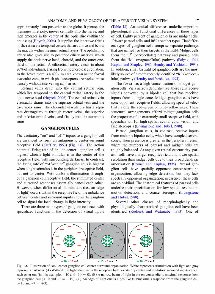
The excitatory “on” and “off” inputs to a ganglion cellare arranged to form an antagonistic center-surroundreceptive field (Kuffler, 1953) (Fig. 1.6). The actionpotential firing rate of an “on-center” ganglion cell ishighest when a light stimulus is in the center of thereceptive field, with surrounding darkness. In contrast,the firing rate of “off-center” ganglion cells is highestwhen a light stimulus is in the peripheral receptive field,but not its center. With uniform illumination through-out a ganglion cell receptive field, the summated centerand surround responses essentially cancel each other.However, when differential illumination (i.e., an edgeof light) occurs within the receptive field, the imbalancebetween center and surround inputs allows the ganglioncell to signal the local change in light intensity.
There are three main types of ganglion cell, each withspecialized functions in the detection of visual inputs
ANATOMY AND PHYSIOLOGY O
Fig. 1.6. Illustration of “on”-center ganglion cell center-surroundrepresents darkness. (A) With diffuse light stimulus in the receptiv
each other out (in this example, þ10 and –10 ¼ 0). (B) A narrow
the ganglion cell (þ10 and –0 ¼ þ10). (C) An edge of light eli
(þ10 and –7 ¼ þ3).
(Table 1.1). Anatomical differences underlie importantphysiological and functional differences in these typesof cell. Eighty percent of ganglion cells are midget cells,10% are parasol cells, and 10% are other types. The differ-ent types of ganglion cells comprise separate pathwaysthat are named for their targets in the LGN. Midget cellsform the “P” (parvocellular) pathway and parasol cellsform the “M” (magnocellular) pathway (Polyak, 1941;Kaplan and Shapley, 1986; Hendry and Yoshioka, 1994).In addition, small bistratified ganglion cells are the mostlikely source of a more recently identified “K” (koniocel-lular) pathway (Hendry and Yoshioka, 1994).
The fovea has a high concentration of midget gan-glion cells. Via a narrow dendritic tree, these cells receivesignals conveyed by a bipolar cell that has receivedinputs from a single cone. Midget ganglion cells havecone-opponent receptive fields, allowing spectral selec-tivity along the red–green or blue–yellow axes. Thesestructural arrangements afford midget ganglion cellsthe properties of an extremely small receptive field, withspecialization for high spatial acuity, color vision, andfine stereopsis (Livingstone and Hubel, 1988).
Parasol ganglion cells, in contrast, receive inputsfrom multiple bipolar cells, which have sampled severalcones. Their presence is greater in the peripheral retina,where the numbers of parasol and midget cells areroughly balanced. At any given retinal eccentricity, par-asol cells have a larger receptive field and lower spatialresolution than midget cells due to their broad dendriticarborization (Croner and Kaplan, 1995). Parasol gan-glion cells have spatially opponent center-surroundorganization, allowing edge detection, but they lackspectrally opponent organization; in essence, these cellsare color-blind. The anatomical features of parasol cellsunderlie their specialization for low spatial resolution,motion detection, and coarse stereopsis (Livingstoneand Hubel, 1988).
Several other classes of morphologically andphysiologically characterized ganglion cell have beenidentified (Rodieck and Watanabe, 1993). One of
HE AFFERENT VISUAL SYSTEM 7
organization. White represents stimulation with light and gray
e field, excitatory center and inhibitory surround inputs cancel
beam of light in the on-center elicits maximal response from
cits a positive (submaximal) response from the ganglion cell

Table 1.1
Features of the ganglion cell types in the P, M, and K pathways
Property P pathway M pathway K pathway
Retinal source Midget cells Parasol cells UnknownReceptive field size Small Large LargeGanglion cells/mm2 Many Few Unknown
Spectral opponency Yes No Some (blue-on)Conduction velocity
of axonsLow High Low/varied
LGN projection target Magnocellular layers Parvocellular layers Intercalated
Fraction of LGNpopulation
80% 10% 10%
V1 Projection target 4Cb 4Ca Layers 2–3, CO blobs
Illustration
P, parvocellular; M, magnocellular; K, koniocellular; LGN, lateral geniculate nucleus; CO, cytochrome oxidase.
Illustrations reprinted from Ghosh et al. (1996), with permission.
8S.PRASAD
AND
S.L.GALETTA

F THE AFFERENT VISUAL SYSTEM 9
these, called small bistratified cells, may be the mainprojection to the koniocellular layers of the LGN(Hendry and Yoshioka, 1994). Relatively little isknown about this pathway, because it has been diffi-cult to study in isolation. Given that koniocellularlayers of the LGN subsequently project to cytochromeoxidase-rich regions of the upper layers of primaryvisual cortex (CO “blobs”), these cells may play a rolein some aspects of color vision (Hendry and Yoshioka,1994). An additional class of ganglion cells are uniquein that they contain the melanopsin pigment and there-fore demonstrate direct responses to light (Hattar et al.,2002). These cells may participate in the pupillary lightreflex and may contribute to the pathophysiology ofphotophobia. Their main function, however, is to medi-ate circadian rhythms, as discussed below in the sectionentitled “Suprachiasmatic Nucleus.”
The axons of ganglion cells travel in the nerve fiberlayer (the innermost retinal layer), enter the opticnerve, travel through the chiasm and tract, and thenfinally synapse in the LGN of the thalamus. Fovealganglion cells send axons directly to the temporalaspect of the optic disc in the papillomacular bundle(Fig. 1.7). The remaining temporal ganglion cell nerveaxons are arranged on either side of the horizontalraphe and form arcuate bundles that course aboveand below the fovea, and finally enter the superiorand inferior portions of the optic nerve. Finally, axonsoriginating nasal to the disc enter the nasal portionof the optic nerve.
ANATOMY AND PHYSIOLOGY O
Arcuatebundles Optic discPapillomacular bundle
Fovea
T N
Horizontalraphe
Fig. 1.7. Arrangement of the retinal nerve fiber layer, com-
prised of ganglion cell axons. The papillomacular bundle
conveys axons from the fovea directly to the temporal margin
(T) of the optic disc. The remainder of temporal ganglion cell
axons are arranged in arcuate bundles above and below the
fovea, arriving at the superior and inferior disc margins.
Finally, axons originating nasal to the disc arrive at its nasal
border (N).
OPTICNERVE
Each optic nerve is comprised of approximately1.2 million retinal ganglion cell axons (in constrast tothe acoustic nerve, for example, which has only 31 000axons) (Bruesch and Arey, 1942). The intraocular seg-ment of the optic nerve head (the optic disc) is typicallylocated 3–4 mm nasal to the fovea and is 1 mm thick.The optic disc has a central depression called the opticcup, which is typically one-third the size of the disc(Jonas et al., 1988). There are no retinal photoreceptorsin the region of the optic disc, which gives rise to themonocular blind spot (Mariotte, 1668).
The optic nerve travels posteriorly through the lam-ina cribrosa to exit the back of the globe, where itabruptly increases in diameter from 3 to 4 mm. In orderto accommodate the rotations of the globe, the lengthof the intraorbital segment of the optic nerve is typi-cally between 25 and 30 mm in length, at least 5 mmlonger than the distance from the globe to the orbitalapex (Glaser and Sadun, 1990). Upon passing throughthe lamina cribrosa, the optic nerve becomes investedwith meninges and also becomes myelinated. Since theoptic nerve is an extension of the central nervous sys-tem, unlike other cranial and peripheral nerves, it ismyelinated by oligodendrocytes rather than Schwanncells. Upon exiting the orbit, the optic nerve entersthe optic canal, within the lesser wing of the sphenoidbone, for approximately 6 mm. The intracanalicularoptic nerve rises at a 45� angle and then exits the opticcanal, where it continues in its intracranial portion forapproximately 17 mm before reaching the chiasm.
In the proximal third of the optic nerve the positionsof ganglion cell axons are rearranged. Macular gan-glion cell axons which initially lie temporally move tothe nerve’s center. Peripheral temporal fibers becomepositioned temporally, both superior and inferior tothe macular fibers. Finally, nasal fibers remain in thenasal portion of the optic nerve.
Axoplasmic transport is essential to the maintenanceof ganglion cell axons (Minckler, 1986). Orthograde axo-nal transport (movement away from the ganglion cellbody, toward the LGN) occurs at both slow and fastspeeds, and relies upon elements of the axon cytoskel-eton (microtubules, neurofilaments, andmicrofilaments).Slow transport (1–4 mm/day) is used for elements ofthe cytoskeleton itself, while fast vesicular transport(400 mm/day) is used for proteins and neurotransmitters,using motor proteins (kinesin and dynein) (Vale et al.,1985). Mitochondria are essential to providing energy inthe form of adenosine triphosphate (ATP) for theseprocesses to occur.
All of the blood supply to the optic nerve is ulti-mately derived from the ophthalmic artery (Fig. 1.8).

Fig. 1.8. Blood supply to the optic nerve. (Reprinted with permission from Patten (2004).)
10 S. PRASAD AND S.L. GALETTA
The anastomotic circle of Zinn–Haller provides circu-lation to the optic nerve head, and is supplied by theposterior ciliary arteries, the pial arteriole plexus,and the peripapillary choroid (Anderson, 1970). Theposterior ciliary arteries each provide a variable, seg-mental supply to portions of the optic nerve belowits head. Distal portions of the optic nerve receiveblood supply from the arterial pial plexus. Within theoptic canal, this vascular network is usually suppliedby the internal carotid artery, and in the intracranialsegment of the optic nerve it is supplied by the internalcarotid, anterior cerebral, or anterior communicatingarteries.
OPTICCHIASM
The chiasm, which has a dumbbell shape when viewedin coronal section, is the site of decussation foraxons from the optic nerve (Fig. 1.9). It lies in the
Suprasellarcistern
Sellaturcica
Pituitary
AComm
Chiasm
ICA
PComm
A B
10 mm
4
45i
ON15mm
8mm
Fig. 1.9. The optic chiasm. (A, sagittal view) The chiasm lies in
sella turcica. (B, ventral view) The chiasm is situated between t
arteries (ACA) and anterior communicating artery (AComm) lie
lie inferiorly. (C) Partial decussation of ganglion cell axons at th
superior nasal fibers decussate posteriorly. Temporal fibers trav
MCA, middle cerebral artery. (Adapted and reprinted with perm
subarachnoid space of the suprasellar cistern, abovethe diaphragma sella and the pituitary gland, inferiorto the hypothalamus, and anterior to the pituitary stalk(infundibulum). The chiasm is typically 10 mm above thepituitary, which rests in the sella turcica within the sphe-noid bone. In most individuals, the chiasm is directlyabove the pituitary, but in 15% it is displaced anteriorly,above the tuberculum sellae (a prefixed chiasm), and in5% it is displaced posteriorly, over the dorsum sellae(a postfixed chiasm) (Bergland et al., 1968; Doyle, 1990).
The chiasmal decussation serves to bring togetherinformation from the halves of each retina that viewthe same portion of the visual field. Therefore, axonsfrom nasal ganglion cells cross and join axons from tem-poral ganglion cells from the contralateral eye. Thereis a greater number of crossed (53%) than uncrossed(47%) fibers. Among crossing fibers, those originatingin the macula lie in a superoposterior position within
Crossingfibers
Opticnerve
Optictract
ACA
C
MCAR
L
the suprasellar cistern, 10 mm above the pituitary gland in the
he two internal cerebral arteries (ICA). The anterior cerebral
superiorly and the posterior communicating arteries (Pcomm)
e optic chiasm. Inferior nasal fibers decussate anteriorly and
el in the lateral aspect of the chiasm and do not decussate.
ission from Hoyt and Luis (1963).)

Hilum6
54
3
21
Medialhorn
Lateralhorn
Parvocellular
Magnocellular
Fig. 1.10. Layers of the lateral geniculate nucleus. Layers 1
and 2 are the magnocellular layers, and layers 3–6 are the par-
vocellular layers. Inputs to the hilum are from the central
visual field, those to the medial horn are from the inferior
visual field, and those to the lateral horn are from the supe-
F THE AFFERENT VISUAL SYSTEM 11
the chiasm. Axons from the inferior nasal retina maybend slightly forward (up to 3 mm) into the contralateraloptic nerve, forming a structure called Wilbrand’s knee(Hoyt, 1970). However, the existence of Wilbrand’s kneehas been questioned, with the suggestion that this anato-mical arrangement may be the artifactual result of long-term monocular enucleation (Horton, 1997).
The optic chiasm lies between several main arteriescomprising the circle of Willis. It is situated betweenthe two internal carotid arteries at their supraclinoidportion, above the two posterior communicatingarteries, and beneath the anterior cerebral arteries andthe anterior communicating artery. These neighboringvessels provide an anastomotic supply to the chiasmvia an inferior group (the superior hypophyseal arteries,which derive from the internal carotid, posterior com-municating, and posterior cerebral arteries) as well asa superior group (branches from the anterior cerebralarteries) (Bergland et al., 1968). The body of the chiasmmay be predominantly supplied by the inferior group,whereas the lateral aspects of the chiasm may receivedual supply from both the superior and inferior groups.
ANATOMY AND PHYSIOLOGY O
rior visual field. (Adapted and reprinted from http://www.
psych.ucalgary.ca/PACE/VA-LAB/.)
OPTIC TRACTSEach optic tract contains axons from the ipsilateral tem-poral retina and the contralateral nasal retina. In theproximal optic tract there is a 90� inward rotation offibers such that inferior retinal axons from each eyebecome positioned laterally and those from the superiorretinas become positioned medially. In the posteriortract, these fibers fan out toward the LGN and inter-digitate into its separate layers. The blood supply tothe optic tract is variable but typically arises fromanastomotic branches of the posterior communicatingand anterior choroidal arteries.
LATERALGENICULATENUCLEUS
Most fibers from the optic tracts synapse in the ipsilat-eral LGN. The number of cells in the LGN is large, withnearly a 1:1 ratio to ganglion cell inputs (Spear et al.,1996). There is retinotopic organization in the LGN,with macular vision represented in the hilum (centralportion), the superior field represented in the lateralhorn, and the inferior field represented in the medialhorn (Kupfer, 1962) (Fig. 1.10). The LGN is arrangedin six neuronal layers, each with monocular inputs.Ganglion cell axons from the ipsilateral eye (temporalretina) synapse in layers 2, 3, and 5, while axons fromthe contralateral eye (nasal retina) synapse in layers1, 4, and 6 (Chacko, 1948). Layers 1 and 2 contain largeneurons (magnocellular layers) which receive parasolganglion cell inputs; layers 3–6 contain small neurons(parvocellular) and receive midget ganglion cell inputs
(Leventhal et al., 1981). In addition, scattered undereach of the six major layers of the LGN in an inter-calated, intralaminar fashion are the so-called konio-cellular (tiny, dust-like) cells.
The blood supply to the LGN is segregated andarrives via the anterior and posterior choroidal arteries.The anterior choroidal artery is a proximal branch ofthe internal carotid artery, and it supplies the medialand lateral horns of the LGN. The posterior choroidalarteries derive from the posterior cerebral arteries andsupply the hilum of the LGN.
The LGN is a critical relay station with dynamic con-trol upon the amount and nature of information that istransmitted to visual cortex (Guillery and Sherman,2002). In addition to retinal afferents, which may com-prise only 5–10% of the synapses in the LGN (Van Hornet al., 2000), the LGN also receives extensive modulatingconnections from the thalamic reticular nucleus andlayer 6 of the visual cortex. The LGN thus provides abottleneck to information flow, filtering visual infor-mation for relevance to the present behavioral state.
The pulvinar is another thalamic nucleus, much largerthan the LGN, that forms a higher-order relay receivingextensive descending cortical projections from both layers5 and 6 of the visual cortex (Chalupa, 1991). While inputsfrom layer 5 are essential to driving pulvinar responses,inputs from layer 6 have more subtle modulating effects(Sherman and Guillery, 1998). The pulvinar has wide-spread connections to most cortical regions and is key to

D
transthalamic corticocortical pathways, capable of modi-fying transmission in accord with requirements of selec-tive visual attention (Petersen et al., 1987; Rafal andPosner, 1987). More specific functions of the pulvinar invisual processing, however, have yet to be elucidated.
SUPERIORCOLLICULI
The superior colliculi play a critical role in generatingorienting eye and head movements to sudden visual(and other sensory) stimuli. They are located in thedorsal midbrain within the tectal plate. The superiorcolliculi are structurally and functionally organizedinto superficial and deep layers. The superficial layerssolely process visual information, with direct retinalinputs comprising a visuotopic map of the contralat-eral field (Cynader and Berman, 1972). Along the ante-rior–posterior axis of each colliculus, the receptivefields of these neurons move from the central visualfield to more peripheral locations. Along the medial–lateral axis, the receptive fields move from the uppervisual field to the lower visual field. The representa-tion of foveal vision is magnified, with over one-thirdof collicular neurons processing inputs from the cen-tral 10� of vision. The superficial layers have efferentconnections to thalamic nuclei; these signals are thenrelayed to cortical visual areas. The deep layers ofthe colliculi receive multimodal sensory inputs andhelp mediate saccadic eye movements through theirefferent connections to ocular motor systems. Inaddition, these layers receive reciprocal connectionsfrom several cortical areas involved in generatingsaccades.
PRETECTALNUCLEI
A portion of the fibers in the optic tract subserve thepupillary light reflex and synapse at the pretectalnuclei in the midbrain. There is consensual innervationto both pretectal nuclei, and each pretectal nucleus hasdual connections to each Edinger–Westphal nucleus.
12 S. PRASAD AN
Fig. 1.11. (A, B) Pathways of the optic radiations, from the latera
fusion tensor imaging (which identifies white-matter tracts). (Re
The Edinger–Westphal nuclei give rise to parasym-pathetic efferent fibers which travel with the oculo-motor nerve and regulate pupillary size via pupillaryconstrictors.
SUPRACHIASMATICNUCLEUS
A newly identified type of retinal ganglion cell containsthe photopigment melanopsin and demonstrates an intrin-sic responsiveness to light (not mediated by rod and conephotoreceptors) (Hattar et al., 2002). These ganglion cellsgive rise to a separate, unmyelinated pathway through theoptic chiasm and tracts, ultimately transmitting light infor-mation directly to the suprachiasmatic nucleus (SCN) atthe base of the anterior hypothalamus (Moore, 1973). Asopposed to most visually responsive brain areas, whereneural responses are transient, the responses in the SCNare sustained for up to 20 seconds. Together with largereceptive fields, these properties allow the SCN to monitorambient light levels reliably. The SCN has efferent connec-tions to the pineal gland, where melatonin secretion occursto drive circadian rhythms.
OPTICRADIATIONS
The second-order neurons of the visual pathway formthe optic radiations extending from the LGN to striate(calcarine) cortex in the occipital lobe. These neuronsare grouped into two major bundles: the temporal radia-tions (which take an anterior course through the tempo-ral pole, termed Meyer’s loop, before turning in aposterior direction) and the parietal radiations (VanBuren and Baldwin, 1958) (Fig. 1.11). Retinotopicarrangement is maintained; temporal radiations repre-sent the contralateral superior field and parietalradiations represent the contralateral inferior field.
The temporal optic radiations receive blood supplyfrom the anterior choroidal artery as well as proximalbranches from the middle cerebral arteries, includingthe lenticulostriate and inferior temporo-occipital arteries.The parietal radiations are supplied by more distal
S.L. GALETTA
l geniculate nucleus to calcarine cortex, demonstrated by dif-
printed with permission from Yamamoto et al. (2007).)

F T
branches of the middle cerebral artery, including theangular and posterior temporal arteries. Distal portionsof the optic radiations, before their entry into visual cor-tex, are supplied by the superior temporo-occipital branchof the middle cerebral artery and the anterior temporaland calcarine branches from the posterior cerebral artery.
ANATOMY AND PHYSIOLOGY O
CALCARINECORTEX (PRIMARYVISUALCORTEX)
The optic radiations arrive at the mesial surface of theoccipital lobe, in the striate (calcarine) cortex. Fasciclesfrom the parietal radiations synapse in the superiorbank of calcarine cortex, while those from the temporalradiations arrive in the inferior bank. These axons makeconnections in cortical layer 4, termed the “stripe ofGennari,” which is plainly visible on gross inspection ofthe brain (Gennari, 1782). There is a 300–400-fold increasein the total number of neurons in primary visual cortex
BA
FOVEA
Fig. 1.12. Retinotopic map of the human striate cortex. (A) Leftcalcarine fissure. The dotted lines demark area V1, showing the up
(B) The gyrus is lifted to reveal the banks of the calcarine sulcus
Note the immense magnification of central vision. (C) The contraloval represents the monocular blind spot and the stippled zone re
from Horton and Hoyt (1991).)
Fig. 1.13. Retinotopic organization of striate cortex using functio
map receptive field eccentricity. Note magnification of the foveal
polar angle. Note that the superior fields are represented in the inf
rior bank. (Reprinted with permission from Dougherty et al. (200
compared to the retina or the LGN, with approximately350 million neurons packed at a density that may be twiceas high as that in other cortical areas (Tolhurst and Ling,1988). Representation of the vertical meridian of thevisual field lies most medially, in the calcarine lips, whilethe horizontal meridian is represented deep within the cal-carine fissure (Gray et al., 1998; Galetta and Grossman,2000). Macular projections make their synapses in theposterior pole of calcarine cortex (Fig.s 1.12 and 1.13).The macular representation is greatly magnified in thevisual cortex retinotopic map; connections from 1 mm2
of retina, representing the central 10� (2% of the totalvisual field), encompass 60% of striate cortex (Tolhurstand Ling, 1988; Horton andHoyt, 1991). The psychophysi-cal correlate of this cortical magnification is extremelyhigh central acuity (spatial resolution of 60–100 cycles/degree). More anterior portions of striate cortex repre-sent the peripheral visual field. The most anterior bankof calcarine cortex represents the temporal 30� of the
HE AFFERENT VISUAL SYSTEM 13
180
315
270
4590
2.551020
40
Coccipital lobe, with striate cortex in the lips and banks of the
per and lower vertical meridian of the contralateral hemifield.
, with its retinotopic map marked in degrees from the fovea.
ateral visual field, marked in degrees from the fovea. The dark
presents the monocular crescent. (Reprinted with permission
nal magnetic resonance imaging. (A) Expanding ring stimuli
representation. (B) Rotating wedge stimuli map receptive field
erior bank, and the inferior fields are represented in the supe-
3).)

D
contralateral field; this area receives solemonocular inputfrom the contralateral eye, since this portion of the visualfield is not represented by the ipsilateral eye.
The main blood supply to visual cortex is providedby the posterior cerebral arteries and its branches (thecalcarine, posterior temporal, and parieto-occipitalarteries) (Smith and Richardson, 1966) (Fig. 1.14). Atthe occipital pole, however, there may be a dual bloodsupply to the area subserving central vision, with ana-stomoses between branches of the posterior cerebralarteries and the superior temporo-occipital branch fromthe middle cerebral artery.
In layer 4 of striate (calcarine) cortex there is anato-mical division of the two functionally segregated LGNinputs. Neurons from the magnocellular layers (M path-way) synapse in cortical layer 4Ca, while those from theparvocellular layers (P pathway) synapse in layer 4Cb. Inaddition, both M and P pathway neurons send collateralinputs to layer 6, which in turn sends reciprocal projec-tions back to the thalamus. Neurons of the K pathwaysend projections directly to the “CO blobs” in layer 3and layer 1 of visual cortex, rather than to layer 4 or 6.
Monocular inputs to primary visual cortex arearranged in ocular dominance columns. The two eyeshave different views of visual space, resulting in a slightdisplacement of their respective retinal images. At thebinocular fixation point (which depends upon the
14 S. PRASAD AN
Fig. 1.14. Occipital blood supply. The posterior cerebral arteries pvia the parieto-occipital arteries, calcarine arteries, and posterior
occipital pole is supplied by branches of the middle cerebral arte
permission from Smith and Richardson (1966).)
amount the eyes are converged), an image is projectedon to anatomically corresponding retinal locations.Objects located in front of or behind the binocular fixa-tion point, however, give rise to noncorresponding reti-nal images (Fig. 1.15). This retinal disparity forms thebasis of cortical calculations of stereoscopic depth, sinceneurons in V1 as well as extrastriate regions are sensitiveto these differences. Some cortical neurons respondmost strongly for objects further than the fixation point,while others are tuned for objects nearer than the fixa-tion point (Barlow et al., 1967; Poggio et al., 1988; Backuset al., 2001). These features allow the monocular two-dimensional projection of visual space to become a rich,three-dimensional perception (Wheatstone, 1838).
V1 neurons are particularly selective for specificorientations of luminance contrast, forming the basisof image contour analysis (Hubel and Wiesel, 1962).In addition, there is the initial processing of color com-position, brightness, and direction of motion (Tootellet al., 1988a, b). The reason that V1 neurons are ableto detect contours is that their receptive fields have alarger, elongated on-center that is constructed fromthe concentric-ring on-center inputs of ganglion cells(Fig. 1.16). When a light stimulus is the preferred orien-tation, it spans the on-centers of these contributing gan-glion cells and therefore elicits a maximal responsefrom the V1 neuron (Hubel and Wiesel, 1962).
S.L. GALETTA
rovide the majority of the blood supply to the occipital cortex,
temporal arteries. In addition, collateral blood supply to the
ry, via the superior temporo-occipital arteries. (Adapted with

Fig. 1.15. Basis for stereoscopic vision. At the binocular fix-ation point there is retinal correspondence of images on the
fovea, but for objects near or far to fixation there is retinal
disparity. Cells in visual cortex are sensitive to the presence
and amount of binocular disparity, thus signalling whether
images are at the point of fixation or in the fore/background.
ANATOMY AND PHYSIOLOGY OF T
FEEDBACKMECHANISMS
Information processing relies not only on feedforwardconnections to higher visual areas but also on reciprocalconnections that transfer information in the reverse
Fig. 1.16. V1 neuron receptive fields. The inputs from adjacent ga
with the proper orientation will maximally stimulate the V1 neur
reduced response from the V1 neuron because of competing exci
direction (Felleman and Van Essen, 1991). These feed-back connections, which far outweigh the feedforwardinputs arriving from the LGN, serve to fine-tune theprocessing of incoming stimuli. Even in V1, therefore,neural responses do not mirror retinal inputs precisely,but are modified by higher inputs to support a coherentperceptual interpretation.
While line segments form the ideal stimulus for a V1neuron given its small, simple receptive field, a singleline segment on a blank background is a rarely encoun-tered visual scene. In natural scenes, multiple contouredges are present, forming diverse object boundaries.An accurate perceptual interpretation therefore relieson the contextual processing of individual line seg-ments within the overall visual scene. The context of astimulus can greatly alter its perceptual salience, withaccompanying changes even in early stages of neuralprocessing, including altered responses in V1 neuronsthemselves (Knierim and van Essen, 1992) (Fig. 1.17).When a stimulus is similar to its surroundings, thecamouflage effect is reflected by a reduction of V1responses corresponding to that receptive field. Onthe other hand, when a stimulus differs from its sur-roundings, a perceptual popout occurs, mediated by apartial restoration of V1 responses (Nothdurft, 1991;Knierim and van Essen, 1992). Top-down and horizon-tal corticocortical connections give rise to contextualmodulation in early visual areas by facilitating selectiveresponses (Callaway, 1998). Surrounding stimuli thatare outside the “classical” receptive field of a neuronmay therefore modulate its activity, through both facil-itatory and inhibitory mechanisms (Allman et al., 1985;Lamme and Roelfsema, 2000). In fact, in the rightsetting, neurons in visual cortex will exhibit responsesto illusory contours, which are perceptually suggested
HE AFFERENT VISUAL SYSTEM 15
nglion cells are the inputs to a V1 neuron. (A) A light stimulus
on. (B) A light stimulus with another orientation will create a
tatory and inhibitory inputs.

Fig. 1.17. (A) The optimal stimulus in the receptive field of a V1 neuron is a line segment. (B) The response of the V1 neuron isdampened when many surrounding stimuli contain a similar stimulus (camouflage effect). (C) A partial restitution of activity
occurs in the V1 neuron when the surrounding stimuli are different from the stimulus processed in its receptive field (popout
effect).
Fig. 1.18. The Kanizsa figure demonstrates the phenomenon
of illusory contours. A superimposed white triangle is seen,
although no physical stimulus exists to define its shape.
Mechanisms of perceptual constancy impose this pattern,
with neural firing that supports the coherent perception
(Kanizsa, 1976).
Fig. 1.19. Schematic of the dorsal and ventral processing
streams. MST, medial superior temporal area; LO, lateral
occipital; FFA, fusiform face area; PPA, parahippocampal
place area.
16 S. PRASAD AND S.L. GALETTA
by surrounding stimuli but not physically present (vonder Heydt et al., 1984) (Fig. 1.18). Ultimately, thesemechanisms are essential to generating figure–groundsegregation and a coherent phenomenal experience ofthe visual scene (Lamme et al., 2000).
HIGHER-ORDERVISUAL CORTEX
The complexity of receptive field properties progres-sively increases from lower-order to higher-order pro-cessing areas. Information is integrated to endowhigher-order neurons with receptive fields of greatlyincreased size and complexity. While early processingareas contain neurons with relatively small receptivefields confined to the contralateral visual hemifield,neurons in higher visual areas have expanded receptivefields that span both hemifields. Projections to these
higher areas lose their strict point-to-point retinotopicarrangement (Livingstone and Hubel, 1983). Representa-tion of the ipsilateral visual hemifield within thesecortical areas is mediated by interhemispheric callosalconnections relaying information from contralateralearly visual areas.
Multiple extrastriate visual areas, each specialized forthe detection of particular attributes of visual scenes, areorganized into two roughly parallel processing streams(Fig. 1.19). Ungerleider and Mishkin (1982) originallyproposed two functionally dichotomous processingstreams, in which the ventral stream mediates visual rec-ognition of objects (the “what” pathway) and the dorsalstream is specialized for processing spatial relationshipsamong objects (the “where” pathway). In addition tovisuospatial processing, the dorsal stream is also involvedwith grasping and manipulating objects (Goodale et al.,1991). The notion of “what” and “where” pathways wasbuilt upon an observed double dissociation in the behaviorof macaques with selective cortical lesions. Lesionsof inferior temporal cortex caused severe deficits ofvisual discrimination (identifying objects, color, patterns,or shapes) without affecting visuospatial performance

F T
(visually guided reaching or judging the distance betweenobjects); conversely, lesions of posterior parietal cortexhad the opposite effects.
The ventral stream begins in layer 4Cb of area V1,which sends projections to the thin and interstriperegions of V2. The thin region mainly represents colorinformation, while the interstripe regions representform (Shipp and Zeki, 1985; Sincich and Horton, 2002).These regions subsequently project to area V4 (Zeki,1980), which ultimately projects to inferotemporal cortex.Here, specialized neurons are involved in visual objectprocessing.
The dorsal stream begins in motion-sensitive compo-nents of layer 4Ca in area V1. From here, projections aresent to the thick stripes in areas V2 and V3 (Shipp andZeki, 1985; Sincich and Horton, 2002). Subsequently,these areas send connections to area V5, and ultimatelythese inputs arrive at the medial superior temporal areacomplex, which participates in higher-order motion anal-ysis (Boussaoud et al., 1990; Tootell et al., 1995). Proces-sing in the dorsal stream occurs at shorter latencies thanprocessing in the ventral stream (Schmolesky et al.,1998), leading some investigators to propose the term“fast brain” for the visual areas in the dorsal stream(Nowak and Bullier, 1997). In fact, axons in the dorsalstream contain more myelin than their counterparts inthe ventral stream (Nowak and Bullier, 1997).
Functional imaging while subjects perform visualprocessing tasks has allowed the characterization ofhighly specialized cortical areas. The lateral occipital (LO)area, within ventral occipitotemporal cortex, has func-tional specialization for processing objects comparedto nonobjects (Tanaka, 1993; Grill-Spector et al., 2001).LO includes the fusiform face area, specialized for faceprocessing (Kanwisher et al., 1997), and the parahippo-campal place area, specialized for processing scenes(Epstein and Kanwisher, 1998). Although the retinalinputs of a visual object will differ according to featuressuch as its position, distance, illuminance, and orienta-tion, the ventral stream extracts invariant features ofan object that enable perceptual constancy across vari-able circumstances (Gross and Mishkin, 1977; Ito et al.,1995). Short-term habituation of these neurons, wherebyrepeated presentations lead to decreased responses,allow signaling of the novelty or significance of an object(Miller and Desimone, 1994).
Ultimately, visual information is sent from bothdorsal and ventral streams to distant cortical areas for thehighest levels of processing. Inputs to entorhinal cortex(via perirhinal and parahippocampal areas) mediate theformation of long-term memories of visual objects.Inputs to prefrontal cortex are critical for visual workingmemory. Direct inputs to the amygdala serve to attachemotional valence to a visual stimulus. Mechanisms of
ANATOMY AND PHYSIOLOGY O
attention interact with processing of visual informationat all stages (Desimone and Duncan, 1995; Treue andMaunsell, 1996; Reynolds et al., 2000).
CONCLUSION
The visual system demonstrates remarkably efficientorganization in its transmission of information with thegoal of accomplishing higher-order, complex visuospa-tial and object identity processing. Anatomical divisionsreflect the segregation of functions at each stage. Com-bined feedforward and feedback connections give riseto powerful computational ability, with high processingefficiency and selective filtering of inputs. The specificanatomical and physiological features of the functionalorganization of the visual system have been the focusof intense research. Current knowledge has been sum-marized in this chapter, and further insights are inevi-table with ongoing modern research efforts.
REFERENCES
Allman J, Miezin F, McGuinness E (1985). Stimulus
specific responses from beyond the classical receptive
field: neurophysiological mechanisms for local-global
comparisons in visual neurons. Annu Rev Neurosci 8:
407–430.Anderson DR (1970). Vascular supply to the optic nerve of
primates. Am J Ophthalmol 70: 341–351.Backus BT, Fleet DJ, Parker AJ et al. (2001). Human cortical
activity correlates with stereoscopic depth perception.
J Neurophysiol 86: 2054–2068.Barlow HB, Blakemore C, Pettigrew JD (1967). The neural
mechanism of binocular depth discrimination. J Physiol
193: 327–342.Bergland RM, Ray BS, Torack RM (1968). Anatomical varia-
tions in the pituitary gland and adjacent structures in 225
human autopsy cases. J Neurosurg 28: 93–99.Boussaoud D, Ungerleider LG, Desimone R (1990). Pathways
for motion analysis: cortical connections of the medial
superior temporal and fundus of the superior temporal
visual areas in the macaque. J Comp Neurol 296: 462–495.Bruesch SR, Arey LB (1942). The number of myelinated
and unmyelinated fibers in the optic nerve of vertebrates.
J Comp Neurol 77: 631.Callaway EM (1998). Local circuits in primary visual cortex
of the macaque monkey. Annu Rev Neurosci 21: 47–74.Chacko LW (1948). The laminar pattern of the lateral genic-
ulate body in the primates. J Neurol Neurosurg Psychiatry
11: 211–224.Chalupa L (1991). Visual function of the pulvinar. In: L Ag
(Ed.), The Neural Basis of Visual Function. CRC Press,
Boca Raton, pp. 140–159.
Croner LJ, Kaplan E (1995). Receptive fields of P and M gan-
glion cells across the primate retina. Vision Res 35: 7–24.Cynader M, Berman N (1972). Receptive-field organization
of monkey superior colliculus. J Neurophysiol 35:187–201.
HE AFFERENT VISUAL SYSTEM 17

18 S. PRASAD AND S.L. GALETTA
Desimone R, Duncan J (1995). Neural mechanisms of selec-
tive visual attention. Annu Rev Neurosci 18: 193–222.Dougherty RF, Koch VM, Brewer AA et al. (2003). Visual
field representations and locations of visual areas V1/2/3
in human visual cortex. J Vis 3: 586–598.Doyle AJ (1990). Optic chiasm position on MR images.
AJNR Am J Neuroradiol 11: 553–555.
Epstein R, Kanwisher N (1998). A cortical representation of
the local visual environment. Nature 392: 598–601.Felleman DJ, Van Essen DC (1991). Distributed hierarchi-
cal processing in the primate cerebral cortex. Cereb
Cortex 1: 1–47.Fishman RS (1973). Kepler’s discovery of the retinal image.
Arch Ophthalmol 89: 59–61.
Flores-Herr N, Protti DA, Wassle H (2001). Synaptic currents
generating the inhibitory surround of ganglion cells in the
mammalian retina. J Neurosci 21: 4852–4863.
Galetta SL, Grossman RI (2000). The representation of
the horizontal meridian in the primary visual cortex.
J Neuroophthalmol 20: 89–91.
Gennari F (1782). De peculiari structura cerebri, nonnolisque
ejus morbis. Parmae: Ex Reg. Typog.
Ghosh KK, Goodchild AK, Sefton AE et al. (1996). Morphol-
ogy of retinal ganglion cells in a new world monkey, the
marmoset Callithrix jacchus. J Comp Neurol 366: 76–92.Glaser J, Sadun AA (1990). Anatomy of the visual sen-
sory system. In: J Glaser (Ed.), Neuro-ophthalmology.
Lippincott, Philadelphia, pp. 61–82.
Goodale MA, Milner AD, Jakobson LS et al. (1991). A neu-
rological dissociation between perceiving objects and
grasping them. Nature 349: 154–156.Gray LG, Galetta SL, Schatz NJ (1998). Vertical and horizon-
tal meridian sparing in occipital lobe homonymous
hemianopias. Neurology 50: 1170–1173.Grill-Spector K, Kourtzi Z, Kanwisher N (2001). The lateral
occipital complex and its role in object recognition.
Vision Res 41: 1409–1422.Gross CG, Mishkin M (1977). The neural basis of stimulus
equivalence across retinal translation. In: S Harnard,
R Doty, J Jaynes et al. (Eds.), Lateralization in the Nervous
System. Academic Press, New York, pp. 109–122.
Guillery RW, Sherman SM (2002). Thalamic relay functions
and their role in corticocortical communication: generali-
zations from the visual system. Neuron 33: 163–175.Hattar S, Liao HW, Takao M et al. (2002). Melanopsin-
containing retinal ganglion cells: architecture, projections,
and intrinsic photosensitivity. Science 295: 1065–1070.Hayreh SS (2006). Orbital vascular anatomy. Eye 20: 1130–1144.Hendry SH, Yoshioka T (1994). A neurochemically distinct
third channel in the macaque dorsal lateral geniculate
nucleus. Science 264: 575–577.Hirsch J, Curcio CA (1989). The spatial resolution capacity
of human foveal retina. Vision Res 29: 1095–1101.
Horton JC (1997). Wilbrand’s knee of the primate optic
chiasm is an artefact of monocular enucleation. Trans
Am Ophthalmol Soc 95: 579–609.
Horton JC, Hoyt WF (1991). The representation of the visual
field in human striate cortex. A revision of the classic
Holmes map. Arch Ophthalmol 109: 816–824.
Hoyt WF (1970). Correlative functional anatomy of the optic
chiasm. Clin Neurosurg 17: 189–208.Hoyt WF, Luis O (1963). The primate chiasm. Details of
visual fiber organization studied by silver impregnation
techniques. Arch Ophthalmol 70: 69–85.Hubel DH, Wiesel TN (1962). Receptive fields, binocular
interaction and functional architecture in the cat’s visual
cortex. J Physiol 160: 106–154.Ito M, Tamura H, Fujita I et al. (1995). Size and posi-
tion invariance of neuronal responses in monkey
inferotemporal cortex. J Neurophysiol 73: 218–226.Jaffe GJ, Caprioli J (2004). Optical coherence tomography to
detect and manage retinal disease and glaucoma. Am J
Ophthalmol 137: 156–169.
Jonas JB, Gusek GC, Naumann GO (1988). Optic disc, cup
and neuroretinal rim size, configuration and correlations
in normal eyes. Invest Ophthalmol Vis Sci 29: 1151–1158.
Kanizsa G (1976). Subjective contours. Sci Am 234: 48–52.Kanwisher N, McDermott J, Chun MM (1997). The fusi-
form face area: a module in human extrastriate cortex
specialized for face perception. J Neurosci 17: 4302–4311.Kaplan E, Shapley RM (1986). The primate retina contains
two types of ganglion cells, with high and low contrast
sensitivity. Proc Natl Acad Sci U S A 83: 2755–2757.Knierim JJ, van Essen DC (1992). Neuronal responses to
static texture patterns in area V1 of the alert macaque
monkey. J Neurophysiol 67: 961–980.
Kuffler SW (1953). Discharge patterns and functional organi-
zation of mammalian retina. J Neurophysiol 16: 37–68.Kupfer C (1962). The projections of the macula in the lateral
geniculate nucleus of man. Am J Ophthalmol 54: 597–609.Lamme VA, Roelfsema PR (2000). The distinct modes of
vision offered by feedforward and recurrent processing.
Trends Neurosci 23: 571–579.Lamme VA, Super H, Landman R et al. (2000). The role of
primary visual cortex (V1) in visual awareness. Vision
Res 40: 1507–1521.Leventhal AG, Rodieck RW, Dreher B (1981). Retinal gan-
glion cell classes in the Old World monkey: morphology
and central projections. Science 213: 1139–1142.
Livingstone M, Hubel D (1988). Segregation of form, color,
movement, and depth: anatomy, physiology, and percep-
tion. Science 240: 740–749.
Livingstone MS, Hubel DH (1983). Specificity of cortico-
cortical connections in monkey visual system. Nature
304: 531–534.
Mariotte LA (1668). A new discovery touching vision. Phil
Trans R Soc Lond 3: 668–671.Maurice DM (1970). The transparency of the corneal stroma.
Vision Res 10: 107–108.
Meyer F (1887). Zur anatomie der orbitalarteien. Morphol
Jahr 12: 414–487.Miller EK, Desimone R (1994). Parallel neuronal mechan-
isms for short-term memory. Science 263: 520–522.Minckler DS (1986). Correlations between anatomic features
and axonal transport in primate optic nerve head. Trans
Am Ophthalmol Soc 84: 429–452.Moore RY (1973). Retinohypothalamic projection in mam-
mals: a comparative study. Brain Res 49: 403–409.

ANATOMY AND PHYSIOLOGY OF THE AFFERENT VISUAL SYSTEM 19
Nothdurft HC (1991). Texture segmentation and pop-out
from orientation contrast. Vision Res 31: 1073–1078.Nowak LG, Bullier J (1997). The timing of information
transfer in the visual system. In: KS Rockland, JH Kaas,
A Peters (Eds.), Extrastriate Visual Cortex in Primates,
Vol. 12. Plenum Press, New York, pp. 205–241.Osterberg G (1935). Topography of the layer of rods and
cones in the human retina. Acta Ophthalmol 13: 1–103.Palczewski K, Kumasaka T, Hori T et al. (2000). Crystal
structure of rhodopsin: a G protein-coupled receptor.
Science 289: 739–745.Patten J (2004). Vision, the visual fields, and the olfactory
nerve. In: J Patten (Ed.), Neurological Differential
Diagnosis. Springer, London.
Petersen SE, Robinson DL, Morris JD (1987). Contributions
of the pulvinar to visual spatial attention. Neuropsycholo-
gia 25: 97–105.
Poggio GF, Gonzalez F, Krause F (1988). Stereoscopic
mechanisms in monkey visual cortex: binocular correla-
tion and disparity selectivity. J Neurosci 8: 4531–4550.
Polyak SL (1941). The Retina. University of Chicago Press,
Chicago.
Rafal RD, Posner MI (1987). Deficits in human visual spatial
attention following thalamic lesions. Proc Natl Acad Sci
U S A 84: 7349–7353.Reynolds JH, Pasternak T, Desimone R (2000). Attention
increases sensitivity of V4 neurons. Neuron 26: 703–714.
Rodieck RW, Watanabe M (1993). Survey of the morphology
of macaque retinal ganglion cells that project to the
pretectum, superior colliculus, and parvicellular laminae
of the lateral geniculate nucleus. J Comp Neurol 338:289–303.
Schmolesky MT, Wang Y, Hanes DP et al. (1998). Signal
timing across the macaque visual system. J Neurophysiol
79: 3272–3278.Sherman SM, Guillery RW (1998). On the actions that one
nerve cell can have on another: distinguishing “drivers”
from “modulators”. Proc Natl Acad Sci U S A 95:7121–7126.
Shipp S, Zeki S (1985). Segregation of pathways leading
from area V2 to areas V4 and V5 of macaque monkey
visual cortex. Nature 315: 322–325.Sincich LC, Horton JC (2002). Divided by cytochrome oxi-
dase: a map of the projections from V1 to V2 in macaques.
Science 295: 1734–1737.Smith CG, Richardson WF (1966). The course and distribu-
tion of the arteries supplying the visual (striate) cortex.
Am J Ophthalmol 61: 1391–1396.Spear PD, Kim CB, Ahmad A et al. (1996). Relationship
between numbers of retinal ganglion cells and lateral
geniculate neurons in the rhesus monkey. Vis Neurosci
13: 199–203.
Tanaka K (1993). Neuronal mechanisms of object recogni-
tion. Science 262: 685–688.Tolhurst DJ, Ling L (1988). Magnification factors and the
organization of the human striate cortex. Hum Neurobiol
6: 247–254.Tootell RB, Hamilton SL, Silverman MS et al. (1988a). Func-
tional anatomy of macaque striate cortex. I. Ocular domi-
nance, binocular interactions, and baseline conditions.
J Neurosci 8: 1500–1530.Tootell RB, Switkes E, SilvermanMS et al. (1988b). Functional
anatomy of macaque striate cortex. II. Retinotopic organi-
zation. J Neurosci 8: 1531–1568.Tootell RB, Reppas JB, Kwong KK et al. (1995). Functional
analysis of humanMT and related visual cortical areas using
magnetic resonance imaging. J Neurosci 15: 3215–3230.Treue S, Maunsell JH (1996). Attentional modulation of
visual motion processing in cortical areas MT and MST.
Nature 382: 539–541.Ungerleider LG, Mishkin M (1982). Two cortical visual sys-
tems. In: DJ Ingle, MA Goodale, RJW Mansfield (Eds.),
Analysis of Visual Behavior. MIT Press, Cambridge,
pp. 549–586.
Vale RD, Reese TS, Sheetz MP (1985). Identification of a
novel force-generating protein, kinesin, involved in
microtubule-based motility. Cell 42: 39–50.Van Buren JM, Baldwin M (1958). The architecture of the
optic radiation in the temporal lobe of man. Brain 81: 15–40.
Van Horn SC, Erisir A, Sherman SM (2000). Relative distri-
bution of synapses in the A-laminae of the lateral genicu-
late nucleus of the cat. J Comp Neurol 416: 509–520.
von der Heydt R, Peterhans E, Baumgartner G (1984). Illusory
contours and cortical neuron responses. Science 224:1260–1262.
Wald G (1933).Vitamin A in the retina. Nature 132: 316.Wald G (1964). The receptors of human color vision: action
spectra of three visual pigments in human cones account
for normal color vision and color-blindness. Science 145:1007–1016.
Wheatstone C (1838). Contributions to the physiology of
vision: I. On some remarkable, and hitherto unobserved,
phenomena of binocular vision. Philos Trans R Soc Lond
128: 371–394.Yamamoto A, Miki Y, Urayama S et al. (2007). Diffusion
tensor fiber tractography of the optic radiation: analysis
with 6-, 12-, 40-, and 81-directional motion-probing
gradients, a preliminary study. AJNR Am J Neuroradiol
28: 92–96.Zeki S (1980). The representation of colours in the cerebral
cortex. Nature 284: 412–418.