Embed Size (px)
Citation preview
566
2006 (2007). The Journal of Arachnology 34:566–577
ANATOMY AND PHYSIOLOGY OF GIANT NEURONS IN THEANTENNIFORM LEG OF THE AMBLYPYGID
PHRYNUS MARGINEMACULATUS
A. J. Spence: Department of Integrative Biology, 3060 Valley Life Science Building#3140, University of California at Berkeley, Berkeley, California 94720-3140, USA.E-mail: [email protected]
E. A. Hebets: School of Biological Sciences, 324 Manter Hall, University ofNebraska, Lincoln, Nebraska 68588, USA.
ABSTRACT. Amblypygids have modified front legs that are not used for locomotion, but rather toprobe the environment in the manner of antennae. These elongate, motile sense organs are referred to asantenniform legs. We have found remarkable replication in structure and function of giant neurons in theantenniform leg of the amblypygid Phrynus marginemaculatus C. L. Koch 1841 when compared withother amblypygids. These neurons have such large diameter axons (several �m) that their action potentialscan be recorded outside the cuticle. Their cell bodies are found in the periphery, in the distal-most segmentsof the antenniform leg, centimeters away from the central nervous system. Primary afferents from senseorgans on the antenniform leg synapse onto some of the giant fibers in these distal segments of the leg.Standard histological techniques and a novel whole mount preparation were used to identify the locationof giant cell bodies within the antenniform leg. We found several new cell bodies in segments 10–20,three of which were predicted by previous electrophysiological studies of another amblypygid, Hetero-phrynus elaphus Pocock 1903. Electrophysiology was used to show that the structure and function of fourof the giant neurons, GN1, 2, 6, and 7, is very similar in P. marginemaculatus and H. elaphus. Hetero-phrynus elaphus inhabits humid tropical forests in South America while P. marginemaculatus individualswere collected from a pine rock hammock in the Florida Keys, USA. The similarity of findings in specieswith such distinct habitats suggests that the giant neurons are required for basic neuromechanical operationof these extended limbs, and are not subject to intense selection via ecological factors.
Keywords: Whip spider, giant neurons, mechanoreceptor, antennae
Giant neurons are a specialization of thenervous system in many animals that has en-abled investigators to observe directly howneuron structure and function bring about be-havior. Having exceptionally large diameteraxons, giant neurons conduct action potentialsvery quickly (on the order of one to tens ofmeters per second). As conduction velocityscales with the square root of diameter, thisincrease in axon diameter and hence conduc-tion velocity is one way in which organismscan reduce the conduction time of nervoussignals. This results in faster turnaround fromperceived stimuli to motor response. Selectionpressure for fast escape behaviors is thoughtto have led to the development of the giantneurons found in squid, fish, crayfish, crickets,flies and cockroaches (Levine & Tracey 1973;Tauber & Camhi 1995; Mizrahi & Libersat1997; Eaton et al. 2001; Jablonski & Straus-
feld 2001). The function of the giant neuronsin the above cases has been demonstratedclearly; their unusually rapid spike conductiontime facilitates fast escape behavior.
Giant neurons and the conspicuous rapidbehaviors they underlie have led to notableadvancements in neuroscience. For example,in teleost fish, giant Mauthner neurons facili-tate rapid evasive turning behavior. As a re-sult, the entire neural pathway from sensoryinput (predator approach angle) to motor out-put (bilateral flexing of trunk muscles) is wellunderstood and has been modeled mathemat-ically (Eaton et al. 2001). Studies of wind-sensitive giant interneurons in the cricket aredemonstrating how dendritic morphology pro-duces complex spatiotemporal responses (Ja-cobs & Theunissen 2000). Finally, the paintedredstart (Myioborus pictus) employs a visualdisplay specialized to trigger giant neuron me-
567SPENCE & HEBETS—GIANT NEURONS IN PHRYNUS MARGINEMACULATUS
diated escape responses, enabling them to for-age more effectively (Jablonski & Strausfeld2001). In all of these examples, giant neuronshave served as a model system for under-standing the neural system in general, at theanatomical, physiological, and behavioral lev-els, and have offered insight into the neuralbasis of behavioral and ecological adaptations.
Amblypygids (Arachnida, Amblypygi),commonly called whip spiders, have a systemof giant neurons in their first pair of legs that,in addition to having unique morphology andsynaptic connectivity, has not yet been linkedto any behavior. Unlike most other arachnids,these nocturnal predators do not use this firstpair of legs for locomotion. The legs insteadare elongated, motile, sensory appendages thatare used to probe the environment in a mannersimilar to insect antennae. The antenniformlegs are very long relative to body size. Atapproximately 5 cm long for an adult Phrynusmarginemaculatus C. L. Koch 1841, the an-tenniform legs are 5 times the width of theprosoma and 2.5 times longer than the walk-ing legs, yet are very thin, measuring only 150�m in diameter at the distal end. They areused in orientation, prey capture, agonisticdisplays, and even courtship displays (Wey-goldt 1972, 1974; Beck & Gorke 1974; Foelix& Hebets 2001; Fowler-Finn & Hebets 2006).The tips of the antenniform legs are coveredwith various types of sensilla (Beck et al.1974, 1977; Foelix et al. 1975; Igelmund1987; Weygoldt 2000; Foelix & Hebets 2001),some of which have been shown (using elec-trophysiology) to have mechanosensory (Ig-elmund & Wendler 1991b) or olfactory (He-bets & Chapman 2000) function. Several othertypes of sensilla are also found on the anten-niform leg tip, some of unknown function, andothers that are morphologically similar to con-tact chemoreceptive or hygroreceptive sensil-la, though this latter function has not beendemonstrated with electrophysiology (Foelixet al. 1975; Beck et al. 1977; Foelix & Troyer1980). The two nerves inside the antenniformleg contain some 20,000 small primary sen-sory axons (typically 100–200 nm in diame-ter) projecting from these sensilla, but theyalso contain several conspicuous giant neu-rons (axon diameter up to 12 �m).
Several features differentiate these giantneurons from those found in any other taxa.The cell bodies, or somata, of these giant neu-
rons are found in the periphery, located in spe-cific segments of the antenniform leg tarsus,in some cases centimeters away from the cen-tral nervous system (CNS). While some ofthese giant neurons are interneurons, othersare proprioceptors. Synapses between primaryafferent neurons and the giant interneuronsalso occur in the periphery, in the antenniformleg, a feature first discovered by Foelix(1975). Some of these synapses are axo-axo-nal, connecting the primary sensory neuron’saxon to the axon of a giant interneuron. Inalmost all other arthropods studied to date,primary afferents project all the way into theCNS before synapsing onto second order neu-rons, making this peripheral integration aunique feature. Equally intriguing is the factthat the primary afferents, in addition to syn-apsing onto the giants, project in parallel allthe way to the CNS. So the animal receivesfast, highly summed information from the gi-ant interneurons, and slower, sense organ spe-cific sensory information in parallel. It is un-known whether primary afferents fromolfactory or contact chemosensory sensillasynapse onto any of the giants, although stim-ulation with common odorants does not elicitspikes in the giants in another species of am-blypygid, Heterophrynus elaphus Pocock1903 (Igelmund & Wendler 1991a, b).
As intriguing as the morphological differ-ences are, even more mysterious is the behav-ioral role of the amblypygid giant neuron sys-tem. The system does not seem to underlieescape or foraging behaviors. Touching theantenniform leg in a manner sufficient to elicitspikes in the mechanosensitive giants does notelicit an escape response of the animal, oreven a reliable retraction of the antenniformleg (Igelmund & Wendler 1991b). In prey cap-ture, touching a prey item with the antenni-form leg does not directly precede the rapidstrike movement, and often a delay of severalseconds occurs between the touch and thestrike (Foelix et al. 2002). Courtship and intra-specific aggressive behaviors are accompaniedby high-speed (�30 Hz) flicking of the anten-niform leg; whether they are mediated by thegiant neurons remains to be seen (Weygoldt2002; Fowler-Finn & Hebets 2006).
Although the majority of the 136 describedspecies of amblypygid inhabit tropical andsubtropical habitats, a few species are foundin the temperate zones and still others inhabit
568 THE JOURNAL OF ARACHNOLOGY
arid and semi-arid regions (Weygoldt 2000).With such a range of habitat types, one mightexpect environmental factors to play an im-portant role in shaping the sensory biology ofecologically disparate species. The purpose ofthis study was to characterize the giant neuronsystem of Phrynus marginemaculatus andcompare it with previously studied amblypy-gids, in an attempt to shed light on the behav-ioral role of these giant fibers. The specificquestion that drove this study was whetherthese giant neurons are broadly used tools,supporting a variety of functions, or whetherthey are more specialized, facilitating species-specific tasks. Prior to this study, only the gi-ant neurons in H. elaphus had been studied indetail using both histology and electrophysi-ology (Igelmund 1984, 1987; Igelmund &Wendler 1991a, b). Our studies suggest thatwhile there are some similarities and differ-ences between species, the giant neurons ap-pear to remain conserved across disparate eco-logical niches.
We chose to study P. marginemaculatus forseveral reasons. While it is in the same familyas H. elaphus (Phrynidae), the species usedfor earlier neurophysiological work, the twogenera do not appear closely related (Wey-goldt 1996). P. marginemaculatus is also thespecies about which we know the most. It hasbeen the subject of several behavioral studiesincluding those focusing on life cycle and de-velopment (Weygoldt 1970), reproductive be-havior (Weygoldt 1969, 1974), male-malecontests, and female-female contests (Wey-goldt 1969; Fowler-Finn & Hebets 2006). Fur-thermore, the habitats of H. elaphus and P.marginemaculatus differ greatly. H. elaphus isfound on the vertical surfaces of large but-tressed trees underneath the dense tropical for-est canopies of South America. Their habitatis extremely heterogeneous both in terms ofphysical structure and biotic composition. Incontrast, P. marginemaculatus is found hori-zontally underneath limestone rocks in the rel-atively open pine rock hammocks of the Flor-ida Keys, USA where the complexity of bothphysical and biotic structure is likely muchlower. Comparing the giant neuron structureand function between P. marginemaculatusand the previously studied H. elaphus not onlyadds to our knowledge of the unique structureof amblypygid giant neurons, but also pro-
vides insights in the behavioral role of thisgiant neuron system.
METHODS
Specimens.—Adult male and female whipspiders (Phrynus marginemaculatus) werecollected from Big Pine Key, Florida (24.67N,81.35W) on 6–9 November 2002 and werebrought back to the laboratory where theywere housed and cared for in an identicalmanner to a previous study (Hebets & Chap-man 2000). Voucher specimens are availablefrom the personal collection of E. Hebets.
The antenniform leg of P. marginemacu-latus comprises a femur (�1 cm long), tibia(�1.7 cm long), and tarsus (�2 cm long), re-sulting in an appendage � 5 cm in length (rel-ative to a body length of 1 cm). The tibia andtarsus are made up of many cylindrical small-er segments, called pseudosegments, givingrise to visible segmental boundaries and re-peated sensory structures, and having lengthon the order of roughly 0.5–1 mm. In keepingwith past convention, the pseudosegments ofthe antenniform leg are labeled with increas-ing numbers starting at the distal-most tip.The tip segment then is 1 (segment is some-times abbreviated S, hence the tip segment isS1), moving proximally with increasing num-ber to the most proximal segment of the tar-sus, at the tarsus-tibia joint, which was typi-cally segment 59 (S59).
Histology.—Standard histological proto-cols were used to stain and image cross- andlongitudinal sections of the antenniform leg.Three techniques were used. The first consist-ed of Propidium Iodide staining of wholemount preparations (Duch et al. 2000). Theantenniform leg was clipped distal to the pa-tella, and dissected in Schneider’s culture me-dium at room temperature (RT). A sliver ofrazor blade was used to shave approximatelythe top third of the cuticle from the distal-most 30 segments of the tarsus. Tissue wasfixed in 3.5% paraformaldehyde in PBS(phosphate buffered saline), and rinsed in sev-eral changes of PBS for 30 min. In two prep-arations, the tissue was incubated in RNase A(Sigma, 0.1 mg/ml, in PBS) for 30 min at 37�C, to reduce background staining. After beingrinsed for 30 min in PBST (PBS � 0.3% Tri-ton-X 100), the tissue was incubated in Pro-pidium Iodide (Sigma, 1:1000 in PBST) for60 min at RT. After final rinses in PBS, the

569SPENCE & HEBETS—GIANT NEURONS IN PHRYNUS MARGINEMACULATUS
tissue was mounted on a slide with Vecta-Shield mounting media (VectaLabs, Inc.).
Methylene blue and Osmium Tetroxidestains were utilized with resin (Ultra Low Vis-cosity Embedding Kit, Polysciences, Inc.) em-bedded tissue. For both, the antenniform legwas cut into short (2–5 mm) tubes and fixedwith an aldehyde based fixative. Osmium Te-troxide staining proceeded as documented inIgelmund & Wendler 1991a. Tissue was cutinto 5, 8, and 10 �m thick sections using aLeitz 1512 microtome. Tissue that had notbeen stained with OsO4 was stained on theslide with 1% methylene blue solution, beforebeing mounted with Entellan mounting media.Slides were imaged with a Leica DM4000Bcompound microscope and DFC480 digitalcamera.
Propidium iodide staining of whole mountpreparations provided adequate contrast to im-age the large cell bodies, was the most rapidimaging technique, and had the advantage ofleaving long intact lengths of leg, which madenoting in which segment each cell body wasfound an easy task. Due to the need to con-serve our limited number of specimens for theelectrophysiological studies, we discontinuedour histological studies upon obtaining imagesadequate to resolve the axons within the nervecross section and the locations of giant cellbodies.
Electrophysiology.—Extracellular record-ings from the antenniform leg were made us-ing a technique similar to that of Igelmund &Wendler (1991a). Animals were anesthetizedwith CO2 or by placing them on a bed of icefor 3 min. Once anesthetized, they were re-strained using strips of dental wax, and cov-ered with a moist Kim-wipe to prevent des-iccation. The antenniform leg was extendedlaterally and woven through four pairs of met-al pins. These were made by cutting one sideof a 16-pin DIP socket into four small plasticpieces, each containing two pins spaced by2.54 mm. Electrochemical connection be-tween each pin and the leg was made using asmall amount of EEG paste. These pins con-stituted four pairs of differential recordingelectrodes, with the spacing between each pairbeing approximately 5 mm, that were con-nected to the positive and negative inputs ofa differential voltage amplifier. As the distal-most 50 segments of the tarsus that we wereinterested in typically measured between 1
and 1.5 cm, we placed the electrodes as closetogether as possible. The amplifier was a cus-tom built, miniature 16 channel extracellularvoltage amplifier, with a gain of 1000. The 4amplified signals were digitized (20 kHz sam-ple rate) by a 16 channel FireWire A/D box(DAQPad, National Instruments, Inc.), and ac-quired directly into MATLAB (Mathworks,Inc.) for analysis.
Multichannel spike waveforms were ana-lyzed in MATLAB using custom scripts thatperformed an amalgam of the most popularmanual and automated sorting techniques(Lewicki 1998; Spence et al. 2003). Briefly,the raw waveforms were bandpass filtered(passband: 300Hz to 5 kHz), occurrences ofspikes were detected and sorted by amplitudeusing an energy window filter, and then thedistributions of propagation times betweenchannels were used to identify unique spiketypes. The cleanest (i.e., not overlapping withother spikes on any other channel) individualspikes were extracted from the raw record-ings, aligned on channel four, and averaged toproduce a spike template (for details, seeSpence et al. 2003). Each of these ‘‘tem-plates’’ for a particular multichannel spikehaving distinct spike amplitude on each chan-nel and distinct propagation time betweenchannels is assumed to originate from an in-dividual giant fiber.
Stimuli were applied manually under a dis-secting microscope. For mechanosensorystimuli, a small plastic rod was used to gentlydeflect bristle hairs at various points along theantenniform leg. Two bouts of 30 sec of stim-ulation were applied at each point. For deflec-tion stimuli, the same rod was used to deflectthe antenniform leg laterally in the plane ofthe animal.
RESULTS
External morphology.—Ten molted, pre-served, or whole mount antenniform legs werequalitatively surveyed with an optical micro-scope for the presence and distribution of sen-silla. Examination confirmed the presence ofbristle, club, porous and rod hair sensilla inaddition to modified tarsal claws, a pit organ,and a plate organ (Igelmund 1987). The leaf-like hairs reported on H. elaphus were not sys-tematically found, with a single leaf-like hairbeing found on only one animal out of 10 ob-served. The rod sensilla are grouped in a sin-

570 THE JOURNAL OF ARACHNOLOGY
gle oval shaped patch on segment 1. The plateorgan was typically found on segment 11,once on segment 13, and in one case two plateorgans appeared: one each on segments 11and 13. A bulbous (and angled relative to theplane perpendicular to the leg axis) segmentalboundary appears at the S22/S23 boundary, asopposed to the S21/22 boundary in H. ela-phus. This boundary likely contains the largeslit sensilla reported for the similar boundaryin H. elaphus, but we were unable to resolvethe slit in the optical microscope.
Histology.—The internal morphology ofthe antenniform leg of P. marginemaculatusis similar to that of H. elaphus. At segment40, the antenniform leg is approximately 140�m in diameter (Figs. 1–3). Visible within thetarsus are 2 tendons, the lumen, a blood vesseland 2 large nerves. The nerves contain severallarge axons, the most readily apparent ofwhich are 2 giant axons located in nerve 1,and 5 others in nerve 2 (Figs. 2, 3). These arein addition to an estimated 20,000–30,000 pri-mary sensory afferents of much smaller di-ameter, of order 0.1 �m, contained in fascicles(Foelix & Troyer 1980). In this segment (40),the largest seven axons have effective radii (�reff) of 1.8, 2.1, 2.3, 2.5, 2.6, 5.1, and 6.8 �mrespectively, where the effective radius iscomputed from
Ar �eff ��
and ‘‘A’’ is the measured cross-sectional areaof the axon (n � 1).
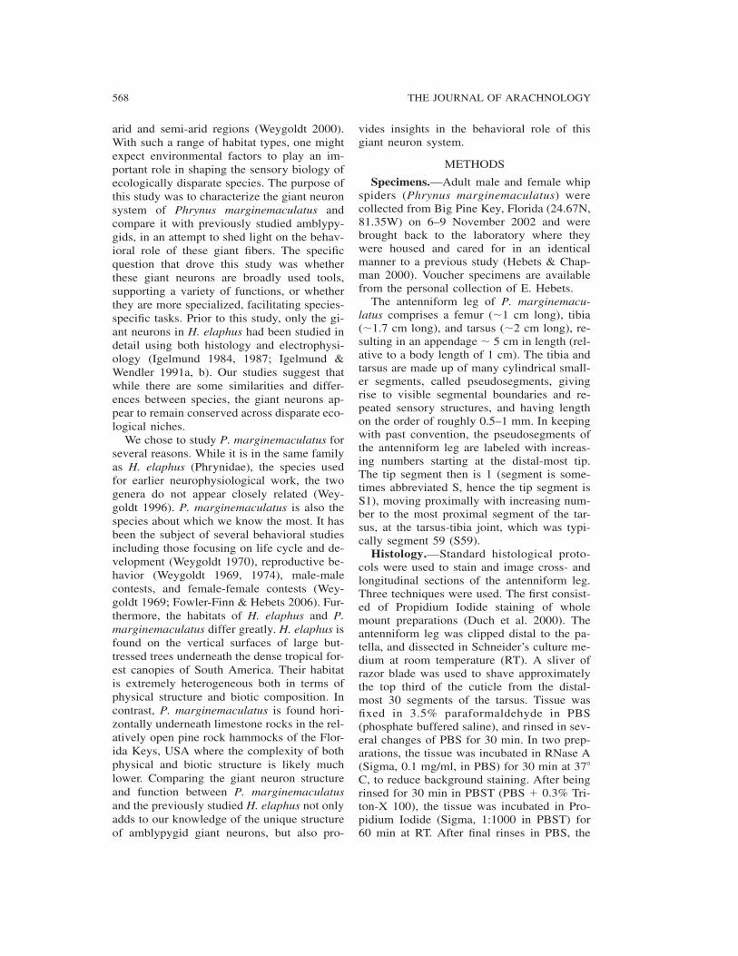
Longitudinal sections of the antenniformleg revealed giant cell bodies in segments 5,6, 10, 11, 12, 14, 15, 16, 20, 22, 23, and 26(Figs. 4–10). These somata are several tens of�m in length and width (the largest was insegment 6, measuring �100 by 50 �m, Fig.4), and are readily identified by their large sizeand distinct structure. They consist of an ovalshaped outer cell body, a circular, outlined nu-cleus with homogenous, lightly stained inte-rior, and an innermost nucleolus, which ap-pears as a spot. The Propidium iodide stainedcell bodies (Figs. 4–7) display a white out-lined nucleus with dark interior, and the nu-cleolus appears as a white spot. The OsmiumTetroxide (Fig. 4) and Methylene Blue (Figs.7, 10) stained cell bodies have the reversecontrast. It is assumed that these cell bodiesgive rise to the giant axons seen in Figs. 1–3.
The cell bodies in segments 11 and 26 wereof slightly smaller size, having narrower andmore elongated cell bodies, but still displayingthe distinct circular nucleus and nucleolus(Figures 4–10). Finding the location of thecell bodies within the antenniform leg is use-ful because these results can be comparedwith multi-site electrophysiological record-ings, establishing a connection between theobserved cytology and sensory physiology.
Electrophysiology.—Large (�50 �V–1.5mV) action potentials of varied amplitude andconduction velocity were recorded across 4positions on the antenniform leg tarsus (Fig.11). The spacing between pins in an individ-ual recording pair was 2.54 mm, and the spac-ing between pairs was approximately 5 mm(see Fig. 11). For the purposes of understand-ing spike timing and conduction velocity, thespike can be thought of as ‘‘at’’ the midpointbetween a pair of recording sites at the timewhen the inner part of its spike waveformcrosses zero. Spontaneous activity from small-er units was common, while mechanical stim-ulation (brushing hairs or bending the leg) wasrequired to elicit bursts of spikes from largerunits. At least 7 distinct types of spikes wereobserved. Four of these were elicited repeat-edly and classified reproducibly (n � 5 dif-ferent animals) using mechanosensory stimu-lation. Stimulation of the bristle hairs at 3different points along the tarsus (Fig. 11) wasadequate to identify spikes corresponding totwo of the giant neurons, which we label GN1and GN2 following the convention of Igel-mund & Wendler (1991a).
The largest spike, corresponding to GN1,had peak-to-peak amplitude 1.3 mV at seg-ment 52 (recording site 4), and an averageconduction velocity of 2.9 m/s. It respondedto deflections of bristle hairs, maximally at thetarsus tip and with reduced sensitivity as thestimulation site was moved proximally (Fig.11). The GN1 spike is generated at the site ofstimulation, and propagates both proximallyand distally inside the neuron, appearing onour most distal recording site at segment 10.GN1 is most likely the largest axon (Figs. 1–3, Axon 1), and one of the cell bodies in seg-ment 5 or 6 (Fig. 4), due to its large spikeamplitude and appearance on our segment 10recording site.
The same bristle hair stimulation traceswere also adequate to identify spikes origi-

571SPENCE & HEBETS—GIANT NEURONS IN PHRYNUS MARGINEMACULATUS
Figures 1–3. Figure 1.—Cross section of the tarsus of the antenniform leg, stained with methylene blue.Labeled are two tendons (te), a blood vessel (bv), and the two nerves (n1 and n2). Enlarged views ofnerves n1 and n2 are seen at right (Figures 2, 3), with outlines of the nerve bundle and largest sevengiant axons. Scale bars in Figures 2 and 3 are 10 �m.
nating from GN2. Although this neuron’sspike also originates at the point of stimula-tion and propagates in both directions, it issmaller in amplitude and conduction velocitythan GN1 (0.41 mV and 2.6 m/s), and doesnot appear on our segment 10 recording site(Fig. 11). This places its cell body betweensegments 10 and 28. These traits suggest thatit is GN2, and based on the similarity of ourresults to those of Igelmund & Wendler(1991a), it seems likely that it consists of oneof the axons of intermediate size (Figs. 1–3,Axons 2–7), and one of the cell bodies foundin segment 23 (Figs. 9, 10). The amplitudesand conduction velocities of both GN1 andGN2 we have found are similar to those foundin H. elaphus (Igelmund 1984). The firingrates of GN1 and GN2 adapted quickly, withrepeated stimulation of the same bristle hairsproducing few spikes.
Two types of spike that responded to de-flection of the antenniform leg near segment20 were identified. Deflection (bending) of thetarsus at other points produced fewer or nospikes from these neurons. We did not localizethe exact segmental boundary for which bend-ing maximally elicits these spikes. The factthat these spikes responded to bending of thetarsus, propagated solely proximally, and didnot appear on our segment 12 recording site(suggesting the cell body is proximal to seg-
ment 12 but distal to segment 28) identifiesthem as GN6 and 7. These spikes had rela-tively small average amplitude and conductionvelocity (GN6: 0.20 mV and 1.8 m/s, GN7:0.16 mV and 1.7 m/s). The amplitude of thesespikes is in good agreement with that foundfor them in H. elaphus, which varied between0.1 and 0.2 mV (Igelmund 1984).
DISCUSSION
Morphology of the antenniform leg of P.marginemaculatus.—We found that P. mar-ginemaculatus has a close replication of thesensory physiology found in H. elaphus. Thetarsus of the antenniform leg is equipped withsimilar classes of sensory organs, and withsimilar distribution. The rod hairs are groupedin a single oval-shaped patch on the first tarsalsegment, which is similar to H. elaphus, butin contrast to H. longicornis Butler 1873, inwhich they are grouped in 3 distinct circularpatches on each of the first 3 segments (Igel-mund 1987). We did not repeatedly find theleaf-like hairs found on specific segments ofH. elaphus (Igelmund 1987). In this manner,P. marginemaculatus is similar to H. longi-cornis and H. batesii Butler 1873, which alsolack the leaf-like hairs (Igelmund 1987).
Internally, the antenniform leg of P. mar-ginemaculatus has a neural architecture thatclosely parallels previously studied amblypy-
572 THE JOURNAL OF ARACHNOLOGY
Figures 4–10.—Representative longitudinal sections of the antenniform leg. Whole mount Propidiumiodide stains of the following segments: (Figure 4) 6 , (Figure 5) 10, (Figure 6) 12, and (Figure 7) 22,respectively. Osmium tetroxide stain of segment 10 (Figure 8, different specimen from Figure 5), andmethylene blue stained sections of segment 23 (Figures 9 and 10). Single cell bodies (arrows) are visiblein segments 6 (Figure 4), 10 (Figures 5 and 8), and 12 (Figure 6). Three are seen in segment 22 (Figure7), and 23 (Figures 9 and 10; these are serial sections through the same tissue; two cell bodies are seenin Figure 9, and a third appears in Figure 10). Cell bodies (cf. panel Figure 4) consist of outer cell bodymembrane (in this case �100 �m wide by 50 �m tall), inner nucleus (white circular line enclosing darkarea, here 26 �m diameter) and innermost nucleolus (inner white spot, 6 �m diameter). Scale bars �50 �m.
gids. The giant axons in P. marginemaculatusare distributed between the two nerves withremarkable similarity to H. elaphus. The larg-est 2 axons, presumably GN1 and GN2, aresituated adjacent to each other in one nerve,while the remaining smaller axons are bun-dled together in the other nerve. The periph-eral giant cell bodies and sensory synapsesfound in whip spiders (Amblypygi), whipscorpions (Uropygi), and harvestmen (Opili-ones) are rare, and to date this type of neuralarchitecture has only been found in a few cas-es within the animal kingdom (Foelix 1975;Foelix & Troyer 1980). Insects and other ar-thropods are thought to have their neuronalcell bodies and the first site of synaptic inte-gration located centrally, either in the brain organglia.
We used a new preparation and stainingmethod to image the giant cell bodies: whole
mount dissection of the antenniform leg fol-lowed by Propidium Iodide labeling (Figs. 4–7). We found that the unique cytology of thegiant neuron somata, especially the homoge-neous, light staining inside the nucleus butoutside the nucleolus, was reproduced withthe Propidium Iodide stain (Figs. 4–7). Thispeculiar staining and large nucleus size ledprevious investigators to ask whether thesecells were polyploid, but a Feulgen stain es-tablished that this was unlikely (Foelix &Troyer 1980). This new preparation provideda more rapid technique to locate in which seg-ment each cell body lies, and resulted in lon-ger, multi-segment pieces of intact antenni-form leg for observation. This more readilyenables observation of structures that spanmultiple segments, such as nerves, tendons,and blood vessels.
Giant cell bodies in segments 10–20, and

573SPENCE & HEBETS—GIANT NEURONS IN PHRYNUS MARGINEMACULATUS
Figure 11.—Four point recording from the tarsus, and spikes from GN1 and GN2. Average spike (darkline) overlaid on individual spikes (gray lines). The number of individual spikes averaged to calculateeach template is shown on the fourth recording site waveform. Peak-to-peak amplitude of average wave-form on fourth site, and average conduction velocity are shown below the first GN1 and GN2 spike types.Propagation direction can be seen in time course between channels and as a reversal of peak order(proximal � positive then negative, distal � negative then positive). Spikes were aligned in time on thefourth recording site, and thus the variation in conduction time is most easily seen in the ‘‘jitter’’ of spikeson channel 1. The geometry of the recording pins is not drawn to scale. Separation between pins withina recording site is 2.54 mm, and the center to center spacing between pairs was approximately 5 mm. Asa result the spacing between pins on the edge of neighboring pairs was also approximately 2.5 mm. Themeasured center to center spacings for this recording are indicated with arrows.
GN3, 4 and 5.—In P. marginemaculatus wehave found giant cell bodies in segments 5, 6,10, 11, 12, 14, 15, 16, 20, 22, 23, and 26. InH. longicornis, H. batesii, and H. elaphus gi-ant cell bodies were found most frequently insegments 1, 5, 6, 13, 19, 20, 21, 22, 25 and101 (Foelix & Troyer 1980; Igelmund & Wen-dler 1991a; Foelix & Hebets 2001). There isremarkable replication in the location of thecell bodies, with those in S5 and 6 being inthe identical segment, and those in the regionof 19–26 likely to be slightly shifted but ho-mologous cell bodies. We found several newgiant cell bodies between segments 10 and 20.This finding agrees with the electrophysiolog-ical results of Igelmund & Wendler (1991a),whose recordings predicted that the cell bod-ies of GN3, 4 and 5 would lie in this region.Now that the segmental location of these giant
neuron cell bodies is known, tracer-fills ofsensory neurons on individual segments canbe pursued to look for connectivity betweenthe giant neurons and the various sensilla. Inaddition to bristles, these sensilla include theclub and porous hairs that are found on thedistalmost �20 segments, which are thoughtto have hygrometric and olfactory function,respectively. This is in addition to the slitsense organs that are found on each tarsal seg-ment, which may sense cuticular stress or seg-mental deflection.
The sensory function of GN3, 4, and 5 isunknown. Although we recorded additionaltypes of spikes that matched examples fromthese neurons in H. elaphus (Igelmund &Wendler 1991a), we could not elicit their ac-tivity with basic mechanosensory or odorstimuli. The odor stimuli used were a leaf-

574 THE JOURNAL OF ARACHNOLOGY
Figure 12.—Four point recording from the tarsus, and spikes from GN6 and GN7. Average peak topeak amplitudes are shown for each recording site, and the average conduction velocity denoted below.Both templates are averages of 100 individual spikes.
alcohol and a leaf-aldehyde, odorants com-monly emitted by plants. This confirms someof the results for H. elaphus, in which me-chanical, olfactory, and even temperature andhygrometric stimuli did not elicit a responsefrom these neurons, apart from a phasic re-sponse of GN5 to tobacco smoke (Igelmund& Wendler 1991a).
Structure and function of giant neurons1, 2, 6 and 7.—GN1 and GN2 are mechano-sensory interneurons that respond to deflec-tions of the bristle hairs. We were able to iden-tify them clearly in P. marginemaculatususing several factors: their spike amplitudeand conduction velocity, response to bristlehair stimulation, spike initiation at the pointof stimulation and propagation in both direc-tions, and the location of their cell bodies im-plied by the recording sites (i.e. GN1 distal tosegment 10, GN2 between segments 10 and28). We qualitatively tested the receptivefields of GN1 and GN2 in P. marginemacu-latus and found agreement with H. elaphus:stimulation at the tip of the antenniform legproduces only GN1 spikes, stimulation at seg-ment 20 produces both GN1 and GN2 spikes,
and nearing segment 45 only GN2 spikes areelicited. Thus it appears P. marginemaculatushas a similar organization of GN1 and 2: GN1covers the distalmost 20 segments, rampingdown its sensitivity moving proximally asGN2 begins to take over, becoming more sen-sitive as segment 40 is approached. GN1 and2 adapt quickly to repeated stimulation of thesame bristles. Given their large (several mm)receptive fields and rapid adaptation, GN1 and2 appear to function as rapid touch detectorsfor the tarsus.
GN6 and 7 are sensory neurons. They func-tion as rapid proprioceptors, giving the animalfeedback in the amount and direction of bend-ing at the segment 22/23 joint. We were ableto identify GN6 and 7 in P. marginemaculatususing similar criteria: their smaller amplitudespikes always originated at the same point(between segments 12 and 28), propagatedsolely proximally, and responded to deflectionof the leg, all of which matches the behaviorpreviously found in H. elaphus. GN6 and 7are thought to be either coupled to a large slitsense organ at the segment 22/23 border, orpart of a separate joint receptor mechanism at
575SPENCE & HEBETS—GIANT NEURONS IN PHRYNUS MARGINEMACULATUS
that boundary. Either connectivity could allowthem to perform their observed proprioceptivefunction.
Comparing histology with multi-site elec-trophysiology, it was found that in H. elaphusthe GN1 cell body is the one found in segment5, GN2 is in segment 23, and GN6 and 7 aretwo of four found in segment 22. Our electro-physiological results suggest that P. margi-nemaculatus has markedly similar structureand function of GN1, 2, 6, and 7. GN1 is oneof the cell bodies in segment 5 or 6, GN2 ismost likely in segment 23, and GN6 and 7 aretwo of those found in segment 22.
The role of the giant neurons in naturalbehavior.—The role of the giant fiber systemof amblypygids in the natural behavior of theanimal remains a mystery. Although there arecases in which giant fibers are not directlylinked to specific behaviors (DiCaprio 2003),typically, in other arthropods, giant neuronsfacilitate rapid escape or predatory behavior(Levine & Tracey 1973; Tauber & Camhi1995; Mizrahi & Libersat 1997). Touching theantenniform leg in a manner that elicits spikesin GN1 or 2, even repeatedly, does not evokea rapid escape response of the amblypygid,while puffs of air directed at trichobothria onthe walking legs usually does. Giant neuronscan underlie rapid predatory behaviors (Gro-nenberg 1995a, b), but while amblypygidsmake rapid prey strikes with their pedipalps,the antenniform legs do not touch the preyimmediately before a strike, and often a pe-riod of seconds will elapse between the lasttouch of the antenniform leg and the strike(pers. obs.).
Rapid tapping and vibratory movements aremade with the antenniform legs during court-ship and intraspecific aggressive behaviors(Weygoldt 2000; Fowler-Finn & Hebets2006). High speed video of aggressive behav-iors (Fowler-Finn & Hebets 2006) has foundthe frequency of tapping to be �30 Hz. Spikesfrom GN1 take on the order of 30 ms to getto the CNS, and so it is possible that the an-imal could use GN1 to receive feedback dur-ing each cycle of the tapping behavior. Whilefeedback at the same rate as the tapping maynot be needed to regulate the behavior, itwould be required in order to react to changeswithin a single cycle. Spikes in the primaryafferents could not provide feedback on thetime scale of a single tapping cycle: applying
local circuit theory to these unmyelinated ax-ons, we predict conduction velocity to scaleas the square root of axon radius (Aidley1998), and so estimate that primary afferentshaving a radius 20 times smaller than the gi-ants would take �140 ms to arrive at the CNS.This would give a maximum feedback driventapping rate of about 7 Hz. However, the rapidadaptation of GN1 to stimulation of the samebristles, and the lack of an obvious need forfeedback, makes this hypothesis (that GN1 ex-ists to provide fast feedback for rapid vibra-tions) unlikely. It is possible that some of theother motor or proprioceptive giants facilitatethis high-speed tapping of the antenniformlegs. As the vibratory tapping occurs duringcourtship and aggressive behaviors, it seemslikely that it signals individual quality, and isused as a basis of assessment of a mate orcompetitor. Whether information about qualityis contained in the frequency of the vibrationor some other component of the signal re-mains open. If the frequency of antenniformleg vibration were to signal the quality of anindividual, however, this could be a source ofevolutionary pressure on the development ofa faster sensorimotor system in the antenni-form leg.
Amblypygids will intermittently exhibitrapid retraction of the antenniform leg whentouched, a behavior that appears highly de-pendent on the animal’s state of alertness(Spence pers. obs.). Given the costs of losingthese appendages (animals missing both legscannot orient or proactively hunt), and per-haps even the costs of being entrapped bythem, fast touch detection and rapid proprio-ceptive feedback may simply be required foradequate maneuverability in such long ap-pendages.
The sense organs and underlying giant fibersystem we have studied in P. marginemacu-latus is remarkably similar to that of H. ela-phus, yet the habitats of these two species, theFlorida Keys for P. marginemaculatus andBrazilian rainforest for H. elaphus, are quitedifferent. One predicts that the environment ofP. marginemaculatus would offer a smallerdiversity of prey, fewer vertical surfaces, moreseasonality, a lack of canopy and correspond-ingly more light, and lower humidity than theBrazilian home of H. elaphus. The similarityin the giant neuron systems across these spe-cies suggest that they are crucial for the mo-
576 THE JOURNAL OF ARACHNOLOGY
tility and basic function of the antenniformlegs, and as such are not under great selectionpressure from these ecological differences.
ACKNOWLEDGMENTS
The authors would like to thank all themembers of the Hebets laboratory, includingGinevra Ryman, Jennifer Wesson, NicoleVanderSal, and Kasey Fowler-Finn. MigumiFuse, Simon Sponberg, Susan Somerville, andmembers of the UC Berkeley ArachnologyDiscussion group provided valuable criticalfeedback. We would also like to acknowledgethe assistance of the Hoy Laboratory, espe-cially Dr. Patricia Rivlin, Prof. Elke Busch-beck, Damien Elias, and Prof. Ron Hoy. Wewould also like to thank the United States De-partment of the Interior Fish and Wildlife Ser-vice at National Key Deer Refuge for a specialuse permit. The experiments in this study fullycomply with the NIH guidelines for animalcare, publication No. 86-23, revised 1985, andwith all state and federal laws.
LITERATURE CITED
Aidley, D.J. 1998. The Physiology of ExcitableCells. Fourth edition. Cambridge UniversityPress, New York. 489 pp.
Beck, L. & K. Gorke. 1974. Tagesperiodik, Re-vierverhalten und Beutefang der GeißelspinneAdmetus pumilio C. L. Koch. Zeitschrift furTierpsychologie 35:173–186.
Beck, L., R. Foelix, E. Godeke & R. Kaiser. 1974.Uber die Haarsensillen der Geißelspinne Adme-tus pumilio (Arach., Amblypygi). Naturwissen-schaften 61:327–328.
Beck, L., R. Foelix, E. Goedeke & R. Kaiser. 1977.Morphologie, Larvantwicklung und Haarsensil-len des Tastbeinpaares der Geißelspinne Heter-ophrynus longicornis Butler (Arach., Amblypy-gi). Zoomorphologie 88:259–276.
DiCaprio, R.A. 2003. Nonspiking and spiking pro-prioceptors in the crab: nonlinear analysis ofnonspiking TCMRO afferents. Journal of Neu-rophysiology 89:1826–1836.
Duch, C., R.J. Bayline & R.B. Levine. 2000. Post-embryonic development of the dorsal longitudi-nal flight muscle and its innervation in Manducasexta. Journal of Comparative Neurology 422:1–17.
Eaton, R.C., R.K.K. Lee & M.B. Foreman. 2001.The Mauthner cell and other identified neuronsof the brainstem escape network of fish. Progressin Neurobiology 63:467–485.
Foelix, R.F. 1975. Occurrence of synapses in pe-ripheral sensory nerves of arachnids. Nature 254:146–148.
Foelix, R.F., I.W. Chu-Wang & L. Beck. 1975. Finestructure of tarsal sensory organs in the whip spi-der Admetus pumilio (Arachnida, Amblypygi).Tissue & Cell 7:331–346.
Foelix, R.F. & E.A. Hebets. 2001. Sensory biologyof whip spiders (Arachnida, Amblypygi). An-drias 15:129–140.
Foelix, R.F. & D. Troyer. 1980. Giant neurons andassociated synapses in the peripheral nervoussystem of whip spiders. Journal of Neurocytol-ogy 9:517–536.
Foelix, R.F., D. Troyer & P. Igelmund. 2002. Pe-ripheral synapses and giant neurons in whip spi-ders. Microscopy Research and Technique 58:272–282.
Fowler-Finn, K.D. & E.A. Hebets. 2006. An ex-amination of agonistic interactions in the whipspider Phrynus marginemaculatus (Arachnida,Amblypygi). Journal of Arachnology 34:62–76.
Gronenberg, W. 1995a. The fast mandible strike inthe trap-jaw ant Odontomachus. 1. Temporalproperties and morphological characteristics.Journal of Comparative Physiology A: Neuroeth-ology, Sensory, Neural, and Behavioral Physi-ology 176:391–398.
Gronenberg, W. 1995b. The fast mandible strike inthe trap-jaw ant Odontomachus. 2. Motor con-trol. Journal of Comparative Physiology A 176:399–408.
Hebets, E.A. & R.F. Chapman. 2000. Electrophys-iological studies of olfaction in the whip spiderPhrynus parvulus (Arachnida, Amblypygi). Jour-nal of Insect Physiology 46:1441–1448.
Igelmund, P. 1984. Elektrophysiologische und mor-phologische Untersuchungen zur Funktion peri-pherer Riesenneurone in den Tastbeinen derGeisselspinne Heterophrynus elaphus Pocock.Doctoral thesis. Cologne University, Cologne,Germany.
Igelmund, P. 1987. Morphology, sense-organs, andregeneration of the forelegs (whips) of the whipspider Heterophrynus elaphus (Arachnida, Am-blypygi). Journal of Morphology 193:75–89.
Igelmund, P. & G. Wendler. 1991a. The giant fibersystem in the forelegs (whips) of the whip spiderHeterophrynus elaphus Pocock (Arachnida, Am-blypygi). Journal of Comparative Physiology A:Neuroethology, Sensory, Neural, and BehavioralPhysiology 168:63–73.
Igelmund, P. & G. Wendler. 1991b. Morphologyand physiology of peripheral giant interneuronsin the forelegs (whips) of the whip spider Het-erophrynus elaphus Pocock (Arachnida, Ambly-pygi). Journal of Comparative Physiology A:Neuroethology, Sensory, Neural, and BehavioralPhysiology 168:75–83.
Jablonski, P.G. & N.J. Strausfeld. 2001. Exploita-tion of an ancient escape circuit by an avianpredator: relationships between taxon-specific
577SPENCE & HEBETS—GIANT NEURONS IN PHRYNUS MARGINEMACULATUS
prey escape circuits and the sensitivity to visualcues from the predator. Brain Behavior and Evo-lution 58:218–240.
Jacobs, G.A. & F.E. Theunissen. 2000. Extractionof sensory parameters from a neural map by pri-mary sensory interneurons. Journal of Neurosci-ence 20:2934–2943.
Levine, J. & D. Tracey. 1973. Structure and func-tion of giant motorneuron of Drosophila melan-ogaster. Journal of Comparative Physiology 87:213–235.
Lewicki, M.S. 1998. A review of methods for spikesorting: the detection and classification of neuralaction potentials. Network: Computation in Neu-ral Systems 9:R53–R78.
Mizrahi, A. & F. Libersat. 1997. Independent cod-ing of wind direction in cockroach giant inter-neurons. Journal of Neurophysiology 78:2655–2661.
Spence, A.J., R.R. Hoy & M.S. Isaacson. 2003. Amicromachined silicon multielectrode for multi-unit recording. Journal of Neuroscience Methods126:119–126.
Tauber, E. & J. Camhi. 1995. The wind-evoked es-cape behavior of the cricket Gryllus bimaculatus:integration of behavioral elements. Journal ofExperimental Biology 198:1895–1907.
Weygoldt, P. 1969. Beobachtungen zur Fort-pflanzungsbiologie und zum Verhalten derGeißelspinne Tarantula marginemaculata C. L.
Koch (Chelicerata, Amblypygi). Zeitschrift furMorphologie der Tiere 64:338–360.
Weygoldt, P. 1970. Lebenszyklus und postembryon-ale Entwicklung der Geißelspinne Tarantulamarginemaculata C.L. Koch (Chelicerata, Am-blypygi) in Laboratorium. Zeitschrift fur Mor-phologie der Tiere 67:58–85.
Weygoldt, P. 1972. Geisselskorpione und Geissel-spinne (Uropygi und Amplypygi). Zeitschrift desKoelner Zoo 15:95–107.
Weygoldt, P. 1974. Kampf und Paarung bei derGeisselspinne Charinus montanus Weygoldt.Zeitschrift fur Tierpsychologie 34:217–223.
Weygoldt, P. 1996. Evolutionary morphology ofwhip spiders: towards a phylogenetic system(Chelicerata: Arachnida: Amblypygi). Journal ofZoological Systematics and Evolutionary Re-search 34:185–202.
Weygoldt, P. 2000. Whip Spiders (Chelicerata: Am-blypygi): Their Biology, Morphology and Sys-tematics. Apollo Books, Stenstrup, Denmark.163 pp.
Weygoldt, P. 2002. Fighting, courtship, and sper-matophore morphology of the whip spider Mu-sicodamon atlanteus Fage, 1939 (Phrynichidae)(Chelicerata, Amblypygi). Zoologischer Anzeig-er 241:245–254.
Manuscript received 12 May 2005, revised 30 May2005.