Embed Size (px)
Citation preview
Anatomy and Physiology II
Face and Head
Review
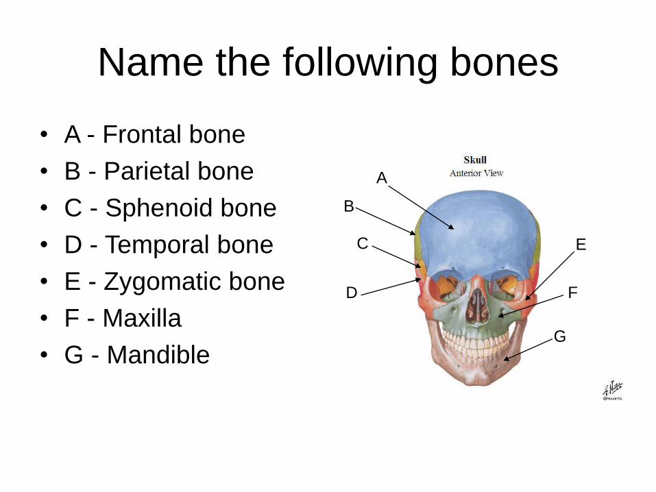
Name the following bones
• A - Frontal bone
• B - Parietal bone
• C - Sphenoid bone
• D - Temporal bone
• E - Zygomatic bone
• F - Maxilla
• G - Mandible
A
D
B
C E
F
G
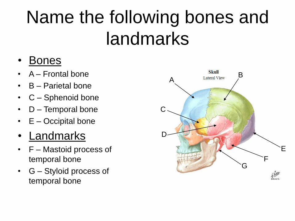
Name the following bones and
landmarks• Bones• A – Frontal bone
• B – Parietal bone
• C – Sphenoid bone
• D – Temporal bone
• E – Occipital bone
• Landmarks• F – Mastoid process of
temporal bone
• G – Styloid process of
temporal bone
A
D
B
C
E
FG
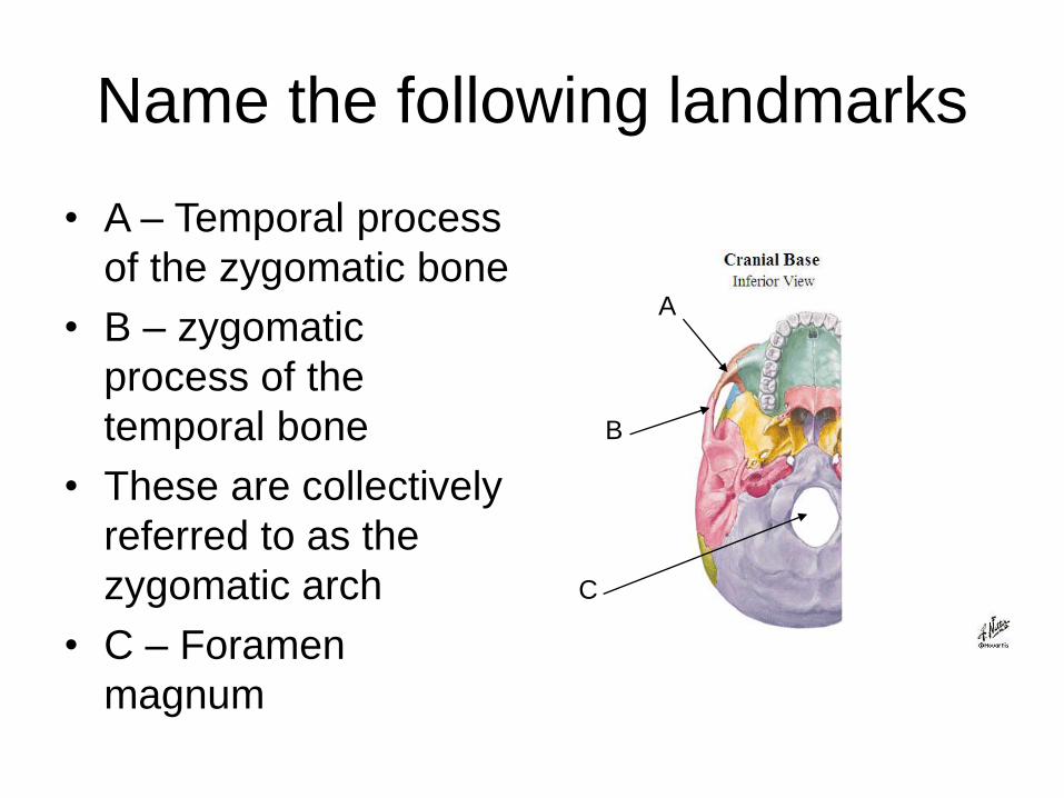
Name the following landmarks
• A – Temporal process
of the zygomatic bone
• B – zygomatic
process of the
temporal bone
• These are collectively
referred to as the
zygomatic arch
• C – Foramen
magnum
A
B
C
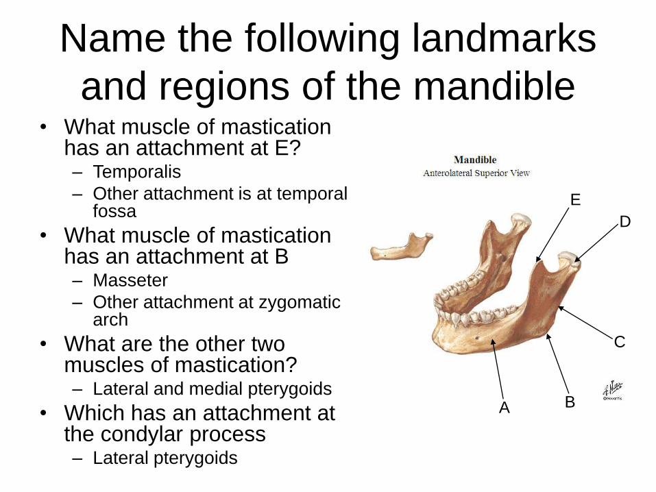
Name the following landmarks
and regions of the mandible
E
D
C
BA
• What muscle of mastication has an attachment at E?– Temporalis
– Other attachment is at temporal fossa
• What muscle of mastication has an attachment at B– Masseter
– Other attachment at zygomatic arch
• What are the other two muscles of mastication?– Lateral and medial pterygoids
• Which has an attachment at the condylar process– Lateral pterygoids
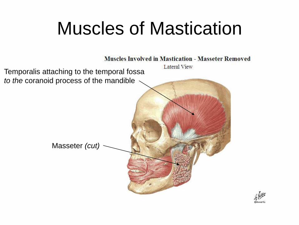
Muscles of Mastication
Temporalis attaching to the temporal fossa
to the coranoid process of the mandible
Masseter (cut)
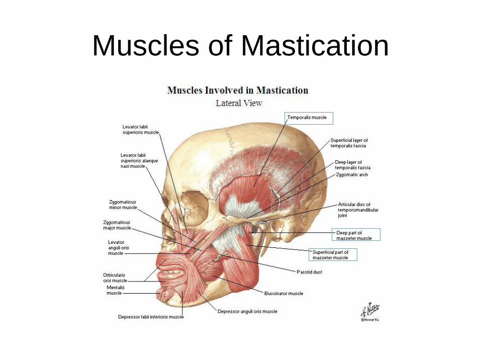
Muscles of Mastication
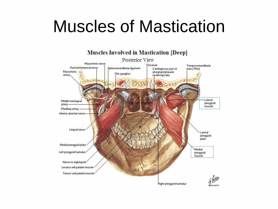
Muscles of Mastication
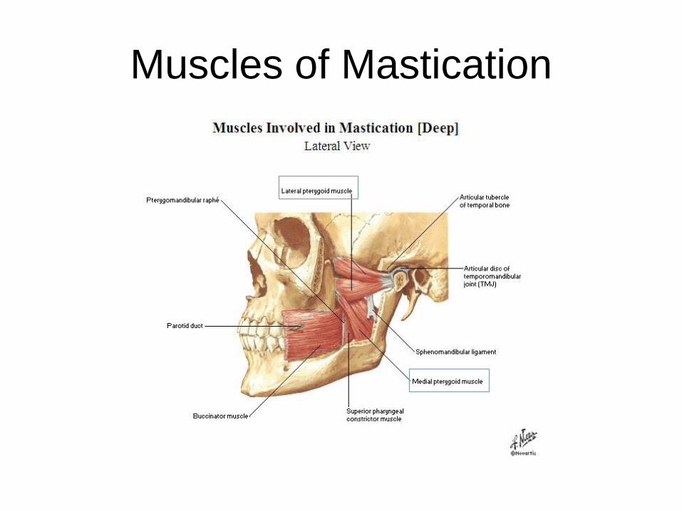
Muscles of Mastication
Anatomy and Physiology II
Pelvis

Bones
• The Pelvis includes the sacrum, coccyx, and the coxalbone – We will focus on the sacrum and coccyx when we look at the
lumbar spine
• The Hip joint includes the coxal bone and the femur
• Coxal Bone– Aka Os Coxa or Innominate Bone
– Three bones that fuse together
• Ilium
• Ischium
• Pubis
• Femur
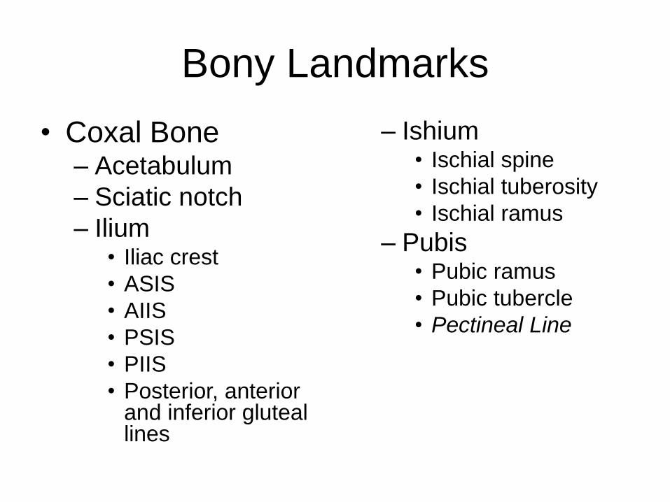
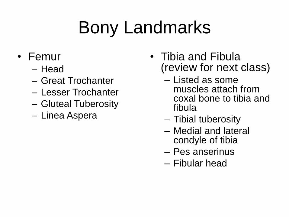
Bony Landmarks
• Coxal Bone– Acetabulum
– Sciatic notch
– Ilium• Iliac crest
• ASIS
• AIIS
• PSIS
• PIIS
• Posterior, anterior and inferior gluteal lines
– Ishium• Ischial spine
• Ischial tuberosity
• Ischial ramus
– Pubis• Pubic ramus
• Pubic tubercle
• Pectineal Line
Bony Landmarks
• Femur– Head
– Great Trochanter
– Lesser Trochanter
– Gluteal Tuberosity
– Linea Aspera
• Tibia and Fibula (review for next class)– Listed as some
muscles attach from coxal bone to tibia and fibula
– Tibial tuberosity
– Medial and lateral condyle of tibia
– Pes anserinus
– Fibular head

Coxal Bone - Lateral View
Ilium
Ischium
Pubis
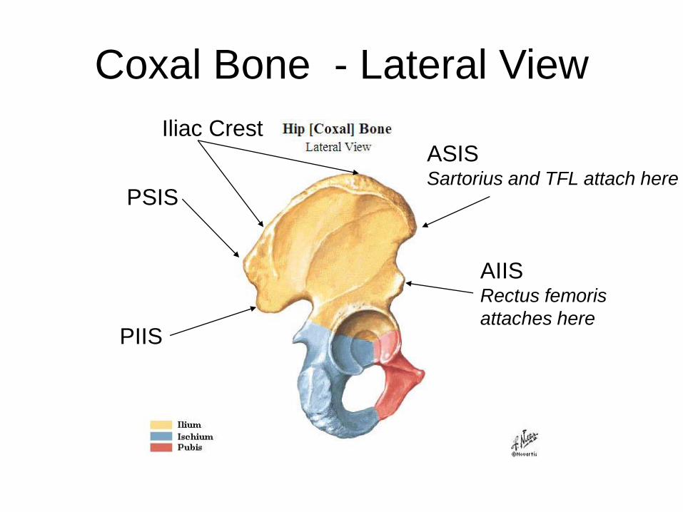
Coxal Bone - Lateral View
PSIS
PIIS
ASISSartorius and TFL attach here
AIISRectus femoris
attaches here
Iliac Crest
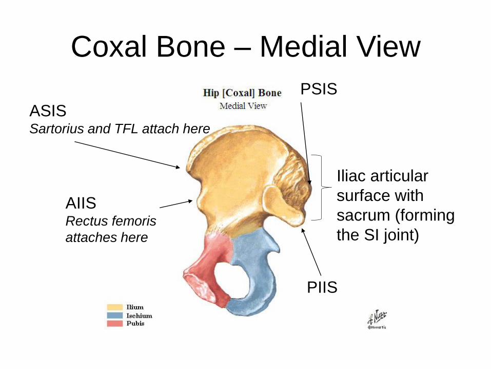
Coxal Bone – Medial View
PSIS
PIIS
ASISSartorius and TFL attach here
AIISRectus femoris
attaches here
Iliac articular
surface with
sacrum (forming
the SI joint)
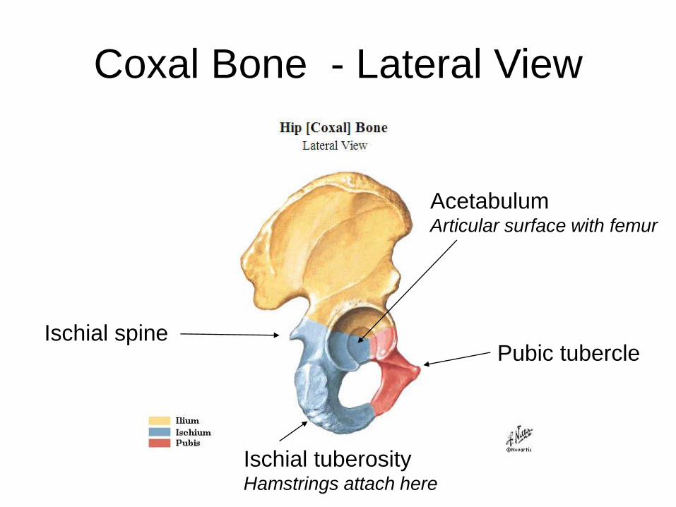
Coxal Bone - Lateral View
Ischial tuberosityHamstrings attach here
Ischial spinePubic tubercle
AcetabulumArticular surface with femur
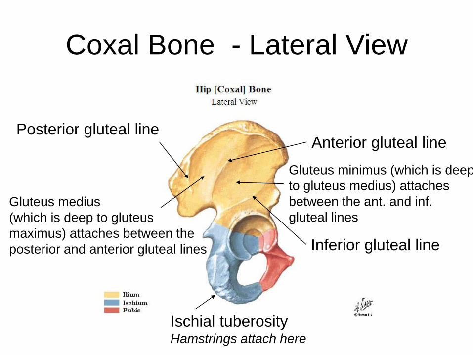
Coxal Bone - Lateral View
Ischial tuberosityHamstrings attach here
Posterior gluteal line
Inferior gluteal line
Anterior gluteal line
Gluteus medius
(which is deep to gluteus
maximus) attaches between the
posterior and anterior gluteal lines
Gluteus minimus (which is deep
to gluteus medius) attaches
between the ant. and inf.
gluteal lines

Proximal Femur – Anterior View
Head of Femur
Lesser Trochanter
(iliopsoas attaches here)
Greater Trochanter
(many muscles attach
here)
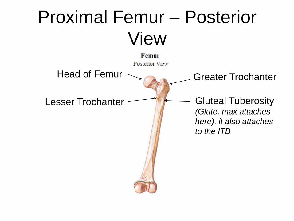
Proximal Femur – Posterior
View
Head of Femur
Lesser Trochanter
Greater Trochanter
Gluteal Tuberosity(Glute. max attaches
here), it also attaches
to the ITB

Pelvis Bone – Lateral View
Sacrotuberous
ligament (links the
ischial tuberosity to the sacrum)
Sacrospinous ligament (links the
ischial spine to the sacrum, stabilizes
the sacrum during trunk flexion)
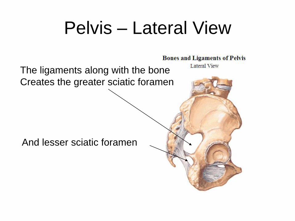
Pelvis – Lateral View
And lesser sciatic foramen
The ligaments along with the bone
Creates the greater sciatic foramen
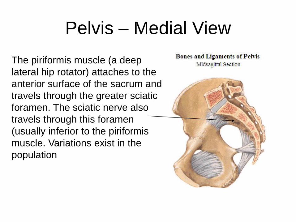
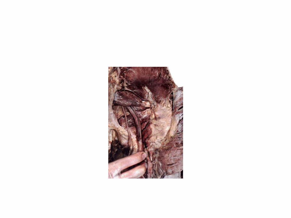
Pelvis – Medial View
The piriformis muscle (a deep
lateral hip rotator) attaches to the
anterior surface of the sacrum and
travels through the greater sciatic
foramen. The sciatic nerve also
travels through this foramen
(usually inferior to the piriformis
muscle. Variations exist in the
population
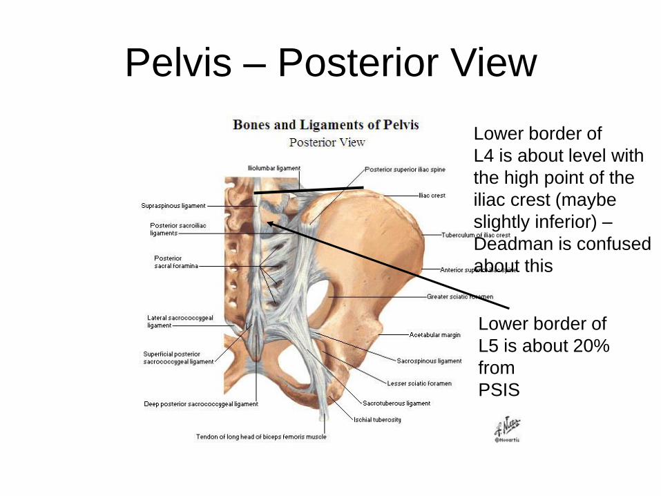
Pelvis – Posterior View
Lower border of
L5 is about 20%
from
PSIS
Lower border of
L4 is about level with
the high point of the
iliac crest (maybe
slightly inferior) –
Deadman is confused
about this
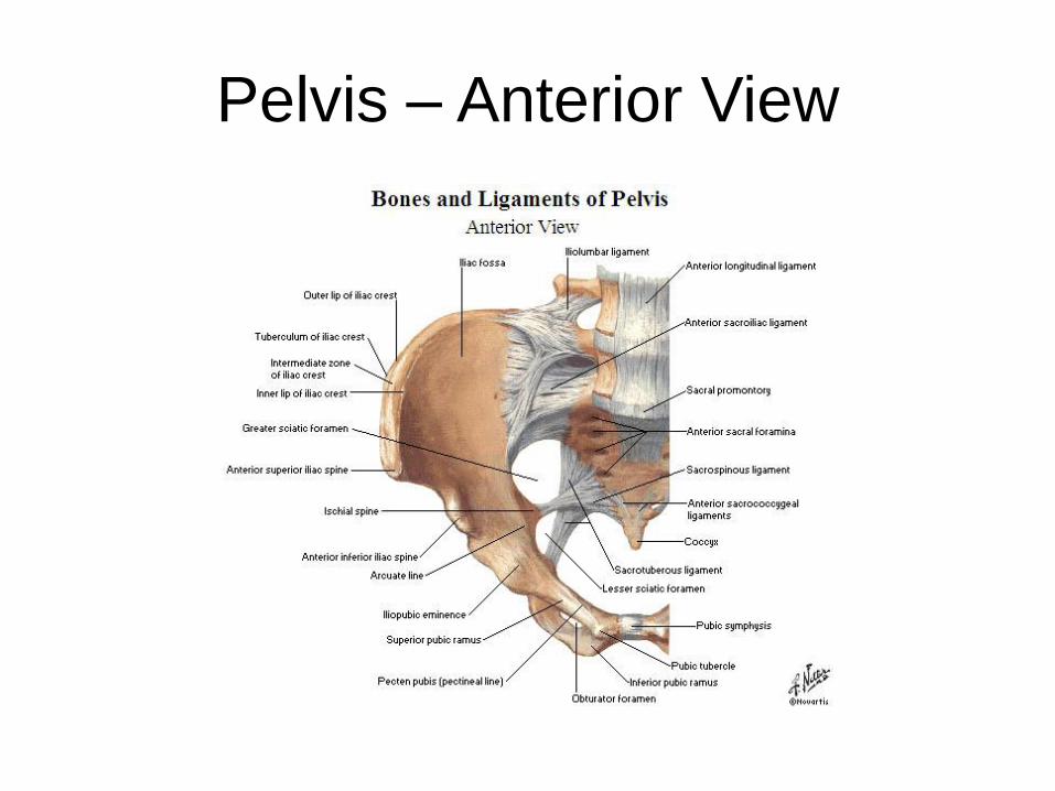
Pelvis – Anterior View

Muscles of the Pelvis
and HipFrom Superficial to Deep
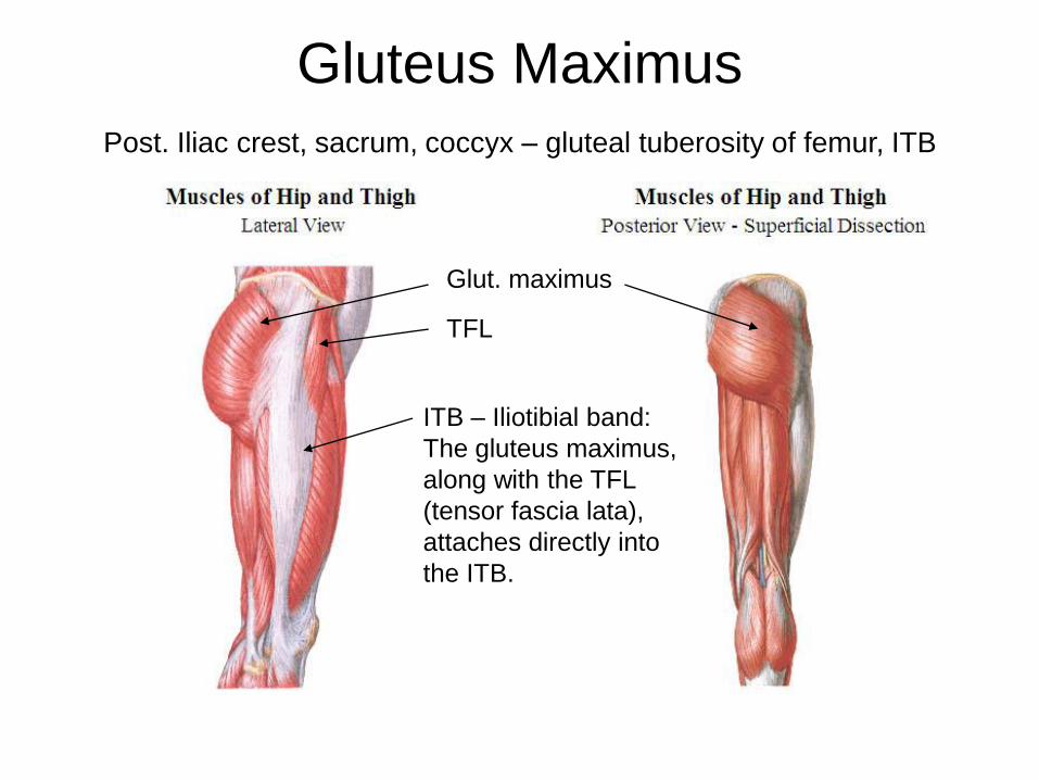
Gluteus Maximus
Post. Iliac crest, sacrum, coccyx – gluteal tuberosity of femur, ITB
ITB – Iliotibial band:
The gluteus maximus,
along with the TFL
(tensor fascia lata),
attaches directly into
the ITB.
TFL
Glut. maximus
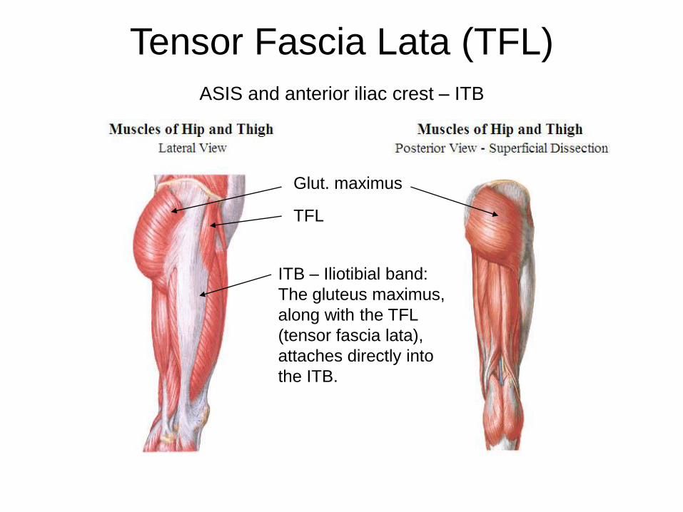
Tensor Fascia Lata (TFL)
ASIS and anterior iliac crest – ITB
ITB – Iliotibial band:
The gluteus maximus,
along with the TFL
(tensor fascia lata),
attaches directly into
the ITB.
TFL
Glut. maximus
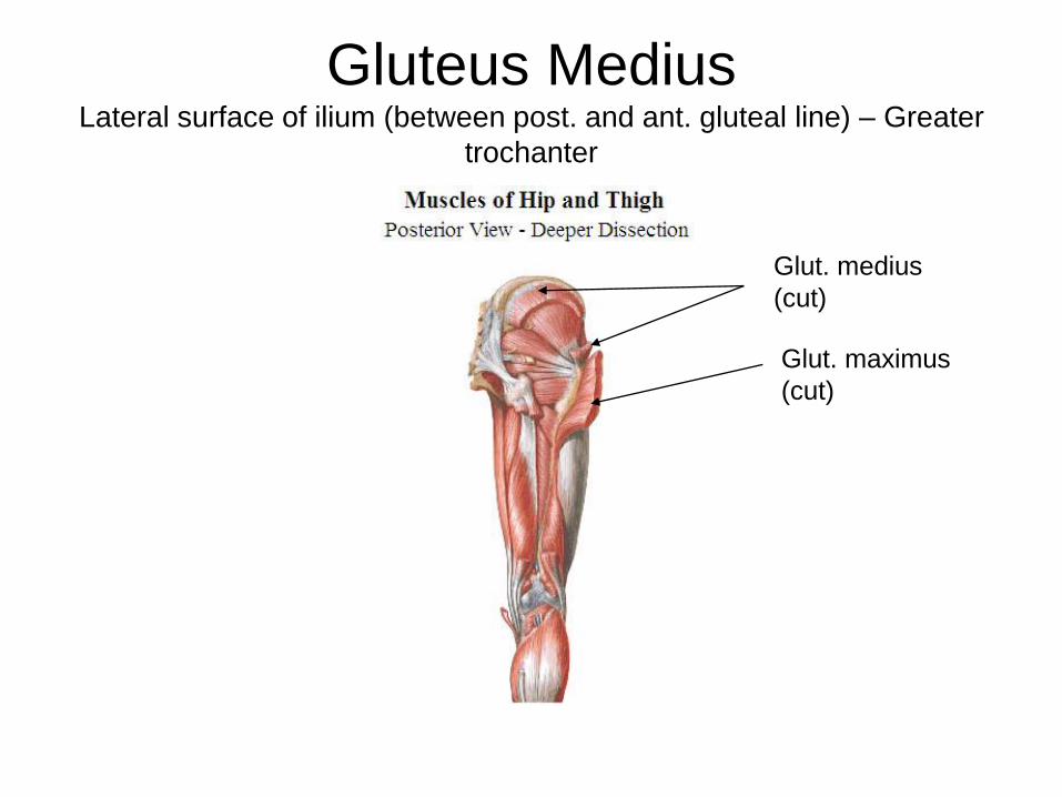
Gluteus MediusLateral surface of ilium (between post. and ant. gluteal line) – Greater
trochanter
Glut. maximus
(cut)
Glut. medius
(cut)

Gluteus MinimusLateral surface of ilium (between ant. and inf. gluteal line) – Greater
trochanter
Glut. minimus
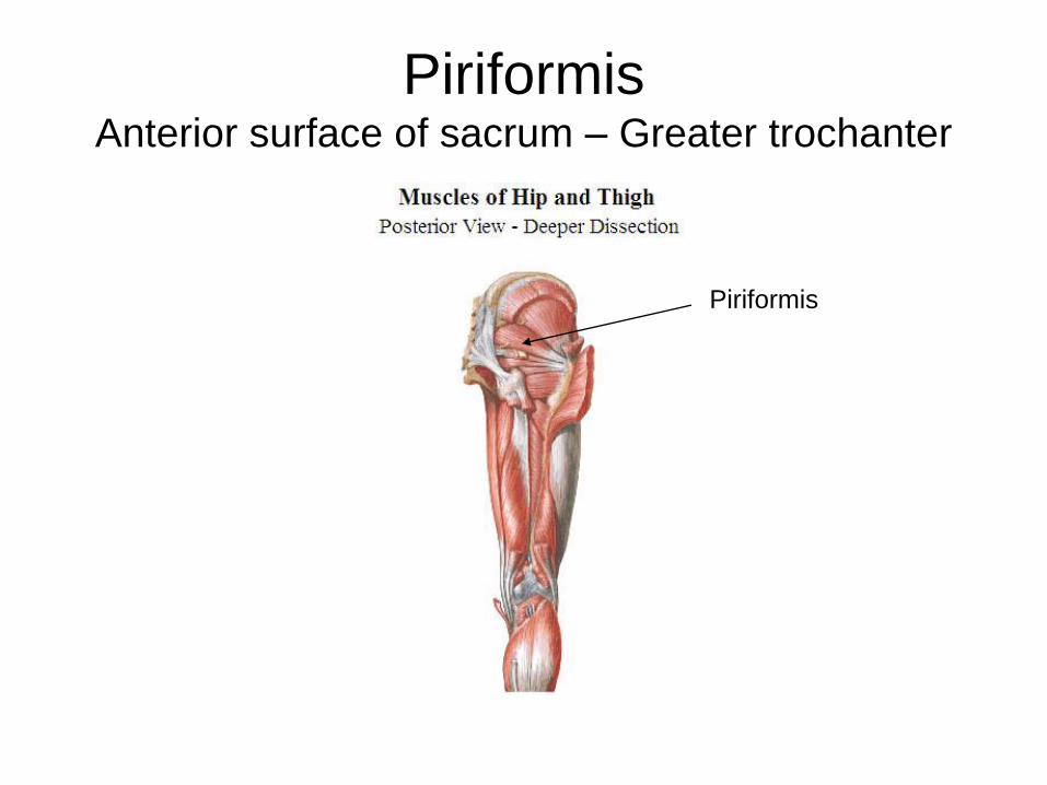
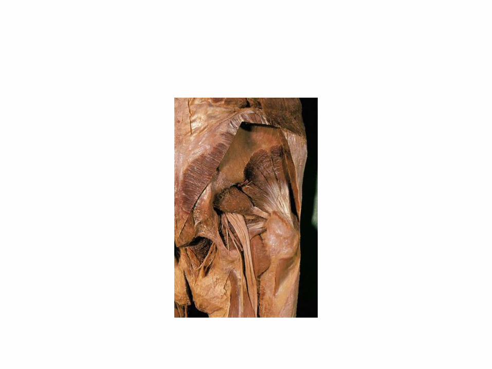
PiriformisAnterior surface of sacrum – Greater trochanter
Piriformis
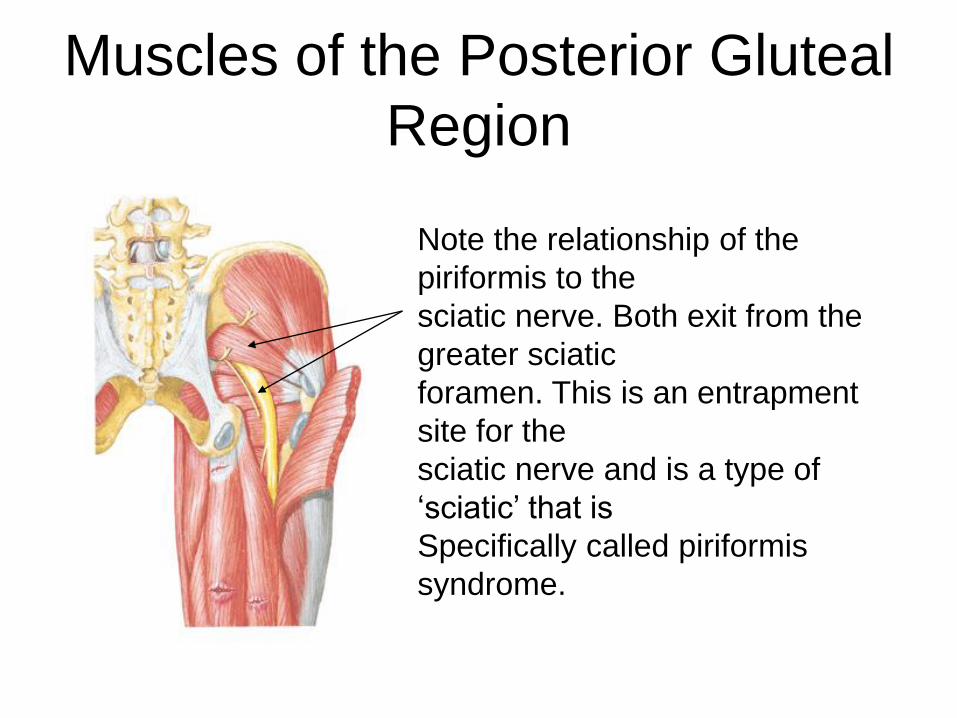
Muscles of the Posterior Gluteal
Region
Note the relationship of the
piriformis to the
sciatic nerve. Both exit from the
greater sciatic
foramen. This is an entrapment
site for the
sciatic nerve and is a type of
‘sciatic’ that is
Specifically called piriformis
syndrome.
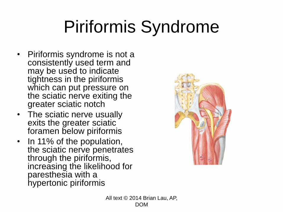
Piriformis Syndrome
• Piriformis syndrome is not a consistently used term and may be used to indicate tightness in the piriformiswhich can put pressure on the sciatic nerve exiting the greater sciatic notch
• The sciatic nerve usually exits the greater sciatic foramen below piriformis
• In 11% of the population, the sciatic nerve penetrates through the piriformis, increasing the likelihood for paresthesia with a hypertonic piriformis
All text © 2014 Brian Lau, AP,
DOM
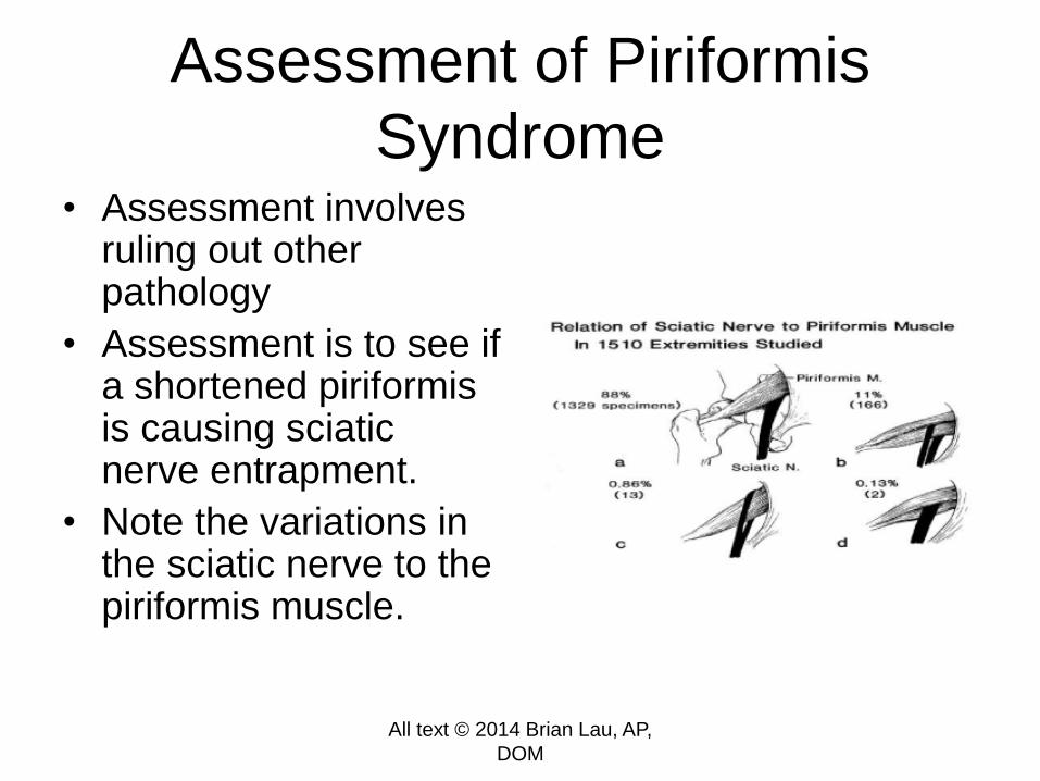
Assessment of Piriformis
Syndrome• Assessment involves
ruling out other pathology
• Assessment is to see if a shortened piriformis is causing sciatic nerve entrapment.
• Note the variations in the sciatic nerve to the piriformis muscle.
All text © 2014 Brian Lau, AP,
DOM


Warning: Cadaver Images
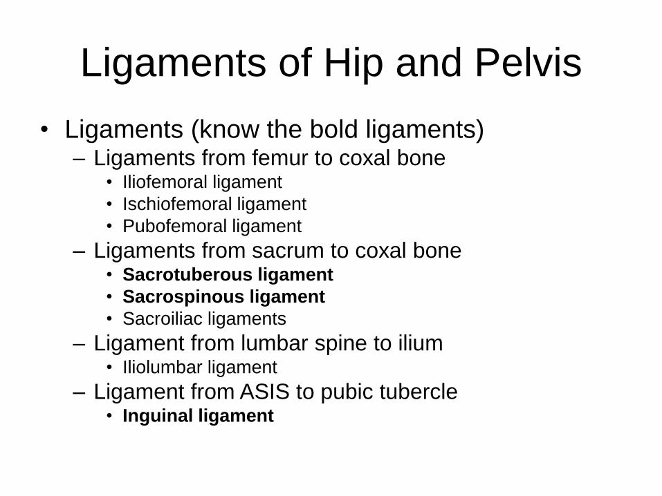
Ligaments of Hip and Pelvis
• Ligaments (know the bold ligaments)– Ligaments from femur to coxal bone
• Iliofemoral ligament
• Ischiofemoral ligament
• Pubofemoral ligament
– Ligaments from sacrum to coxal bone• Sacrotuberous ligament
• Sacrospinous ligament
• Sacroiliac ligaments
– Ligament from lumbar spine to ilium• Iliolumbar ligament
– Ligament from ASIS to pubic tubercle• Inguinal ligament
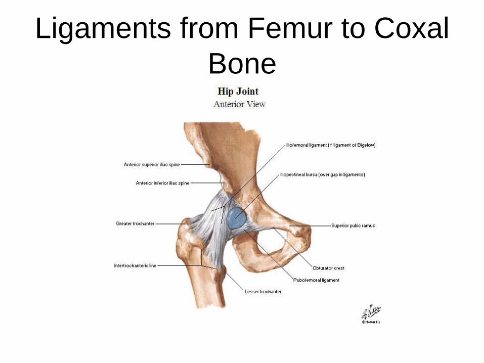
Ligaments from Femur to Coxal
Bone
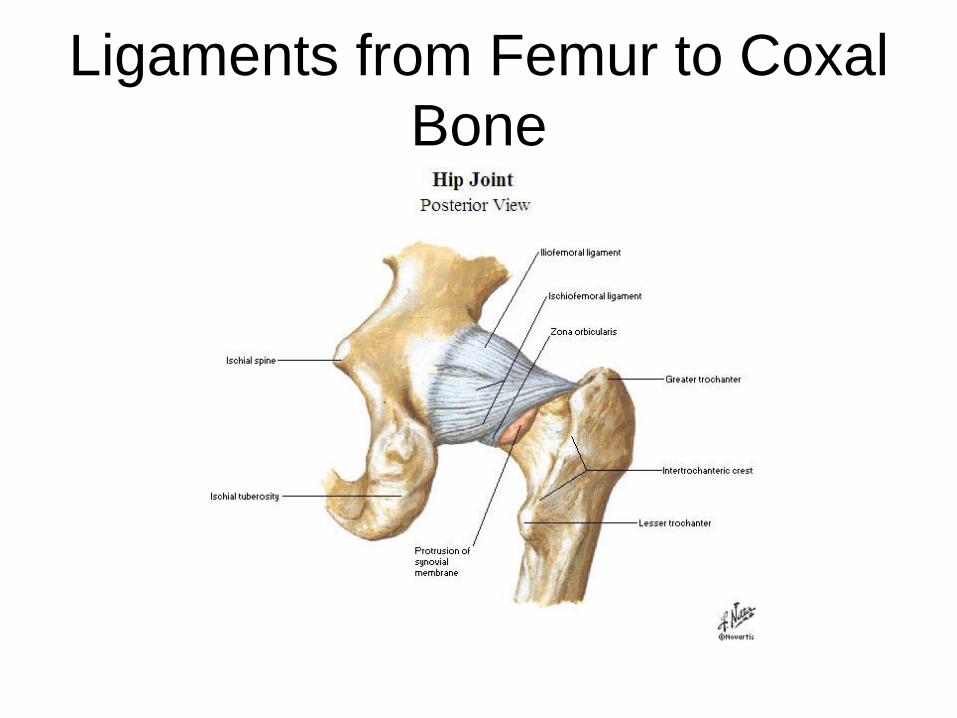
Ligaments from Femur to Coxal
Bone

Ligaments from Sacrum to
Coxal Joint
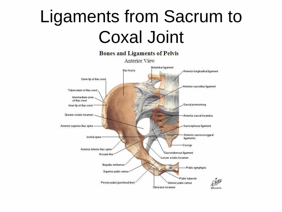
Ligaments from Sacrum to
Coxal Joint
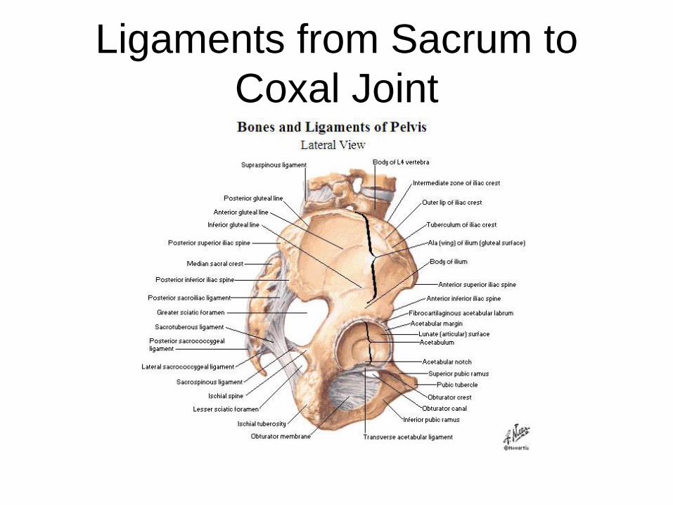
Ligaments from Sacrum to
Coxal Joint

Ligaments from Lumbar Spine
to Ilium

Ligaments from ASIS to Pubic
Bone – Inguinal Ligament
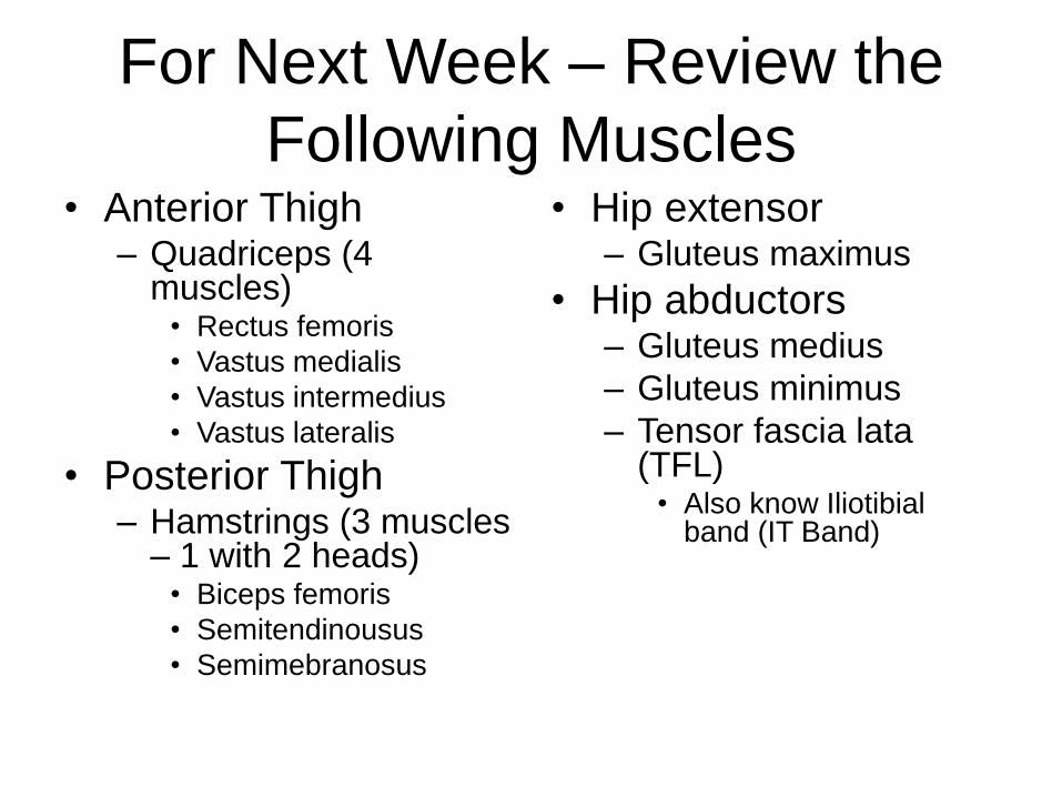
For Next Week – Review the
Following Muscles• Anterior Thigh
– Quadriceps (4 muscles)
• Rectus femoris
• Vastus medialis
• Vastus intermedius
• Vastus lateralis
• Posterior Thigh– Hamstrings (3 muscles
– 1 with 2 heads)• Biceps femoris
• Semitendinousus
• Semimebranosus
• Hip extensor– Gluteus maximus
• Hip abductors– Gluteus medius
– Gluteus minimus
– Tensor fascia lata(TFL)
• Also know Iliotibial band (IT Band)
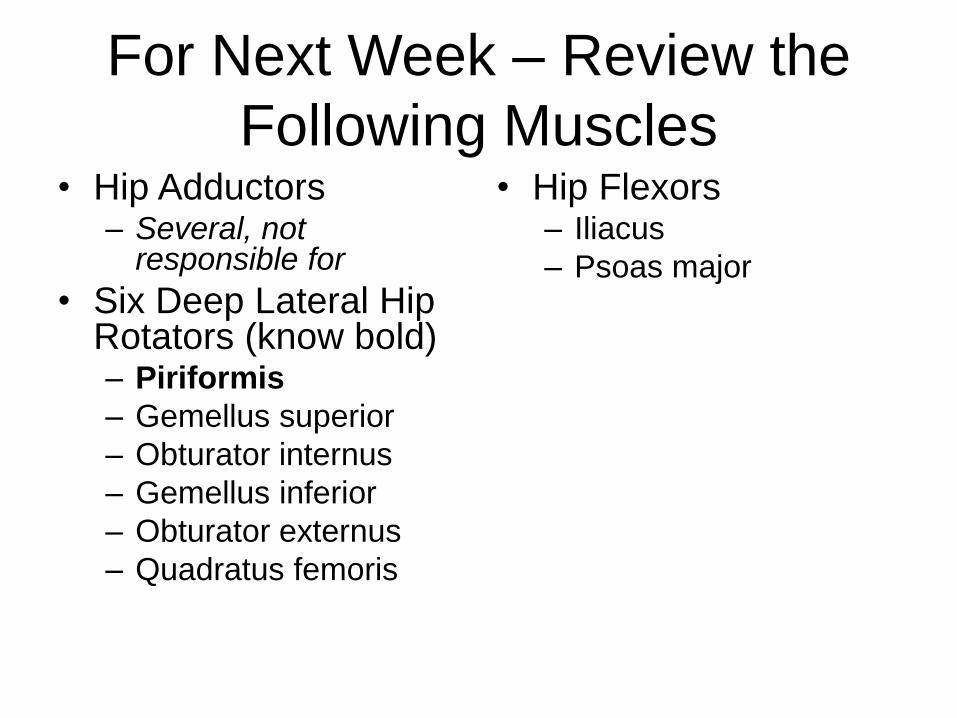
For Next Week – Review the
Following Muscles• Hip Adductors
– Several, not responsible for
• Six Deep Lateral Hip Rotators (know bold)– Piriformis
– Gemellus superior
– Obturator internus
– Gemellus inferior
– Obturator externus
– Quadratus femoris
• Hip Flexors– Iliacus
– Psoas major