Embed Size (px)
Citation preview
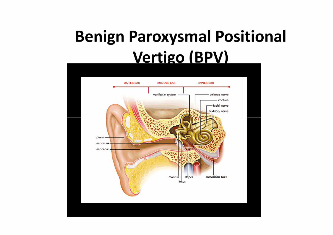
Anatomy andPathophysiology for ICD-10
Module 11
Ear and Mastoid

Ear Anatomy
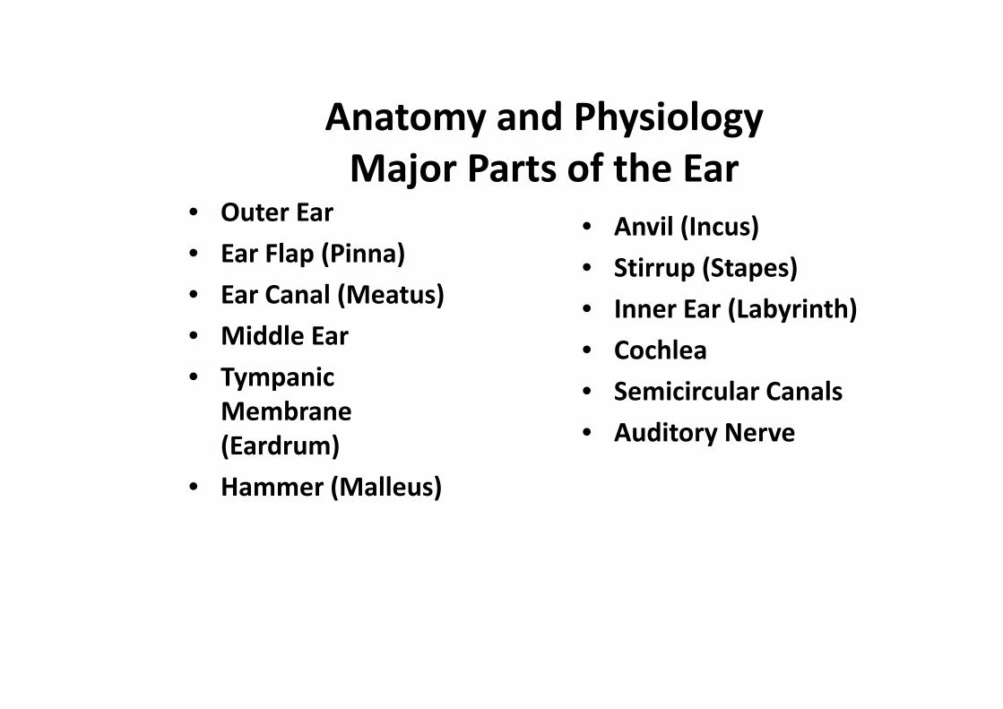
• Outer Ear
• Ear Flap (Pinna)
• Ear Canal (Meatus)
• Middle Ear
• Anvil (Incus)
• Stirrup (Stapes)
• Inner Ear (Labyrinth)
• Cochlea
Anatomy and PhysiologyMajor Parts of the Ear
• TympanicMembrane(Eardrum)
• Hammer (Malleus)
• Cochlea
• Semicircular Canals
• Auditory Nerve
Outer Ear
– The outer ear or external ear is a visible portion of the ear,which serves as a protective organ for the eardrum. Itcollects and guides the sound waves into the middle ear.The outer ear consists of the following two partsThe outer ear consists of the following two parts

Ear Flap (Pinna)
Ear Flap (Pinna)
– The visible part of the ear that resides outside of thehead
– The function of the pinna:
• Collect sound
• Perform spectral transformations to incoming sounds• Perform spectral transformations to incoming soundswhich enable the process of vertical localization totake place
– Acts as a funnel, amplifying the sound and directing it tothe auditory canal
– The pinna works differently for low and high frequencysounds
Ear Flap (Pinna)
– For low frequencies, it behaves similarly to a reflectordish, directing sounds toward the ear canal
– For high frequencies, however, its value is thought to bemore sophisticated
– Some of the sounds that enter the ear travel directly to– Some of the sounds that enter the ear travel directly tothe canal
– Others reflect off the contours of the pinna first
– A delay translates into phase cancellation
• Where the frequency component whose wave periodis twice the delay period is virtually eliminated.
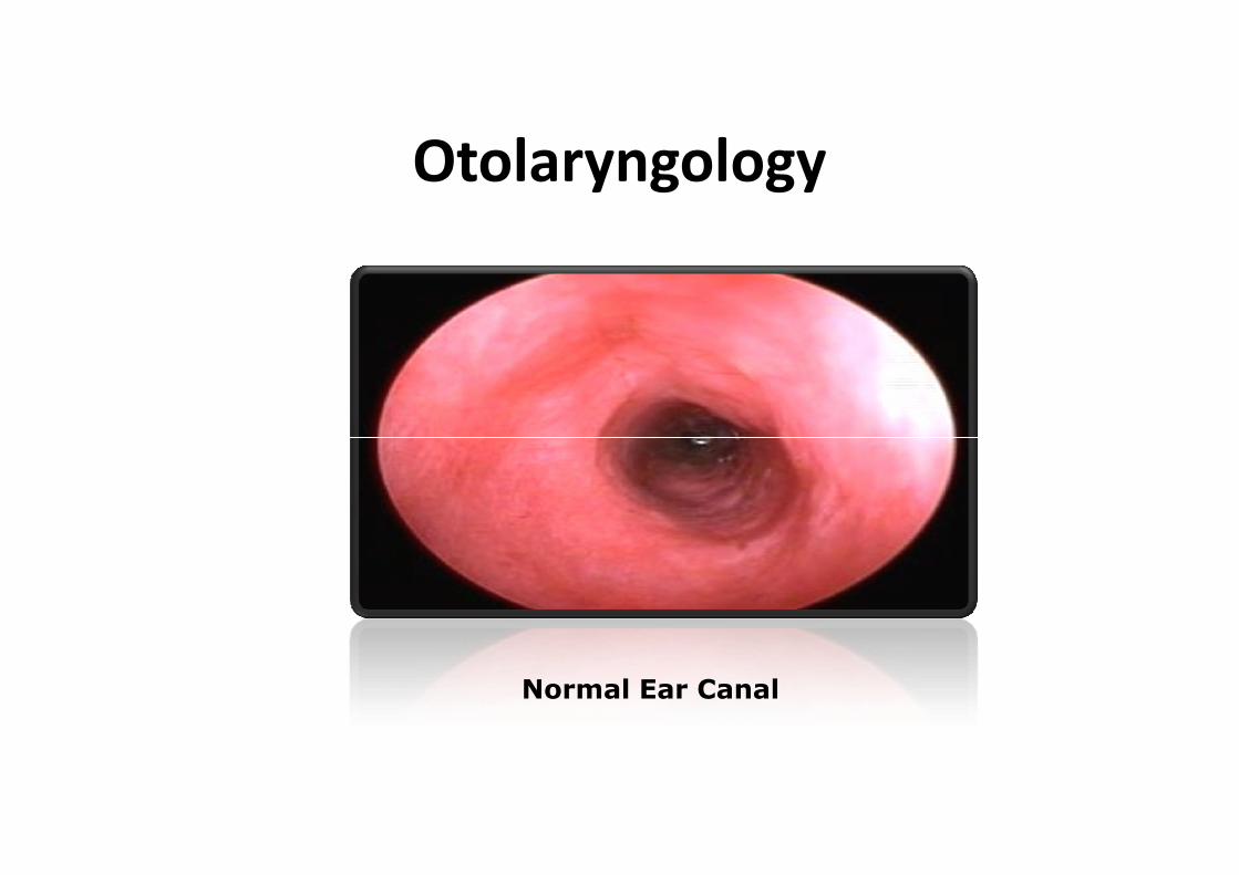
Otolaryngology
Normal Ear Canal

Ear Canal (meatus)
– The ear canal is a tube running from the outerear to the middle ear
– The human ear canal extends from the pinna tothe eardrum
– It is approximate 35 mm in length and 5 to 10mm in diameter
– The human ear canal is divided into two parts
• The fibrocartilaginous part forms the outerthird of the canal
• anterior and lower wall are cartilaginous,whereas its superior and back wall arefibrous
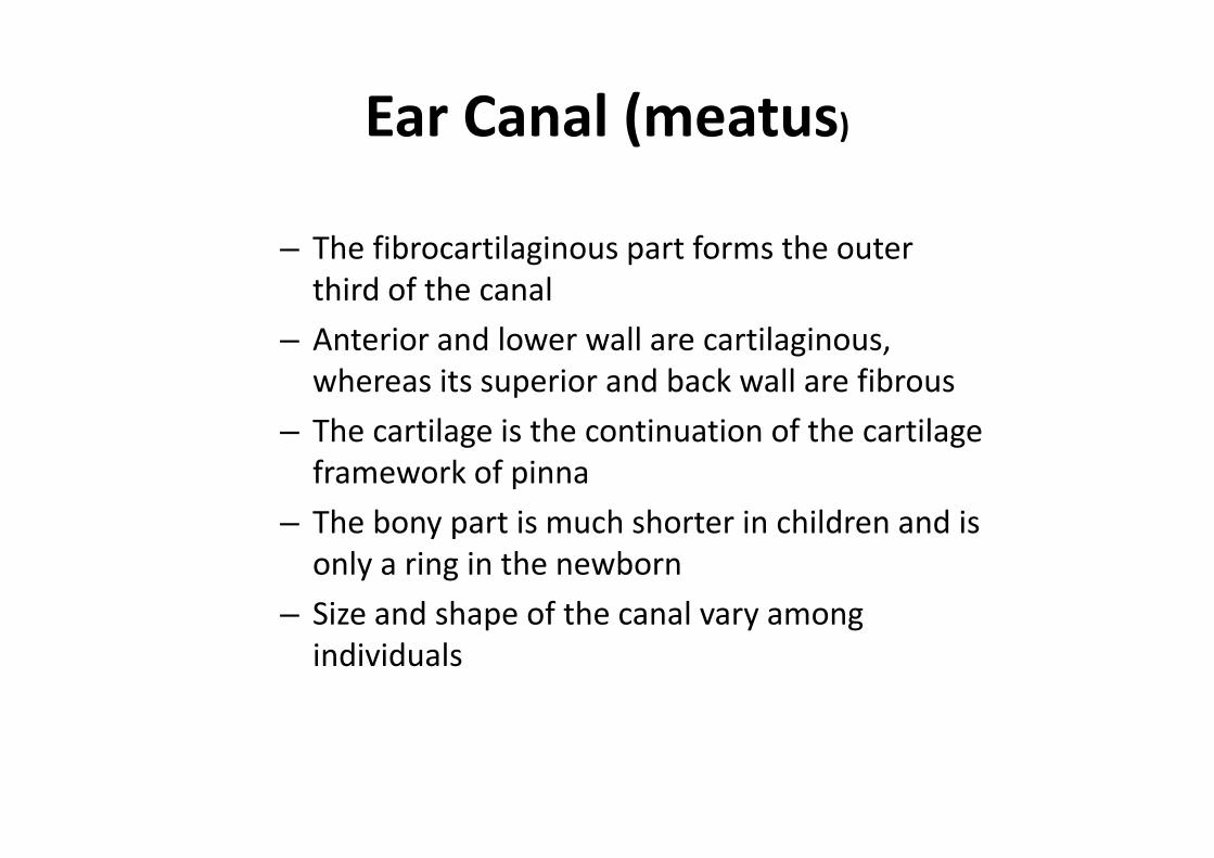
Ear Canal (meatus)
– The fibrocartilaginous part forms the outerthird of the canal
– Anterior and lower wall are cartilaginous,whereas its superior and back wall are fibrous
– The cartilage is the continuation of the cartilage– The cartilage is the continuation of the cartilageframework of pinna
– The bony part is much shorter in children and isonly a ring in the newborn
– Size and shape of the canal vary amongindividuals
Ear Canal (meatus)
– Earwax, also known as cerumen, is a yellowish,waxy substance secreted in the ear canals
– Assist in cleaning and lubrication
– Provides some protection from bacteria,fungus, and insectsfungus, and insects
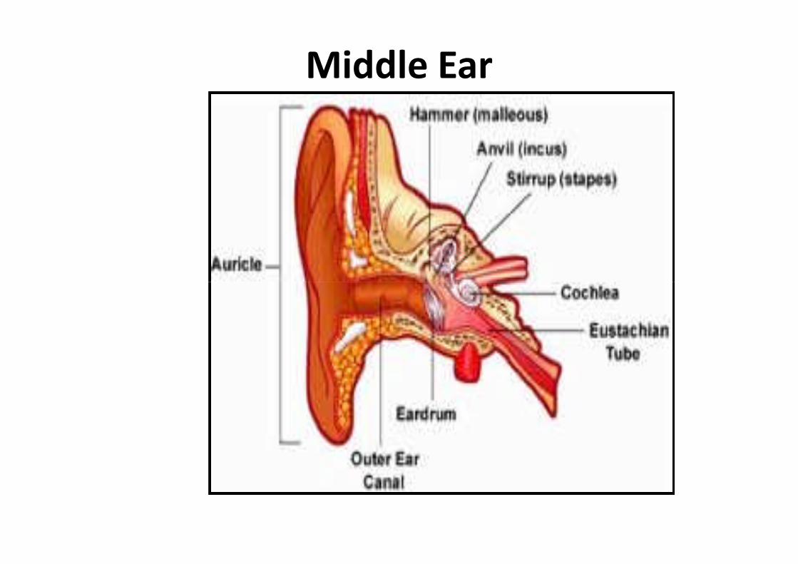
Middle Ear
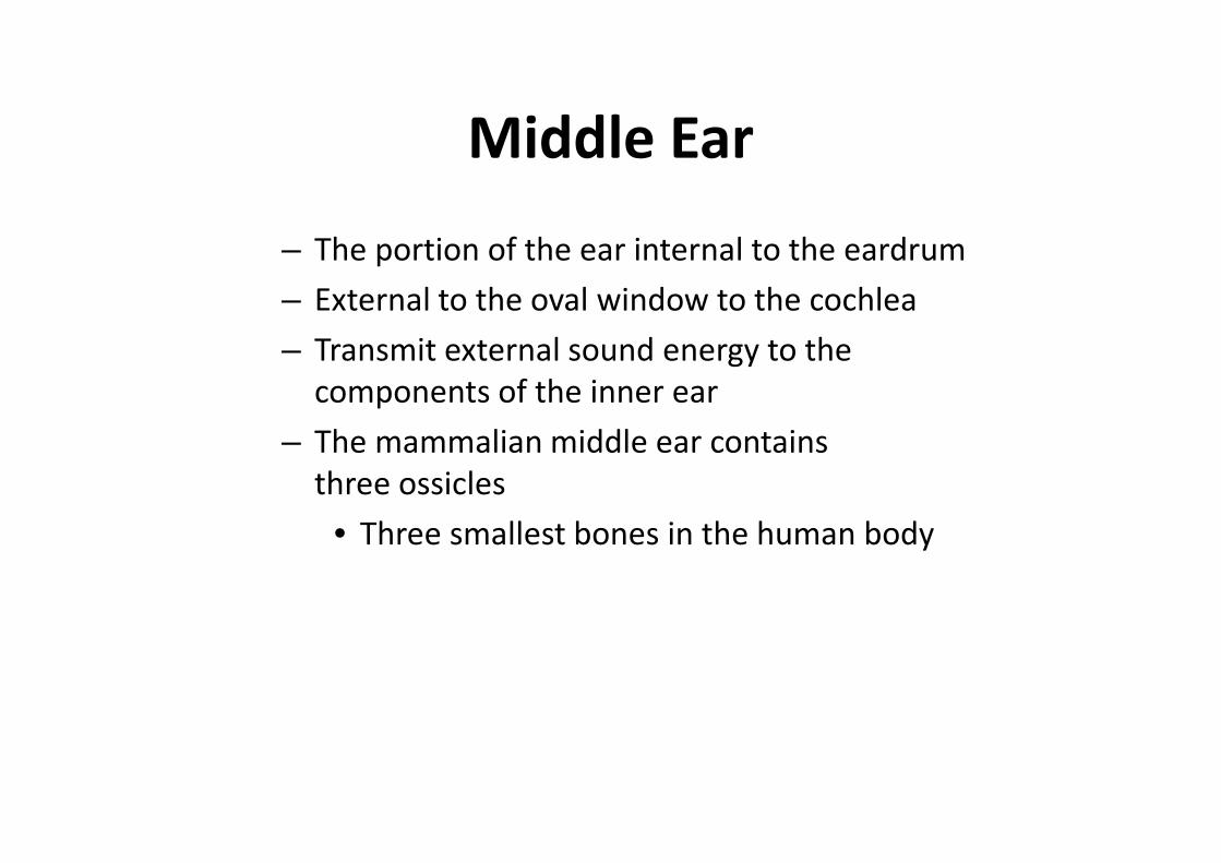
Middle Ear
– The portion of the ear internal to the eardrum
– External to the oval window to the cochlea
– Transmit external sound energy to thecomponents of the inner ear
– The mammalian middle ear contains– The mammalian middle ear containsthree ossicles
• Three smallest bones in the human body
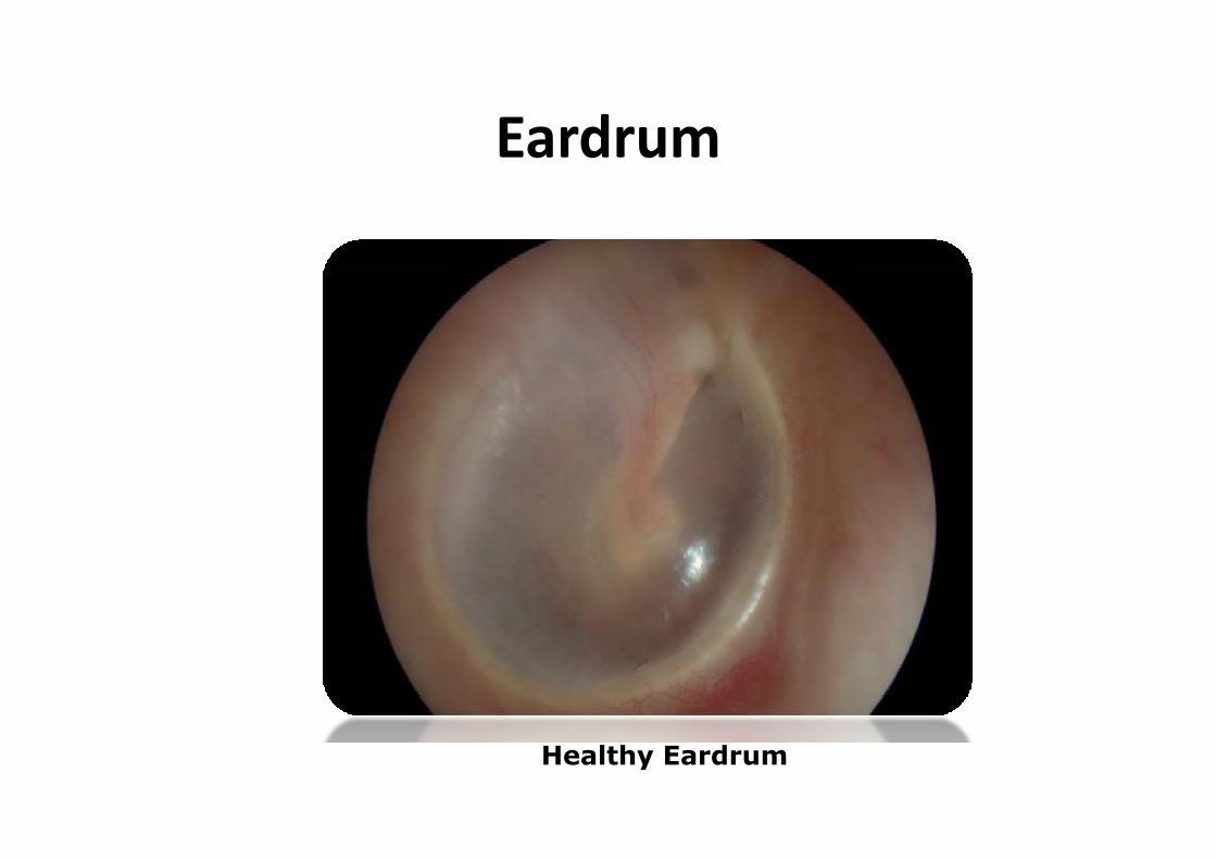
Eardrum
Healthy Eardrum
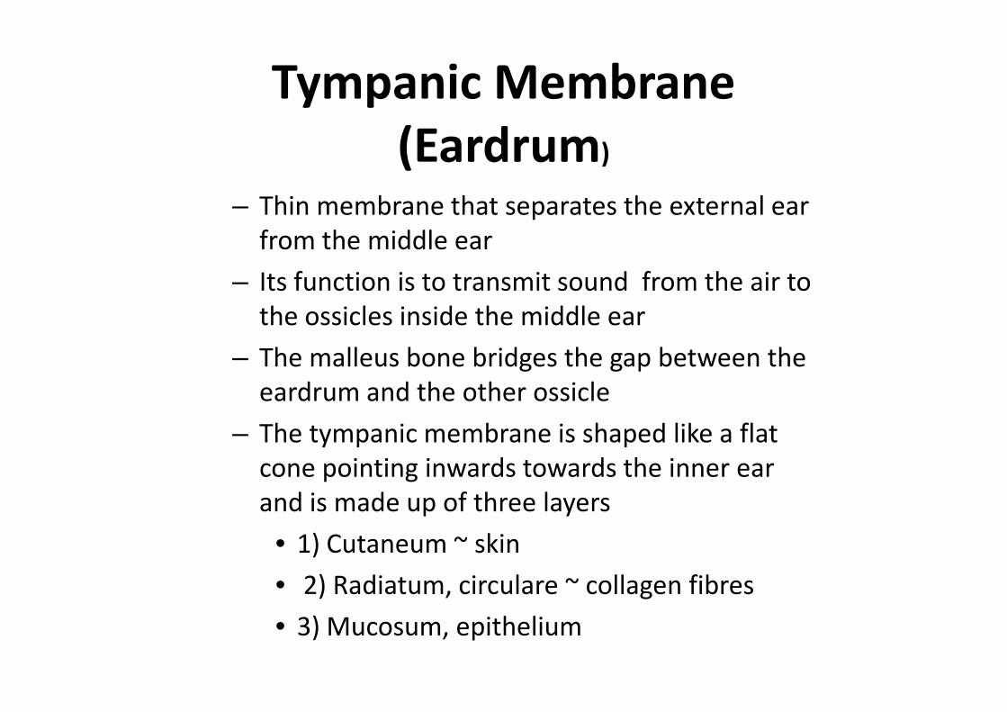
Tympanic Membrane(Eardrum)
– Thin membrane that separates the external earfrom the middle ear
– Its function is to transmit sound from the air tothe ossicles inside the middle ear
– The malleus bone bridges the gap between the– The malleus bone bridges the gap between theeardrum and the other ossicle
– The tympanic membrane is shaped like a flatcone pointing inwards towards the inner earand is made up of three layers
• 1) Cutaneum ~ skin
• 2) Radiatum, circulare ~ collagen fibres
• 3) Mucosum, epithelium
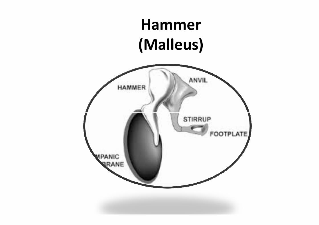
Hammer(Malleus)
Hammer(Malleus)
– Hammer-shaped small bone or ossicle of the middleear which connects with the incus and is attached tothe inner surface of the eardrum
– Transmits the sound vibrations from the eardrum tothe incusthe incus
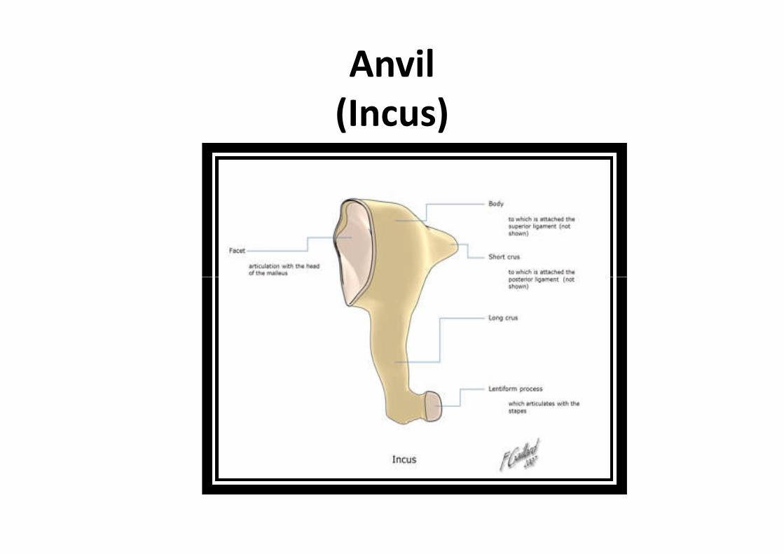
Anvil(Incus)
Anvil(Incus)
– Is the anvil-shaped small bone or ossicle in the middleear
– Connects the malleus to the stapes
– Transmits sound vibrations from the malleus tothe stapesthe stapes
Stapes(Stirrups)
NormalStape
AbnormalStape
Stapes(Stirrups)
– Transmits the sound vibrations from the incus to themembrane of the inner ear
– Stabilized by the stapedius muscle, which is innervatedby the facial nerve
– The stapes is the smallest bone in the body– The stapes is the smallest bone in the body
– Pronounced stay-peas

Inner Ear(Labyrinth)
Inner Ear(Labyrinth)
– The innermost part of the vertebrate ear
– System of passages comprising two main functionalparts
• The cochlea, dedicated to hearing
• The vestibular system, dedicated to balance• The vestibular system, dedicated to balance
Cochlea
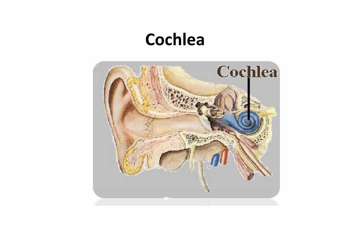
Cochlea– Is the auditory portion of the inner ear
– It is a spiral-shaped cavity in the bony labryinth
– A core component of the cochlea is the Organ of Corti,the sensory organ of hearing
– Filled with a watery liquid, which moves in response tothe vibrations coming from the middle earthe vibrations coming from the middle ear
– The walls of the hollow cochlea are made of bone,with a thin, delicate lining of epithelial tissue
Middle Ear
– The malleus (hammer) articulates with the incus and isattached to the tympanic membrane (eardrum), fromwhich vibrational sound pressure motion is passed
– The incus (anvil) is connected to both the other bones
– The stapes (stirrup) articulates with the incus and is– The stapes (stirrup) articulates with the incus and isattached to the membrane of the fenestra ovalis, theelliptical or oval window or opening between themiddle ear and thevestibule of the inner ear
Semicircular Canal
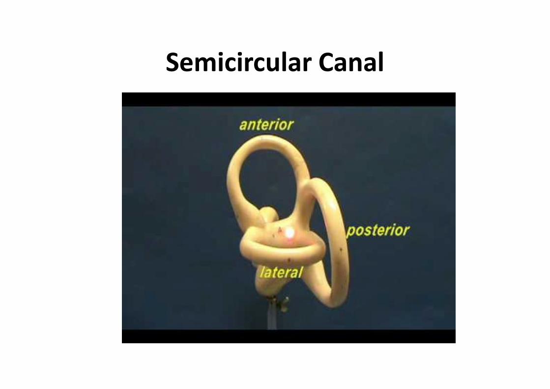
Semicircular Canal
– Three half-circular, interconnected tubes locatedinside each ear
– The three canals are the horizontal semicircularcanal (also known as the lateral semicircularcanal), superior semicircular canal (also known as thecanal), superior semicircular canal (also known as theanterior semicircular canal), and the posteriorsemicircular canal
– Each canal is filled with a fluid called endolymph andcontains a motion sensor with little hairs
– The semicircular canals are a component of the bonylabryinth

Auditory Nerve

Auditory Nerve
– A nerve in the head that carries signals fromthe cochlea of the inner ear to the brain
– A sensory nerve, one which conducts tothe brain information about the environment
– Arises from within the cochlea and extends to– Arises from within the cochlea and extends tothe brainstem
Disease and Treatments of the Ear

Impacted Cerumen
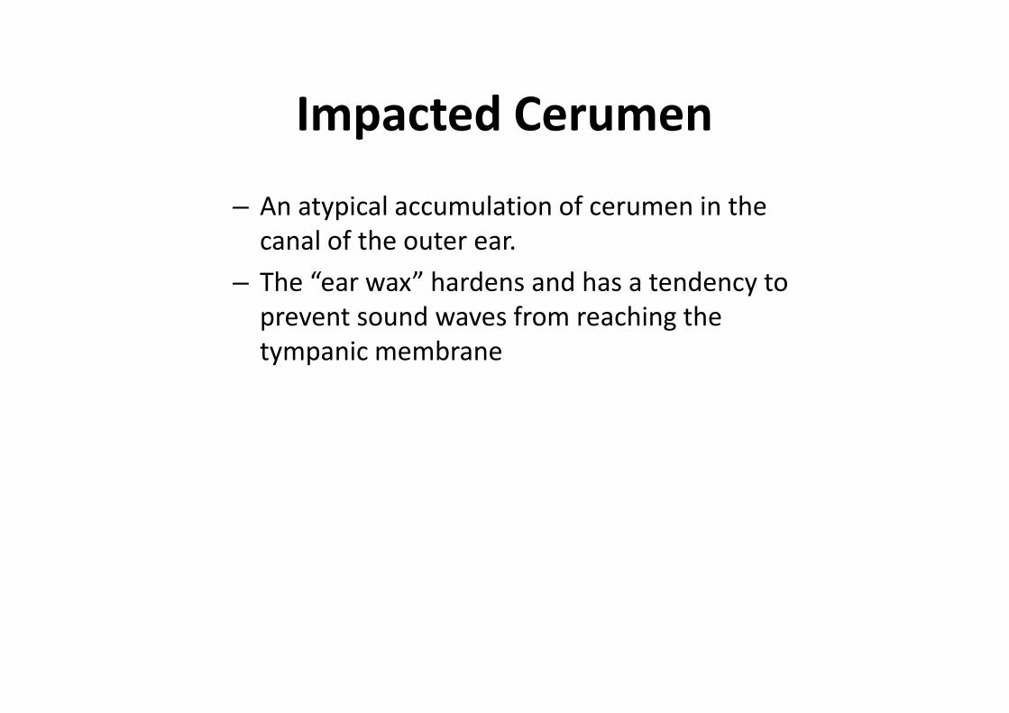
Impacted Cerumen
– An atypical accumulation of cerumen in thecanal of the outer ear.
– The “ear wax” hardens and has a tendency toprevent sound waves from reaching thetympanic membranetympanic membrane

Impacted Cerumen Signs andSymptoms
– If the secretions accumulates excessively, agradual loss of hearing may occur and thepatient may have a feelig that the ear isplugged and may experience tinnitus or anplugged and may experience tinnitus or anearache (otalgia)
– Impacted cerumen is a common cause ofconductive hearing loss
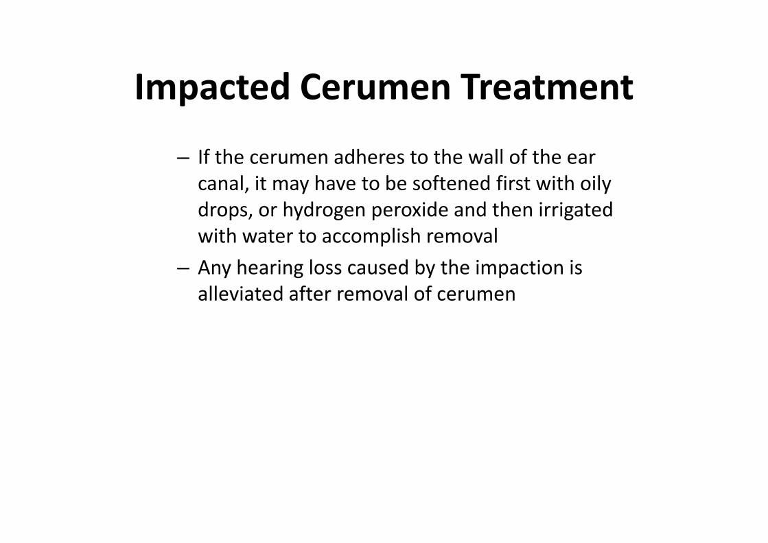
Impacted Cerumen Treatment
– If the cerumen adheres to the wall of the earcanal, it may have to be softened first with oilydrops, or hydrogen peroxide and then irrigatedwith water to accomplish removal
– Any hearing loss caused by the impaction is– Any hearing loss caused by the impaction isalleviated after removal of cerumen
Impacted Cerumen Prognosis
– The prognosis for removal is positive
– Hearing usually improves once the ear canal isclear of the impacted ear wax.
– Recurrence is likely so periodic examinationsmay be necessarymay be necessary
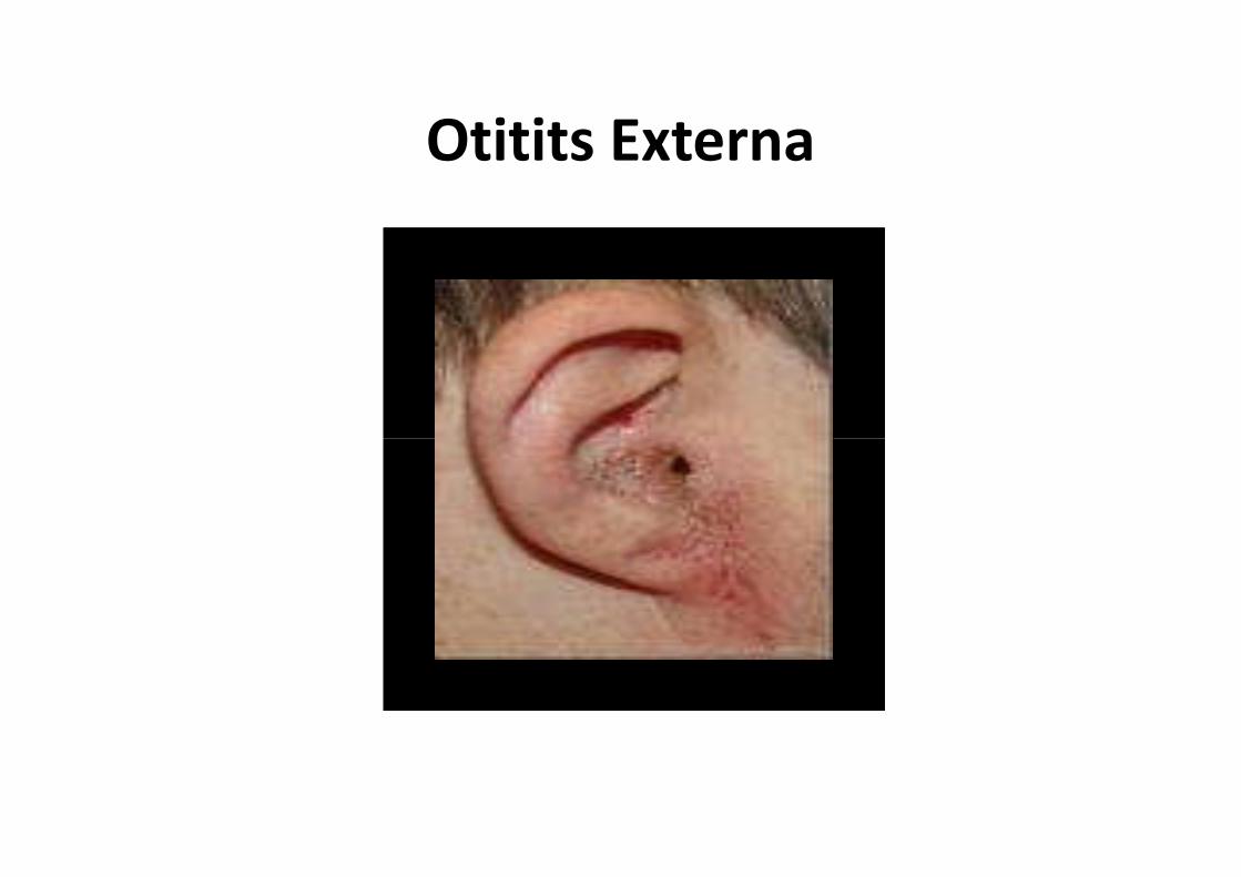
Otitits Externa
Otitis Externa
– Inflammation of the external ear canal
– Condition is usually accompanied by an infectiousprocess
Otitis Externa Signs andSymptoms
– Severe pain
– Red, swollen ear canal
– Hearing loss
– Fever
– Pruritus– Pruritus
– Drainage from the ear may be either wax orpurulent
Otitis Externa Diagnosis
– Otologic examination and a history ofsymptoms confirm the diagnosis.
– If a bacterial infection is suspected, a culture ofthe material found in the canal may be neededto determine how to properly treat theto determine how to properly treat theinfection
Otitis Externa Treatment
– The ear canal must be kept clean and free fromwater
– Antibiotic or steroid eardrops and systemicantibiotics may be prescribed
– Tends to recur and can become chronic– Tends to recur and can become chronic
Otitis Externa Prognosis
– Prognosis is positive with treatment
– Chroinic otitis externa may develop withrepeated irritation by earphones, earplugs, orhearing aids

Otitis Externa Prevention
– Keep the ear clean and dry
– Keep earphones, earplugs, and hearing aidsclean
– Use of another’s earphones or earplugs isdiscourageddiscouraged
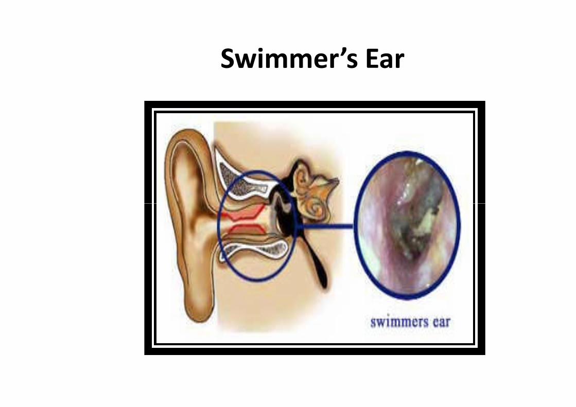
Swimmer’s Ear
Swimmer’s Ear
– Inflammation and resulting infection of theouter ear canal after water has been entrappedduring

Swimmer’s Ear Signs andSymptoms
– Severe pain
– Red, swollen ear canal
– Hearing loss
– Fever
– Pruritis– Pruritis
– Drainage that may be watery or purulent

Swimmer’s Ear Treatment
– Keep ear canal clean and dry after swimming
– Antibiotic or steroid eardrops and systemicantibiotics
– Can become chronic for those with repeatedexposure to waterexposure to water

Otitis Media
Otitis Media
– Most common reason for visits to physicians bychildren and can be experienced by adults
– Classified as either serous or suppurative
– Nonsuppurative, the fluid is relatively clear andsterilesterile
– Suppurative, the fluid is purulent
– The only symptom maybe a feeling of fullnessor pressure and some degree of hearing loss
Otitis Media Diagnosis
– Otoscopy reveals the presence of a fluid-filledmiddle ear
– Pearl-gray eardrum is inflamed and may bebulging
– Fluid bubbles may be visible through the– Fluid bubbles may be visible through themembrane
– If a culture is taken, white blood count wouldbe elevated
– Measurement with tympanogram
Otitis Media Treatment
– Analgesics and deongestants
– Antibiotics
– Surgical evacuation of fluid for the more severecases
– Myringotomy tubes– Myringotomy tubes
– Removal of hypertrophic adenoids is atherapeutic measure

Otosclerosis

Otosclerosis
– Primarily affects the stapes or third bone orossicle in the middle ear
– Movement is impaired, which causesdiminished conduction of sound waves andresults in hearing lossresults in hearing loss
Otosclerosis Signs andSymptoms
– Abnormal growth of spongy bone forms aroundthe oval window
– Ankylosis produces conductive deafness
– Gradual hearing loss
– Loss of low or soft sounds– Loss of low or soft sounds
– Tinnitus (ringing in the ear)
– Onset begins after puberty and before 35 yearsof age
Otosclerosis Treatment
– The only treatment that cures otosclerosis is asurgical procedure called a Stapedectomy
– Removes the diseased stapes and replaces itwith a prosthesis
Benign Paroxysmal PositionalVertigo (BPV)
Benign Paroxysmal PositionalVertigo
– Benign positional vertigo is usually a vestibularsystem disorder. The patient complains ofhis/her head spinning, becoming worse withmovement of the heardhis/her head spinning, becoming worse withmovement of the heard
Benign Paroxysmal PositionalVertigo Signs and Symptoms– Spinning sensation with movement of the head
– Dizziness
– Feel that their body is moving with eyes open
– Nausea, vomitting, and involuntary eyemovement
Benign Paroxysmal PositionalVertigo Diagnosis
– History and examination
– Audiogram or other means of hearing testing
– Testing to rule out CNS involvement
– CT, MRI scan of the head– CT, MRI scan of the head
– MRA scan of the brain blood vessels
– Caloric stimulation test
Benign Paroxysmal PositionalVertigo Treatment
– Antihistamines (Antivert, Dramamine)
– Anticholinergics (Scopolamine)
– Benzodiazepines (Compazine)
– Exercises where the individual repeatedly turns– Exercises where the individual repeatedly turnsthe head from side to side may also be helpful
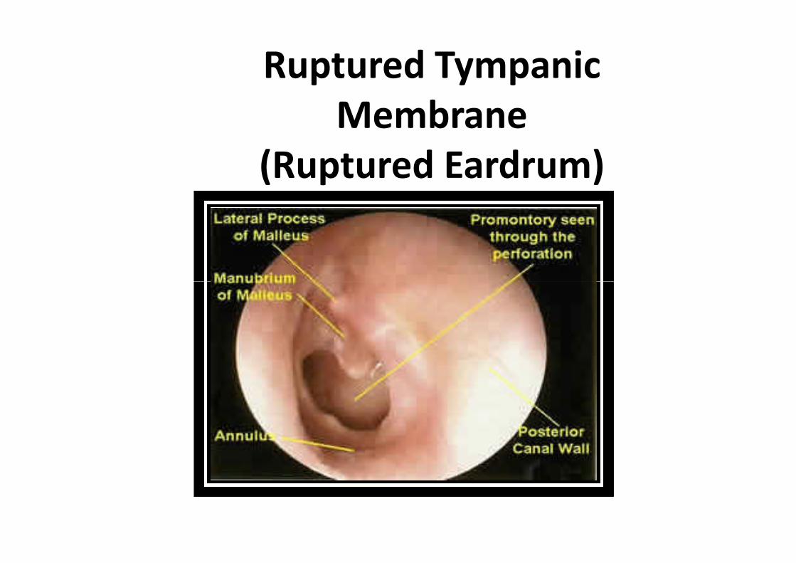
Ruptured TympanicMembrane
(Ruptured Eardrum)
Ruptured TympanicMembrane
(Ruptured Eardrum)– Any type of tear or injury to the eardrum
causes a breach in the integrity of themembrane
Results in pressure, force, or insult from the– Results in pressure, force, or insult from theexterior aspect, or it may be caused byincreased pressure within the middle ear
Ruptured TympanicMembrane Signs and
Symptoms– Slight pain
– Partial loss of hearing
– Discharge or bleeding from the ear
Ruptured TympanicMembrane Diagnosis
– Visual examination of the ear with an otoscopeconfirms the diagnosis
– Audiometry
Ruptured TympanicMembrane Treatment
– Antibiotic
– Patch may be applied to the eardrum to aid inhealing and improve hearing
– Tympanoplasty (involves actual grafting oftissue for eardrum repair)tissue for eardrum repair)
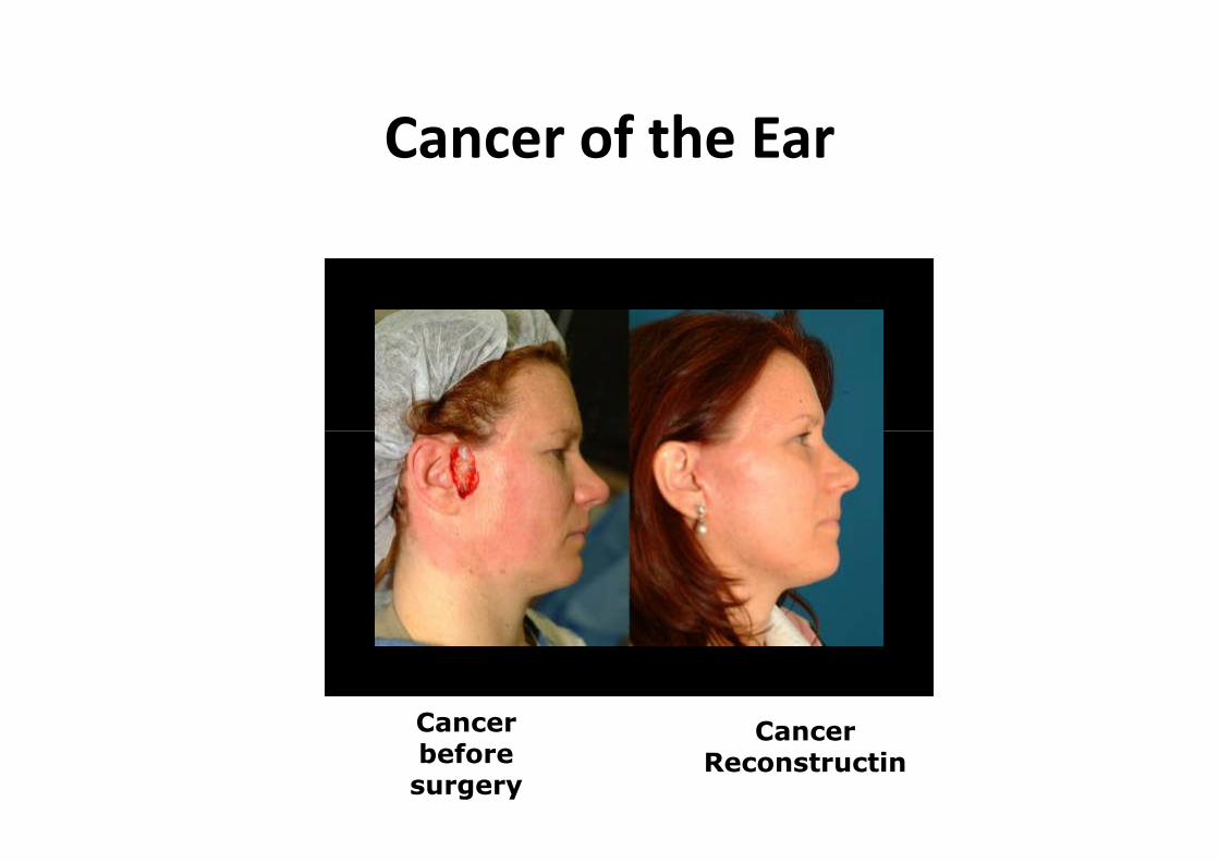
Cancer of the Ear
Cancerbeforesurgery
CancerReconstructin

Cancer of the Ear
– Tumors of the ear can occur in any part of theear and may be benign or malignant
– Include:
• Cutaneous tumrs, ceruminal glandneoplasms, acoustic and facial neuromasneoplasms, acoustic and facial neuromasand glomus tumors
– Most metastasize to the ear in secondary earcancer
Signs and Symptoms
– Progressive hearing loss
– Chronic otic discharge
– Mass or lesion on ear exam
– Pain
– Pulsatile tinnitus– Pulsatile tinnitus
Diagnosis
– First identified because of the symptoms
– Biopsy
– Ct scan
– MRI used to evaluate the extent of the disease

Treatment
– Surgical excision
– Radiation therapy
– Nerve graft may be performed after surgicalexcision of a neuroma


![Automatic Cochlea Multi-modal Images Segmentation · 2018-04-03 · Automatic Cochlea Multi-modal Images Segmentation Al-Dhamari, CI2018 Methods: Cochlea Model 9 [5] Gerber et al,](https://img.pdfslide.us/doc/110x75/5f8e42f1fe0c2a0180250f2a/automatic-cochlea-multi-modal-images-segmentation-2018-04-03-automatic-cochlea.jpg)