Embed Size (px)
Citation preview

Anatomopathological Session
Case 4 – 84-Year Old Female with Precordial Pain and Cardiac Arrest with Pulseless Electrical ActivityBruna Affonso Madaloso e Luiz Alberto BenvenutiInstituto do Coração (InCor) - HC-FMUSP, São Paulo, SP - Brasil
Mailing Address: Vera Demarchi Aiello •bloco I, Cerqueira César. Postal Code 05403-000, São Paulo, SP - BrazilE-mail: [email protected], [email protected]
KeywordsHypertension; Chest Pain; Dissection; Aorta, Thoracic;
Plaque, Atherosclerotic.
An 84-year old woman, born in Itamuji (Minas Gerais) and from São Paulo (SP), with arterial hypertension, history of atrial fibrillation, and former smoker, has sought for emergency medical care due to a prolonged precordial pain (Mar 03, 2010).
The patient started medical treatment at InCor in June, 2005, when she received treatment for arrhythmia, dyspnea on exertion and precordial pain, which occurred in prolonged episodes, without precise triggers, during the previous six years. She was administrating warfarin 2.5mg in alternate days and, daily doses of captopril 150mg, amiodarone 200mg, isosorbide mononitrate 20mg, spirinolactone 25mg, metoprolol 100mg, furosemide 40mg and cilostazol 100mg.
ECG (Jun 24, 2005) showed atrial fibrillation rhythm, mean heart rate of 89bpm, QRS duration 85ms, Qtc = 470 ms, SÂQRS 0° (Figure 1).
Test results (Jun 24, 2005) showed: hemoglobin 12.6 g / dL, hematocrit 39%, white blood cells 5.800/mm³, sodium 142mEq/L, potassium 5.8mEq/L and serology to detect positive (Elisa) and negative (indirect immunofluorescence) Chagas disease.
Physical exam (Jun 29, 2005) showed irregular 88bpm pulse rate, 150 x 110mmHg blood pressure, symmetric pulse, pulmonary auscultation was normal, cardiac auscultaiton showed arrhythmic heart sounds and hyperphonesis of 2nd heart sound in aortic area. There were no abnormalities in the examination of abdomen and lower limbs. The following medication was interrupted: amiodarone, isosorbide mononitrate and cilostazol.
Two months later (August, 2005) showed large hematomas on abdomen and lower limbs, although INR is within the therapeutic range, 2, 32, and oral anticoagulant was interrupted for a period of time.
Echocardiogram (Jan, 2006) revealed left ventricle hyper t rophy (11mm pos te r io r wa l l and 12mm interventricular septum), left ventricle with normal dimensions (47 x 32mm) and normal ejection fraction (59%); left atrium diameter was 41mm, enlargement of aortic root diameter (43mm) and aorta arch size was normal (28mm). Mitral annular calcification was present; there were no valvular changes.
New ECG (Feb, 2006) revealed sinus bradycardia (43mm) (Figure 2).
Long-term electrocardiogram using Holter system (Jan 02, 2007), revealed sinus rhythm, 15 isolated ventricular extrasystoles, and 2 pairs; 2,878 atrial extrasystoles (229 pairs, 9 atrial tachycardias: the most rapid with 175bpm frequency and the longest with 15 beats). The complaint of “chest burn” was related to atrial extrasystoles, and there were no changes in the ventricular repolarization.
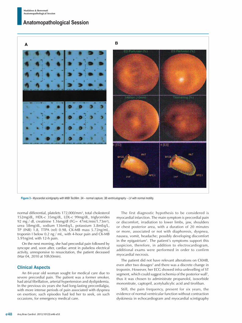
Myocardial scintigraphy with MIBI Tec99m and adenosine administration (Feb 13, 2007) did not indicate abnormalities on myocardial capture of the radiotracer and ejection fraction evaluated by tomography (SPECT) was 48%, without segmental alteration of motility (Figure A and Figure 3B).
She had sought for medical care in August, 2007 due to epigastric pain and “dark colored” vomit.
The physical exam (Aug 15, 2007) revealed revealed an eupneic and slightly pale patient 106bpm heart rate, 156 x 100mmHg blood pressure and, no other changes, except for arrhythmic heart sounds.
Laboratory exams on that same day revealed: hemoglobin 12.7g/dL, 40% hematocrit, 8,800 white blood cells/mm³ with normal differential, 240,00 platelets/mm³, urea 86gm/dL, creatinine 1.51mg/dL, sodium 140mEq/L, potassium 5.0mEq/L, ionized calcium 1.19mmol/L, ionized magnesium 1.3mmol/L, TP (INR) 7.83, TTP (rel) 2.43, dimer-D 116ng/mL.
High digestive endoscopy (Aug 15, 2007) revealed moderate hemorrhagic erosive pangastritis (Figure 4). Three fresh frozen plasma units were administered, TP (INR) was recorded at 3.0 and TTP (rel) to 1.5. The patient was discharged with proton pump inhibitor on the next day.
Section Editor: Alfredo José Mansur ([email protected])
Associate Editors: Desidério Favarato ([email protected])
Vera Demarchi Aiello ([email protected])
DOI: 10.5935/abc.20130178
e46

Anatomopathological Session
Madaloso & BenvenutiAnatomopathological Session
Arq Bras Cardiol. 2013;101(3):e46-e53
Figure 2 - ECG. Sinus bradycardia.
Figure 1 - ECG. Atrial fibrillation.
Laboratorial evaluation of outpatient (Apr 2008) revealed total cholesterol 235mg/dL, HDL-c 33mg/dL, LDL-c 171mg / dL, triglycerides 156mg/dL, fasting glycemia 108mg/dL, TSH 2.09µu/mL, free T4 1.7ng/dL, sodium 144mEq/L, potassium 4.9mEq/L, urea 63mg/dL, creatinine 1.22mg/dL and INR 2.0.
Synvastatin 40mg and digoxin 0.125mg were prescribed.Control tests on the following year (2009) revealed total
cholesterol 198mg/dL, HDL-c 36mg/dL, LDL-c96 mg/dL, triglycerides 120mg/dL, INR 2.7 and no changes on hepatic enzymes or creatine phosphokinase.
Long term electrocardiogram using Holter system (May 2009) revealed atrial fibrillation rhythm with heart rate ranging from 45bpm to 143bpm. Palpitations were associated with the increased heart rate.
The patient progresses with occasional precordial pain and dyspnea on exertion.
She sought for medical care on the night of March 03, 2010 complaining of pain on anterior wall chest area, irradiating
over upper limbs and abdomen, with nausea and cold sweating, which began 3 hours earlier.
The physical examination revealed mean heart rate at 79bpm, blood pressure 127 x 91mmHg; except for arrhythmic heart sounds, no abnormalities were reported in the physical examination.
ECG during pain (Mar 03, 2010, 21:27) revealed atrial fibrillation rhythm, mean rate at 121bpm, left ventricle overload and alterations on ventricular repolarization using strain or digital pattern (Figure 5).
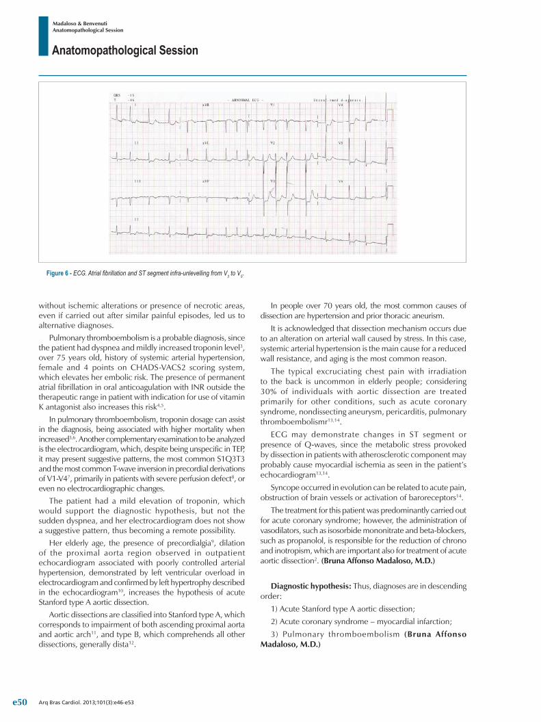
ECG after the use of isosorbide (Mar 03, 2010; 22:5) showed decreased heart rate, now at 103bpm, and onset of ST infra-unlevelling from V3 to V5 (Figure 6).
This led to pain relief and the administration of acetylsalicylic acid 100mg, tirofiban 0.625mg in half hour, followed by 0.33mg/h; captopril 12.5mg 8/8h and propanolol 40mg every 8 hours.
Laboratory tests (Mar 03, 2010 at 23h) revealed hemoglobin 11.9g/dL, hematocrit 38%, white blood cells 9,900/mm³, with
e47
Anatomopathological Session
Madaloso & BenvenutiAnatomopathological Session
Arq Bras Cardiol. 2013;101(3):e46-e53
Figure 3 - Myocardial scintigraphy with MIBI Tec99m. 3A – normal capture; 3B ventriculography – LV with normal motility.
normal differential, platelets 172,000/mm³, total cholesterol 152mg/dL, HDL-c 35mg/dL, LDL-c 99mg/dL, triglycerides 92 mg / dl, creatinine 1.16mg/dl (FG= 47mL/min/1.73m²), urea 58mg/dL, sodium 136mEq/L, potassium 3.8mEq/L, TP (INR) 1.8, TTPA (rel) 0.98, CK-MB mass 5.72ng/mL, troponin I below 0.2 ng / mL, with 4-hour pain and CK-MB 5.91ng/mL with 12-h pain.
On the next morning, she had precordial pain followed by syncope and, soon after, cardiac arrest in pulseless electrical activity, unresponsive to resuscitation, the patient deceased (Mar 04, 2010 at 10h30min).
Clinical AspectsAn 84-year old woman sought for medical care due to
severe precordial pain. The patient was a former smoker, had atrial fibrillation, arterial hypertension and dyslipidemia. In the previous six years she had long-lasting precordialgia, with more intense periods of pain associated with dyspnea on exertion; such episodes had led her to seek, on such occasions, for emergency medical care.
The first diagnostic hypothesis to be considered is myocardial infarction. The main symptom is precordial pain or discomfort, irradiation to lower limbs, jaw, shoulders or chest posterior area, with a duration of 20 minutes or more, associated or not with diaphoresis, dyspnea, nausea, vomit, headache; possibly developing discomfort in the epigastrium1. The patient’s symptoms support this suspicion, therefore, in addition to electrocardiogram, additional exams were performed in order to confirm myocardial necrosis.
The patient did not have relevant alterations on CKMB, even after two dosages2 and there was a discrete change in troponin. However, her ECG showed infra-unlevelling of ST segment, which could suggest ischemia of the posterior wall1, thus it was chosen to administrate propanolol, isosorbide mononitrate, captopril, acetylsalicylic acid and tirofiban.
Still, the pain frequency, present for six years, the evidence of normal ventricular function without contraction dyskinesia in echocardiogram and myocardial scintigraphy
e48

Anatomopathological Session
Madaloso & BenvenutiAnatomopathological Session
Arq Bras Cardiol. 2013;101(3):e46-e53
Figure 4 - High digestive endoscopy. Moderate hemorrhagic erosive pangastritis.
Figure 5 - ECG. Atrial fibrillation. Left ventricular overload with strain pattern.
e49
Anatomopathological Session
Madaloso & BenvenutiAnatomopathological Session
Arq Bras Cardiol. 2013;101(3):e46-e53
Figure 6 - ECG. Atrial fibrillation and ST segment infra-unlevelling from V3 to V5.
without ischemic alterations or presence of necrotic areas, even if carried out after similar painful episodes, led us to alternative diagnoses.
Pulmonary thromboembolism is a probable diagnosis, since the patient had dyspnea and mildly increased troponin level3, over 75 years old, history of systemic arterial hypertension, female and 4 points on CHADS-VACS2 scoring system, which elevates her embolic risk. The presence of permanent atrial fibrillation in oral anticoagulation with INR outside the therapeutic range in patient with indication for use of vitamin K antagonist also increases this risk4,5.
In pulmonary thromboembolism, troponin dosage can assist in the diagnosis, being associated with higher mortality when increased3,6. Another complementary examination to be analyzed is the electrocardiogram, which, despite being unspecific in TEP, it may present suggestive patterns, the most common S1Q3T3 and the most common T-wave inversion in precordial derivations of V1-V47, primarily in patients with severe perfusion defect8, or even no electrocardiographic changes.
The patient had a mild elevation of troponin, which would support the diagnostic hypothesis, but not the sudden dyspnea, and her electrocardiogram does not show a suggestive pattern, thus becoming a remote possibility.
Her elderly age, the presence of precordialgia9, dilation of the proximal aorta region observed in outpatient echocardiogram associated with poorly controlled arterial hypertension, demonstrated by left ventricular overload in electrocardiogram and confirmed by left hypertrophy described in the echocardiogram10, increases the hypothesis of acute Stanford type A aortic dissection.
Aortic dissections are classified into Stanford type A, which corresponds to impairment of both ascending proximal aorta and aortic arch11, and type B, which comprehends all other dissections, generally dista12.
In people over 70 years old, the most common causes of dissection are hypertension and prior thoracic aneurism.
It is acknowledged that dissection mechanism occurs due to an alteration on arterial wall caused by stress. In this case, systemic arterial hypertension is the main cause for a reduced wall resistance, and aging is the most common reason.
The typical excruciating chest pain with irradiation to the back is uncommon in elderly people; considering 30% of individuals with aortic dissection are treated primarily for other conditions, such as acute coronary syndrome, nondissecting aneurysm, pericarditis, pulmonary thromboembolismr13,14.
ECG may demonstrate changes in ST segment or presence of Q-waves, since the metabolic stress provoked by dissection in patients with atherosclerotic component may probably cause myocardial ischemia as seen in the patient’s echocardiogram13,14.
Syncope occurred in evolution can be related to acute pain, obstruction of brain vessels or activation of baroreceptors14.
The treatment for this patient was predominantly carried out for acute coronary syndrome; however, the administration of vasodilators, such as isosorbide mononitrate and beta-blockers, such as propanolol, is responsible for the reduction of chrono and inotropism, which are important also for treatment of acute aortic dissection2. (Bruna Affonso Madaloso, M.D.)
Diagnostic hypothesis: Thus, diagnoses are in descending order:
1) Acute Stanford type A aortic dissection;
2) Acute coronary syndrome – myocardial infarction;
3) Pulmonary thromboembolism (Bruna Affonso Madaloso, M.D.)
e50

Anatomopathological Session
Madaloso & BenvenutiAnatomopathological Session
Arq Bras Cardiol. 2013;101(3):e46-e53
Figure 7 - Internal view of ascending aorta, aortic arch and beginning of descending thoracic aorta. The entry orifice of dissection is located at the isthmus level, in ulcerated atherosclerotic plaque (arrow). Observe the aortic wall delamination, with presence of false lumen and adventitial hemorrhage in the initial segment of descending thoracic aorta (asterisk).
NecropsyAfter opening the chest cavity, it was observed a severe pleural
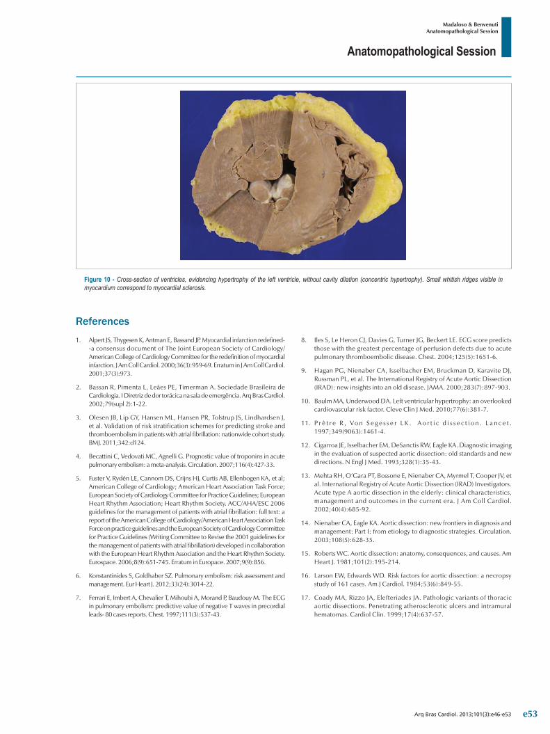
effusion on the left, consisting of 1.7 liters of blood clot. There was hemorrhagic infiltration of both pulmonary hila and adventitial hematoma at the level of distal segment of aortic arch and proximal descending thoracic aorta. The aortic opening revealed an entry site of acute dissection located in the aortic isthmus, in a region of complicated atherosclerotic plaque (Figure 7). Dissection was progressing anterogradely throughout the aorta, extending through iliac arteries, without detecting a re-entry orifice. There was no retrograde dissection. Severe atherosclerosis was detected in aorta, with numerous yellowish, ulcerated plaques, showing extensive calcification, spread throughout the vessel (Figure 8). The histological examination of aorta confirmed acute dissection, with cleavage in the outer third of the media; there was no inflammatory infiltrate in the aortic wall (Figure 9). The pericardial sac had no effusion. Cardiac examination evidenced concentric hypertrophy of the left ventricle, of moderate/severe level, with mild myocardial sclerosis (Figure 10). There were no infarction areas. The macroscopic examination of epicardial coronary arteries revealed atherosclerosis with extensive calcification areas and luminal stenosis; however, no severely obstructive injuries. Other findings in autopsy consisted in calcification of mitral valve annulus, renal ischemic changes, with focal areas of healed infarction, mild hepatic steatosis, and subserosal uterine leiomyoma, measuring 1.4cm diameter. (Luiz Alberto Benvenuti, M.D.)
Anatomopathological diagnosis – Systemic atherosclerosis; concentric hypertrophy of the left ventricle; benign nephrosclerosis; calcification of mitral valve annulus; uterine leiomyoma; acute dissection of aorta, ruptured to left pleural cavity (cause of death). (Luiz Alberto Benvenuti, M.D.)
CommentsCase of a female patient, 84 years old, with systemic arterial
hypertension and chronic atrial fibrillation, who sought for emergency medical care due to prolonged precordial pain, followed by syncope and cardiac arrest. Necropsy confirmed the hypertensive heart disease diagnosis in compensated phase, i.e. with left ventricle hypertrophy without cavity dilation. Death was caused by hemothorax, secondary to acute dissection rupture of aorta. Aorta dissection occurs when there is wall delamination, generally in the outer third of the media, forming a false path to blood, where it penetrates through intimal orifice. Generally it is associated with systemic arterial hypertension, such as in this case, but Marfan syndrome, other diseases in connective tissue, aortic valve congenital anomalies, aorta coarctation, pregnancy or thoracic trauma may be predisposing factors15. According to DeBakey classification, the patient’s dissection was type III, i.e. with entry orifice located beyond the ascending aorta, subtype B, i.e. without retrograde dissection of ascending aorta. Such type of dissection (III B) corresponds to 16% of the total, remembering that the most common is type I dissection, which entry orifice is located in the ascending aorta and dissection progresses to descending aorta (54% of cases)16. According to Stanford classification, dissection would be type B, i.e. without involving the ascending aorta.
Unlike the usual, in this case, dissection occurred in aorta impaired by severe atherosclerosis. Curiously, the entry orifice was located in an ulcerated atherosclerotic plaque, requiring a differential diagnosis with penetrating atherosclerotic ulcer. However, in the latter there is no true progressive dissection of the wall, as in this case, and lesion is localized, with possible formation of aneurysm and local rupture17. (Luiz Alberto Benvenuti, M.D.)
e51

Anatomopathological Session
Madaloso & BenvenutiAnatomopathological Session
Arq Bras Cardiol. 2013;101(3):e46-e53
Figure 8 - Internal view of abdominal aorta evidencing severe atherosclerosis, with numerous complicated plaques, showing ulceration and extensive calcification. Note the aortic wall dissection, present in the distal segment of abdominal aorta (arrow).
Figure 9 – Histological section of aorta showing delamination of wall at the level of outer third of the media, with false lumen partially filled with thrombus (arrow). Note the presence of atherosclerotic plaque in intimal layer (asterisk).
e52
Anatomopathological Session
Madaloso & BenvenutiAnatomopathological Session
Arq Bras Cardiol. 2013;101(3):e46-e53
Figure 10 - Cross-section of ventricles, evidencing hypertrophy of the left ventricle, without cavity dilation (concentric hypertrophy). Small whitish ridges visible in myocardium correspond to myocardial sclerosis.
1. Alpert JS, Thygesen K, Antman E, Bassand JP. Myocardial infarction redefined--a consensus document of The Joint European Society of Cardiology/American College of Cardiology Committee for the redefinition of myocardial infarction. J Am Coll Cardiol. 2000;36(3):959-69. Erratum in J Am Coll Cardiol. 2001;37(3):973.
2. Bassan R, Pimenta L, Leães PE, Timerman A. Sociedade Brasileira de Cardiologia. I Diretriz de dor torácica na sala de emergência. Arq Bras Cardiol. 2002;79(supl 2):1-22.
3. Olesen JB, Lip GY, Hansen ML, Hansen PR, Tolstrup JS, Lindhardsen J, et al. Validation of risk stratification schemes for predicting stroke and thromboembolism in patients with atrial fibrillation: nationwide cohort study. BMJ. 2011;342:d124.
4. Becattini C, Vedovati MC, Agnelli G. Prognostic value of troponins in acute pulmonary embolism: a meta-analysis. Circulation. 2007;116(4):427-33.
5. Fuster V, Rydén LE, Cannom DS, Crijns HJ, Curtis AB, Ellenbogen KA, et al; American College of Cardiology; American Heart Association Task Force; European Society of Cardiology Committee for Practice Guidelines; European Heart Rhythm Association; Heart Rhythm Society. ACC/AHA/ESC 2006 guidelines for the management of patients with atrial fibrillation: full text: a report of the American College of Cardiology/American Heart Association Task Force on practice guidelines and the European Society of Cardiology Committee for Practice Guidelines (Writing Committee to Revise the 2001 guidelines for the management of patients with atrial fibrillation) developed in collaboration with the European Heart Rhythm Association and the Heart Rhythm Society. Eurospace. 2006;8(9):651-745. Erratum in Europace. 2007;9(9):856.
6. Konstantinides S, Goldhaber SZ. Pulmonary embolism: risk assessment and management. Eur Heart J. 2012;33(24):3014-22.
7. Ferrari E, Imbert A, Chevalier T, Mihoubi A, Morand P, Baudouy M. The ECG in pulmonary embolism: predictive value of negative T waves in precordial leads- 80 cases reports. Chest. 1997;111(3):537-43.
8. Iles S, Le Heron CJ, Davies G, Turner JG, Beckert LE. ECG score predicts those with the greatest percentage of perfusion defects due to acute pulmonary thromboembolic disease. Chest. 2004;125(5):1651-6.
9. Hagan PG, Nienaber CA, Isselbacher EM, Bruckman D, Karavite DJ, Russman PL, et al. The International Registry of Acute Aortic Dissection (IRAD): new insights into an old disease. JAMA. 2000;283(7):897-903.
10. Baulm MA, Underwood DA. Left ventricular hypertrophy: an overlooked cardiovascular risk factor. Cleve Clin J Med. 2010;77(6):381-7.
11. P r ê t r e R , Vo n S e g e s s e r L K . A o r t i c d i s s e c t i o n . L a n c e t . 1997;349(9063):1461-4.
12. Cigarroa JE, Isselbacher EM, DeSanctis RW, Eagle KA. Diagnostic imaging in the evaluation of suspected aortic dissection: old standards and new directions. N Engl J Med. 1993;328(1):35-43.
13. Mehta RH, O’Gara PT, Bossone E, Nienaber CA, Myrmel T, Cooper JV, et al. International Registry of Acute Aortic Dissection (IRAD) Investigators. Acute type A aortic dissection in the elderly: clinical characteristics, management and outcomes in the current era. J Am Coll Cardiol. 2002;40(4):685-92.
14. Nienaber CA, Eagle KA. Aortic dissection: new frontiers in diagnosis and management: Part I: from etiology to diagnostic strategies. Circulation. 2003;108(5):628-35.
15. Roberts WC. Aortic dissection: anatomy, consequences, and causes. Am Heart J. 1981;101(2):195-214.
16. Larson EW, Edwards WD. Risk factors for aortic dissection: a necropsy study of 161 cases. Am J Cardiol. 1984;53(6):849-55.
17. Coady MA, Rizzo JA, Elefteriades JA. Pathologic variants of thoracic aortic dissections. Penetrating atherosclerotic ulcers and intramural hematomas. Cardiol Clin. 1999;17(4):637-57.
References
e53