Embed Size (px)
Citation preview
Analele Universităţii din Oradea - Fascicula Biologie Tom. XVII, Issue: 2, 2010, pp. 219-224
219
ANATOMICAL STRUCTURE OF AFRICAN VIOLET(Saintpaulia ionantha L.)
VITRO- AND EXVITROPLANTLETS
Adriana PETRUŞ-VANCEA*, Angela Monica ŞIPOŞ*
* University of Oradea, Faculty of Science, Biology Department, RomaniaCorresponding author: Adriana Petruş-Vancea, University of Oradea, Faculty of Science, Department of Biology, 1 Universităţii, 410087Oradea, Romania, phone: 0040723143399, fax: 0040259408461, e-mail: [email protected]
Abstract. In this article we study the histo-anatomical structure of vegetative organs of African violet (Saintpaulia ionantha L.)vitro- and exvitroplantlets in comparison with similar aspects at the same organs of the greenhouse plants (control lot). Thephytoinoculs vitroculture period was 120 days, the ex vitro acclimatization for the exvitroplantlets needed 30 days, and thegreenhouse cultivar was 2 years old. Finally, we found that only rootlets of the vitroplantlets had a primary structure because atstemlets level has been identified the cambium presence still the vitroculture period. The cortical parenchyma cells at vitro- andexvitroplantlets was larger and less compact in comparison with those of control lot. Also, in the vitroplantlet rootles and stemletsthe report cortex:central cylinder was much higher and vascular bundle was very poorly represented that at exvitroplantlets, butespecially in comparison with these aspects in the plants grown in natural conditions. The spongy parenchyma at leaflets from invitro culture was composed of fewer cell layers which was larger and less compact in comparison with those of exvitroplantlet leafhomologous layers and with the same layers from the leaf of greenhouse plants. At in vitro leaflets the peryphloemic protectivemechanical tissue was at an early forming stage. However, we consider these differences as being due to the plants normalontogenetic development.
Keywords: vitroculture, acclimatization, Saintpaulia, anatomical structure
INTRODUCTION
The in vitro regenerated plantlets have somehistological changes, together with morphological,physiological and biochemical ones in comparison withplants grown in the natural life. These changes may be adirect consequence of the major and specificvitroculture conditions (especially high humidity, theculture medium rich in inorganic ions, vitamins andsugars, with the increased osmotic lever, constantatmosphere, without air currents and O2 or CO2, but richin ethylene, with additional lighting, the heterotrophicnutrition, possible mixotrophic) [6]. Since theseplantlets transferring in the natural conditions, theymust pass through a functional adaptation stage werethe histo-anatomical structure and ultrastructure ofexvitroplantlets suffer gradual changes for successfulsupport the septic regime.
At the moment of transferring into the septicmedium an important role in exvitroplantlet survivals isvitroleaflet histo-anatomy, especially the epidermalcells and that mesophyll. Histological studies haveshown that the plum and apple [2, 3] or birch [12]leaflets regenerated in vitro had a poorly developedpalisade tissue in comparison with those grown innatural conditions. Moreover, there is a positivecorrelation between the mesophyll cells numberdecrease and changing the stomata structure andfunction [11, 15].
In present research we aimed to identify the hysto-anatomical changes of vegetative organs of Saintpauliaplants grown in vitro and their structural organizationsoccurring during the acclimatization of septic medium,compared with those grown in natural conditions of life,then to find new procedures to improve rate of survivalin this final stage of micropropagations.
MATERIALS AND METHODS
The plant material consisted of vegetative organs(roots, stems and leafs) of some African violetsvitroplantlets, which are at a vitroculture stage of 120days on average mineral base (MB) Murashige - Skoog(MS) [5] modified by us, namely without glycine, withthe addition of 1 mg/l vitamins (thiaminehydrochloride, pyridoxine HCl and nicotinic acid)instead of 0.1 mg/l and 0,5 mg/l according to originalrecipes, 100 mg/l meso-inositol, 20 g/l sucrose (insteadof 30 g/l sucrose in the recommendations of theauthors) and only 7 g/l agar, instead of 10 g/l in theoriginal recipe, the culture medium was deprived ofregulators growth, environmental pH was adjusted,prior autoclaving at value of 5.7, culture wasperformed in containers of 2/7 cm, placed on shelves litby fluorescent light, white with a luminance of 1700lux and a 16-h day length response light/24 h, in a heattreatment between 23oC± 2oC, during light period and20oC± 2oC, in the dark period. Cross sections made tohighlight the anatomy of vitroplantlets organs weremade at the moment of transferring to soil; they had abeam waist of propagul 2.5-3.0 cm.
Exvitroplantlets were grown for 30 days in Topsoil substrate, which was produced by frame cultures,in a biobase [14], distributed in specially incubatorsarranged by us for this activities [13]. Lighting andtemperature regime in the growth mediumexvitroculture was identical to that provided duringvitroculture.
Plants from greenhouse - which served as control- came from a culture which was realised in septicmedium, in the second year of life.
Anatomical structure of organs was studied onsections made by hand practiced trough fresh plantmaterial with anatomical razor, in the transverse plane
Petruş-Vancea, A., Şipoş, A.M. - Anatomical Structure Of African Violet (Saintpaulia ionantha L.) Vitro- And Exvitroplantlets
220
and colored with 'Congo Red' and green iodine [1]. Thelocation of section was at middle of roots or stem leghtand the leafs was collected from plant middle. For eachexperimental variant were made and examinedmicroscopically every 30 sections per sample. Therepresentative images were photographed. I used themanual drawing of the anatomy observed under themicroscope for their presentation as explicit. Becausewe haven’t identified major changes between thevegetative organs structures of ex vitro cultivatedplants and those of in vivo, for this we have made asingle schematic representation of each one.
RESULTS
A. Issues relating to anatomical structure ofcontrol plant organs, harvested from greenhouse
a) The anatomical structure of adventitious rootsfrom greenhouse plants
African violet under natural life conditions - takenas control in these experiments - were also generated invitro, and then acclimatized to the septic medium. Atthe moment of the use they were in an advancedgrowth stage inside the greenhouse (2 years), reasonwhy the rootlets of these plants were adventitious, notembryonic origin.
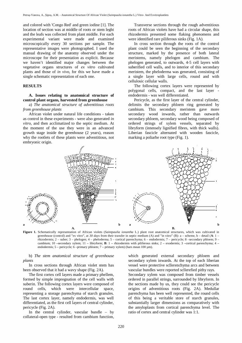
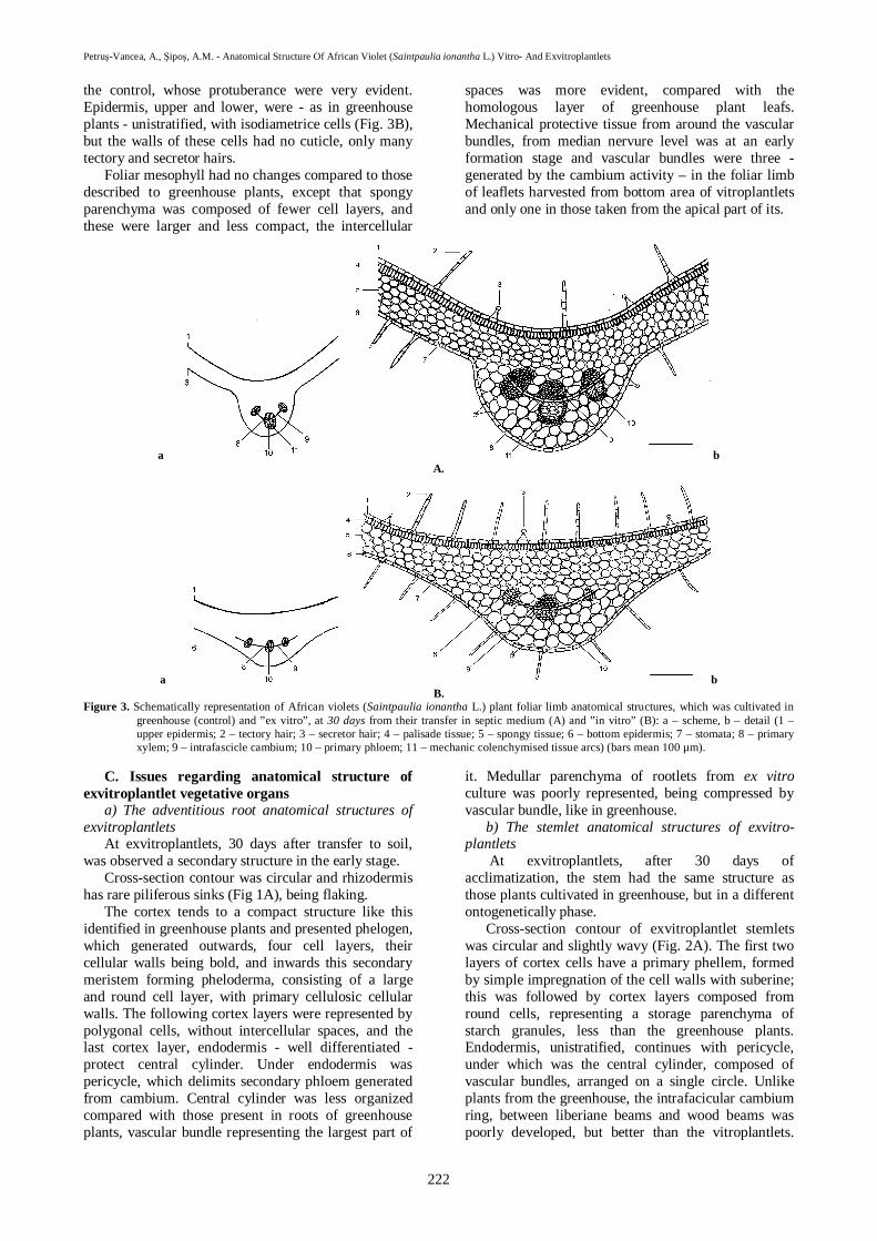
Transverse sections through the rough adventitiousroots of African violets have had a circular shape, thisrhizodermis presented some flaking phenomens andwere identified rare piliferous sinks (Fig. 1A).
In cross section through the roots of the controlplant could be seen the beginning of the secondarystructure, marked by the presence of both lateralmeristems, namely phelogen and cambium. Thephelogen generated, to outwards, 4-5 cell layers withsuberified cell walls, and to interior of this secondarymeristem, the pheloderma was generated, consisting ofa single layer with large cells, round and withcellulosic cellular walls.
The following cortex layers were represented bypolygonal cells, compact, and the last layer -endodermis - was well differentiated.
Pericycle, as the first layer of the central cylinder,delimits the secondary phloem ring generated bycambium. This secondary meristem gave moresecondary wood inwards, rather than outwardssecondary phloem, secondary wood being composed ofordered strings of xylem vessels, separated bylibryform (intensely lignified fibres, with thick walls).Liberian fascicle alternated with wooden fascicle,marking a poliarhe root type (Fig. 1).
a b a bA. B.
Figure 1. Schematically representation of African violets (Saintpaulia ionantha L.) plant root anatomical structures, which was cultivated ingreenhouse (control) and ”ex vitro”, at 30 days from their transfer in septic medium (A) and ”in vitro” (B): a – scheme, b – detail (A: 1 –rhizodermis; 2 – suber; 3 – phelogen; 4 – pheloderma; 5 – cortical parenchyma; 6 – endodermis; 7 – pericycle; 8 –secondary phloem; 9 –cambium; 10 –secondary xylem; 11 – libryform; B: 1 – rhizodermis with piliferous sinks; 2 – exodermis; 3 –cortical parenchyma; 4 –endodermis; 5 – pericycle; 6 –primary phloem; 7 – primary xylem) (bars mean 100 µm).
b) The stem anatomical structure of greenhouseplants
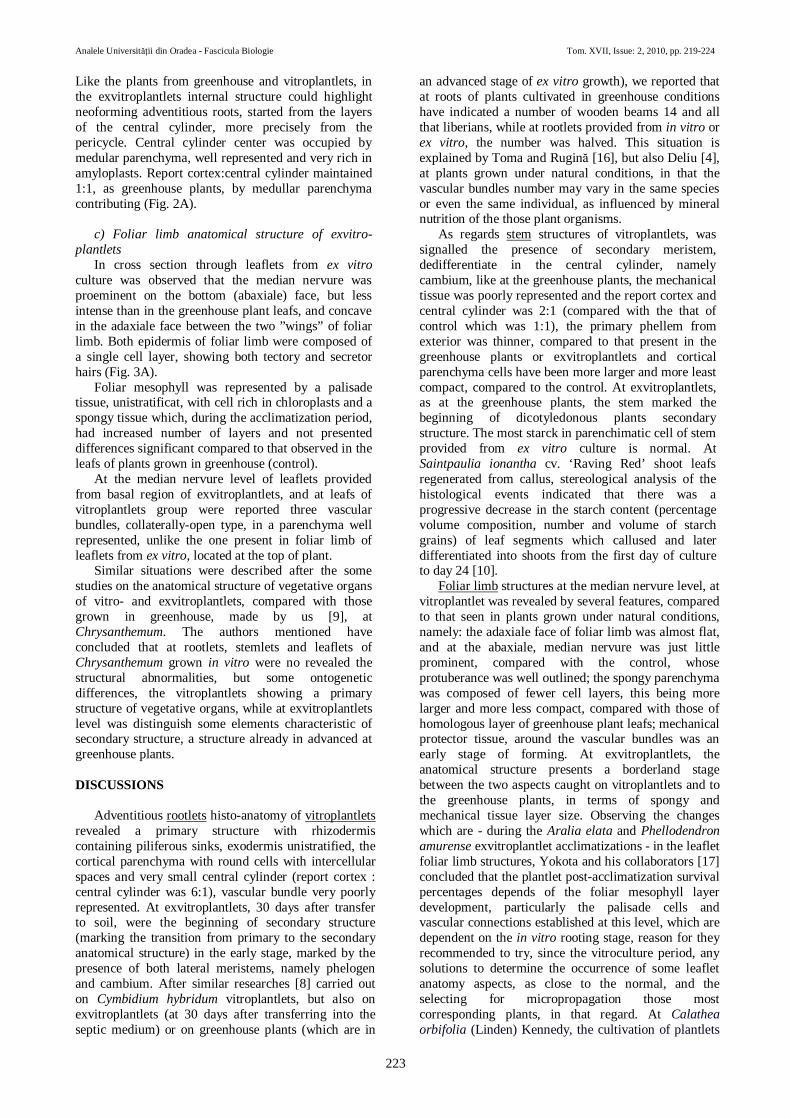
In cross sections through African violet stem hasbeen observed that it had a wavy shape (Fig. 2A).
The first cortex cell layers made a primary phellem,formed by simple impregnation of the cell walls withsuberin. The following cortex layers were composed ofround cells, which were intercellular space,representing a storage parenchyma of starch granules.The last cortex layer, namely endodermis, was welldifferentiated, as the first cell layers of central cylinder,pericycle (Fig. 2A).
In the central cylinder, vascular bundle – bycollateral-open type - resulted from cambium function,
which generated external secondary phloem andsecondary xylem inwards. At the top of each liberianvessel were protective scllerenchyma arcs and betweenvascular bundles were reported scllerefied pithy rays.Secondary xylem was composed from timber vesselsordered in parallel strings, surrounded by libryform. Inthe sections made by us, they could see the pericycleorigins of adventitious roots (Fig. 2A). Medullarparenchyma has been well represented, the round cellsof this being a veritable store of starch granules,substantially larger dimensions as comparatively withthe amyloplasts from cortical parenchyma level. Theratio of cortex and central cylinder was 1:1.

Analele Universităţii din Oradea - Fascicula Biologie Tom. XVII, Issue: 2, 2010, pp. 219-224
221
c) The foliar limb anatomical structures ofgreenhouse plants (control)
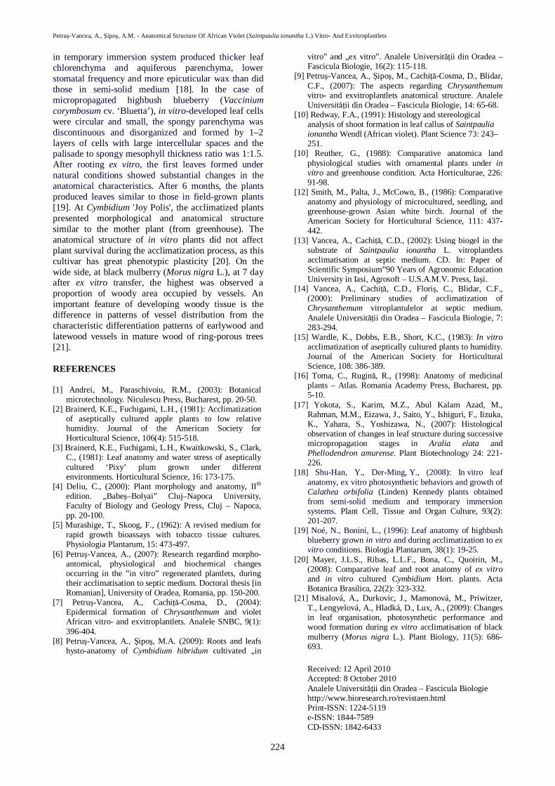
In cross section made through of hypostomaticeleafs of control African violet plants was observed thatthe median nervure is very much proeminent at thelower face (abaxiale) of foliar limb, but the adaxialenervure part, between the two ”wings” of foliar limb,presented a concavity. Upper epidermis cells werelarge and rectangular; they are many tectory hairs,pluricellular, unbranched and longer, described by us[7], and secretor hairs, with a constitution identical tothose present and Chrysanthemums. The leaves werepubescent.
Otherwise, be carried out sections through leafsfrom bottom propagul regions were observed three
vascular bundles, while in the median leaf nervureharvested from the apical propagul area was revealedonly a single vascular bundle, which signifies that thetwo beams was ulterior differentiated in plantontogenetically development, the fact no identified atChrysanthemums, where the leaf structures was notaffected by leaf locations on the stem. After Deliu [4],the vascular bundle numbers can be different to thesame species or even to the same individual.
Procambium generated primary xylem to thesuperior face of foliar limb and primary phloem to theinferior of this. Vascular bundles were protected by acollenchymas tissue following additional cellulosedeposition at the cellular wall levels (Fig. 3A).
a b a bA. B.
Figure 2. Schematically representation of African violets (Saintpaulia ionantha L.) plant stem anatomical structures, which was cultivated ingreenhouse (control) and ”ex vitro”, at 30 days from their transfer in septic medium (A) and ”in vitro” (B): a – scheme, b – detail (1 –suber; 2 – cortical parenchyma; 3 – endodermis; 4 – pericycle; 5 – scllerenchyma arcs; 6 – starch granules; 7 – secondary phloem; 8 –cambium; 9 – secondary xylem; 10 – scllerefied pithy rays; 11 – medullar parenchyma; 12 – adventitious roots) (bars mean 100 µm).
B. Issues regarding anatomical structure ofvitroplantlet vegetative organs
a) The adventitious rootlet anatomical structures ofvitroplantlets
At vitroplantlets, anatomical structure has beenhighlighted as being in a primary form. The cross-section contour was circular, rhizodermis withpiliferous sinks, exodermis unistratified, the corticalparenchyma poorly organized, consisting of oval cellswith thin and cellulosic walls, with intercellular spacesmore larger than that seen in greenhouse plants,presented endodermis in interior, which has cellswhose walls had no thickening (Fig. 1B).
Immediately below endodermis was presentpericycle, which delimit a small central cylinder (thereport cortex : central cylinder was 6:1), with vascularbundle poorly represented, and in central the extremelylow medullar parenchyma (Fig. 1B).
b) The stemlet anatomical structures of vitro-plantlets
African violet vitroplantlet stemlet cross sectionscontours were circular, slightly wavy (Fig. 2B).
The primary phellem from exterior was thinner andthe cortical parenchyma cells, oval and thin, beinglarger and less compact, with lower starch deposit,
compared to that seen in plants grown in thegreenhouse.
Under cortex was present endodermis unistratified,followed by pericycle (Fig. 2B), then the centralcylinder composed of vascular bundles, with anintrafacicular cambium ring, between phloem andxylem, less represented that control group. Secondarymeristem which was dedifferentiate in the centralcylinder, namely cambium, appeared to in vitroplantlets too, in which case, the mechanic tissue waspoorly represented, and the ratio between the cortexand central cylinder was 2:1. In place instead, to thecentral cylinder level, distinguish rays directedoutwards, which marked the adventitious rootneoforming to stemlet level. Certainly, adventitiousroots neoforming was present in greenhouse plants, butthe process was decreased as intensity, probabilityperformance of sections which capture the process, inthis case, being lower.
c) The foliar limb anatomical structures of vitro-plantlets
The foliar limb of vitroplantlet leaflets had the samebifacial dorsiventrale structure, as those of controlplants. Instead, the foliar limb adaxiale face - atvitroleaflets - was almost flat, and at the abaxiale, themedian nervure was least prominent, compared with
Petruş-Vancea, A., Şipoş, A.M. - Anatomical Structure Of African Violet (Saintpaulia ionantha L.) Vitro- And Exvitroplantlets
222
the control, whose protuberance were very evident.Epidermis, upper and lower, were - as in greenhouseplants - unistratified, with isodiametrice cells (Fig. 3B),but the walls of these cells had no cuticle, only manytectory and secretor hairs.
Foliar mesophyll had no changes compared to thosedescribed to greenhouse plants, except that spongyparenchyma was composed of fewer cell layers, andthese were larger and less compact, the intercellular
spaces was more evident, compared with thehomologous layer of greenhouse plant leafs.Mechanical protective tissue from around the vascularbundles, from median nervure level was at an earlyformation stage and vascular bundles were three -generated by the cambium activity – in the foliar limbof leaflets harvested from bottom area of vitroplantletsand only one in those taken from the apical part of its.
a bA.
a bB.
Figure 3. Schematically representation of African violets (Saintpaulia ionantha L.) plant foliar limb anatomical structures, which was cultivated ingreenhouse (control) and ”ex vitro”, at 30 days from their transfer in septic medium (A) and ”in vitro” (B): a – scheme, b – detail (1 –upper epidermis; 2 – tectory hair; 3 – secretor hair; 4 – palisade tissue; 5 – spongy tissue; 6 – bottom epidermis; 7 – stomata; 8 – primaryxylem; 9 – intrafascicle cambium; 10 – primary phloem; 11 – mechanic colenchymised tissue arcs) (bars mean 100 µm).
C. Issues regarding anatomical structure ofexvitroplantlet vegetative organs
a) The adventitious root anatomical structures ofexvitroplantlets
At exvitroplantlets, 30 days after transfer to soil,was observed a secondary structure in the early stage.
Cross-section contour was circular and rhizodermishas rare piliferous sinks (Fig 1A), being flaking.
The cortex tends to a compact structure like thisidentified in greenhouse plants and presented phelogen,which generated outwards, four cell layers, theircellular walls being bold, and inwards this secondarymeristem forming pheloderma, consisting of a largeand round cell layer, with primary cellulosic cellularwalls. The following cortex layers were represented bypolygonal cells, without intercellular spaces, and thelast cortex layer, endodermis - well differentiated -protect central cylinder. Under endodermis waspericycle, which delimits secondary phloem generatedfrom cambium. Central cylinder was less organizedcompared with those present in roots of greenhouseplants, vascular bundle representing the largest part of
it. Medullar parenchyma of rootlets from ex vitroculture was poorly represented, being compressed byvascular bundle, like in greenhouse.
b) The stemlet anatomical structures of exvitro-plantlets
At exvitroplantlets, after 30 days ofacclimatization, the stem had the same structure asthose plants cultivated in greenhouse, but in a differentontogenetically phase.
Cross-section contour of exvitroplantlet stemletswas circular and slightly wavy (Fig. 2A). The first twolayers of cortex cells have a primary phellem, formedby simple impregnation of the cell walls with suberine;this was followed by cortex layers composed fromround cells, representing a storage parenchyma ofstarch granules, less than the greenhouse plants.Endodermis, unistratified, continues with pericycle,under which was the central cylinder, composed ofvascular bundles, arranged on a single circle. Unlikeplants from the greenhouse, the intrafacicular cambiumring, between liberiane beams and wood beams waspoorly developed, but better than the vitroplantlets.
Analele Universităţii din Oradea - Fascicula Biologie Tom. XVII, Issue: 2, 2010, pp. 219-224
223
Like the plants from greenhouse and vitroplantlets, inthe exvitroplantlets internal structure could highlightneoforming adventitious roots, started from the layersof the central cylinder, more precisely from thepericycle. Central cylinder center was occupied bymedular parenchyma, well represented and very rich inamyloplasts. Report cortex:central cylinder maintained1:1, as greenhouse plants, by medullar parenchymacontributing (Fig. 2A).
c) Foliar limb anatomical structure of exvitro-plantlets
In cross section through leaflets from ex vitroculture was observed that the median nervure wasproeminent on the bottom (abaxiale) face, but lessintense than in the greenhouse plant leafs, and concavein the adaxiale face between the two ”wings” of foliarlimb. Both epidermis of foliar limb were composed ofa single cell layer, showing both tectory and secretorhairs (Fig. 3A).
Foliar mesophyll was represented by a palisadetissue, unistratificat, with cell rich in chloroplasts and aspongy tissue which, during the acclimatization period,had increased number of layers and not presenteddifferences significant compared to that observed in theleafs of plants grown in greenhouse (control).
At the median nervure level of leaflets providedfrom basal region of exvitroplantlets, and at leafs ofvitroplantlets group were reported three vascularbundles, collaterally-open type, in a parenchyma wellrepresented, unlike the one present in foliar limb ofleaflets from ex vitro, located at the top of plant.
Similar situations were described after the somestudies on the anatomical structure of vegetative organsof vitro- and exvitroplantlets, compared with thosegrown in greenhouse, made by us [9], atChrysanthemum. The authors mentioned haveconcluded that at rootlets, stemlets and leaflets ofChrysanthemum grown in vitro were no revealed thestructural abnormalities, but some ontogeneticdifferences, the vitroplantlets showing a primarystructure of vegetative organs, while at exvitroplantletslevel was distinguish some elements characteristic ofsecondary structure, a structure already in advanced atgreenhouse plants.
DISCUSSIONS
Adventitious rootlets histo-anatomy of vitroplantletsrevealed a primary structure with rhizodermiscontaining piliferous sinks, exodermis unistratified, thecortical parenchyma with round cells with intercellularspaces and very small central cylinder (report cortex :central cylinder was 6:1), vascular bundle very poorlyrepresented. At exvitroplantlets, 30 days after transferto soil, were the beginning of secondary structure(marking the transition from primary to the secondaryanatomical structure) in the early stage, marked by thepresence of both lateral meristems, namely phelogenand cambium. After similar researches [8] carried outon Cymbidium hybridum vitroplantlets, but also onexvitroplantlets (at 30 days after transferring into theseptic medium) or on greenhouse plants (which are in
an advanced stage of ex vitro growth), we reported thatat roots of plants cultivated in greenhouse conditionshave indicated a number of wooden beams 14 and allthat liberians, while at rootlets provided from in vitro orex vitro, the number was halved. This situation isexplained by Toma and Rugină [16], but also Deliu [4],at plants grown under natural conditions, in that thevascular bundles number may vary in the same speciesor even the same individual, as influenced by mineralnutrition of the those plant organisms.
As regards stem structures of vitroplantlets, wassignalled the presence of secondary meristem,dedifferentiate in the central cylinder, namelycambium, like at the greenhouse plants, the mechanicaltissue was poorly represented and the report cortex andcentral cylinder was 2:1 (compared with the that ofcontrol which was 1:1), the primary phellem fromexterior was thinner, compared to that present in thegreenhouse plants or exvitroplantlets and corticalparenchyma cells have been more larger and more leastcompact, compared to the control. At exvitroplantlets,as at the greenhouse plants, the stem marked thebeginning of dicotyledonous plants secondarystructure. The most starck in parenchimatic cell of stemprovided from ex vitro culture is normal. AtSaintpaulia ionantha cv. ‘Raving Red’ shoot leafsregenerated from callus, stereological analysis of thehistological events indicated that there was aprogressive decrease in the starch content (percentagevolume composition, number and volume of starchgrains) of leaf segments which callused and laterdifferentiated into shoots from the first day of cultureto day 24 [10].
Foliar limb structures at the median nervure level, atvitroplantlet was revealed by several features, comparedto that seen in plants grown under natural conditions,namely: the adaxiale face of foliar limb was almost flat,and at the abaxiale, median nervure was just littleprominent, compared with the control, whoseprotuberance was well outlined; the spongy parenchymawas composed of fewer cell layers, this being morelarger and more less compact, compared with those ofhomologous layer of greenhouse plant leafs; mechanicalprotector tissue, around the vascular bundles was anearly stage of forming. At exvitroplantlets, theanatomical structure presents a borderland stagebetween the two aspects caught on vitroplantlets and tothe greenhouse plants, in terms of spongy andmechanical tissue layer size. Observing the changeswhich are - during the Aralia elata and Phellodendronamurense exvitroplantlet acclimatizations - in the leafletfoliar limb structures, Yokota and his collaborators [17]concluded that the plantlet post-acclimatization survivalpercentages depends of the foliar mesophyll layerdevelopment, particularly the palisade cells andvascular connections established at this level, which aredependent on the in vitro rooting stage, reason for theyrecommended to try, since the vitroculture period, anysolutions to determine the occurrence of some leafletanatomy aspects, as close to the normal, and theselecting for micropropagation those mostcorresponding plants, in that regard. At Calatheaorbifolia (Linden) Kennedy, the cultivation of plantlets
Petruş-Vancea, A., Şipoş, A.M. - Anatomical Structure Of African Violet (Saintpaulia ionantha L.) Vitro- And Exvitroplantlets
224
in temporary immersion system produced thicker leafchlorenchyma and aquiferous parenchyma, lowerstomatal frequency and more epicuticular wax than didthose in semi-solid medium [18]. In the case ofmicropropagated highbush blueberry (Vacciniumcorymbosum cv. ‘Bluetta’), in vitro-developed leaf cellswere circular and small, the spongy parenchyma wasdiscontinuous and disorganized and formed by 1–2layers of cells with large intercellular spaces and thepalisade to spongy mesophyll thickness ratio was 1:1.5.After rooting ex vitro, the first leaves formed undernatural conditions showed substantial changes in theanatomical characteristics. After 6 months, the plantsproduced leaves similar to those in field-grown plants[19]. At Cymbidium 'Joy Polis', the acclimatized plantspresented morphological and anatomical structuresimilar to the mother plant (from greenhouse). Theanatomical structure of in vitro plants did not affectplant survival during the acclimatization process, as thiscultivar has great phenotypic plasticity [20]. On thewide side, at black mulberry (Morus nigra L.), at 7 dayafter ex vitro transfer, the highest was observed aproportion of woody area occupied by vessels. Animportant feature of developing woody tissue is thedifference in patterns of vessel distribution from thecharacteristic differentiation patterns of earlywood andlatewood vessels in mature wood of ring-porous trees[21].
REFERENCES
[1] Andrei, M., Paraschivoiu, R.M., (2003): Botanicalmicrotechnology. Niculescu Press, Bucharest, pp. 20-50.
[2] Brainerd, K.E., Fuchigami, L.H., (1981): Acclimatizationof aseptically cultured apple plants to low relativehumidity. Journal of the American Society forHorticultural Science, 106(4): 515-518.
[3] Brainerd, K.E., Fuchigami, L.H., Kwaitkowski, S., Clark,C., (1981): Leaf anatomy and water stress of asepticallycultured ‘Pixy’ plum grown under differentenvironments. Horticultural Science, 16: 173-175.
[4] Deliu, C., (2000): Plant morphology and anatomy, IIth
edition. „Babeş–Bolyai” Cluj–Napoca University,Faculty of Biology and Geology Press, Cluj – Napoca,pp. 20-100.
[5] Murashige, T., Skoog, F., (1962): A revised medium forrapid growth bioassays with tobacco tissue cultures.Physiologia Plantarum, 15: 473-497.
[6] Petruş-Vancea, A., (2007): Research regardind morpho-antomical, physiological and biochemical changesoccurring in the ”in vitro” regenerated plantlets, duringtheir acclimatisation to septic medium. Doctoral thesis [inRomanian], University of Oradea, Romania, pp. 150-200.
[7] Petruş-Vancea, A., Cachiţă-Cosma, D., (2004):Epidermical formation of Chrysanthemum and violetAfrican vitro- and exvitroplantlets. Analele SNBC, 9(1):396-404.
[8] Petruş-Vancea, A., Şipoş, M.A. (2009): Roots and leafshysto-anatomy of Cymbidium hibridum cultivated „in
vitro” and „ex vitro”. Analele Universităţii din Oradea –Fascicula Biologie, 16(2): 115-118.
[9] Petruş-Vancea, A., Şipoş, M., Cachiţă-Cosma, D., Blidar,C.F., (2007): The aspects regarding Chrysanthemumvitro- and exvitroplantlets anatomical structure. AnaleleUniversităţii din Oradea – Fascicula Biologie, 14: 65-68.
[10] Redway, F.A., (1991): Histology and stereologicalanalysis of shoot formation in leaf callus of Saintpauliaionantha Wendl (African violet). Plant Science 73: 243–251.
[10] Reuther, G., (1988): Comparative anatomica landphysiological studies with ornamental plants under invitro and greenhouse condition. Acta Horticulturae, 226:91-98.
[12] Smith, M., Palta, J., McCown, B., (1986): Comparativeanatomy and physiology of microcultured, seedling, andgreenhouse-grown Asian white birch. Journal of theAmerican Society for Horticultural Science, 111: 437-442.
[13] Vancea, A., Cachiţă, C.D., (2002): Using biogel in thesubstrate of Saintpaulia ionantha L. vitroplantletsacclimatisation at septic medium. CD. In: Paper ofScientific Symposium”90 Years of Agronomic EducationUniversity in Iasi, Agrosoft – U.S.A.M.V. Press, Iaşi.
[14] Vancea, A., Cachiţă, C.D., Floriş, C., Blidar, C.F.,(2000): Preliminary studies of acclimatization ofChrysanthemum vitroplantulelor at septic medium.Analele Universităţii din Oradea – Fascicula Biologie, 7:283-294.
[15] Wardle, K., Dobbs, E.B., Short, K.C., (1983): In vitroacclimatization of aseptically cultured plants to humidity.Journal of the American Society for HorticulturalScience, 108: 386-389.
[16] Toma, C., Rugină, R., (1998): Anatomy of medicinalplants – Atlas. Romania Academy Press, Bucharest, pp.5-10.
[17] Yokota, S., Karim, M.Z., Abul Kalam Azad, M.,Rahman, M.M., Eizawa, J., Saito, Y., Ishiguri, F., Iizuka,K., Yahara, S., Yoshizawa, N., (2007): Histologicalobservation of changes in leaf structure during successivemicropropagation stages in Aralia elata andPhellodendron amurense. Plant Biotechnology 24: 221-226.
[18] Shu-Han, Y., Der-Ming, Y., (2008): In vitro leafanatomy, ex vitro photosynthetic behaviors and growth ofCalathea orbifolia (Linden) Kennedy plants obtainedfrom semi-solid medium and temporary immersionsystems. Plant Cell, Tissue and Organ Culture, 93(2):201-207.
[19] Noé, N., Bonini, L., (1996): Leaf anatomy of highbushblueberry grown in vitro and during acclimatization to exvitro conditions. Biologia Plantarum, 38(1): 19-25.
[20] Mayer, J.L.S., Ribas, L.L.F., Bona, C., Quoirin, M.,(2008): Comparative leaf and root anatomy of ex vitroand in vitro cultured Cymbidium Hort. plants. ActaBotanica Brasilica, 22(2): 323-332.
[21] Misalová, A., Durkovic, J., Mamonová, M., Priwitzer,T., Lengyelová, A., Hladká, D., Lux, A., (2009): Changesin leaf organisation, photosynthetic performance andwood formation during ex vitro acclimatisation of blackmulberry (Morus nigra L.). Plant Biology, 11(5): 686-693.
Received: 12 April 2010Accepted: 8 October 2010Analele Universităţii din Oradea – Fascicula Biologiehttp://www.bioresearch.ro/revistaen.htmlPrint-ISSN: 1224-5119e-ISSN: 1844-7589CD-ISSN: 1842-6433