Embed Size (px)
Citation preview

IOSR Journal of Research & Method in Education (IOSR-JRME)
e-ISSN: 2320–7388, p- ISSN: 2320-737x Volume 10, Issue 5 Ser. VI (Sep. – Oct. 2020), PP 50-63
www.iosrjournals.org
DOI: 10.9790/7388-1005065063 www.iosrjournals.org 50 | Page
Anatomical Motion Knee Joint - A new design concept of
prosthetic knee joint for Knee Disarticulation (KD) amputation
Bhuyar L.R.1, Maner Aqeel R.
2, Indalkar A.G.
3
1,2 & 3(Dept. of Prosthetics and Orthotics, AIIPMR, India)
Abstract: Background: The Knee Disarticulation (KD) amputation provides an excellent quality stump with the
properties such as end weight bearing or partial end weight bearing, complete lever of femur bone,
proprioception, condylar suspension for the socket, etc. yet it is not preferred by the surgeons primarily because
of prosthetic fitting issues and also because of lack of awareness about the advantages of such amputation.
Also, the prosthetic field is less studied when it comes to KD as compared to the other levels. The prosthetic
options available are also not as vast as compared to the other levels. Due to all of the above reasons, the
literature and studies available about KD are also less.
Materials and Methods: As the AMKJ is conceptual design and actual working model has not been prepared,
for now the resources used are the books to study the normal human knee joint and CAD software to prepare a
digital model of the proposed knee joint.
Results: The simulated model of this conceptual design makes the idea very much clear about the concept,
motion, stability, the centre of rotation etc. of the actual design.
Conclusion: From the CAD simulation of the design, the expected effectiveness of the proposed knee joint
design in recreating the near natural motion of the lost knee joint due to the KD amputation, using simple
mechanics can be concluded.
Key Words: Knee Disarticulation, Amputation, Prosthetic Knee joint.
----------------------------------------------------------------------------------------------------------------------------- ----------
Date of Submission: 10-10-2020 Date of Acceptance: 26-10-2020
----------------------------------------------------------------------------------------------------------------------------- ----------
I. Introduction The ANATOMICAL MOTION PROSTHETIC KNEE JOINT (AMPKJ) is a new concept of
mechanical knee joint designs for KD amputation, designed by studying the anatomy and biomechanics of the
human knee joint and the attempt is made to mimic each and every function available thus. But as the human
knee is the most complex joint of the body hence, complete replication of each and every aspect of it needs yet
more research and innovative thinking. The AMKJ design can also be modified further and also the concepts
completely different from this design can be thought of.
II. Relevant Biomechanics
2 main considerations regarding the biomechanics of the human knee joint are made viz. the surface
joint motion of the knee joint and the Instantaneous Centre of Rotation (ICR) of the knee joint.
In a normal knee, the instant center pathway for the tibiofemoral joint is semicircular. The reason is
that the radius of curvature of the femoral condyles gradually reduces from the distal end, which articulates at
low flexion angles, to the posterior-superior, which articulates in high flexion. In addition, the motion at the
articulating surfaces is a combination of rolling and sliding, lowering the instant centers in the femur toward the
contact point. During normal knee motion in the sagittal plane from full extension to full flexion, the instant
center pathway of the midsagittal plane moves posteriorly, indicating a combination of rolling and sliding
between the articular surfaces. The unique mechanism prevents the femur from rolling off the posterior aspect of
the tibia plateau as the knee goes into increased flexion (Draganich et al., 1987; Fu et al., 1994; Kapandji, 1970).
The motion shown in Figure B is characteristic of the medial side of the knee where the anterior-posterior
displacement of the femur on the tibia is small, and there is almost complete sliding of the femur on the tibia. If
there were pure rolling, the femoral condyle would displace off the posterior of the tibial plateau (Fig. C). Figure
D represents the lateral side where the contact point displaces to the very posterior of the tibia by a combination
of rolling and sliding. The mechanism that prevents complete roll-off is the link formed between the tibial and
femoral attachment sites of the anterior and posterior cruciate ligaments and the geometry of the femoral
condyles (Fu et al., 1994).

Anatomical Motion Knee Joint - A new design concept of prosthetic knee joint for ..
DOI: 10.9790/7388-1005065063 www.iosrjournals.org 51 | Page
III. Normal Human Gait
Each limb blends the patterns of motion, passive force, and muscular control into a sequence of activity (called a
gait cycle or a stride), which is repeated endlessly until the desired destination is reached. The two limbs
perform in a reciprocal manner, offset by 50% of the gait cycle. The head, neck trunk and pelvis are self
contained passengers riding on the limb's locomotor system.
Task I: weight acceptance.
This is the first determinant of the ability to walk. Two objectives determine the events that occur during this
task: the establishment of a stable limb for weight bearing and the minimization of the shock of floor impact.
The last phase of swing and first two stance phases are dedicated to optimum weight acceptance
Phase 8 - terminal swing. To prepare the swinging limb for stance, hip flexion is interrupted, the knee extends,
and the ankle remains dorsiflexed.
Rapid, intense action by the hamstring muscles (Semimembranosus, semitendinosus, biceps femoris long head)
stops hip flexion. These muscles then reduce their intensity and allow the quadriceps to extend the knee. The
continuation of mild hamstring action prevents knee hyperextension from the residual tibial momentum.
Pretibial muscle action supports the dorsiflexed foot.

Anatomical Motion Knee Joint - A new design concept of prosthetic knee joint for ..
DOI: 10.9790/7388-1005065063 www.iosrjournals.org 52 | Page
Phase 1 - initial contact.
Floor contact by the heel is the critical event (Fig. 5.3). Its purpose is to initiate the heel rocker. The significant
postures are ankle dorsiflexion and full knee extension. Anterior tibialis controls the foot determines heel rocker
effectiveness.
Phase 2 - loading response (Initial double stance). This is a highly demanding phase of gait. The limb is
destabilized by the heel rocker and then supported by strong extensor muscular response. There are three critical
events.
Anatomical Motion Knee Joint - A new design concept of prosthetic knee joint for ..
DOI: 10.9790/7388-1005065063 www.iosrjournals.org 53 | Page
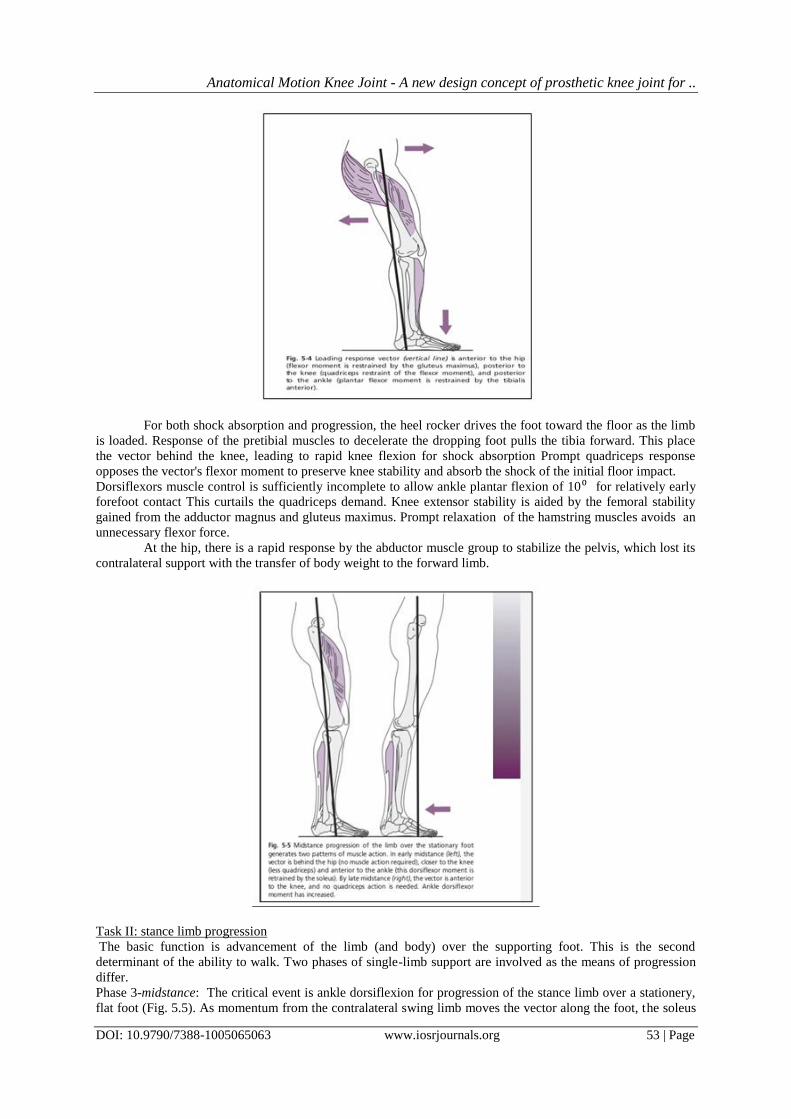
For both shock absorption and progression, the heel rocker drives the foot toward the floor as the limb
is loaded. Response of the pretibial muscles to decelerate the dropping foot pulls the tibia forward. This place
the vector behind the knee, leading to rapid knee flexion for shock absorption Prompt quadriceps response
opposes the vector's flexor moment to preserve knee stability and absorb the shock of the initial floor impact.
Dorsiflexors muscle control is sufficiently incomplete to allow ankle plantar flexion of 10⁰ for relatively early
forefoot contact This curtails the quadriceps demand. Knee extensor stability is aided by the femoral stability
gained from the adductor magnus and gluteus maximus. Prompt relaxation of the hamstring muscles avoids an
unnecessary flexor force.
At the hip, there is a rapid response by the abductor muscle group to stabilize the pelvis, which lost its
contralateral support with the transfer of body weight to the forward limb.
Task II: stance limb progression
The basic function is advancement of the limb (and body) over the supporting foot. This is the second
determinant of the ability to walk. Two phases of single-limb support are involved as the means of progression
differ.
Phase 3-midstance: The critical event is ankle dorsiflexion for progression of the stance limb over a stationery,
flat foot (Fig. 5.5). As momentum from the contralateral swing limb moves the vector along the foot, the soleus
Anatomical Motion Knee Joint - A new design concept of prosthetic knee joint for ..
DOI: 10.9790/7388-1005065063 www.iosrjournals.org 54 | Page
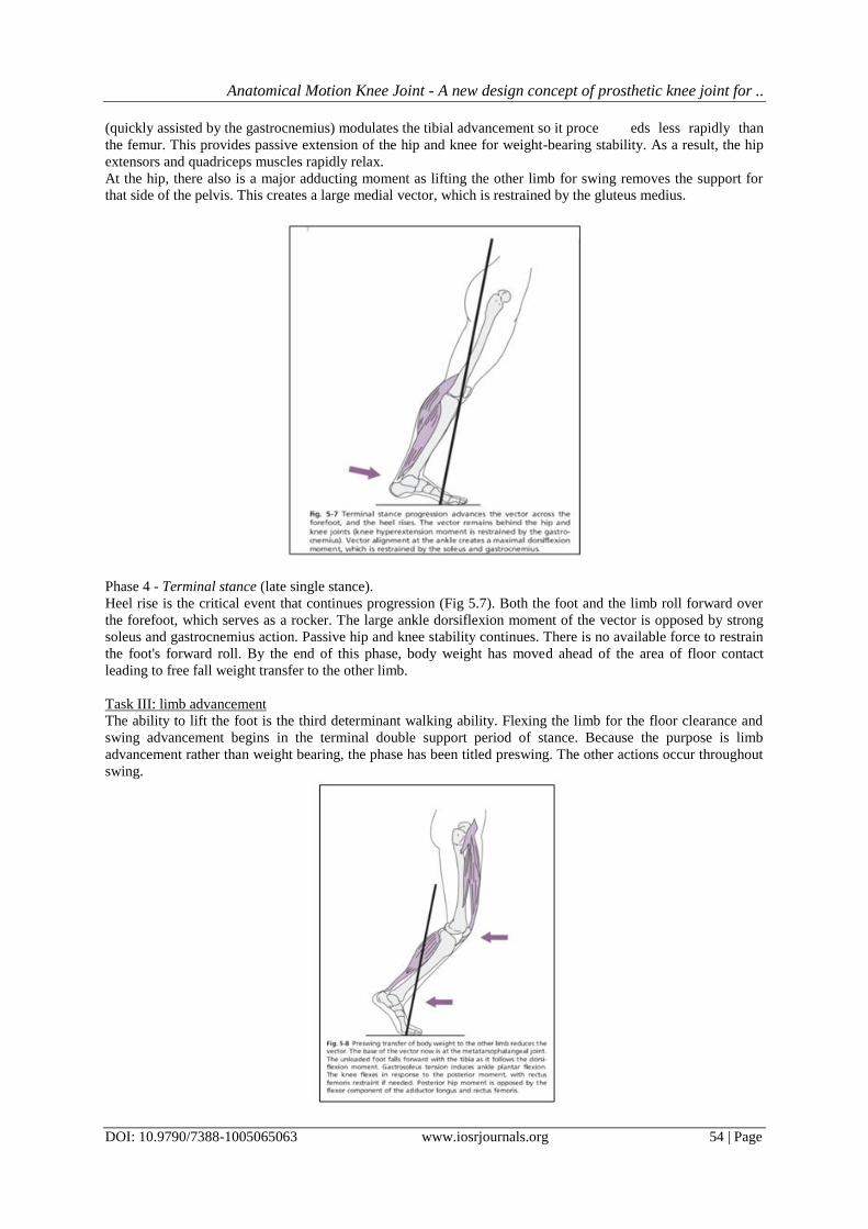
(quickly assisted by the gastrocnemius) modulates the tibial advancement so it proce eds less rapidly than
the femur. This provides passive extension of the hip and knee for weight-bearing stability. As a result, the hip
extensors and quadriceps muscles rapidly relax.
At the hip, there also is a major adducting moment as lifting the other limb for swing removes the support for
that side of the pelvis. This creates a large medial vector, which is restrained by the gluteus medius.
Phase 4 - Terminal stance (late single stance).
Heel rise is the critical event that continues progression (Fig 5.7). Both the foot and the limb roll forward over
the forefoot, which serves as a rocker. The large ankle dorsiflexion moment of the vector is opposed by strong
soleus and gastrocnemius action. Passive hip and knee stability continues. There is no available force to restrain
the foot's forward roll. By the end of this phase, body weight has moved ahead of the area of floor contact
leading to free fall weight transfer to the other limb.
Task III: limb advancement
The ability to lift the foot is the third determinant walking ability. Flexing the limb for the floor clearance and
swing advancement begins in the terminal double support period of stance. Because the purpose is limb
advancement rather than weight bearing, the phase has been titled preswing. The other actions occur throughout
swing.

Anatomical Motion Knee Joint - A new design concept of prosthetic knee joint for ..
DOI: 10.9790/7388-1005065063 www.iosrjournals.org 55 | Page
Phase 5-preswing. Passive knee flexion to 40⁰ is the critical event because this is the primary contributor to
foot clearance of the floor in swing (Fig. 5.8).
Following floor contact by the other foot, body weight is rapidly transferred to that limb to catch the forward
fall. This unloads the trailing limb, allowing several small forces to be effective. As the limbs trailing posture
reduces the foot's floor contact to the anterior margins of the metatarsal heads and the toes (fourth rocker), there
is no stabilizing force, so the foot as well as the leg is free to roll forward. This is accelerated by the rapid ankle
plantar flexion stimulated by the release of the tension stored in the eccentrically stretched soleus and
gastrocnemius. Passive knee flexion is initiated. Unloading the limb also releases the tension in the hip flexors.
This force combined with adductor longus action initiates early hip flexion and assists knee flexion.
Phase 6 - initial swing.
The critical event is knee flexion sufficient for the toe to clear the floor as the thigh advances. This involves
total limb flexion. Hip flexion may be a passive continuation of the preswing events or result from direct action
by the illiacus, sartorious and gracilis. Attainment of full knee flexion largely depends on the imbalance between
the forward momentum of the femur generated by the hip flexion and inertia of the tibia. Active assistance is
also is provided by the biceps femoris, short head. Brisk activation of the pretibial muscle initiates ankle
dorsiflexion, but the arc is incomplete in initial swing (Fig. 5.9).

Anatomical Motion Knee Joint - A new design concept of prosthetic knee joint for ..
DOI: 10.9790/7388-1005065063 www.iosrjournals.org 56 | Page
Phase 7- midswing.
Ankle dorsiflexion to neutral is the critical event for floor clearance at this time. Additional hip flexion and
partial knee extension advance the limb. The relative vertical posture of the lower leg requires pretibial muscle
support of the ankle (Fig. 5.10).
Phase 8 –terminal swing.
Forward swing of the limb for step length is accomplished by knee extension. The other actions relate to
preparing the limb for stance as previously described.
IV. The Anatomical Motion Prosthetic Knee Joint (Ampkj) Design
This new design of the prosthetic knee joint named as „ANATOMICAL MOTION PROSTHETIC KNEE
JOINT (AMPKJ)‟ consists of following two components:
• Femoral Chondyle Guide or FC-Guide
• Tibial Chondyle Rider or TC-Rider.
The FC-Guide
Anatomical Motion Knee Joint - A new design concept of prosthetic knee joint for ..
DOI: 10.9790/7388-1005065063 www.iosrjournals.org 57 | Page
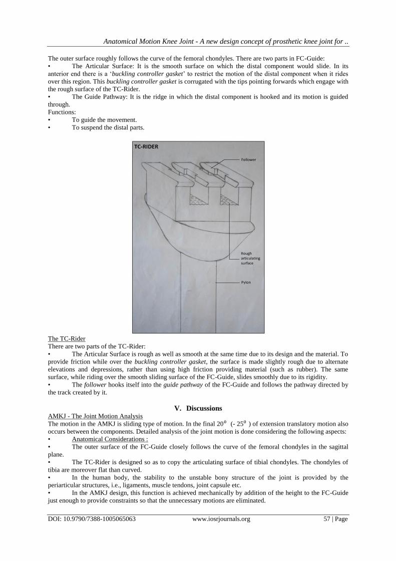
The outer surface roughly follows the curve of the femoral chondyles. There are two parts in FC-Guide:
• The Articular Surface: It is the smooth surface on which the distal component would slide. In its
anterior end there is a „buckling controller gasket‟ to restrict the motion of the distal component when it rides
over this region. This buckling controller gasket is corrugated with the tips pointing forwards which engage with
the rough surface of the TC-Rider.
• The Guide Pathway: It is the ridge in which the distal component is hooked and its motion is guided
through.
Functions:
• To guide the movement.
• To suspend the distal parts.
The TC-Rider
There are two parts of the TC-Rider:
• The Articular Surface is rough as well as smooth at the same time due to its design and the material. To
provide friction while over the buckling controller gasket, the surface is made slightly rough due to alternate
elevations and depressions, rather than using high friction providing material (such as rubber). The same
surface, while riding over the smooth sliding surface of the FC-Guide, slides smoothly due to its rigidity.
• The follower hooks itself into the guide pathway of the FC-Guide and follows the pathway directed by
the track created by it.
V. Discussions AMKJ - The Joint Motion Analysis
The motion in the AMKJ is sliding type of motion. In the final 20⁰ (- 25⁰ ) of extension translatory motion also
occurs between the components. Detailed analysis of the joint motion is done considering the following aspects:
• Anatomical Considerations :
• The outer surface of the FC-Guide closely follows the curve of the femoral chondyles in the sagittal
plane.
• The TC-Rider is designed so as to copy the articulating surface of tibial chondyles. The chondyles of
tibia are moreover flat than curved.
• In the human body, the stability to the unstable bony structure of the joint is provided by the
periarticular structures, i.e., ligaments, muscle tendons, joint capsule etc.
• In the AMKJ design, this function is achieved mechanically by addition of the height to the FC-Guide
just enough to provide constraints so that the unnecessary motions are eliminated.
Anatomical Motion Knee Joint - A new design concept of prosthetic knee joint for ..
DOI: 10.9790/7388-1005065063 www.iosrjournals.org 58 | Page
• Another function of the ligaments is to hold the two parts together, i.e., to provide the suspension to the
distal part. This is also achieved in the design through the hooking of the follower to the guide pathway.
• Biomechanical Considerations:
• ROM: More than 90⁰ (needed to be experimentally tested. Estimated ROM is about 130⁰) • Kinematics :
The total motion of the AMKJ is divided into two parts
• Motion of the TC-Rider over the smooth sliding surface
• Motion of the TC-Rider over the buckling controller gasket
Motion of the TC-Rider over the smooth surface: From about 20⁰ (-25⁰ ) of flexion to further complete flexion,
even though the structure of design is designed so as to provide purely sliding motion, the articulating surface of
TC-Rider may or may not contact the corresponding surface of the FC-Guide.
Motion primarily takes place at the joint formed by the guide pathway and the follower.
Rest all structures function to provide stability as well as to direct the motion as assistance to the primary
structures.
Motion of the TC-Rider over the buckling controller gasket: motion during 0⁰ - 20⁰ ( - 25⁰) is modified due to
two factors – (i) Guide pathway ridge modification to provide translatory motion and (ii) engaging of the rough
surface of TC-Rider with the buckling controller gasket.
These two factors work together to stop the motion when the surfaces are engaged and allow the same when
they are separate.
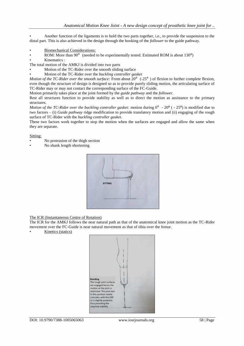
Sitting:
• No protrusion of the thigh section
• No shank length shortening
The ICR (Instantaneous Centre of Rotation)
The ICR for the AMKJ follows the near natural path as that of the anatomical knee joint motion as the TC-Rider
movement over the FC-Guide is near natural movement as that of tibia over the femur.
• Kinetics (statics)
Anatomical Motion Knee Joint - A new design concept of prosthetic knee joint for ..
DOI: 10.9790/7388-1005065063 www.iosrjournals.org 59 | Page
During standing, the weight bearing line and the normal reaction force/ Ground Reaction Force (GRF) line are
aligned in one line or in the way to provide a slight extension moment which somewhat mimics the locking
mechanism of the normal knee.
The joint is positioned in a few degrees ahead of 0⁰ of flexion (maybe as small as 1⁰). The rough surfaces are engaged to provide high friction and resist and restrict motion.
The motion is coronal plane which can be termed as „laxity‟ is eliminated due to the constraints provided by the
small leverage of the added height of the FC-Guide.
• Dynamic Analysis or Gait Analysis
• Task I : Weight Acceptance
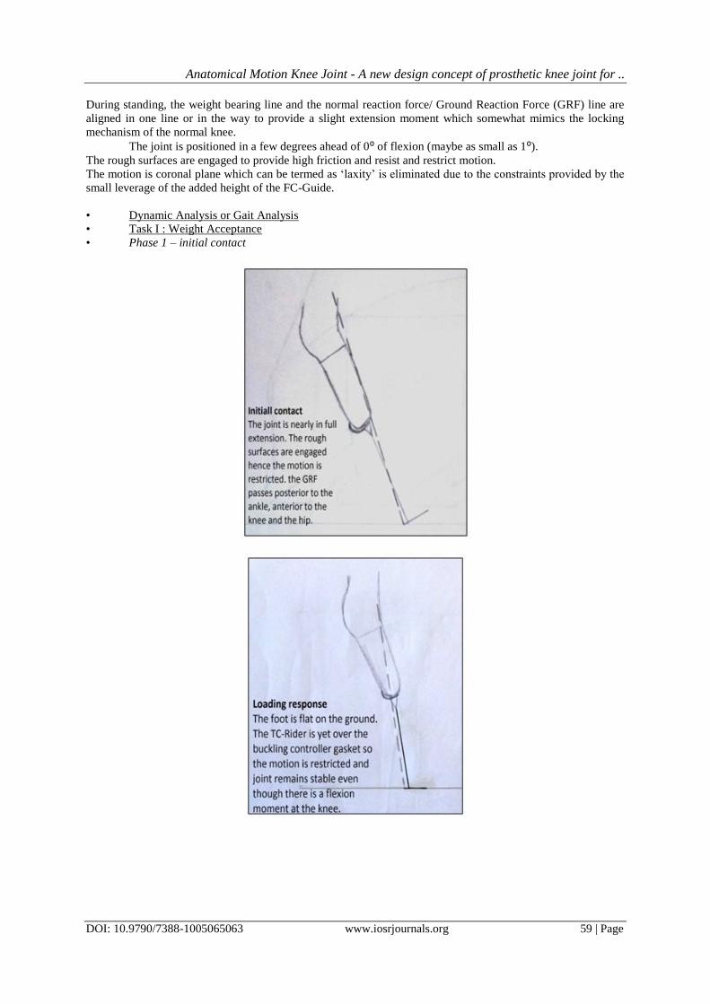
• Phase 1 – initial contact

Anatomical Motion Knee Joint - A new design concept of prosthetic knee joint for ..
DOI: 10.9790/7388-1005065063 www.iosrjournals.org 60 | Page
The heel touches the ground. The GRF vector passes through the heel – posterior to the ankle – anterior to the
knee and hip.
Here the motion is controlled voluntarily by the hip extensors. Thus an extensor moment is present at the knee.
Also, due to the heel contact and weight bearing, the distal segment is subjected to the upward force creating a
translatory motion at the joint and thus, engaging the rough surfaces. The motion at the joint is restricted.
The knee is positioned in full extension or in slight (about 5⁰) of flexion.
At the same time, a plantar flexion moment is generated at the ankle joint. True plantar flexion occurs if
articulated foot is used or the action is simulated by the bumper or the flexibility of the structure according to
the type of prosthetic foot used.
• Phase 2 – loading response
The heel rocker initiates knee flexion as the GRF vector progressively moves behind the knee joint and the sole
of the foot makes contact with the ground.
This knee flexion is restricted flexion is restricted due to the engaging of the rough surfaces, thus, acting as the
quadriceps which controls buckling in anatomical knee.
Hip extensors also provide some assistance for avoiding buckling.
• Task II : Stance limb progression
• Phase 3 – midstance
Once the knee is successful in taking the limb safely into the stance phase without buckling the stance phase is
completed moreover passively than actively.
The GRF vector is in the line with the knee joint axis during initial midstance and progressively moves in front
of it as the stance phase advances towards the terminal stance phase.
• Phase 4 – terminal stance phase
Heel rise is achieved.
The GRF is at metatarsal head level. Hip flexors act to break the stability of the knee joint. As the weight is
relieved, the rough surfaces disengage and the joint components are free to move.

Anatomical Motion Knee Joint - A new design concept of prosthetic knee joint for ..
DOI: 10.9790/7388-1005065063 www.iosrjournals.org 61 | Page
• Task III : Limb Advancement
• Phase 5 – preswing
The weight is transferred to the contralateral limb and the prosthetic side is ready to go into the swing phase.
The TC-Rider articulating surface is now over the
smooth sliding surface of FC-Guide.
Knee is flexed maximum to the degree possible passively. As there is no active movement, the degree of knee
flexion (about 40⁰) achieved is the final, which will be used for foot clearance during the swing.
• Phase 6 – initial swing
Main event is pelvic stability and pelvic tilt by the hip abductors action of the contralateral side providing foot
clearance.
Knee starts to extend under the action of gravity.
Anatomical Motion Knee Joint - A new design concept of prosthetic knee joint for ..
DOI: 10.9790/7388-1005065063 www.iosrjournals.org 62 | Page
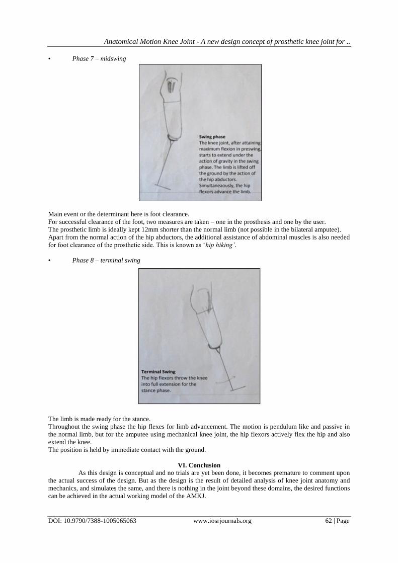
• Phase 7 – midswing
Main event or the determinant here is foot clearance.
For successful clearance of the foot, two measures are taken – one in the prosthesis and one by the user.
The prosthetic limb is ideally kept 12mm shorter than the normal limb (not possible in the bilateral amputee).
Apart from the normal action of the hip abductors, the additional assistance of abdominal muscles is also needed
for foot clearance of the prosthetic side. This is known as „hip hiking’.
• Phase 8 – terminal swing
The limb is made ready for the stance.
Throughout the swing phase the hip flexes for limb advancement. The motion is pendulum like and passive in
the normal limb, but for the amputee using mechanical knee joint, the hip flexors actively flex the hip and also
extend the knee.
The position is held by immediate contact with the ground.
VI. Conclusion
As this design is conceptual and no trials are yet been done, it becomes premature to comment upon
the actual success of the design. But as the design is the result of detailed analysis of knee joint anatomy and
mechanics, and simulates the same, and there is nothing in the joint beyond these domains, the desired functions
can be achieved in the actual working model of the AMKJ.
Anatomical Motion Knee Joint - A new design concept of prosthetic knee joint for ..
DOI: 10.9790/7388-1005065063 www.iosrjournals.org 63 | Page
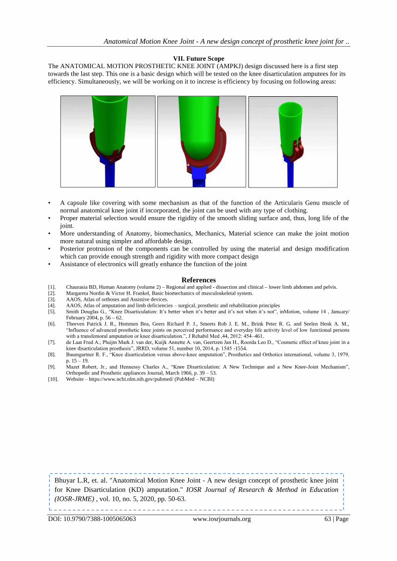
VII. Future Scope
The ANATOMICAL MOTION PROSTHETIC KNEE JOINT (AMPKJ) design discussed here is a first step
towards the last step. This one is a basic design which will be tested on the knee disarticulation amputees for its
efficiency. Simultaneously, we will be working on it to increse is efficiency by focusing on following areas:
• A capsule like covering with some mechanism as that of the function of the Articularis Genu muscle of
normal anatomical knee joint if incorporated, the joint can be used with any type of clothing.
• Proper material selection would ensure the rigidity of the smooth sliding surface and, thus, long life of the
joint.
• More understanding of Anatomy, biomechanics, Mechanics, Material science can make the joint motion
more natural using simpler and affordable design.
• Posterior protrusion of the components can be controlled by using the material and design modification
which can provide enough strength and rigidity with more compact design
• Assistance of electronics will greatly enhance the function of the joint
References [1]. Chaurasia BD, Human Anatomy (volume 2) – Regional and applied - dissection and clinical – lower limb abdomen and pelvis.
[2]. Margareta Nordin & Victor H. Frankel, Basic biomechanics of musculoskeletal system.
[3]. AAOS, Atlas of orthoses and Assistive devices. [4]. AAOS, Atlas of amputation and limb deficiencies – surgical, prosthetic and rehabilitation principles
[5]. Smith Douglas G., “Knee Disarticulation: It‟s better when it‟s better and it‟s not when it‟s not”, inMotion, volume 14 , January/ February 2004, p. 56 – 62.
[6]. Theeven Patrick J. R., Hemmen Bea, Geers Richard P. J., Smeets Rob J. E. M., Brink Peter R. G. and Seelen Henk A. M.,
“Influence of advanced prosthetic knee joints on perceived performance and everyday life activity level of low functional persons with a transfemoral amputation or knee disarticulation.”, J Rehabil Med ,44, 2012: 454–461.
[7]. de Laat Fred A.; Pluijm Mark J. van der, Kuijk Annette A. van, Geertzen Jan H., Roorda Leo D., “Cosmetic effect of knee joint in a
knee disarticulation prosthesis”, JRRD, volume 51, number 10, 2014, p. 1545 -1554. [8]. Baumgartner R. F., “Knee disarticulation versus above-knee amputation”, Prosthetics and Orthotics international, volume 3, 1979,
p. 15 – 19.
[9]. Mazet Robert, Jr., and Hennessy Charles A., “Knee Disarticulation: A New Technique and a New Knee-Joint Mechanism”, Orthopedic and Prosthetic appliances Journal, March 1966, p. 39 – 53.
[10]. Website – https://www.ncbi.nlm.nih.gov/pubmed/ (PubMed – NCBI)
Bhuyar L.R, et. al. "Anatomical Motion Knee Joint - A new design concept of prosthetic knee joint
for Knee Disarticulation (KD) amputation." IOSR Journal of Research & Method in Education
(IOSR-JRME) , vol. 10, no. 5, 2020, pp. 50-63.