Embed Size (px)
Citation preview
THE UNIVERSITY OF NEW SOUTH
WALES
SCHOOL OF MEDICAL SCIENCES DEPARTMENT OF ANATOMY
ANAT 3141
Functional Anatomy 2
Session 2, 2011
Class Notes and Lecture Summaries
1
UNIVERSITY OF NEW SOUTH WALES SCHOOL OF MEDICAL SCIENCES, DEPARTMENT OF ANATOMY
FUNCTIONAL ANATOMY II - ANAT3141
SESSION 2, 2011 Staff involved in the course: Course Organiser: Dr Dzung H. Vu, Room 305, Goodsell Building email: [email protected] Information about the course: Course relationships: This course covers the musculoskeletal anatomy of the trunk and lower limb. Functional Anatomy I covers musculoskeletal anatomy of the head, neck and upper limb. These courses build on Level II offerings in introductory anatomy and histology, and complement level III courses in neuroanatomy and visceral anatomy. Course objectives: To provide the student with an understanding of the musculoskeletal anatomy of the trunk and lower limb and the functional principles underlying joint movements in this region. Teaching strategies: The course is delivered in lecture format complemented by lab classes using prosected specimens and radiographs Approaches to learning in the course: It is suggested that students attend the lectures and lab classes. Attendance of lab classes are compulsory. Contents of the lectures and class notes are examinable. NOTE that in the spot test, students should be able to IDENTIFY all anatomical structures listed in the learning activities of the practicals. The written exam will cover all materials presented in the lectures and all questions and discussions listed in the lab classes. Expected learning outcomes: A student who has completed the course should have a good knowledge of the functional anatomy of the trunk and lower limb, and an understanding of some biomechanical aspects of walking. Course Structure: There will be two lectures and one lab class per week
Day Lecture Lab class Monday 9-10 Biomed D 10-12:30 pm Dissecting RoomTuesday 5-6pm Biomed A Wednesday 10-12:30 pm Dissecting Room
Assessment in the course Assessment strategy: Practical aspects of the course will be assessed by two lab-based exams (‘spot tests’). Theoretical aspects will be assessed by a final exam incorporating multiple choice and essay components. Assessment details: Mark allocation is shown below
Continuous assessment 10% Mid-session Spot Test 20% Final Spot Test 20% Final Examination 50%
Assessment dates will be announced but both spot test and written exams will be during exam week. The final examination will consist of 50 multiple choice questions and 3 short answer questions (MCQ’s and Questions are worth 50% of the total marks). Pass mark for the course is 50% but students who achieved less than 42% in one component (either spot test or written exam) will be deemed as having failed the course even if the total mark is 50%. At the discretion of the Course Authority and Assessment committee, further assessment will only be given to some students who are borderline (>47%) in the first attempt or who was absent for legitimate reasons. The date and time for re-assessment will be announced but it will be a few weeks after exam week, as they are not meant for students to study for the course after the first exam. There will be absolutely only ONE re-assessment, students who do not present for this re-assessment will be given their original fail marks. There will beNO EXCEPTION, so please do not book your trips or holidays until the final results are disclosed. About this manual and learning process: This manual is designed to be a guide for tutors and students and NOT a comprehensive teaching text. You will be given hand-outs during the lectures, which are not comprehensive teaching text but only guides to help you follow the lectures, to clarify some difficult concept and to give you some emphasis on important issues. Thus they are not posted on the web. Those who miss the lecture can obtain lecture materials from the relevant lab class. Student’s are expected to be prepared for the practical classes by reading textbook and lecture, and they must fully understand all learning activities listed in this manual. Students should bring lecture materials or textbook to the dissecting room to facilitate learning in the lab.
2
Relevant resources for students enrolled in the course Textbook details: There is no text that covers all the material in the course. A good textbook of human
anatomy will be useful, such as: MOORE "Clinically Oriented Anatomy" or SNELL, R.S. "Clinical Anatomy for Medical Students" Recommended: YOKOCHI, ROHEN & WEINREB "Photographic Anatomy of the Human Body" or ROHEN ,YOKOCHI & LUTJEN-DRECOLL “Color Atlas of Anatomy” Administrative matters Official communications: All students in course ANAT3131/3531 are advised that email is now the
official means by which the School of Medical Sciences at UNSW will communicate with you. Occupational Health and Safety: For full details, refer to the Faculty of Medicine URL:
http://ohs.med.unsw.edu.au/ You must wear laboratory coat and closed shoes in the laboratory and bring gloves to all lab classes
TO HANDLE WET AND DRY SPECIMENS DURING TUTORIAL/LAB CLASSES. Applications for Consideration. Students who miss an assessment through illness or misadventure
must submit an application for consideration within three working days to Student Central. Request for special consideration is available at https://my.unsw.edu.au/student/atoz/consideration.pdf
Grievance: See https://my.unsw.edu.au/student/atoz/Complaints.html Attendance. Students are required to attend each lecture & laboratory class unless given special
permission. Provision of an appropriate medical certificate will be required for Special Consideration.
Rules of use of the Dissecting Room You may enter and view specimens in the Dissecting Room 101 only in the presence of your tutor
and/or during your designated laboratory class hours. You are not allowed to take visitors into the Dissection Room.
In the Dissecting Room, you must: never eat or drink; never put anything in your mouth, e.g., biros
or pencils that you may have picked up from the table; wear a laboratory coat; wear covered shoes, not thongs; wear latex or vinyl gloves when touching wet specimens (gloves are available from Union Arcade Shop); use blunt forceps only to handle specimens and probes to point to structures, and never pull at any parts of the specimen; as far as possible, avoid inhaling preservative solutions for prolonged periods (if you feel in need of fresh air, ask permission to leave the laboratory for a few minutes); and at the end of your laboratory: cover wet specimens with the towels provided; replace stools under the tables in your cubicle; wash your hands and instruments.
Great care should always be exercised when handling specimens, in order to preserve their delicate structure. Much work has gone into the prosection of each specimen before it is ready for use in class. A damaged specimen cannot be replaced immediately, so it will not be available to students for a long time.
You are learning from human material prepared from people who have generously donated their
bodies for the benefit of science. Apart from caring for the specimens, it is important for all students learning Anatomy to have and show utmost respect for the specimens at all times, in the Dissecting Room and in the Anatomy Museum. It is illegal for any anatomical material to be removed from the premises of the Department of Anatomy for any purpose whatsoever (except of course, for the funeral).
3
Preservative solution. Most anatomy specimens are stored in 2% phenoxyethanol, which is classified as non-toxic. You should always wear gloves when handling specimens. Detailed information about phenoxyenthanol is posted on the Dissecting Room notice board. A few specimens (brain tissue, etc.) are stored in formaldehyde, which is toxic if ingested, and corrosive to the eye; it can also be absorbed through the skin. Formaldehyde is reported to cause allergic skin and respiratory effects. The potential for adverse health effects, however, is markedly reduced at the concentrations used for embalming and storage of specimens in the Dissecting Room. Moreover, the air in the Dissecting room is continuously changed and specimens are not stored in formalin anymore. Essentially, you should prevent any preservative solution from coming in direct contact with your eyes, skin or mouth. First Aid kit is available in the dissecting room. All anatomy specimens are microchipped for identification and record-keeping.
Revision Facilities are available in the Anatomy Museum (normally open 9 am - 5 pm, Monday to Friday). Please do not remove the museum jars from their shelves.
Photography and video recording is not permitted in the Dissecting Room or the Anatomy Museum.
4
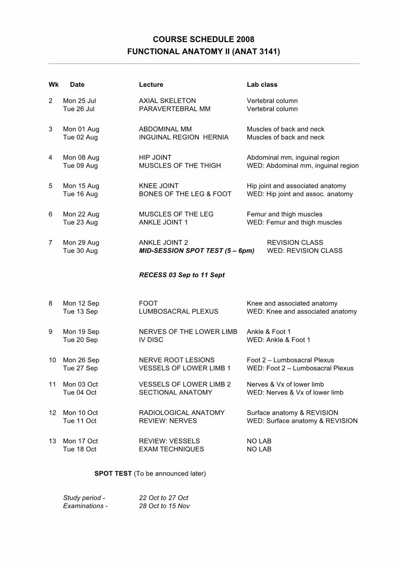
COURSE SCHEDULE 2008 FUNCTIONAL ANATOMY II (ANAT 3141)
Wk Date Lecture Lab class 2 Mon 25 Jul AXIAL SKELETON Vertebral column Tue 26 Jul PARAVERTEBRAL MM Vertebral column
3 Mon 01 Aug ABDOMINAL MM Muscles of back and neck Tue 02 Aug INGUINAL REGION HERNIA Muscles of back and neck
4 Mon 08 Aug HIP JOINT Abdominal mm, inguinal region Tue 09 Aug MUSCLES OF THE THIGH WED: Abdominal mm, inguinal region
5 Mon 15 Aug KNEE JOINT Hip joint and associated anatomy Tue 16 Aug BONES OF THE LEG & FOOT WED: Hip joint and assoc. anatomy
6 Mon 22 Aug MUSCLES OF THE LEG Femur and thigh muscles Tue 23 Aug ANKLE JOINT 1 WED: Femur and thigh muscles
7 Mon 29 Aug ANKLE JOINT 2 REVISION CLASS Tue 30 Aug MID-SESSION SPOT TEST (5 – 6pm) WED: REVISION CLASS RECESS 03 Sep to 11 Sept
8 Mon 12 Sep FOOT Knee and associated anatomy Tue 13 Sep LUMBOSACRAL PLEXUS WED: Knee and associated anatomy
9 Mon 19 Sep NERVES OF THE LOWER LIMB Ankle & Foot 1 Tue 20 Sep IV DISC WED: Ankle & Foot 1
10 Mon 26 Sep NERVE ROOT LESIONS Foot 2 – Lumbosacral Plexus Tue 27 Sep VESSELS OF LOWER LIMB 1 WED: Foot 2 – Lumbosacral Plexus 11 Mon 03 Oct VESSELS OF LOWER LIMB 2 Nerves & Vx of lower limb Tue 04 Oct SECTIONAL ANATOMY WED: Nerves & Vx of lower limb
12 Mon 10 Oct RADIOLOGICAL ANATOMY Surface anatomy & REVISION Tue 11 Oct REVIEW: NERVES WED: Surface anatomy & REVISION
13 Mon 17 Oct REVIEW: VESSELS NO LAB Tue 18 Oct EXAM TECHNIQUES NO LAB SPOT TEST (To be announced later) Study period - 22 Oct to 27 Oct Examinations - 28 Oct to 15 Nov
LAB CLASS 1: VERTEBRAL COLUMN LEARNING ACTIVITIES: 1. Spinal curvatures: Observe an articulated vertebral column. Note the normal curvatures of the
spinal column in the sagittal plane and identify the primary and secondary curvatures. What is lordosis, kyphosis and scoliosis?
2. Note that the inclination of the spinous processes (SP) increases from the cervical to the thoracic
region then is much less in the lumbar region. The spinous process of L4 is at the level with the highest point of the iliac crest, and the spinous process of S2 is level with a line joining the PSIS's.
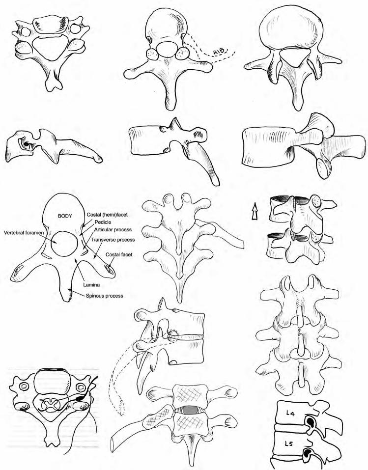
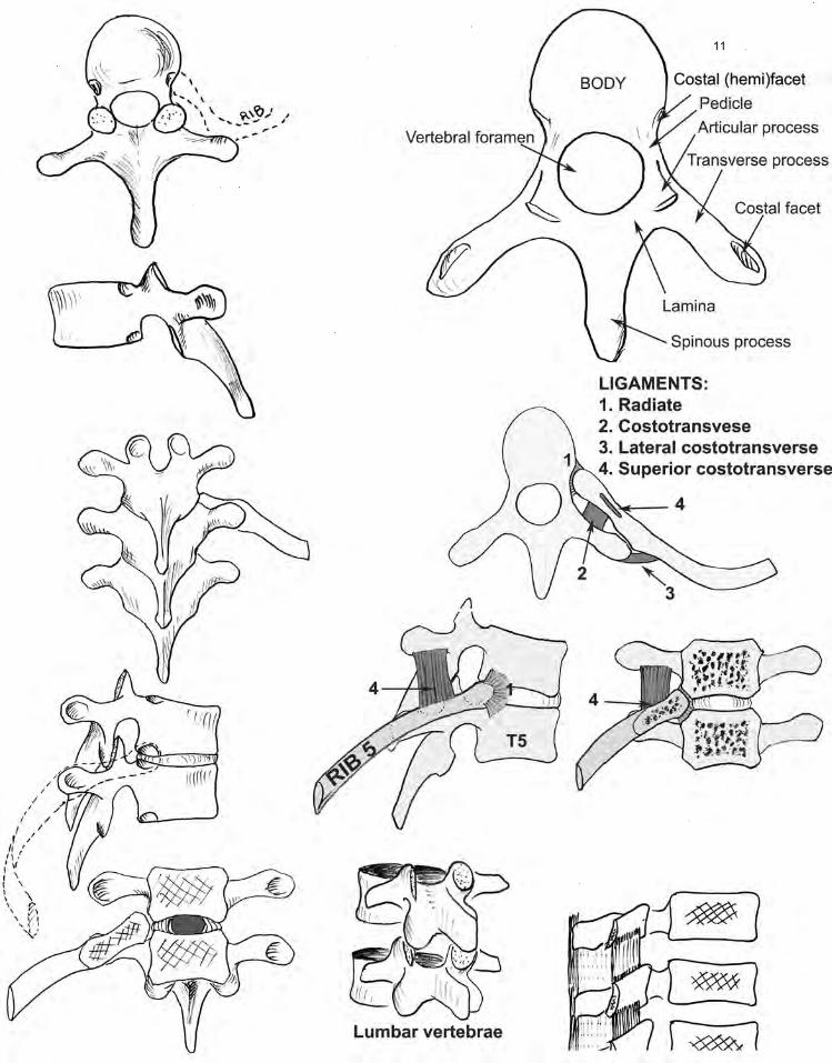
3. On a mid-thoracic vertebra, identify the main parts of a typical vertebra: body, pedicle, lamina,
transverse process, spinous process, superior and inferior articular processes, vertebral notch. Put 2 vertebrae together and see what structures form the zygapophyseal joint (facet joint), and what structures make up the intervertebral foramen. Observe the change in the orientation of the facet joint in the cervical, thoracic and lumbar vertebrae.
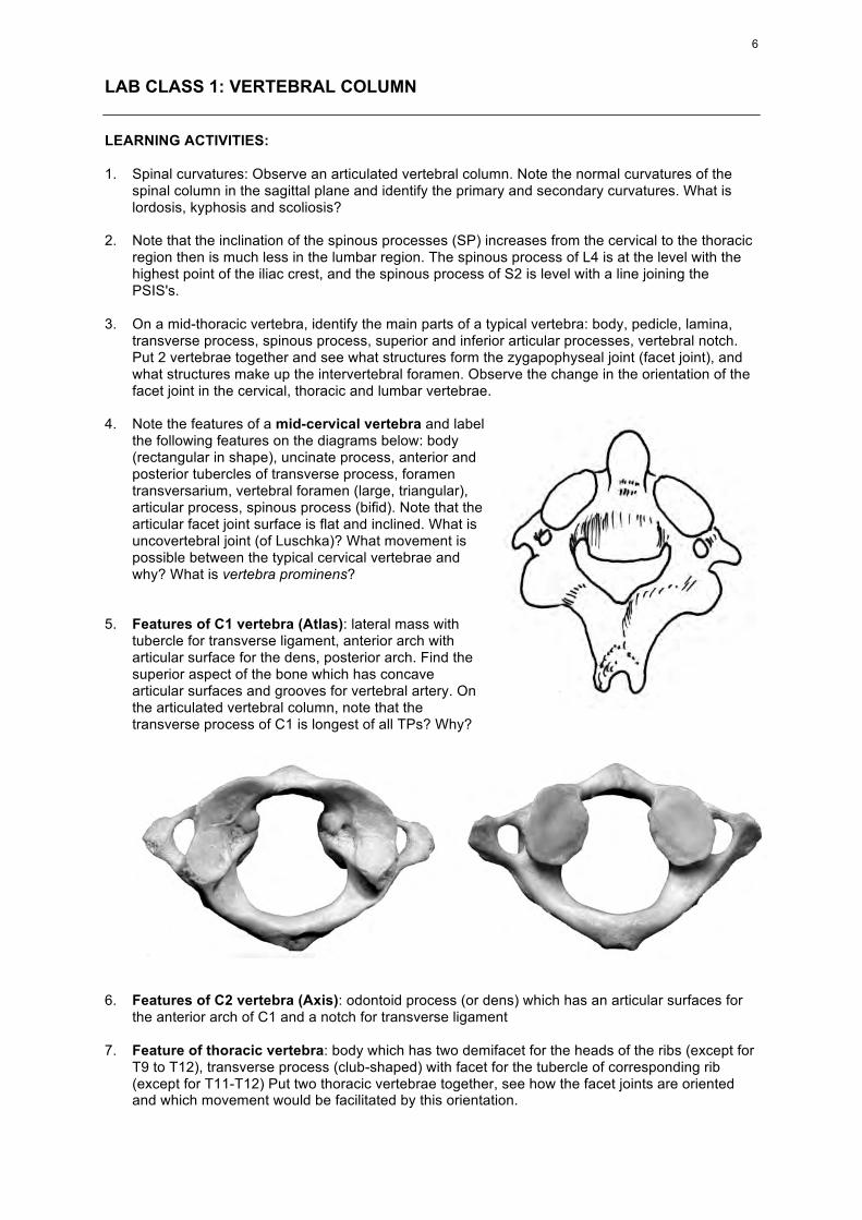
4. Note the features of a mid-cervical vertebra and label
the following features on the diagrams below: body (rectangular in shape), uncinate process, anterior and posterior tubercles of transverse process, foramen transversarium, vertebral foramen (large, triangular), articular process, spinous process (bifid). Note that the articular facet joint surface is flat and inclined. What is uncovertebral joint (of Luschka)? What movement is possible between the typical cervical vertebrae and why? What is vertebra prominens?
5. Features of C1 vertebra (Atlas): lateral mass with
tubercle for transverse ligament, anterior arch with articular surface for the dens, posterior arch. Find the superior aspect of the bone which has concave articular surfaces and grooves for vertebral artery. On the articulated vertebral column, note that the transverse process of C1 is longest of all TPs? Why?
6. Features of C2 vertebra (Axis): odontoid process (or dens) which has an articular surfaces for
the anterior arch of C1 and a notch for transverse ligament 7. Feature of thoracic vertebra: body which has two demifacet for the heads of the ribs (except for
T9 to T12), transverse process (club-shaped) with facet for the tubercle of corresponding rib (except for T11-T12) Put two thoracic vertebrae together, see how the facet joints are oriented and which movement would be facilitated by this orientation.
6
8. Put a rib against a thoracic vertebra: note how the rib
articulates with the body (costovertebral joint) and with transverse process (costotransverse joint)
9. Features of lumbar vertebra: body (large, kidney-
shaped), transverse process with accessory process, short spinous process. Note direction of the articular processes and their facet joint, and the mamillary process on the superior articular process. What movement would be difficult with the orientation of the facet joints? What is pars interarticularis and could result from fracture at that point?
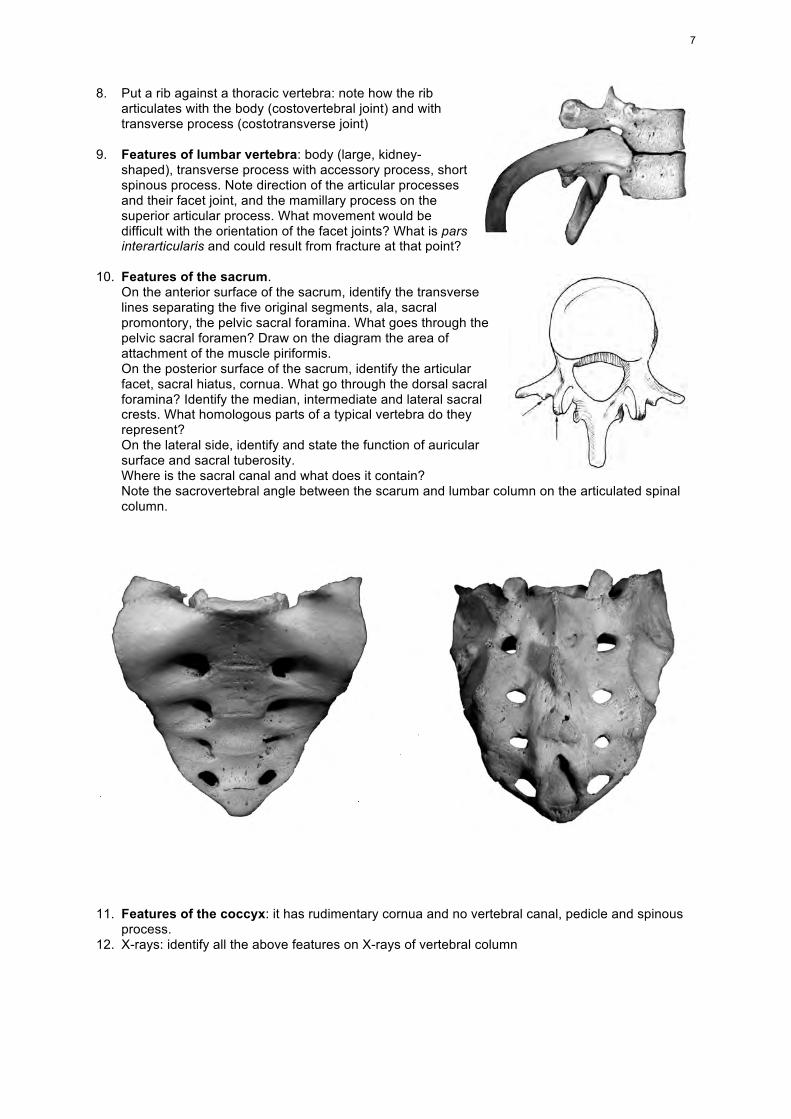
10. Features of the sacrum.
On the anterior surface of the sacrum, identify the transverse lines separating the five original segments, ala, sacral promontory, the pelvic sacral foramina. What goes through the pelvic sacral foramen? Draw on the diagram the area of attachment of the muscle piriformis. On the posterior surface of the sacrum, identify the articular facet, sacral hiatus, cornua. What go through the dorsal sacral foramina? Identify the median, intermediate and lateral sacral crests. What homologous parts of a typical vertebra do they represent? On the lateral side, identify and state the function of auricular surface and sacral tuberosity. Where is the sacral canal and what does it contain? Note the sacrovertebral angle between the scarum and lumbar column on the articulated spinal column.
11. Features of the coccyx: it has rudimentary cornua and no vertebral canal, pedicle and spinous
process. 12. X-rays: identify all the above features on X-rays of vertebral column
7
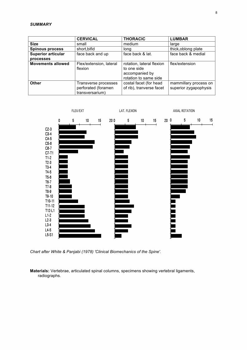
SUMMARY CERVICAL THORACIC LUMBAR Size small medium large Spinous process short,bifid long thick,oblong plate Superior articular processes
face back and up face back & lat. face back & medial
Movements allowed Flex/extension, lateral flexion
rotation, lateral flexion to one side accompanied by rotation to same side
flex/extension
Other Transverse processes perforated (foramen transversarium)
costal facet (for head of rib), tranverse facet
mammillary process on superior zygapophysis
Chart after White & Panjabi (1978) 'Clinical Biomechanics of the Spine'. Materials: Vertebrae, articulated spinal columns, specimens showing vertebral ligaments,
radiographs.
8
9
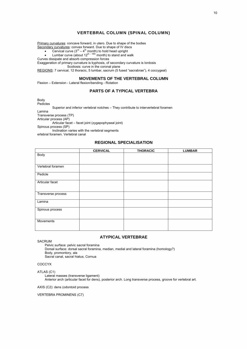
VERTEBRAL COLUMN (SPINAL COLUMN)
Primary curvatures: concave forward, in utero. Due to shape of the bodies Secondary curvatures: convex forward. Due to shape of IV discs
• Cervical curve (3rd – 4th month) to hold head upright • Lumbar curve (about 12th – 18th month) to stand and walk
Curves dissipate and absorb compression forces Exaggeration of primary curvature is kyphosis, of secondary curvature is lordosis
Scoliosis: curve in the coronal plane REGIONS: 7 cervical, 12 thoracic, 5 lumbar, sacrum (5 fused “sacrabrae”), 4 coccygeal)
MOVEMENTS OF THE VERTEBRAL COLUMN Flexion – Extension - Lateral flexion/bending - Rotation
PARTS OF A TYPICAL VERTEBRA
Body Pedicles
Superior and inferior vertebral notches – They contribute to intervertebral foramen Lamina Transverse process (TP) Articular process (AP)
Articular facet – facet joint (zygapophyseal joint) Spinous process (SP) Inclination varies with the vertebral segments ertebral foramen. Vertebral canal
REGIONAL SPECIALISATION CERVICAL THORACIC LUMBAR Body
Vertebral foramen
Pedicle
Articular facet
Transverse process
Lamina
Spinous process
Movements
ATYPICAL VERTEBRAE
SACRUM Pelvic surface: pelvic sacral foramina Dorsal surface: dorsal sacral foramina, median, medial and lateral foramina (homology?) Body, promontory, ala Sacral canal, sacral hiatus, Cornua
COCCYX ATLAS (C1)
Lateral masses (transverse ligament) Anterior arch (articular facet for dens), posterior arch. Long transverse process, groove for vertebral art.
AXIS (C2): dens (odontoid process VERTEBRA PROMINENS (C7)
10
11
JOINTS OF THE VERTEBRAL BODIES ANT LONG LIG POST LONG LIG upper end continuous with membrana tectoria. INTERVERTEBRAL DISCS 1/5 of the total length of the vert. column, thicker in C & L
JOINTS OF VERTEBRAL ARCHES ARTICULAR CAPSULE: Thin & loose, attached to margins of articular facet. LIGAMENTUM FLAVUM: between laminae, SUPRASPINOUS LIG: Connect apices of SP’s (in series with Lig nuchae) INTERSPINOUS LIG: between SP's INTERTRANSVERSE LIG
MOVEMENTS OF THE VERTEBRAL COLUMN FLEXION most extensive in C & L EXTENSION Free in C & L, restricted in T LAT FLEXION Always associated with some rotation. Most free in C & L CIRCUMDUCTION limited ROTATION freer in upper T, least in L ARTICULAR FACETS
CERVICAL: Upward inclination allows free flex & ext. THORACIC: Rotation is freer (Art. facets correspond to the surface of a cylinder with the centre near
the centre of vertebral body) but all mvts are limited (because of ribs & sternum) LUMBAR: Extension free & wider in range than flexion Rotation limited by the direction of articular facets
CRANIOVERTEBRAL JOINTS
LATERAL ATLANTO-AXIAL JOINTS Fibrous capsule: thin & loose
MEDIAN ATLANTO-AXIAL JOINT: Pivot joint.
TRANSVERSE LIG OF C1 + longitudinal bands = cruciform lig of the atlas.
ACCESSORY ATLANTOAXIAL Lig: Near the base of dens to C1 and Occipital bone (near XII canal)
MOVEMENT AT THE ATLANTO-AXIAL JOINTS rotation of C1 upon C2, extent being limited by alar and accessory atlantoaxial ligaments
ATLANTO-OCCIPITAL JOINTS
Ellipsoid. Articular surfaces can be considered as part of the surface of a sphere Fibrous capsules ANT ATL-OCC MEMBR. Ant arch of CI to margin of foramen magnum POST ATL-OCC MEMBRANE Post arch of CI to margin of foramen magnum
MOVEMENTS AT ATL-OCC JOINTS Flex & ext, slight lateral tilting
LIGTS CONNNECTING AXIS WITH OCCIPITAL BONE
MEMBRANA TECTORIA upwards extension of post long lig, blends with cranial dura ALAR LIGAMENTS dens to medial side of occipital condyles APICAL LIG OF THE DENS apex of dens to margin of foramen magnum ACCESSORY ATLANTOAXIAL Lig: Near the base of dens to C1 and Occipital bone (near XII canal) LIGAMENTUM NUCHAE
JOINTS OF VERTEBRAE & RIBS COSTOVERTEBRAL JOINT: Vertebral bodies with head of rib Ligaments: radiate & interarticular COSTOTRANSVERSE JOINT: TP & rib
Ligaments: costotransverse, lateral costotransverse, superior costotrransverse
Dr D. Vu 2008
12
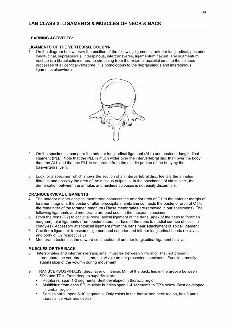
LAB CLASS 2: LIGAMENTS & MUSCLES OF NECK & BACK LEARNING ACTIVITIES: LIGAMENTS OF THE VERTEBRAL COLUMN 1. On the diagram below, draw the position of the following ligaments: anterior longitudinal, posterior
longitudinal, supraspinous, interspinous, intertransverse, ligamentum flavum. The ligamentum nuchae is a fibroelastic membrane stretching from the external occipital crest to the spinous processes of all cervical vertebrae, it is homologous to the supraspinous and interspinous ligaments elsewhere.
2. On the specimens, compare the anterior longitudinal ligament (ALL) and posterior longitudinal
ligament (PLL). Note that the PLL is much wider over the intervertebral disc than over the body than the ALL and that the PLL is separated from the middle portion of the body by the basivertebral vein.
3. Look for a specimen which shows the section of an intervertebral disc. Identify the annulus
fibrosus and possibly the area of the nucleus pulposus. In the specimens of old subject, the demarcation between the annulus and nucleus pulposus is not easily discernible.
CRANIOCERVICAL LIGAMENTS 4. The anterior atlanto-occipital membrane connects the anterior arch of C1 to the anterior margin of
foramen magnum, the posterior atlanto-occipital membrane connects the posterior arch of C1 to the remainder of the foramen magnum (These membranes are removed in our specimens). The following ligaments and membrane are best seen in the museum specimen.
5. From the dens (C2) to occipital bone: apical ligament of the dens (apex of the dens to foramen magnum), alar ligaments (from posterolateral surface of the dens to medial surface of occipital condyles). Accessory atlantoaxial ligament (from the dens near attachjment of apical ligament
6. Cruciform ligament: transverse ligament and superior and inferior longitudinal bands (to clivus and body of C2 respectively)
7. Membrana tectoria is the upward continuation of anterior longitudinal ligament to clivus MUSCLES OF THE BACK 8. Interspinales and intertransversarii: small muscles between SP’s and TP’s, not present
throughout the vertebral column, not visible on our prosected specimens. Function: mostly stabilisation of the volumn during movement
9. TRANSVERSOSPINALIS: deep layer of intrinsic Mm of the back, lies in the groove between
SP’s and TP’s. From deep to superficial are: • Rotatores: span 1-2 segments. Best developed in thoracic region • Multifidus: from each SP, multiple bundles span 1-4 segments to TP’s below. Best developed
in lumbar region • Semispinalis: span 8-10 segments. Only exists in the thorax and neck region, has 3 parts
thoracis, cervicis and capitis
13
10. ERECTOR SPINAE or SACROSPINALIS: superficial layer of the intrinsic mm From a broad tendon attached to the sacrum, lumbar spines and iliac crest, it splits into 3 columns. Iliocostalis is the most lateral, Longissimus is the largest & longest, Spinalis the most medial. Spinalis is variable, blends with semispinalis and you do not need to identify it.
SPINALIS LONGISSIMUS ILIOCOSTALIS Thoracis Thoracis Lumborum Insertion: (thoracic TPs + ribs) (lower 6 ribs) Cervicis Cervicis Thoracis Insertion: (TPs of T4-C2) (upper 6 ribs) Capitis Capitis Cervicis Insertion: (mastoid process) (TPs of C4-6)
11. Identify the thoracolumbar fascia which covers the intrinsic Mm of the vertebral column and
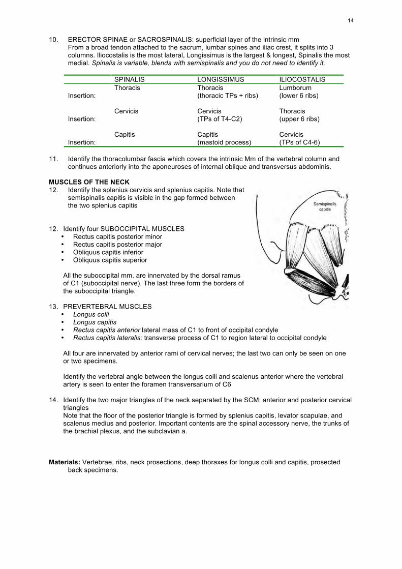
continues anteriorly into the aponeuroses of internal oblique and transversus abdominis. MUSCLES OF THE NECK 12. Identify the splenius cervicis and splenius capitis. Note that
semispinalis capitis is visible in the gap formed between the two splenius capitis
12. Identify four SUBOCCIPITAL MUSCLES
• Rectus capitis posterior minor • Rectus capitis posterior major • Obliquus capitis inferior • Obliquus capitis superior
All the suboccipital mm. are innervated by the dorsal ramus of C1 (suboccipital nerve). The last three form the borders of the suboccipital triangle.
13. PREVERTEBRAL MUSCLES
• Longus colli • Longus capitis • Rectus capitis anterior lateral mass of C1 to front of occipital condyle • Rectus capitis lateralis: transverse process of C1 to region lateral to occipital condyle
All four are innervated by anterior rami of cervical nerves; the last two can only be seen on one or two specimens. Identify the vertebral angle between the longus colli and scalenus anterior where the vertebral artery is seen to enter the foramen transversarium of C6
14. Identify the two major triangles of the neck separated by the SCM: anterior and posterior cervical
triangles Note that the floor of the posterior triangle is formed by splenius capitis, levator scapulae, and scalenus medius and posterior. Important contents are the spinal accessory nerve, the trunks of the brachial plexus, and the subclavian a.
Materials: Vertebrae, ribs, neck prosections, deep thoraxes for longus colli and capitis, prosected
back specimens.
14
LECTURE: PARAVERTEBRAL MUSCLES LIGAMENTUM NUCHAE: interspinous = supraspinous ligaments. Attached to external occipital crest THORACOLUMBAR FASCIA: trilaminar in the lumbar region, covers quadratus lumborum (i) and
erector spinae (ii), from TP (iii) INTRINSIC MUSCLES OF THE BACK: TRANSVERSOSPINALIS (between TPs and SPs)
• Rotators: span 1-2 vertebrae • Multifidus: spans 1-4 vertebrae • Semispinalis: spans 8-10 vertebrae
S. thoracis S. cervicis: reaches C2 S. capitis: reaches occipital bone, between superior and inferior nuchal lines (covered
by trapezius) ERECTOR SPINAE From a broad tendon which is attached to sacrum, lumbar spines and iliac crest. Splits into three columns, each can be described in three sections:
• Spinalis (Cannot be differentiate from semispinalis)• Longissimus: L. thoracis, cervicis and capitis • Iliocostalis: . lumborum, thoracis and cervicis
Iliocostalis:
• lumborum: from sacrum to angles of lower 6 ribs • thoracis: lower 6 ribs to upper ribs • cervicis: ribs 3-6 to TPs of C4-C6
Longissimus: • thoracis: from sacrum to TPs of all thoracic vertebrae and lower 10 ribs • cervicis: TPs of upper 4 Thoracic vertebrae to TPs of lower 4 cervical vertebrae • capitis: FROM TPs of upper 4 Thoracic vertebrae, AP of lower 4 cervical vertebrae TO
mastoid process IN THE NECK SUBOCCIPITAL MUSCLES
• Rectus capitis posterior minor: posterior tubercle of C1 to below inf nuchal line. • Rectus capitis posterior major: spine of C2 to below inferior nuchal line. • Obliquus capitis inferior: spine of C2 to TP of atlas • Obliquus capitis superior: TP of C1 to above the inferior nuchal line.
SPLENIUS
• S. cervicis: SP of T3-T6 to TPs of C1-C3 • S. capitis: Lig nuchae and SP of T1-T3 to below superior nuchal line
PREVERTEBRAL MUSCLES
• Longus colli: bodies of T1-T3 to TPs of C3-C6 and to bodies of C2 • Longus capitis: TPs of C3-C6 to basilar part of occipital. • Rectus capitis anterior: lateral mass of C1 to front of occipital condyle • Rectus capitis lateralis: TP of C1 to lateral to occipital condylle
15
LAB CLASS 3: ABDOMINAL MUSCLES & INGUINAL REGION LEARNING ACTIVITIES 1 On the hip bone, identify the iliac crest, anterior superior iliac spine (ASIS), pubic crest, pubic
tubercle, pecten pubis. On the articulated skeleton, identify the pubic symphysis, the sternum, costal cartilages of the first 10 ribs. Note that ribs 11 and 12 are floating ribs.
POSTERIOR ABDOMINAL WALL 2. Identify the muscles of the posterior abdominal wall: psoas major, psoas minor (if present) and
quadratus lumborum and the iliolumbar ligament. Follow the psoas tendon until its femoral attachment on lesser trochanter together with iliacus tendon.
Identify the genitofemoral nerve which penetrates the psoas major, the femoral nerve and obturator nerve on the lateral and medial sides of the lower part of the psoas
ANTEROLATERAL MUSCLES OF THE ABDOMINAL WALL 3. External oblique: note that its muscle fibres are attached to the lower 8 ribs and anterior half of
iliac crest and end in the external oblique aponeurosis. What is the direction of the muscle fibres? Follow the external oblique aponeurosis, it joins the contralateral aponeurosis at the midline (linea alba), its lateral border runs roughly vertical from 9th costal cartilage to the level of the umbilicus then veers towards the ASIS, its lower border running from ASIS to pubic tubercle is free and called inguinal ligament. Turn to the back of the specimen to identify the posterior b order of the muscle which is free and not attached to thoracolumbar fascia.
4. Internal oblique: Look for its posterior attachment on the thoracolumbar fascia in the back, together with transversus abdominis. The muscle fibres are attached to the anterior 2/3 of the iliac crest and lateral third of inguinal ligament. The upper fibres run upwards toward the midline, the lower fibres become fibrous and arch over spermatic cord to attach to pecten pubis and pubic crest.
5. Transversus abdominis: fibres run horizontally and forwards from the thoracolumbar fascia, the anterior 2/3 of the iliac crest, lateral 2/3 of inguinal ligament, and the lower 6 costal cartilages. Look for conjoint tendon where the aponeuroses of internal oblique and transversus abdominis join at the edge of rectus
6. Rectus abdominis: from xiphoid process to pubic crest. Identify the linea semilunaris, linea alba, and tendinous intersections on its anterior surface. Identify the rectus sheath, note that it is deficient posteriorly below the arcuate line. What form the rectus sheath. What form the linea alba? Of the arteries which run posterior to rectus abdominis in the rectus sheath, you can easily identify the inferior epigastric artery. Where does it originate?
7. Discuss with your tutor the actions of the anterolateral abdominal muscles. Why do they flex the vertebral column, increase intraabdominal pressure and rotate the trunk by working in pairs?
8. Discuss the action of rectus abdominis. INGUINAL CANAL 9. On the anterior surface of the specimen of anterior abdominal wall, identify the inguinal
ligament, superficial inguinal ring (a gap in the external oblique aponeurosis), spermatic cord as it ascend from the testis to enter the superficial inguinal ring
10. What is fascia transversalis? (It has been removed in our prosected specimens) 11. Turn to the posterior surface of the specimen, identify the inguinal ligament, conjoint tendon,
aponeurosis of transversus abdominis, deep inguinal ring which is lateral to the inferior epigastric artery (See diagrams next page). The inguinal canal zigzags between the superficial and deep inguinal rings and contains spermatic cord in the male and round ligament in the female. If the group has a specimen with intact vas deferens, follow it through the inguinal canal to the back of the bladder and note its relation with the inferior epigastric artery.
12. Indirect inguinal hernia is the protrusion of abdominal contents (usually bowels) through the
inguinal canal. Direct inguinal hernia is the protrusion at the of Hesselbach’s triangle (see diagram next page). The landmark separating the 2 types of hernia is the inferior epigastric artery, hence the importance of this artery for the differentiation of direct and indirect inguinal hernia in surgery
16
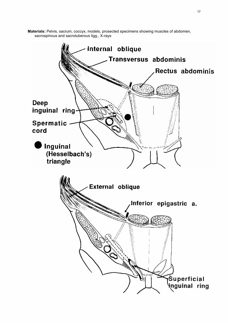
Materials: Pelvis, sacrum, coccyx, models, prosected specimens showing muscles of abdomen,
sacrospinous and sacrotuberous ligg., X-rays
17
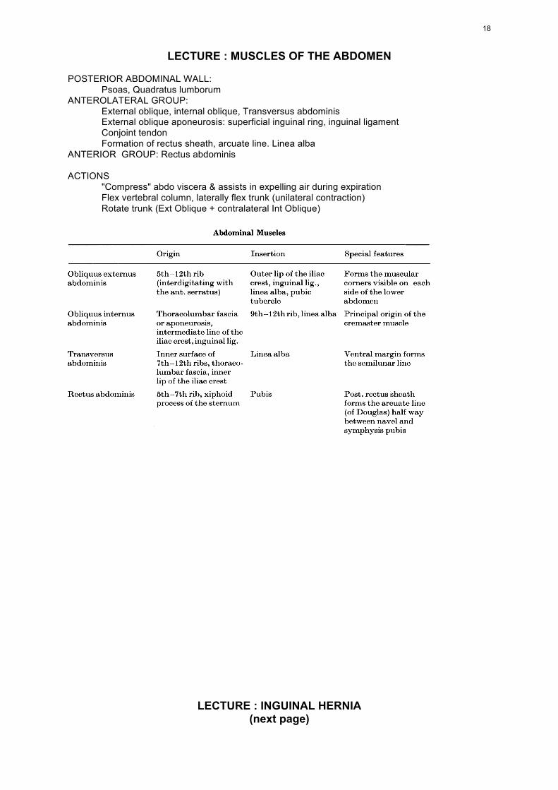
LECTURE : MUSCLES OF THE ABDOMEN POSTERIOR ABDOMINAL WALL:
Psoas, Quadratus lumborum ANTEROLATERAL GROUP:
External oblique, internal oblique, Transversus abdominis External oblique aponeurosis: superficial inguinal ring, inguinal ligament Conjoint tendon Formation of rectus sheath, arcuate line. Linea alba
ANTERIOR GROUP: Rectus abdominis ACTIONS
"Compress" abdo viscera & assists in expelling air during expiration Flex vertebral column, laterally flex trunk (unilateral contraction) Rotate trunk (Ext Oblique + contralateral Int Oblique)
LECTURE : INGUINAL HERNIA (next page)
18
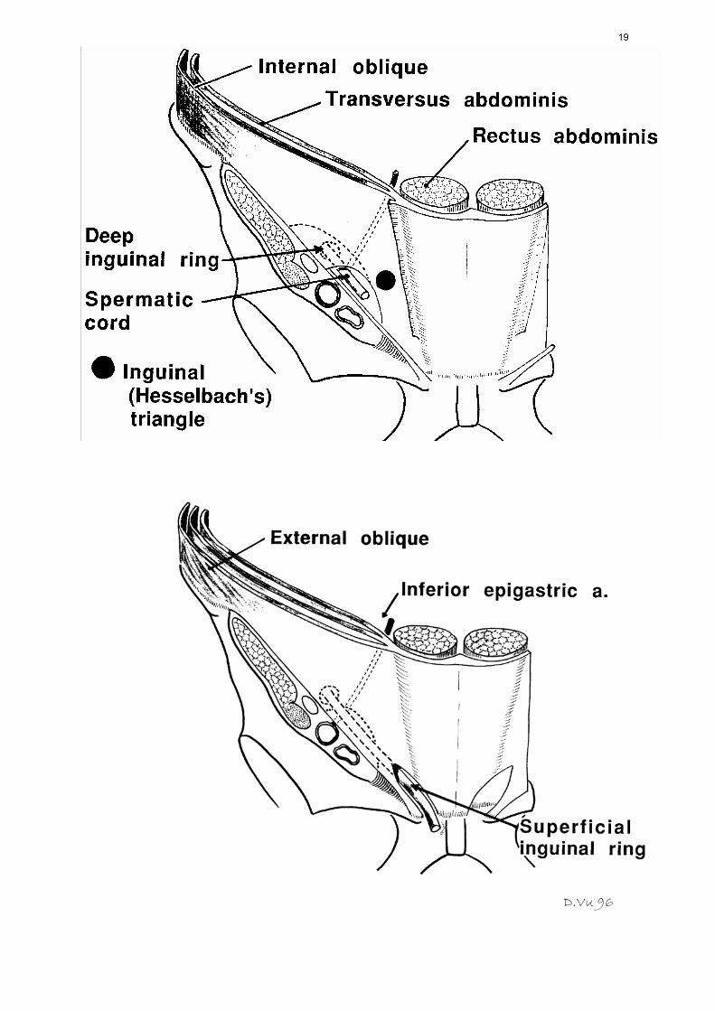
D.Vu 96
19
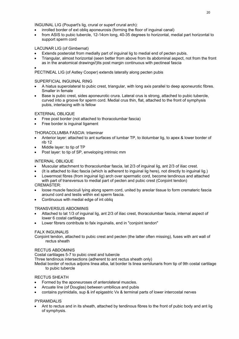
INGUINAL LIG (Poupart's lig, crural or superf crural arch): • inrolled border of ext obliq aponeurosis (forming the floor of inguinal canal) • from ASIS to pubic tubercle, 12-14cm long, 40-35 degrees to horizontal, medial part horizontal to
support sperm cord LACUNAR LIG (of Gimbernat) • Extends posterolat from medially part of inguinal lig to medial end of pecten pubis. • Triangular, almost horizontal (seen better from above from its abdominal aspect, not from the front
as in the anatomical drawings!)Its post margin continuous with pectineal fascia • PECTINEAL LIG (of Astley Cooper) extends laterally along pecten pubis SUPERFICIAL INGUINAL RING • A hiatus superolateral to pubic crest, triangular, with long axis parallel to deep aponeurotic fibres.
Smaller in female • Base is pubic crest, sides aponeurotic crura. Lateral crus is strong, attached to pubic tubercle,
curved into a groove for sperm cord. Medial crus thin, flat, attached to the front of symphysis pubis, interlacing with is fellow
EXTERNAL OBLIQUE • Free post border (not attached to thoracolumbar fascia) • Free border is inguinal ligament THORACOLUMBA FASCIA: trilaminar • Anterior layer: attached to ant surfaces of lumbar TP, to iliolumbar lig, to apex & lower border of
rib 12 • Middle layer: to tip of TP • Post layer: to tip of SP, enveloping intrinsic mm INTERNAL OBLIQUE • Muscular attachment to thoracolumbar fascia, lat 2/3 of inguinal lig, ant 2/3 of iliac crest. • (It is attached to iliac fascia (which is adherent to inguinal lig here), not directly to inguinal lig.) • Lowermost fibres (from inguinal lig) arch over spermatic cord, become tendinous and attached
with part of transversus to medial part of pecten and pubic crest (Conjoint tendon) CREMASTER: • loose muscle fasciculi lying along sperm cord, united by areolar tissue to form cremateric fascia
around cord and testis within ext sperm fascia. • Continuous with medial edge of int obliq TRANSVERSUS ABDOMINIS • Attached to lat 1/3 of inguinal lig, ant 2/3 of iliac crest, thoracolumbar fascia, internal aspect of
lower 6 costal cartilages • Lower fibrers contribute to falx inguinalis, end in "conjoint tendon" FALX INGUINALIS Conjoint tendon, attached to pubic crest and pecten (the latter often missing), fuses with ant wall of
rectus sheath RECTUS ABDOMINIS Costal cartilages 5-7 to pubic crest and tubercle Three tendinous intersections (adherent to ant rectus sheath only) Medial border of rectus adjoins linea alba, lat border Is linea semilunaris from tip of 9th costal cartilage
to pubic tubercle RECTUS SHEATH • Formed by the aponeuroses of anterolateral muscles. • Arcuate line (of Douglas) between umbilicus and pubis • contains pyrimidalis, sup & inf epigastric Vx & terminal parts of lower intercostal nerves PYRAMIDALIS • Ant to rectus and in its sheath, attached by tendinous fibres to the front of pubic body and ant lig
of symphysis.
20
• It lat border inclining medially, its pointed end embedded in linea alba midway between umbilicus and pubis. Anson found it absent in 17.7%
LINEA ALBA Narrow below the umbilicus, broader in supraumbilical part (because recti diverge) TRANSVERSALIS FASCIA • Thin areolar stratum between transversus and extraperitoneal fat • Fuses with ant lamina of thoracolumbar fascia, continuous with fascia iliaca. • It descends with femoral Vx as the femoral sheath DEEP INGUINAL RING • in fasc transversalis • midway between ASIS and symphysis, about 1/2 inch above inguinal lig. INGUINAL CANAL contains spermatic cord/round lig and ilioinguinal nerve Oblique, about 4cm long, slants inferomedially, parallel to inguinal lig Strength: two rings do not coincide. Post wall strengthened by falx inguinalis & reflected inguinal lig
directly behind superf ring. Int obliq overlaps deep ring. Fibres of int obliq & transversus arching over the canal are constantly active in standing, any increase
in intra-abdominal pressure augments contraction of int oliq (and possibly transvesus)
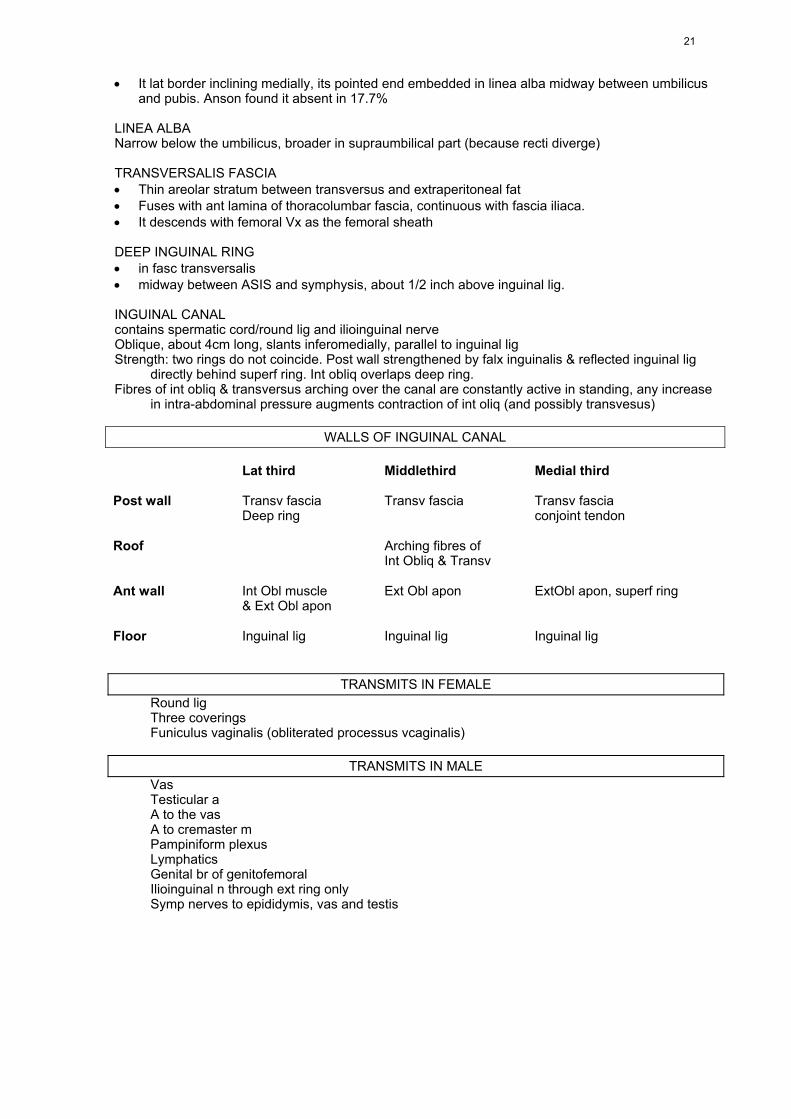
WALLS OF INGUINAL CANAL Lat third Middlethird Medial third Post wall Transv fascia Transv fascia Transv fascia Deep ring conjoint tendon Roof Arching fibres of Int Obliq & Transv Ant wall Int Obl muscle Ext Obl apon ExtObl apon, superf ring & Ext Obl apon Floor Inguinal lig Inguinal lig Inguinal lig
TRANSMITS IN FEMALE Round lig Three coverings Funiculus vaginalis (obliterated processus vcaginalis)
TRANSMITS IN MALE
Vas Testicular a A to the vas A to cremaster m Pampiniform plexus Lymphatics Genital br of genitofemoral Ilioinguinal n through ext ring only Symp nerves to epididymis, vas and testis
21
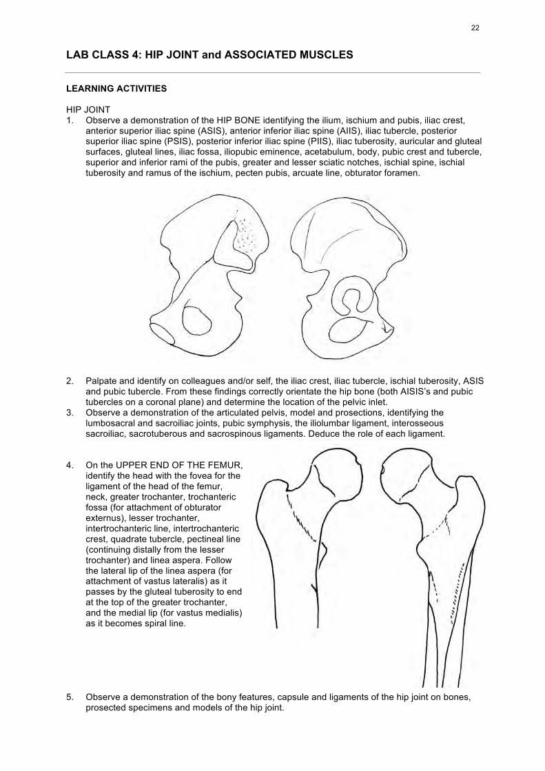
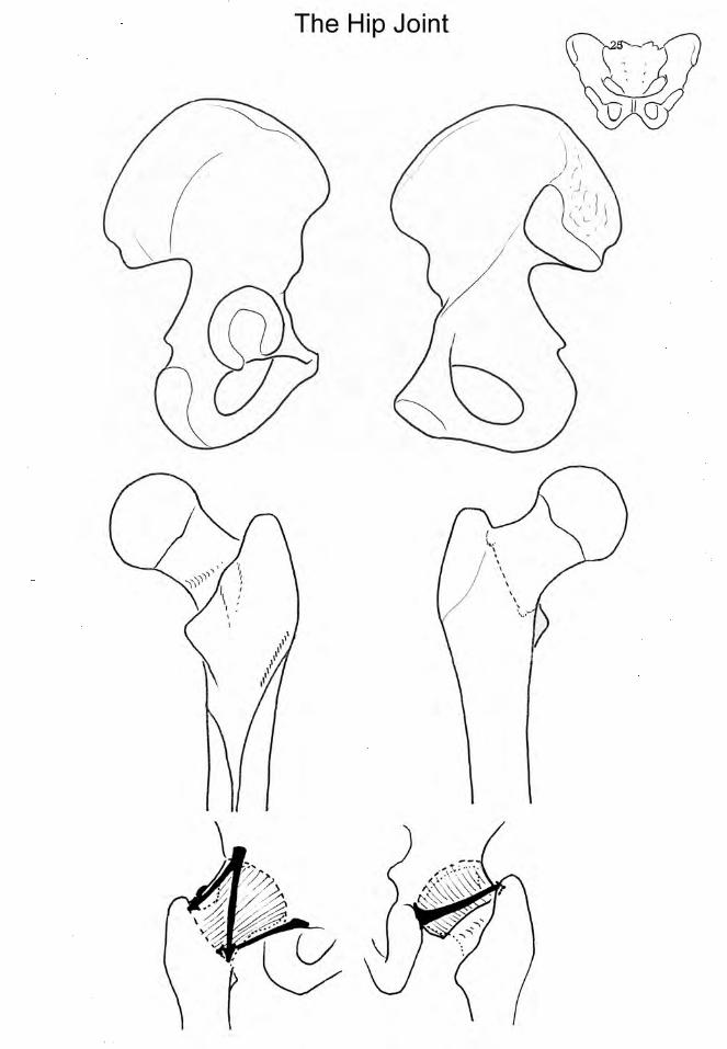
LAB CLASS 4: HIP JOINT and ASSOCIATED MUSCLES LEARNING ACTIVITIES HIP JOINT 1. Observe a demonstration of the HIP BONE identifying the ilium, ischium and pubis, iliac crest,
anterior superior iliac spine (ASIS), anterior inferior iliac spine (AIIS), iliac tubercle, posterior superior iliac spine (PSIS), posterior inferior iliac spine (PIIS), iliac tuberosity, auricular and gluteal surfaces, gluteal lines, iliac fossa, iliopubic eminence, acetabulum, body, pubic crest and tubercle, superior and inferior rami of the pubis, greater and lesser sciatic notches, ischial spine, ischial tuberosity and ramus of the ischium, pecten pubis, arcuate line, obturator foramen.
2. Palpate and identify on colleagues and/or self, the iliac crest, iliac tubercle, ischial tuberosity, ASIS
and pubic tubercle. From these findings correctly orientate the hip bone (both AISIS’s and pubic tubercles on a coronal plane) and determine the location of the pelvic inlet.
3. Observe a demonstration of the articulated pelvis, model and prosections, identifying the lumbosacral and sacroiliac joints, pubic symphysis, the iliolumbar ligament, interosseous sacroiliac, sacrotuberous and sacrospinous ligaments. Deduce the role of each ligament.
4. On the UPPER END OF THE FEMUR,
identify the head with the fovea for the ligament of the head of the femur, neck, greater trochanter, trochanteric fossa (for attachment of obturator externus), lesser trochanter, intertrochanteric line, intertrochanteric crest, quadrate tubercle, pectineal line (continuing distally from the lesser trochanter) and linea aspera. Follow the lateral lip of the linea aspera (for attachment of vastus lateralis) as it passes by the gluteal tuberosity to end at the top of the greater trochanter, and the medial lip (for vastus medialis) as it becomes spiral line.
5. Observe a demonstration of the bony features, capsule and ligaments of the hip joint on bones,
prosected specimens and models of the hip joint.
22
6. Examine prosected specimens and models of the hip joint. Identify the acetabular labrum, transverse acetabular ligament (bridging the acetabular notch and providing some attachment for the ligament of the head of femur), the iliofemoral (Y-shaped), pubofemoral and ischiofemoral ligaments, and the ligament of the head of the femur. Note that the inferior band of the iliofemoral ligament and ischiofemoral ligament twists around the neck of the femur. Explain why they are responsible for the stability of the hip joint.
7. Participate in group discussion on potential movements at the hip joint, on the basis of conformation of bony surfaces and the disposition of ligaments. What factors affect the stability of the hip joint? In what position is it most likely to dislocate and why? What movement is resisted by the (i) iliofemoral ligament and (ii) the pubofemoral ligament?
8. With colleagues study radiographs of the pelvis and hip joint and identify bony features listed in L.A.'s 1 and 4.
GLUTEAL MUSCLES 9. Observe the posterior aspect of the specimen. Identify the gluteus maximus, note in particular that
only ¼ of the inferior muscle fibres are attached to the gluteal tuberosity on the femur, the rest is attached to fascia lata and iliotibial tract. When this muscle is retracted, the following muscles are visible almost on the same plane: gluteus maximus, medius and minimus, piriformis, obturator internus (tendon), superior and inferior gamelli and quadratus femoris. Revise with your colleague their bony attachments.
10. Note the superior gluteal vessels and nerve emerging above the piriformis, the inferior gluteal vessels and nerve, sciatic nerve, posterior femoral cutaneous nerve, pudendal vessels and nerve below the piriformis.
11. Observe the anterior aspect of the specimen. Identify the tensor fasciae latae attaching to the fascia lata and iliotibial tract and covering the gluteus minimus.
12. What are the pelvitrochanteric muscles? Why are they called lateral rotators of the hip joint? What is the main function of the gluteus medius and how would you demonstrate its paralysis (Trendelenberg’s test)? What would result from simultaneous contraction of gluteus maximus and tensor fasciae latae?
13. Identify all the features of the hip bone, femur and the hip joint on X-rays Materials: Articulated skeleton, hip bone, articulated pelvis, femur, tibia, prosected specimens, radiographs.
23
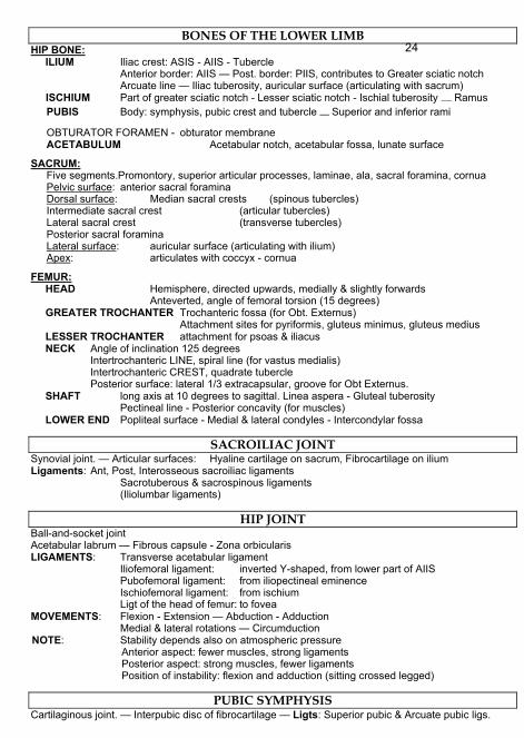
BONES OF THE LOWER LIMB HIP BONE:
ILIUM Iliac crest: ASIS - AIIS - Tubercle Anterior border: AIIS — Post. border: PIIS, contributes to Greater sciatic notch Arcuate line — Iliac tuberosity, auricular surface (articulating with sacrum)
ISCHIUM Part of greater sciatic notch - Lesser sciatic notch - Ischial tuberosity — Ramus PUBIS Body: symphysis, pubic crest and tubercle — Superior and inferior rami
OBTURATOR FORAMEN - obturator membrane ACETABULUM Acetabular notch, acetabular fossa, lunate surface
SACRUM: Five segments.Promontory, superior articular processes, laminae, ala, sacral foramina, cornua Pelvic surface: anterior sacral foramina Dorsal surface: Median sacral crests (spinous tubercles) Intermediate sacral crest (articular tubercles) Lateral sacral crest (transverse tubercles) Posterior sacral foramina Lateral surface: auricular surface (articulating with ilium) Apex: articulates with coccyx - cornua
FEMUR: HEAD Hemisphere, directed upwards, medially & slightly forwards Anteverted, angle of femoral torsion (15 degrees) GREATER TROCHANTER Trochanteric fossa (for Obt. Externus) Attachment sites for pyriformis, gluteus minimus, gluteus medius LESSER TROCHANTER attachment for psoas & iliacus NECK Angle of inclination 125 degrees Intertrochanteric LINE, spiral line (for vastus medialis) Intertrochanteric CREST, quadrate tubercle Posterior surface: lateral 1/3 extracapsular, groove for Obt Externus. SHAFT long axis at 10 degrees to sagittal. Linea aspera - Gluteal tuberosity Pectineal line - Posterior concavity (for muscles) LOWER END Popliteal surface - Medial & lateral condyles - Intercondylar fossa
SACROILIAC JOINT
Synovial joint. — Articular surfaces: Hyaline cartilage on sacrum, Fibrocartilage on ilium Ligaments: Ant, Post, Interosseous sacroiliac ligaments Sacrotuberous & sacrospinous ligaments (Iliolumbar ligaments)
HIP JOINT Ball-and-socket joint Acetabular labrum — Fibrous capsule - Zona orbicularis LIGAMENTS: Transverse acetabular ligament Iliofemoral ligament: inverted Y-shaped, from lower part of AIIS Pubofemoral ligament: from iliopectineal eminence Ischiofemoral ligament: from ischium Ligt of the head of femur: to fovea MOVEMENTS: Flexion - Extension — Abduction - Adduction Medial & lateral rotations — Circumduction NOTE: Stability depends also on atmospheric pressure
Anterior aspect: fewer muscles, strong ligaments Posterior aspect: strong muscles, fewer ligaments Position of instability: flexion and adduction (sitting crossed legged)
PUBIC SYMPHYSIS
Cartilaginous joint. — Interpubic disc of fibrocartilage — Ligts: Superior pubic & Arcuate pubic ligs.
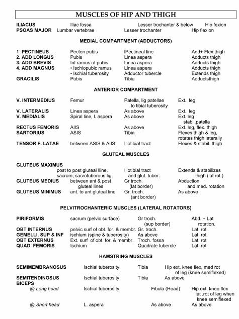
MUSCLES OF HIP AND THIGH ILIACUS Iliac fossa Lesser trochanter & below Hip fexion PSOAS MAJOR Lumbar vertebrae Lesser trochanter Hip flexion
MEDIAL COMPARTMENT (ADDUCTORS)
1 PECTINEUS Pecten pubis lPectineal line Add+ Flex thigh 2. ADD LONGUS Pubis Linea aspera Adducts thigh 3. ADD BREVIS Inf ramus of pubis Linea aspera Adducts thigh 4. ADD MAGNUS • Ischiopubic ramus Linea aspera Adducts thigh • Ischial tuberosity Adductor tubercle Extends thigh GRACILIS Pubis Tibia Adductsthigh
ANTERIOR COMPARTMENT
V. INTERMEDIUS Femur Patella, lig patellae Ext. leg to tibial tuberosity V. LATERALIS Linea aspera As above Ext. leg V. MEDIALIS Spiral line, l. aspera As above Ext. leg stabil.patella RECTUS FEMORIS AIIS As above Ext. leg, flex. thigh SARTORIUS ASIS Tibia Flexes thigh & leg, rotates thigh laterally TENSOR F. LATAE between ASIS & AIIS Iliotibial tract Flexes & stabil. thigh
GLUTEAL MUSCLES
GLUTEUS MAXIMUS post to post gluteal lline, Iliotibial tract Extends & stabilizes sacrum, sacrotuberous lig. and glut. tuber. thigh (lat rot.) GLUTEUS MEDIUS between ant & post Gr troch. Abduction gluteal lines (lat border) and med. rotation GLUTEUS MINIMUS ant. to ant gluteal line Gr. troch. As above (ant border)
PELVITROCHANTERIC MUSCLES (LATERAL ROTATORS) PIRIFORMIS sacrum (pelvic surface) Gr troch. Abd. + Lat (sup border) rotation. OBT INTERNUS pelvic surf of obt. for. & membr. Gr. troch. Lat. rot GEMELLI, SUP & INF ischium (spine & tuberosity) As above Lat. rot. OBT EXTERNUS Ext. surf of obt. for. & membr. Troch. fossa Lat. rot QUAD. FEMORIS Ischium Quadrate tubercle Lat. rot
HAMSTRING MUSCLES SEMIMEMBRANOSUS Ischial tuberosity Tibia Hip ext, knee flex, med rot of leg (knee semiflexed) SEMITENDINOSUS Ischial tuberosity Tibia As above BICEPS @ Long head Ischial tuberosity Fibula (Head) Hip ext, knee flex lat .rot of leg when knee semiflexed @ Short head L. aspera As above As above
24
25
THE HIP JOINT
• Ball-and-socket joint • Head of femur: > ½ sphere, anteverted (14 degrees) • Acetabulum: directed obliquely forward (40 degrees to sagittal) and downward (60 degrees
to horizontal) • Neck of the femur: angle of inclination (125 degrees)
ACETABULUM: • Lunate surface (articular) and acetabular fossa (non-articular) • Acetabul ar notch • Acetabular labrum deepens the bony acetabulum • Transverse ligament of the acetabulum bridges the gap of acetabular notch FIBROUS CAPSULE: • Strong • Attac hment: margin of acetabular labrum and transverse acetabular ligament, intertrochanteric
line, posterior aspect of femoral neck • Composed of longitudinal fibres and circular fibres (zona orbicularis) SYNOVIAL MEMBRANE Ensheathes ligament of the head of the femur LIGAMENTS: 1. Transverse acetabular ligament 2. Iliofemoral lig (Y-shaped lig): inverted Y, thick vertical bands and thin central part, attached to
lower part of AIIS and intertrochanteric line 3. Pubofemoral ligament: from body of pubis and adjacent part of superior ramus, to lower
surface of femoral neck 4. Ischiofemoral lig: from ischium (behind and below acetabulum) to greater trochanter, deep to
iliofemoral ligament 5. Ligament of the head of femur: from each side of acetabular notch and floor of acetabular
fossa to fovea on femoral head NOTES THAT: • Stability of the hip joint depends on: bony configuration, atmospheric pressure, and ligaments • Anterior aspect: fewer muscles, strong ligaments. Posterior aspect: strong muscles, fewer
ligaments • Ligaments SPIRAL AROUND THE NECK: extension winds up these ligaments and holds the
femur tightly into acetabulum (close-pack position). Position of instability: hip flexed and abducted
• Compression force across the hip joint produced by body weight AND the force of hip abductors. Rydell (1973) recorded force up to x5 times body weight in running. Normal neck fractures when femoral head is loaded > 12-15 times body weight!
26
LAB CLASS 5: FEMUR AND THIGH LEARNING ACTIVITIES THE HIP BONE and THE FEMUR 1. Identify the following features of the femur: lesser trochanter, pectineal line, linea aspera, spiral
line, adductor tubercle, popliteal surface, the femoral condyle and intercondylar notch. 2. Review the following features of the hip bone: ASIS, AIIS, pubic tubercle, ischipubic ramus,
ischiopubic ramus. ANTERIOR COMPARTMENT OF THE THIGH (Femoral nerve) 3. Identify the following structures attaching to and near the ASIS: inguinal ligament, tensor fascia
latae, sartorius. Follow the sartorius to its attachment on the tibia and note that it crosses anterior to the hip joint and posteromedial to the knee joint, hence it flexes the hip and knee joints and laterally rotates the thigh.
Observe that at the front of the inguinal region, under the inguinal ligament are iliacus and psoas and pectineus.
4. The bulk of the front of the thigh is formed by the four heads of the quadriceps. Follow the rectus femoris from its attachment on AIIS, retract it to one side to see the other three heads, the vastus lateralis, vastus medialis and vastus intermedius between them. You cannot see the reflected head of the rectus femoris above the acetabulum. Trace the vastus lateralis to the posterior attachment on the linea aspera and note that it ascends up to the greater trochanter. Trace the attachment of the vastus medialis to its posterior attachment on the medial lip of linea aspera and spiral line. All four heads attach together as the patellar tendon onto the patella, note that the vastus medialis extend further down to the medial side of the patella. This part of the muscle has the fibres running more horizontally, it is called vastus medialis obiquus and helps prevent lateral dislocation of the patella. The rectus femoris crosses in front ogf the hip joint so it also flexes the hip joint in addition to working with the rest of the quadriceps to extend the knee.
THE MEDIAL COMPARTMENT OF THE THIGH (Obturator nerve) 5. Observe the anterior aspect of the specimens of the lower limb and thigh. Identify the pectineus,
follow it to their attachment below the lesser trochanter and note its relation there with the ilio-psoas tendon. Where is the muscle attached to the hip bone and femur? On the medial aspect of the thigh, identify the gracilis. Note its proximal and distal attachments on the ischiopubic ramus and the tibia. Identify the adductors arranged in three layers: adductor longus, brevis and magnus. Review their bony attachments. Identify the two parts of adductor magnus, the adductor part (attached to the linea aspera) and the hamstring part (attached to the adductor tubercle of the femur, supplied by the sciatic nerve), and the adductor hiatus between the latter and the adductor.
6. Why is the ischiofemoral head of adductor magnus called “hamstring part”? Which muscle(s) would most likely be affected in a "groin strain" injury?
THE HAMSTRINGS (Sciatic nerve) 7. Identify on the hip bone the ischial tuberosity, ischiopubic ramus. Compare it with the diagram
below to locate the muscle attachments on the bone. 8. Identify the three hamstrings. The semimembranosus and semitendinosus are on the medial side.
Note their aponeurosis and tendons, their relations with each other, and in particular their attachments to the tibia. Note that the semimembranosus is attached to the edge of the tibial plateau and sends an expansion upwards across the knee joint, the oblique popliteal ligament. The semitendinosus is attached further down on the anteromedial aspect of the tibia. See how it is attached near the tendons of sartorius and gracilis to form the so-called pes anserinus (goose feet)
9. Identify the biceps on the lateral side with its long and short heads and its attachment on the fibula. 10. Discuss with your tutor the actions of bi-articular muscles like the hamstrings, how flexion of the
hip joint limits extension of the knee joint and expose the ham strings to injury. FEMORAL TRIANGLE 11. Identify the structures forming the boundaries of the femoral triangle:
• Base: inguinal ligament from ASIS to PT. • Walls: medial borders of sartorius and adductor longus. • Floor: pectineus and iliopsoas • Roof: cribriform fascia, penetrated by great saphenous v.
27
12. Identify its contents: femoral nerve, artery, vein and canal (lateral to medial). Femoral sheath is
continuous with the fascia transversalis, contains femoral artery & vein and canal but not the nerve.
13. Identify the femoral canal. Observe that in the space behind the inguinal ligament are found from lateral to medial: iliacus and psoas and the femoral nerve, pectineus. The medial part of this is occupied by the femoral sheath which has three compartments for the femoral artery, femoral vein and an empty compartment, femoral canal. Hernia through this empty compartment is called femoral hernia.
ADDUCTOR CANAL (SUSARTORIAL CANAL) 14. It begins from the apex of femoral triangle and ends at the adductor hiatus, bounded laterally by
vastus medialis, posteriorly by adductors longus and magnus and roofed by sartorius and a strong aponeurosis connecting the vastus medialis and adductors.
15. Its contents: femoral artery and vein, two branches of femoral nerve (saphenous nerve and nerve to vastus medialis
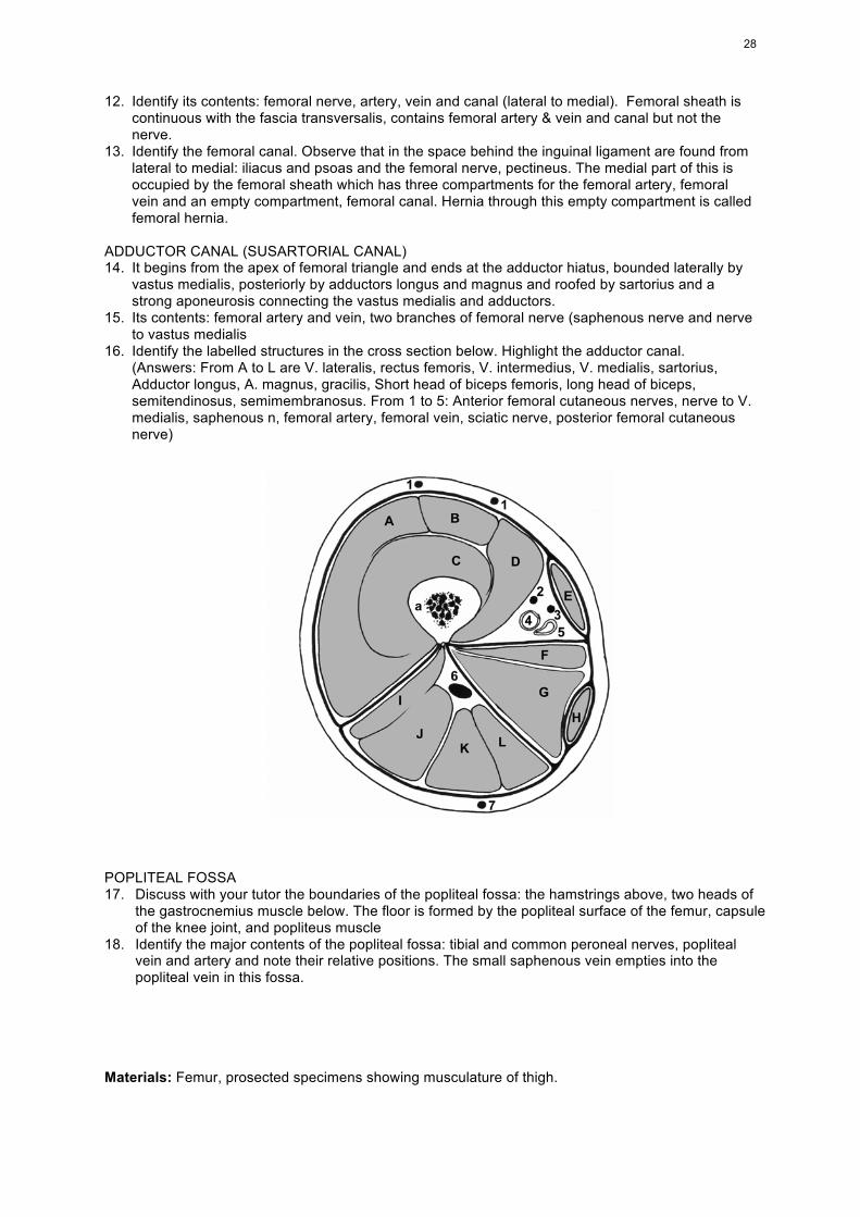
16. Identify the labelled structures in the cross section below. Highlight the adductor canal. (Answers: From A to L are V. lateralis, rectus femoris, V. intermedius, V. medialis, sartorius,
Adductor longus, A. magnus, gracilis, Short head of biceps femoris, long head of biceps, semitendinosus, semimembranosus. From 1 to 5: Anterior femoral cutaneous nerves, nerve to V. medialis, saphenous n, femoral artery, femoral vein, sciatic nerve, posterior femoral cutaneous nerve)
POPLITEAL FOSSA 17. Discuss with your tutor the boundaries of the popliteal fossa: the hamstrings above, two heads of
the gastrocnemius muscle below. The floor is formed by the popliteal surface of the femur, capsule of the knee joint, and popliteus muscle
18. Identify the major contents of the popliteal fossa: tibial and common peroneal nerves, popliteal vein and artery and note their relative positions. The small saphenous vein empties into the popliteal vein in this fossa.
Materials: Femur, prosected specimens showing musculature of thigh.
28
BONES OF THE LOWER LIMB HIP BONE:
ILIUM Iliac crest: ASIS - AIIS - Tubercle Anterior border: AIIS — Post. border: PIIS, contributes to Greater sciatic notch Arcuate line — Iliac tuberosity, auricular surface (articulating with sacrum)
ISCHIUM Part of greater sciatic notch - Lesser sciatic notch - Ischial tuberosity — Ramus PUBIS Body: symphysis, pubic crest and tubercle — Superior and inferior rami
OBTURATOR FORAMEN - obturator membrane ACETABULUM Acetabular notch, acetabular fossa, lunate surface
SACRUM: Five segments.Promontory, superior articular processes, laminae, ala, sacral foramina, cornua Pelvic surface: anterior sacral foramina Dorsal surface: Median sacral crests (spinous tubercles) Intermediate sacral crest (articular tubercles) Lateral sacral crest (transverse tubercles) Posterior sacral foramina Lateral surface: auricular surface (articulating with ilium) Apex: articulates with coccyx - cornua
FEMUR: HEAD Hemisphere, directed upwards, medially & slightly forwards Anteverted, angle of femoral torsion (15 degrees) GREATER TROCHANTER Trochanteric fossa (for Obt. Externus) Attachment sites for pyriformis, gluteus minimus, gluteus medius LESSER TROCHANTER attachment for psoas & iliacus NECK Angle of inclination 125 degrees Intertrochanteric LINE, spiral line (for vastus medialis) Intertrochanteric CREST, quadrate tubercle Posterior surface: lateral 1/3 extracapsular, groove for Obt Externus. SHAFT long axis at 10 degrees to sagittal. Linea aspera - Gluteal tuberosity Pectineal line - Posterior concavity (for muscles) LOWER END Popliteal surface - Medial & lateral condyles - Intercondylar fossa
SACROILIAC JOINT
Synovial joint. — Articular surfaces: Hyaline cartilage on sacrum, Fibrocartilage on ilium Ligaments: Ant, Post, Interosseous sacroiliac ligaments Sacrotuberous & sacrospinous ligaments (Iliolumbar ligaments)
HIP JOINT Ball-and-socket joint Acetabular labrum — Fibrous capsule - Zona orbicularis LIGAMENTS: Transverse acetabular ligament Iliofemoral ligament: inverted Y-shaped, from lower part of AIIS Pubofemoral ligament: from iliopectineal eminence Ischiofemoral ligament: from ischium Ligt of the head of femur: to fovea MOVEMENTS: Flexion - Extension — Abduction - Adduction Medial & lateral rotations — Circumduction NOTE: Stability depends also on atmospheric pressure
Anterior aspect: fewer muscles, strong ligaments Posterior aspect: strong muscles, fewer ligaments Position of instability: flexion and adduction (sitting crossed legged)
PUBIC SYMPHYSIS
Cartilaginous joint. — Interpubic disc of fibrocartilage — Ligts: Superior pubic & Arcuate pubic ligs.
MUSCLES OF HIP AND THIGH ILIACUS Iliac fossa Lesser trochanter & below Hip fexion PSOAS MAJOR Lumbar vertebrae Lesser trochanter Hip flexion
MEDIAL COMPARTMENT (ADDUCTORS)
1 PECTINEUS Pecten pubis lPectineal line Add+ Flex thigh 2. ADD LONGUS Pubis Linea aspera Adducts thigh 3. ADD BREVIS Inf ramus of pubis Linea aspera Adducts thigh 4. ADD MAGNUS • Ischiopubic ramus Linea aspera Adducts thigh • Ischial tuberosity Adductor tubercle Extends thigh GRACILIS Pubis Tibia Adductsthigh
ANTERIOR COMPARTMENT
V. INTERMEDIUS Femur Patella, lig patellae Ext. leg to tibial tuberosity V. LATERALIS Linea aspera As above Ext. leg V. MEDIALIS Spiral line, l. aspera As above Ext. leg stabil.patella RECTUS FEMORIS AIIS As above Ext. leg, flex. thigh SARTORIUS ASIS Tibia Flexes thigh & leg, rotates thigh laterally TENSOR F. LATAE between ASIS & AIIS Iliotibial tract Flexes & stabil. thigh
GLUTEAL MUSCLES
GLUTEUS MAXIMUS post to post gluteal lline, Iliotibial tract Extends & stabilizes sacrum, sacrotuberous lig. and glut. tuber. thigh (lat rot.) GLUTEUS MEDIUS between ant & post Gr troch. Abduction gluteal lines (lat border) and med. rotation GLUTEUS MINIMUS ant. to ant gluteal line Gr. troch. As above (ant border)
PELVITROCHANTERIC MUSCLES (LATERAL ROTATORS) PIRIFORMIS sacrum (pelvic surface) Gr troch. Abd. + Lat (sup border) rotation. OBT INTERNUS pelvic surf of obt. for. & membr. Gr. troch. Lat. rot GEMELLI, SUP & INF ischium (spine & tuberosity) As above Lat. rot. OBT EXTERNUS Ext. surf of obt. for. & membr. Troch. fossa Lat. rot QUAD. FEMORIS Ischium Quadrate tubercle Lat. rot
HAMSTRING MUSCLES SEMIMEMBRANOSUS Ischial tuberosity Tibia Hip ext, knee flex, med rot of leg (knee semiflexed) SEMITENDINOSUS Ischial tuberosity Tibia As above BICEPS @ Long head Ischial tuberosity Fibula (Head) Hip ext, knee flex lat .rot of leg when knee semiflexed @ Short head L. aspera As above As above
LAB CLASS 6: KNEE JOINT AND ASSOCIATED ANATOMY
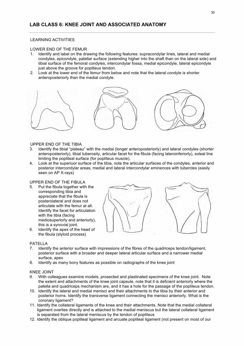
LEARNING ACTIVITIES LOWER END OF THE FEMUR 1. Identify and label on the drawing the following features: supracondylar lines, lateral and medial
condyles, epicondyle, patellar surface (extending higher into the shaft than on the lateral side) and tibial surface of the femoral condyles, intercondylar fossa, medial epicondyle, lateral epicondyle just above the groove for popliteus tendon.
2. Look at the lower end of the femur from below and note that the lateral condyle is shorter anteroposteriorly than the medial condyle.
UPPER END OF THE TIBIA 3. Identify the tibial “plateau” with the medial (longer anteroposteriorly) and lateral condyles (shorter
anteroposteriorly), tibial tuberosity, articular facet for the fibula (facing lateroinferiorly), soleal line limiting the popliteal surface (for popliteus muscle).
4. Look at the superiuor surface of the tibia, note the articular surfaces of the condyles, anterior and posterior intercondylar areas, medial and lateral intercondylar eminences with tubercles (easily seen on AP X-rays)
UPPER END OF THE FIBULA 5. Put the fibula together with the
corresponding tibia and appreciate that the fibula is posterolateral and does not articulate with the femur at all. Identify the facet for articulation with the tibia (facing mediosuperiorly and anteriorly), this is a synovial joint.
6. Identify the apex of the head of the fibula (styloid process)
PATELLA 7. Identify the anterior surface with impressions of the fibres of the quadriceps tendon/ligament,
posterior surface with a broader and deeper lateral articular surface and a narrower medial surface, apex
8. Identify as many bony features as possible on radiographs of the knee joint KNEE JOINT 9. With colleagues examine models, prosected and plastinated specimens of the knee joint. Note
the extent and attachments of the knee joint capsule, note that it is deficient anteriorly where the patella and quadriceps mechanism are, and it has a hole for the passage of the popliteus tendon.
10. Identify the lateral and medial menisci and their attachments to the tibia by their anterior and posterior horns. Identify the transverse ligament connecting the menisci anteriorly. What is the coronary ligament?
11. Identify the collateral ligaments of the knee and their attachments. Note that the medial collateral ligament overlies directly and is attached to the medial meniscus but the lateral collateral ligament is separated from the lateral meniscus by the tendon of popliteus
12. Identify the oblique popliteal ligament and arcuate popliteal ligament (not present on most of our
30
specimens) which reinforce the posterior capsule. 13. Identify the anterior cruciate ligament (ACL) and posterior cruciate ligament (PCL). Review their
attachments to the tibia and femur, note that they are named after their tibial attachment (eg. anterior cruciate ligament is attached to the anterior intercondylar area) and that they cross (hence the name “cruciate”) when viewed from the side, but they lie in two planes when viewed from above.
14. Identify the posterior meniscofemoral ligament which lies on the posterior aspect of the PCL. The anterior meniscofemoral ligament is not visible on our prosections or models.
15. Discuss the abnormal movement of the tibia in case of rupture of ACL and PCL (drawer sign) and rupture of medial collateral ligament (varus instability)
BURSAE 16. Of all the bursae around the knee joint, identify the location of the suprapatellar, prepatellar,
superficial and deep infrapatellar bursae. Be aware of the bursae between overlapping muscles and tendons and between tendons and bone such as those around the pes anserinus and
MUSCLES OF THE KNEE JOINT 17. Review the function of vastus medialis obliquus in stabilising the patella 18. Identify the iliotibial tract which is attached to a small tubercle lateral to the tibial tuberosity.
Identify it as a round long raised strip on the lateral side of your patella. Move the knee joint specimen to see it sliding anteroposterior over the lateral femoral condyle during flexion-extension of the knee. What is the Iliotibial band syndrome (or iliotibial band friction) syndrome?
19. Popliteus: identify the muscle and discuss its attachment and action in “unlocking” the knee joint at the beginning of knee flexion
20. Based on what has been covered in the lecture, discuss with your tutor the complex movement of the femur and tibia during flexion-extension of the knee joint, and the role of popliteus in “unlocking” the knee joint at the beginning of knee flexion
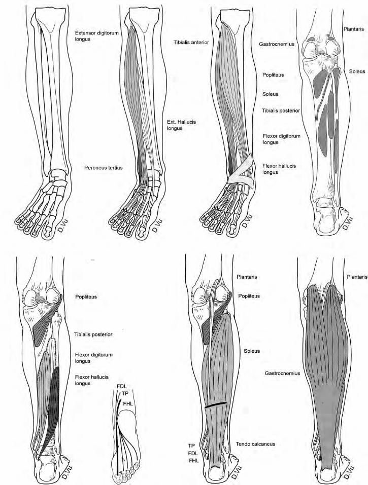
MUSCLES OF THE LATERAL COMPARTMENT OF THE LEG 21. Identify the lateral surface of the tibia which, after giving attachment to the peroneus longs
(fibularis longus) and peroneus brevis (fibularis brevis), rotates in the lower quarter of the bone to become posterior. In the lower end of the fibula, identify the triangular articular surface for the talus facing medially, and the groove for the peroneal tendons peronei posterior to it.
22. Identify the peroneus longus and brevis, follow the tendons distally to see them strapped down by the superior and inferior peroneal retinacula. The peroneus brevis tendon is attached to the posterior end of the 5th metatarsal, the peroneus longus tendon hooks around the cuboid and traverse the sole of the foot to attach to the first metatarsal.
MUSCLES OF THE ANTERIOR COMPARTMENT OF THE LEG 23. Study the cross section of the leg below to appreciate that the so-called anterior compartment is
in actual fact anterolateral in position. 24. Study the deep dissection of the leg to identify the proximal tibiofibular joint (synovial joint),
interosseous membrane and the distal tibiofibular joint (fibrous joint). 25. Identify the tibialis anterior, extensor digitorum longus, and extensor hallucis which begins deep to
the other two and peroneus tertius. Review their proximal and distal attachments. Follow their tendons to see them passing under the superior and inferior extensor retinaculum.
26. What is “shin splint”? MUSCLES OF THE POSTERIOR COMPARTMENT OF THE LEG 27. Identify the superficial muscles which form the triceps surae: lateral gastrocnemius, medial
gastrocnemius and soleus. Follow them to their common tendon: tendo calcaneus (Achilles tendon). Review their attachments. What is retrocalcaneal bursitis? Identify the plantaris, follow its long tendon to its attachment on the medial side of the tendo calcaneus. Note that the gastrocnemius crosses the knee joint and flexes the knee joint while the soleus does not.
28. Identify the three deep muscles: tibialis posterior which is attached to tibia and fibula and interosseous membrane, covered by the flexor hallucis longus (attached to the fibula) and the flexor digitorum longus (attached to the tibia). The tendon of tibialis posterior emerge under the flexor digitorum longus above the ankle. Follow the tendons till they pass through the “tarsal tunnel” under the flexor retinaculum which bound the following structures in order from above downward: tibialis posterior tendon, flexor digitorum longus tendon, posterior tibial artery, tibial nerve, flexor hallucis tendon
31
29. Xrays: identify all the above features of
the knee joint on X-rays
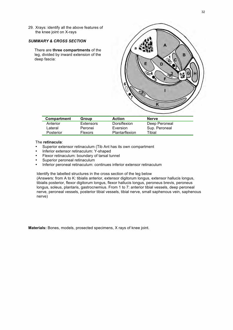
SUMMARY & CROSS SECTION There are three compartments of the
leg, divided by inward extension of the deep fascia:
Compartment Group Action Nerve Anterior Extensors Dorsiflexion Deep Peroneal Lateral Peronei Eversion Sup. Peroneal Posterior Flexors Plantarflexion Tibial
The retinacula:
• Superior extensor retinaculum (Tib Ant has its own compartment • Inferior extensor retinaculum: Y-shaped • Flexor retinaculum: boundary of tarsal tunnel • Superior peroneal retinaculum • Inferior peroneal retinaculum: continues inferior extensor retinaculum
Identify the labelled structures in the cross section of the leg below (Answers: from A to K: tibialis anterior, extensor digitorum longus, extensor hallucis longus,
tibialis posterior, flexor digitorum longus, flexor hallucis longus, peroneus brevis, peroneus longus, soleus, plantaris, gastrocnemius. From 1 to 7: anterior tibial vessels, deep peroneal nerve, peroneal vessels, posterior tibial vessels, tibial nerve, small saphenous vein, saphenous nerve)
Materials: Bones, models, prosected specimens, X rays of knee joint.
32
THE KNEE JOINT 1. Femoropatellar joint : plane joint - gliding 2. Femorotibial joint : synovial joint (hinge + some rotation) ARTICULAR SURFACES
FEMUR: medial condyle - Lateral condyle (groove for popliteus tendon) - Intercondylar notch Patellar articular surface (lateral side higher) - Tibial surfaces
PATELLA: lateral facet broader TIBIA: Medial condyle - Lateral condyle (smaller, rounder) - Intercondylar area & eminences -
Fibular facet. Position of close packing: full extension
CAPSULE Anteriorly: blends with expansions from vasti, absent at the front of the joint Posteriorly: blends with origins of heads of gastrocnemius, gap for popliteus (which is
intracapsular at its origin), strengthened by oblique popliteal ligament. Attached to periphery of menisci (Coronary ligament)
SYNOVIAL MEMBRANE: Suprapatellar bursa - Cruciate ligaments are extrasynovial
LIGAMENTS
Ligamentum patellae Oblique popliteal ligament (expansion from semimembranosus) Arcuate popliteal ligament COLLATERAL LIGAMENTS
- Tibial collat lig: firmly attached to edge of medial meniscus - Fibular coll lig: not attached to meniscus
Relations to biceps & popliteus tendons Both taut on extension
CRUCIATE LIGAMENTS, ant & post: named after their TIBIAL attachments
MENISCI Medial (C-shaped) and lateral (almost circular) Meniscofemoral ligament - Transverse ligament Distorted & move during flexion-extension, follow femoral condyles in rotation
BURSAE
Suprapatellar, prepatellar, superficial & deep infrapatellar Between capsule & heads of gastrocnemius, popliteal, semimembranosus
MOVEMENTS
Flexion, extension, medial & lateral rotations Conjunct rotation (integral with flexion-extension) - Role of popliteus in "unlocking" the joint at the
start of flexion Muscles producing movements at the knee joint (Refer to your text)
Clinical aspects
- Instability due to ligamentous damage (lateral/medial stress test, drawer sign) - Injury to the meniscus (medial meniscus more commonly damaged)
LAB CLASS 7: ANKLE AND FOOT 1
LEARNING ACTIVITIES BONES OF THE FOOT 1. With colleagues, examine an articulated foot and identify the tarsal bones and their features
described below: talus and calcaneus (hindfoot), navicular, 3 cuneiforms, cuboid (midfoot). Similar to the hand, distal to the tarsus are the metatarsals and phalanges (forefoot). The joint between the hindfoot and midfoot is called midtarsal joint/transverse tarsal joint/Chopart’s joint, the joint between the midfoot and forefoot tarsalmetatarsal joint/Lisfranc’s joint
2. TALUS: the body/dome which has a middle groove and is wider anteriorly, and posterior calcanean articular facet for the calcaneus. The head, connected with the body by the neck, articulates with the calcaneus anteriorly, with the anterior and middle facets of the calcaneus inferiorly. Identify the tarsal canal which is formed by the sulcus tali and sulcus calcanei . It contains the interosseous ligament and widens laterally into sinus tarsi. Posteriorly it has a posterior process with a deep groove for the tendon of flexor hallucis longus (FHL)
3. CALCANEUS: Articulates with the cuboid anteriorly and has a prominent calcaneal tuberosity posteriorly. The superior surface has the posterior surface for the body of talus, separated by sulcus calcanei from the anterior and middle surfaces for the head and neck of talus. The inferior surface has a round anterior tubercle and two lateral and medial tubercles at the posterior end, the last two are weightbearing areas of the heel. Sustentaculum tali is a shelf of bone supporting the talus, it is grooved on its underside by the tendon of FHL. The tendon of flexor digitorum longus runs along the medial side of the sustentaculum. Put the talus on the calcaneus to see the continuity of the groove for FHL from the talus to the sustentaculum.
4. NAVICULAR articulates with three cuneiform anteriorly, has a prominent tubercle for attachment of tibialis posterior.
5. CUNEIFORMS: wider on the dorsal side thus responsible for the transverse tarsal arch, numbered from the medial one (which articulates with the 1st metatarsal)
6. CUBOID: has a deep groove for tendon of peroneus longus, articulates with the 4th and 5th metatarsals
7. METATARSALS. Underneath the head of the 1st metatarsal are two sesamoid bones. The 2nd is the least mobile because it is attached to the shorter middle cuneiform and jammed on the sides by the other cuneiforms. The 5th has a tuberosity at its base for the tendon of peroneus brevis.
8. PHALANGES ARCHES OF THE FOOT 9. Identify the arches of the foot. The medial longitudinal arch is the higher of the two, and consists
of the calcaneus, talus, navicular, cuneiforms and medial three metatarsals. The lateral longitudinal arch consists of the calcaneus, cuboid, and lateral two metatarsals. Remember that the skeleton of the foot may be relatively flexible if the plantar aponeurosis is slack, but can be converted into a more rigid, weight-bearing arch by tension on the aponeurosis, brought about by dorsiflexion of the toes. The transverse tarsal arch is a half dome in the mid part at the level of the cuneiforms.
DISTAL TIBIOFIBULAR JOINT 10. Is a fibrous joint (syndesmosis) with a strong interosseus ligament. Identify on the model and
specimen the anterior and posterior tibiofibular ligaments, inferior transverse ligament ANKLE JOINT/ TALOCRURAL JOINT 11. A hinge joint between the tibial and fibular malleoli and the body of talus. Observe the large
triangular articular surface on the talus for the fibular malleolus and the comma-shaped surface for the tibial malleolus. Therefore the lateral malleolus extend further distally than the medial, this can be confirmed on your own feet.
12. Lateral collateral ligament: identify the anterior and posterior talofibular ligaments, the calcaneofibular ligaments
13. Medial collateral ligament: identify the deltoid ligament. You can only see the superficial layer which is attached to the neck of talus, sustentaculum tali, spring ligament, navicular and the back of talus. The deep layer has been described differently by different authors but in composed of fibres running from the tibia to the talus.
35
14. The joint is most stable in dorsiflexion because the wider part of the talar dome is forced in between the malleoli. The range of movement is variable, but it is ca 20-30o in dorsiflexion and 30-50o in plantar flexion.
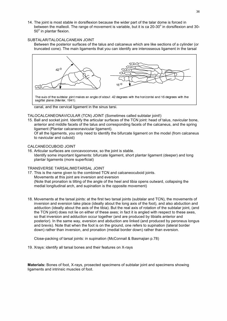
SUBTALAR/TALOCALCANEAN JOINT Between the posterior surfaces of the talus and calcaneus which are like sections of a cylinder (or
truncated cone). The main ligaments that you can identify are interosseous ligament in the tarsal
canal, and the cervical ligament in the sinus tarsi. TALOCALCANEONAVICULAR (TCN) JOINT (Sometimes called subtalar joint!) 15. Ball and socket joint. Identify the articular surfaces of the TCN joint: head of talus, navicular bone,
anterior and middle facets of the talus and corresponding facets of the calcaneus, and the spring ligament (Plantar calcaneonavicular ligament).
Of all the ligaments, you only need to identify the bifurcate ligament on the model (from calcaneus to navicular and cuboid)
CALCANEOCUBOID JOINT 16. Articular surfaces are concavoconvex, so the joint is stable. Identify some important ligaments: bifurcate ligament, short plantar ligament (deeper) and long
plantar ligaments (more superficial)
TRANSVERSE TARSAL/MIDTARSAL JOINT 17. This is the name given to the combined TCN and calcaneocuboid joints. Movements at this joint are inversion and eversion (Note that pronation is tilting of the angle of the heel and tibia opens outward, collapsing the
medial longitudinal arch, and supination is the opposite movement) 18. Movements at the tarsal joints: at the first two tarsal joints (subtalar and TCN), the movements of
inversion and eversion take place (ideally about the long axis of the foot), and also abduction and adduction (ideally about the axis of the tibia). But the real axis of rotation of the subtalar joint, (and the TCN joint) does not lie on either of these axes; in fact it is angled with respect to these axes, so that inversion and adduction occur together (and are produced by tibialis anterior and posterior). In the same way, eversion and abduction are linked (and produced by peroneus longus and brevis). Note that when the foot is on the ground, one refers to supination (lateral border down) rather than inversion, and pronation (medial border down) rather than eversion.
Close-packing of tarsal joints: in supination (McConnail & Basmajian p.78) 19. Xrays: identify all tarsal bones and their features on X-rays Materials: Bones of foot, X-rays, prosected specimens of subtalar joint and specimens showing ligaments and intrinsic muscles of foot.
36
37
38
LAB CLASS 8: FOOT 2 – LUMBOSACRAL PLEXUS LEARNING ACTIVITIES FASCIA OF THE FOOT: 1. Plantar aponeurosis: homologous to palmar aponeurosis but it has no muscle acting on it. It
runs forward from the calcaneus to attach on the plantar ligaments of the metatarso-phalangeal joints Dorsal fascia is thin
INTRINSIC MUSCLES OF THE FOOT 2. Dorsal muscle of the foot: I dentify the only one is extensor digitorum brevis on specimens and
palpate it on your own feet. A separate belly for the hallux may be distinguished, sometimes referred to as extensor hallucis brevis.
3. The plantar muscles of the foot are divided into four layers. Identify the layers and understand
the general topography of intrinsic muscles of the foot, NO knowledge of detailed attachments is required.
4. LAYER 1- Mnemonic: AFA Flexor digitorum brevis (comparable to FDSuperficialis in the hand, its tendons split around flexor
digitorum longus tendons) Abductor hallucis and abductordigiti minimi 5. NEUROVASCULAR PLANE between layers 1 & 2, containing medial & lateral plantar vessels
and nerves 6. LAYER 2 - Two tendons and associated muscles Flexor hallucis longus crossing deep to flexor digitorum longus (FDL) Attached to FDL are the flexor accessorius (quadratus plantae) posteriorly and the 4 lumbricals
anteriorly 7. LAYER 3 - Mnemonic: FAF Flexor hallucis brevis and flexor digiti minimi brevis Adductor hallucis 8. LAYER 4 – 2 tendons and interossei Tendons of peroneus longus and tibialis posterior Dorsal interossei (DAB) and plantar interossei (PAD). Their arrangement follows the same
principles as in the hand, except that the axis of the foot is through the 2nd metatarsal. SUMMARY OF MUSCLES ACTIONS Muscles acting on the ankle joint
DORSIFLEXION PLANTAR FLEXION Extensor hallucis longus Extensor digitorum longus Peroneus tertius Tibialis anterior
Gastrocnemius, Soleus, Plantaris Flexor hallucis longus Peronei Tibialis posterior
Muscles acting on the subtalar and midtarsal joint
INVERSION & ADDUCTION EVERSION & ABDUCTION Tibialis ant & post Flexor hallucis longus Flexor digitorum longus
Peronei longus, brevis and tertius Extensor digitorum longus (lateral part)
39
9. SOME COMMON DISORDERS OF THE FOOT • Flat foot (pes planus), pes cavus (very high longitudinal arch) • Simple Clubfeet (Talipes): talipes equinus (marked plantar flexion like a horse – T. calcaneus:
marked dorsiflexion) • Combined talipes: T. equinovarus (heel also bent inward) – T. equinovalgus (heel also bent
outward – T. calcaneovarus – T. calcaneovalgus • Hallux valgus: long axis of big toe turn laterally, usually resulting in bunion
LUMBOSACRAL PLEXUS 10. Note that the lumbar plexus is deep to the psoas major and formed by the ventral rami of spinal
nerves L1-4 Identify the following nerves on the posterior abdominal wall of deep abdomen specimens,
referring to lecture handout for details. • Iliohypogastric n and ilioinguinal (L1): they are homologous to intercostal nerve and its
collateral branch • Genitofemoral (L1,2): pierces psoas major • Lateral femoral cutaneous nerve (L2,3): runs deep to the inguinal ligament near the ASIS and
emerges superficial to sartorius into the thigh • Femoral nerve (L2,3,4) emerges from the lateral border of psoas • Obturator nerve (L2,3,4) emerges from the medial border of psoas, passes through obturator
canal with obturator vessels to enter the thigh • Lumbosacral trunk (L4,5): join the sacral plexus • Direct branches to psoas, quadratus lumborum (not seen on our specimens)
11. Sacral plexus: is in front of piriformis and continues directly into the sciatic nerve. The following
branches can be identified on most of our specimens: • Sciatic nerve (L4-S3) made up of tibial nerve and common peroneal nerve. The latter
innervate the short head of biceps and may run through or above piriformis • Posterior femoral cutaneous nerve (S1,2,3). Look at the posterior aspect of the thigh to
identify this nerve running medial to the sciatic nerve • Pudendal nerve (S2,3,4) • Superior gluteal nerve • Inferior gluteal nerve: the last two can be seen on the posterior aspect of the gluteal region
proximal to and distal to piriformis • Other small branches (refer to textbook and lecture handout) are not visible
Materials: Bones of foot, foot models, prosected specimens showing ligaments and intrinsic muscles of foot, posterior abdominal wall showing lumbosacral plexus
40
LECTURE & NOTES: THE FOOT FASCIA OF THE FOOT Thin on the dorsum. Plantar fascia thickened in the centre to form Plantar aponeurosis DORSUM OF THE FOOT :EXTENSOR DIGITORUM BREVIS Calcaneus to proximal phalanx of big toe and tendons of ext dig longus (toes 2-4) PLANTAR MUSCLES (Detailed attachments NOT required)
FIRST LAYER all from posterior part of calcaneus 1. ABDUCTOR HALLUCIS 2. ABD. DIGITI MINIMI 3. FLEXOR DIGITORUM BREVIS toes 2-5 - Attachment similar to Flex dig Superf in hand
SECOND LAYER muscles associated with tendons of Flex. hall long & Flex dig. long. 1. QUADRATUS PLANTAE from calcaneus to tendon of flexor digitorum longus 2. LUMBRICAL MUSCLES From FDLongus to dorsal digital expansion (Cf. hand)
THIRD LAYER Small muscles of the hallux and minimus (flexor hallucis brevis, adductor hallucis, flexor digiti minimi brevis)
FOURTH LAYER - PLANTAR & DORSAL INTEROSSEI (Cf. hand) - Tendons of peroneus longus & tibialis post.
RETINACULA SUPERIOR EXTENSOR RETINACULUM
between lower end of tibia and fibula. Tibialis ant has its own synovial sheath INFERIOR EXTENSOR RETINACULUM
Y-shaped. Uper surface of calcaneus i) to tibial malleolus and ii) to plantar aponeurosis FLEXOR RETINACULUM
Tip of medial malleolus to medial process of calcaneus and plantar aponeurosis. From medial to lateral: Tibialis post, flex dig longus, post tibial Vx, tibial n, flex hall longus
SUPERIOR PERONEAL RETINACULUM Back of lat. malleolus to lat. surface of calcaneus INFERIOR PERONEAL RETINACULUM
Continuous with inf extensor retinaculum, to lateral surface of calcaneus. Some fibres attached to peroneal trochlea, forming a septum between 2 peroneal tendons
ARCHES OF THE FOOT LONGITUDINAL ARCHES • MEDIAL ARCH: Calcaneus, talus, navicular, 3 cuneiforms, 3 metatarsals. Higher, more mobile.
Ligaments: all ligaments on the plantar aspect, plantar aponeurosis.Muscles: TibPost, FDL, FHL, intrinsic mm of foot
• LATERAL ARCH: Calcaneus, cuboid, 2 MT. Lower, less mobile. TRANSVERSE ARCH: foot is like a half-dome. Peroneus longus has an important function in maintaining transverse arch NOTE: • INVERSION & EVERSION: Axis (See prac notes) runs from the back of calcaneus (forwards,
upwards & medially) to the sup & medial aspect of the neck of talus. Thus Inversion is associated with adduction & slight planter flexion.
• PRONATION & SUPINATION: Tilting of the calcaneus. Pronation when medial border of the foot is closer to the ground (Compare with the hand)
41
LUMBAR PLEXUS:
• Formed by ventral rami of L1-L4 and a branch from T12 (subcostal nerve) • Formed in the substance of Psoas major, anterior to the transverse processes of lumber
vertebrae • Branches:
• Iliohypogastric and ilioinguinal nerves (L1) • Genitofemoral n (L1-2) • Lateral cutanerous n of the thigh (L2-3) • Femoral n (L2-4) • Obturator nerve (L2-4) • From L4: a branch to form lumbosacral trunk (with L5)
Relations of branches: All branches are formed from within the substance of psoas and emerge
through its anterior surface: genitofemoral n from its medial border at the level of pelvic brim: obturator n from its lateral border: all the other branches.
SACRAL PLEXUS
• Formed by lumbosacral trunk and the ventral rami of S1-S4 • Located in front of the piriformis and behind the ureter & internal ilac Vx and their branches
The sacral roots emerge from the ventral sacral foramina, run on the piriformis and converge into the sciatic nerve at the lower part of the greater sciatic notch (Thus the sciatic nerve escape from the pelvic under the piriformis when viewed from the gluteal region)
The superior and inferior gluteal arteries pass through the plexus Branches:
• Nerve to piriformis (S1-2) • Superior gluteal nerve (L4-5 S1) • Inferior gluteal nerve (L5 S1-2) • Nerve to quadratus femoris and gemellus inferior (L4-5 S1) • Nerve to obturator internus and gemellus superior (L5 S1-2) • Posterior cutaneous nerve of the thigh (S1-3) • Perforating cutaneous nerve (S2-3) • Pudendal nerve (S2-4)
• Sciatic nerve (L4-S4), made up of the tibial nerve and the common peroneal nerve
COOCYGEAL PLEXUS (not important)
Formed by S4-5 Lies on the coccygeus muscle Gives branches to the coccygeus and levator ani
42
LAB CLASS 9: VESSELS AND NERVES OF THE LOWER LIMB LEARNING ACTIVITIES VESSELS OF THE LOWER LIMB: 1 Observe a demonstration identifying position and extent of superior and inferior gluteal, femoral,
profunda femoris, popliteal, anterior and posterior tibial and dorsalis pedis arteries. The great and small saphenous veins and the femoral vein are also indicated. The femoral sheath, femoral canal, saphenous hiatus, adductor canal and femoral rings are described. Positions of superficial and deep inguinal lymph nodes are also indicated.
2. Arteries of the thigh. Identify the femoral artery, note its relations with the femoral vein and nerve
in the femoral triangle, and its relations with the vein and two nerves in the adductor canal. Note that it becomes popliteal artery when it passes through adductor hiatus. Try to find its descending genicular branch(es)
3. Identify the profunda femoris. Note that it runs deep to adductor longus. Identify its major
branches: • Medial circumflex femoral artery: seen in the femoral triangle • Lateral circumflex femoral artery: seen in the femoral triangle • Perforating arteries that can be seen on the back of the thigh as they emerge from the
adductor magnus 4. Note that blood supply to the head of the femur comes from arteries running up the neck to the
femoral head from an arterial circle at the neck which if formed mainly by the 2 circumflex femoral arteries. That is why fracture of the neck of the femur can lead to avascular necrosis of the head
5. Obturator artery accompanies obturator nerve. It is usually removed in our prosections 6. Turn to the back of the thigh to identify the popliteal artery. Note the arrangement of the popliteal
artery and vein and the tibial and common peroneal nerves in the popliteal fossa. Its genicular branches are difficult to find
7. In the gluteal region, try to identify the superior and inferior gluteal arteries accompanied by
nerves of the same names 8. In the back of the leg, identify the posterior tibial artery accompanied by the tibial nerve, find the
origin of the anterior tibial artery just below popliteus muscle and the peroneal artery which is closely related to the flexor hallucis longus.
Follow the posterior tibial artery until it divides into medial and lateral plantar arteries. These
arteries accompany nerves of the same names, the medial one runs along the meidal border of the foot, the lateral one crosses to the lateral side of the foot between muscle layers 1 and 2. Refer to lecture handouts or textbook diagrams to see how the latter form the plantar arch if you cannot find it on the specimens.
9. Look at the front of the leg prosections, retract the tibialis anterior and the extensor digitorum to
find the anterior tibial artery accompanied by the deep peroneal nerve.
Identify the continuation of the anterior tibial artery at the ankle: dorsalis pedis artery. Refer to lecture handouts or textbook diagrams to see the location of arcuate artery and its dorsal metatarsal arteries if you cannot find them on the specimens.
10. Listen to a brief demonstration of your tutor on the anastomoses around the knee joint and ankle.
You only need to be aware of them, no detail knowledge of the component and location of these anastomoses is expected.
11. Identify the great and small saphenous veins and participate in a tutorial discussion of the
venous drainage of the lower limb and the anatomy of varicose veins.
43
NERVES OF THE LOWER LIMB 12. Referring to lecture materials, review the nerves of the lower limb. This manual only list the
nerves and their branches that you can identify on the specimens and is not a comprehensive review of the nerves. Identify the following nerves: • Femoral nerve and its muscular and cutaneous branches: in femoral triangle. Look for the two
nerves in the adductor canal, and follow the saphenous nerve down to the medial border of the foot.
• Obturator nerve: note that the adductor brevis separate its anterior from its posterior branches. Identify some muscular branches. What is the skin area supplied by the obturator nerve?
• Superior and inferior gluteal nerves: in the gluteal region (See above) • Posterior femoral cutaneous nerve on the medial of (or sometimes posterior to) the sciatic
nerve • Sciatic nerve: trace it from its emergence under the piriformis. Sometimes, the 2 components
split early in the upper thigh or even in the pelvis. Identify a few muscular branches to the hamstrings and the hamstring part of adductor longus
• Tibial nerve: continues the direction of the sciatic. Identify some of its muscular branches. Follow it down to the tarsal tunnel when it divides into medial and lateral plantar nerve. Compare the medial plantar nerve to median nerve and the lateral plantar to ulnar nerve in the hand, their pattern of innervation is only slightly different from that in the hand. Review the relations of structures passing under the flexor retinaculum
• Common peroneal nerve: observe it crossing the neck of the fibula where it can be injured. You may see the sural nerve which receives contribution from both the tibial and common peroneal nerve. It eventually runs down the lateral border of the foot
• Superficial peroneal nerve can be found between the two peronei. Follow it to the lower third of the leg when it becomes subcutaneous and innervate the skin of most of the dorsum of the foot
• Deep peroneal nerve can be found on the interosseous membrane when you separate the tibialis anterior from the extensors (See above). You may find the cutaneous branch which lies on the lateral side of the dorsalis pedis artery and supplies the skin between the 1st and 2nd toes
13. Identify major arteries and vfeins of the lower limb on an giograms provided. REVIEW THE MOTOR AND CUTANEOUS SUPPLIES OF THE NERVES Refer to lecture materials and textbook, draw up in your own time a summary of the muscle
groups and skin areas supplied by the nerves of the lower limb. Materials: Deep thigh prosections showing the femoral artery and branches, free lower limbs, superficial and deep leg dissections, plastinated specimens of the foot.
44
DERMATOMES: Revise the pattern of dermatomes, i.e. the areas of skin supplied by individual spinal nerves. Note that you sit on S3, and walk on S1.
45
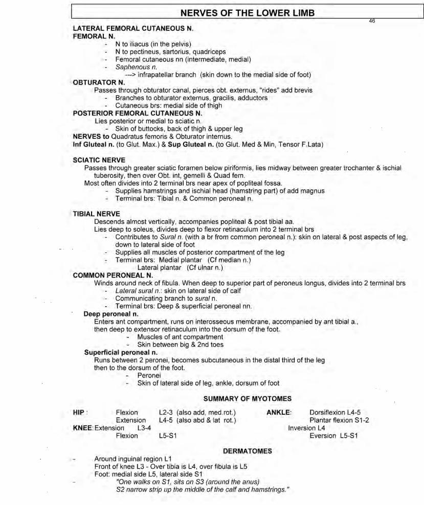
46
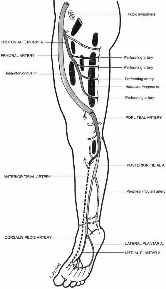
FUNCTIONAL NOTES: FUNCTIONAL MUSCLE GROUPS
HIP JOINT
• Angle of the neck with axis of the shaft: 120-125 degrees. Coxa vara predisposes to dislocation • Anteversion of the neck (20 degrees between axis of the neck and frontal plane. Increased
anteversion predisposes to anterior dislocation HIP FLEXORS They lie in the frontal plane, ANTERIOR to the centre of the hip joint
• Psoas and Iliacus • Tensor fasciae latae • Sartorius • Rectus femoris
Others are not as efficient flexors and also produce adduction/abduction or medial/lateral rotation: • Pectineus • Adductors longus and brevis • Gracilis • The most anterior fibres of all 3 gluteal muscles
HIP EXTENSORS They lie in the frontal plane, POSTERIOR to the centre of the hip joint
• Gluteal muscles (which also produce abduction) • Gluteus maximus (which is only important when running or walking up a slope, it is not
involved in normal walking on flat ground) • Most posterior fibres of gluteus medius and gluteus minimus • Hamstrings (which also produce adduction)
When the pelvis is tilted posteriorly, the iliofemoral ligament stabilises the pelvis When the pelvis is tilted anteriorly, it is stabilised by the hamstrings, then hamstrings AND gluteal
muscles. HIP ABDUCTORS
• The most powerful abductor is gluteus medius (Trendelenburg’s sign) • Tensor fasciae latae and gluteus maximus (by pulling on fascia lata on the lateral side of the
thigh) • Gluteus minimus • Piriformis
HIP ADDUCTORS Powerfull adductors are:
• Three adductors • Gracilis • The fibres of Gluteus maximus which are attached to gluteal tuberosity
Weaker adductors are: • Hamstrings • Pectineus • Quadratus femoris • Obturator externus • Obturtor internus and gemelli
LATERAL ROTATORS OF THE HIP JOINT
• Pelvitrochanteric muscles (6) • Gluteus maximus • Posterior fibres of gluteus medius and minimus • Quadratus femoris
48
MEDIAL ROTATORS OF THE HIP JOINT NOTE: The axis of rotation of the thigh is a line joining the centre of the femoral head and the centre
of the femoral condyle! For example: relate the adductor longus with this axis, and it is obvious the muscle medially rotates the thigh (not laterally rotates it as it appears to do at first glance!)
• Gluteus minimus • Anterior fibres of gluteus medius • Tensor fasciae latae • Hamstring part of adductor magnus
INVERSION OF MUSCLE ACTIONS
Actions of a muscle depend on its relative position with the axis of the movement Example 1: Piriformis is a lateral rotator. But when the femur is flexed, it becomes a pur
abductor Example 2: Quadratus femoris flexes the hip when the femur is extended and extends it when
the femur is flexed
KNEE JOINT KNEE FLEXORS
• Hamstrings • Gracilis • Sartorius • Gastrocnemius • Popliteus
NOTE: Monoarticular muscles (eg short head of biceps) always has the same efficiency. Efficiency of biarticular muscles depends on the position of one joint. Eg. the more the hip is flexed
(stretching the hamstrings), the more efficient the hamstrings are as flexors of the knee. MEDIAL ROTATORS OF THE KNEE Muscles attaching to the medial aspect of the tibia:
• Semimembranosus • Sartorius, gracilis, semitendinosus (attaching as pes anserinus) • Popliteus
LATERAL ROTATORS OF THE KNEE
• Biceps (attaching to the fibular) • Tensor fasciae latae (attaching to the iliotibial tract)
ANKLE JOINT
DORSIFLEXORS Muscles of the anterior compartment of the leg
PLANTAR FLEXORS • Muscles of the posterior compartment of the leg • Peroneus longus and peroneus brevis
INVERSION
• Muscles passing through tarsal tunnel (Muscles of anterior compartment of the leg) • Tibialis posterior
EVERSION
• Muscles of the lateral compartment of the leg (Peroneus longus & P. brevis) • Extensor digitorum longus • Peroneus tertius
D.Vu 2008
49
LAB CLASS 10: SURFACE ANATOMY (Courtesy of Dr Toby Arnold & Prof. David Tracey) Instructions: Swimming costumes or leotards should be worn. Work in mixed groups of three or four
students. One student should be a model, one should mark the features with a grease pencil, and one should read out the instructions.
Identify each of the following: A. HIP and THIGH: 1) Iliac crest and its two ends, the anterior and posterior superior iliac spines (ASIS & PSIS). The
latter is indicated by a dimple. The tubercle of the iliac crest is a prominence of the outer lip, 5 cm posterior to the anterior spine.
2) Anterior inferior iliac spine (AIIS), felt deeply, 2.5 cm below and medial to the superior spine. 3) Pubic symphysis, pubic crest and pubic tubercle. The pubic bones meet in the midline at the
symphysis where they are united by fibrocartilage. On each side of the symphysis, the pubic crest can be felt on the upper border of the pubis extending laterally for about 3 cm on each side as far as the pubic tubercle.
4) Pubic arch. This can be felt below the symphysis, in the male by palpation from the scrotum, in the female immediately below the mons pubis in front of the clitoris. It is formed by the diverging inferior rami of the pubic bones of the two sides.
5) Head of the femur is deep to the midinguinal point: midway between the ASIS and the pubic symphysis. The femoral head can be felt moving when the limb is rotated.
6) Femoral artery can be felt pulsating on firm pressure just distal to the midinguinal point. Just lateral to it is the femoral nerve.
7) Greater trochanter of the femur. Prominent lateral convexity at the junction of buttock and thigh. The top of the greater trochanter should be on line between the ASIS and ischial tuberosity.
8) Ischial tuberosity. Above the medial part of the gluteal sulcus. (Sit on your fingers to palpate). 9) Sacrum and coccyx. The tip of the coccyx can be felt in the upper part of the gluteal cleft, the deep
groove that separates the buttocks. Above this level there is a triangular flattened area which corresponds to the greater part of the sacrum.
Note that the summit of the greater trochanter is situated approximately at the same level as the centre of the hip joint, the upper margin of the symphysis pubis, and the tip of the coccyx.
10) Iliotibial tract, a vertical thickening of the deep fascia on the lateral side of the thigh, best felt when the hip is flexed and the knee extended. Its anterior edge is marked by a line from the ASIS to the lateral edge of the patella, and its posterior edge by a line from the tubercle of the iliac crest to the furrow descending parallel to the anterior edge and about 3-5 cm behind it. The furrow indicates the lateral intermuscular septum.
11) Quadriceps muscle: flex the limb at the hip and extend it at the knee. Identify rectus femoris, vastus lateralis, and vastus medialis.
12) Sartorius muscle: flex the laterally rotated limb at the hip. Sartorius runs from ASIS to the medial side of the knee, but is difficult to see.
13) Tensor fasciae latae muscle, forming a vertical bulge lateral to the ASIS, where the sartorius originates. In the acute angle between these two muscles, feel the tendon of rectus femoris passing down on the capsule of the hip joint.To palpate tensor fasciae latae, plant the foot on the ground and attempt to medially rotate it.
14) Adductor longus tendon, felt descending from the angle between the pubic crest and symphysis. It is more easily felt if the thigh is slightly flexed, and then adducted against resistance.
15) Gracilis and adductor magnus as above, difficult to palpate. 16) Hamstring muscles: flex the knee against resistance when lying prone (face downwards). The
semitendinosus emerges together with the long head of biceps femoris, at the middle of the gluteal fold. Just above the knee they form the triangular upper border of the popliteal fossa. At the medial border of the popliteal fossa, the tendon of semimembranosus is more medial, broader and deeper than the tendon of semitendinosus, which is more cordlike and superficial. On the lateral side of the knee, biceps femoris tendon can be seen attaching to the head of the fibula. 17) Gluteal muscles: gluteus maximus is superficial and forms the prominence of the buttock. With the subject lying face down, extend the hip joint against resistance. The lower margin of the muscle can be palpated below the lateral part of the gluteal fold. Gluteus medius lies superficially in the floor of the gluteal depression, above the greater trochanter. Together with tensor fasciae latae, it covers the deeper gluteus minimus. Abduct the hip (or rotate the thigh medially) against resistance and feel the muscle.
18) Sciatic nerve. Runs from a point midway between the greater trochanter and ischial tuberosity down the middle of the posterior thigh.
50
B. KNEE and LEG: 1) Medial femoral condyle, the rounded prominence on the medial side of the knee, more obvious
when the knee is flexed. Its most prominent point, the medial epicondyle, is near the posterosuperior edge.
2) Lateral femoral condyle, a less obvious prominence surmounted by the lateral epicondyle. 3) Patella, a large sesamoid bone forming the rounded prominence in front of the knee when the leg
is extended. When standing the patella is freely movable from side to side, i.e. the quadriceps is slack. When the leg is flexed, the patella moves distally, disclosing the patellar surface of the femur, a broad visible groove between the condyles. Remember the base is proximal, the apex distal.
4) Adductor tubercle of the femur, about 1 cm above the medial epicondyle. Place the tip of your right index finger on the medial end of the base of your left patella; the adductor tubercle can be felt with the little finger.
5) Tibial tuberosity, connected to the apex of the patella by the patellar ligament. 6) Medial tibial condyle, superomedial to the tuberosity. 7) Lateral tibial condyle, somewhat more prominent, superolateral to the tuberosity. 8) Line of the knee joint, felt as a transverse sulcus between femoral and tibial condyles on each side. 9) Medial and lateral collateral ligaments on either side of the knee joint, taut in extension. 10) Anterior border of tibia, subcutaneous from tibial tuberosity above to the front of medial malleolus
below. 11) Head of the fibula, level with the tibial tuberosity and lateral to the lateral tibial condyle. Note the
lack of movement at the proximal tibiofibular joint. 12) Common peroneal nerve, tucked under the medial edge of the biceps tendon. It can be rolled
against the lateral side of the head of the fibula. 13) Popliteal artery can be felt pulsating in the midline of the upper half of the popliteal fossa with the
model prone and the knee passively flexed to about 45 degrees. 14) Medial and lateral gastrocnemius muscles: their converging heads form the lower border of the
diamond-shaped popliteal fossa. 15) Soleus muscle can be seen extending further laterally and inferiorly than the gastrocnemius
muscles. 16) Peroneus longus and brevis stand out just anterior to soleus, when the model stands on his toes
(plantar flexion), or when the foot is everted against resistance. They cover the lateral side of the fibula.
C. ANKLE and FOOT: Medially: 1) Medial malleolus, whose medial surface is subcutaneous. 2) Sustentaculum tali: just below the medial malleolus. You may be able to feel the tendon of flexor
hallucis longus when the great toe is flexed. 3) Tuberosity of the navicular: the prominence 3 cm antero-inferior to the medial malleolus, about
halfway down the medial surface of the foot. Provides distal attachment for tibialis posterior. Remember: AHANDT Achilles tendon, Flexor hallucis longus, Posterior tibial artery, Tibial nerve, Flexor digitorum longus,
Tibialis posterior. Laterally: 4) Lateral malleolus; its tip is 2cm below and 1 cm behind that of the medial malleolus. Note the
triangular subcutaneous lateral surface. The apex is above, where it provides an attachment for the anterior intermuscular septum.
5) Peroneal trochlea: a small elevation on the calcaneus about 2cm below the tip of the lateral malleolus. It separates the tendons of peroneus longus (posteriorly) and peroneus brevis (anteriorly). Difficult to palpate.
6) Tuberosity of the 5th metatarsal is about 3cm anterior to the peroneal trochlea; provides attachment for peroneus brevis.
7) With the foot resting on the ground, dorsiflex the toes and note the hollow that appears immediately in front of the lateral malleolus.
8) Head of the talus can be felt antero-medially in this hollow, articulating with the navicular deep to the extensor tendons.
9) Body of the talus is felt in slight passive dorsiflexion, just anterior to the distal edge of the front of the tibia. The two are separated by the line of the talocrural (ankle) joint.
51
Dorsally: 10) Extensor digitorum brevis: its belly forms the anterior boundary of the hollow mentioned above. In
some individuals, the medial part of the muscle is distinguishable as extensor hallucis brevis. 11) Peroneus tertius: in active dorsiflexion, its tendon can be seen or felt on the medial border of the
hollow. 12) Peroneus longus and brevis: the muscle bellies are just in front of the lateral edge of soleus. The
tendons of these muscles stand out in plantar flexion and eversion, from behind the lateral malleolus to the lateral edge of the foot. That of peroneus brevis passes above the peroneal trochlea to the tuberosity of the 5th metatarsal; that of peroneus longus passes below the trochlea to the edge of the foot, where it winds under the cuboid, obscured by abductor digiti minimi.
Remember: AHANDT 13) Tibialis anterior: its tendon provides the upper boundary of the hollow in front of the medial
malleolus. Prominent in resisted dorsiflexion and inversion. 14) Extensor hallucis longus tendon 15) Dorsalis pedis artery (continuation of anterior tibial a.) 16) Peroneal nerve: the superficial peroneal lies superficial to tendons of EDL, deep peroneal lies
deep to them. 17) Extensor digitorum longus tendons 18) Peroneus tertius tendon (see #11 above). Finally note the superficial veins, arising from the 19) Dorsal venous arch: 20) Great saphenous vein, running anterior to the medial malleolus, and emptying into the femoral
vein in the femoral triangle 21) Small saphenous vein, running posterior to the lateral malleolus, and emptying into the popliteal
vein a little above the knee joint. Modified from notes by Dr. M. (Toby) Arnold D. DERMATOMES Revise the dermatomes; make sure that you can draw a rough sketch of the anterior and
posterior aspect of the lower limb with the dermatomes and cutaneous distributions of cutaneous nerves such as the sural and saphenous.
Materials: Video of surface anatomy, lower limb; skin pencils and skeletons.
52
CROSS SECTIONAL ANATOMY This lab class may be completed in your own time, using the cross-sectional anatomy program
available on computers in the museum and room 111. Another useful tool is one of the atlases of sectional anatomy, such as:
McGrath & Mills "Atlas of Sectional Anatomy" (Karger, 1984). Ellis, Logan & Dixon "Human Cross-Sectional Anatomy - Atlas of body sections and CT images"
(Butterworth-Heinemann, 1991). Spitzer & Whitlock "Atlas of the Visible Human Male - Reverse engineering of the human body"
(Jones & Bartlett). Identify as many of the following musculoskeletal features as you can. The description assumes that
you navigate from above downwards, and only describes structures which have not been mentioned at previous (upper) levels.
Vertebral column. Scan down the vertebral column from C7-L5, noting how the size of the vertebral body increases from above down. Note also the relation between the size of the vertebral canal and that of the spinal cord at different levels of the vertebral column, and the orientation of facet joints. In particular, note the spinal nerves (C7, 1280); orientation of facet joints in the thoracic spine (T9-T10 IVD, 1490; T11-T12 IVD 1153); costovertebral joints (T10, 1500). Note how the the orientation of the facet joints has changed in the lumbar region (L3, 1665) so that the superior articular facets face medially rather than laterally, preventing axial rotation.
Abdomen LV1 (1596) Note rectus abdominis (RA), external oblique muscle, and transversus abdominis (deep
to RA but unlabelled). Note latissimus dorsi and posteriorly the psoas, quadratus lumborum and postvertebral muscles including erector spinae and transversospinalis.
LV2 (1629) As above: note also the orientation of the facet joint, with the inferior articular process of LV1 (facing laterally).
LV3 (1665) Note that the external and internal oblique as well as transversus abdominis are all clearly seen. Again, note the facet joint between L2 and L3.
Pelvis LV5 (1755) The lamina of L5 can be seen, as can the ilium with gluteus medius externally and iliacus
internally. Psoas and other abdominal muscles can still be seen, as can the postvertebral muscles.
L5-S1 (1775) Note S1 with the sacroiliac joint and the sacroiliac ligament. Note also the upper part of gluteus maximus.
SV2 (1797) The sacroiliac joint and sacroiliac ligament can be seen more clearly. Note also the three gluteal muscles (maximus, medius and minimus) and the iliopsoas muscle.
Co1 (1850) At this level, the gluteal muscles are clear and obturator internus can be seen. Co2 (1875) Note the hip joint with the acetabulum and head of femur. The ligament of the head of the
femur can even be seen in the fovea! Note also the femoral artery and vein lying on iliopsoas, with sartorius, rectus femoris and tensor fascia latae laterally, and pectineus medially. Deep to the gluteal muscles, some of the lateral rotators can be seen - superior gemellus, obturator internus, and the tendon of piriformis.
Co3 (1895) The neck and greater trochanter of the femur can be seen, with iliopsoas, rectus femoris, tensor fascia latae and sartorius anteriorly. Note the pubis and pubic symphysis, obturator internus medial to the ischium, and inferior gemellus behind the neck of the femur.
Materials: Cross sectional anatomy program (Anatomy Museum).
53