Embed Size (px)
Citation preview
J. Anat. (1998) 192, pp. 59–72, with 4 figures Printed in the United Kingdom 59
Analysis of interdigital spaces during mouse limb development
at intervals following amniotic sac puncture
H.-H. CHANG, Y. TSE AND M. H. KAUFMAN
Department of Anatomy, University Medical School, Edinburgh, UK
(Accepted 30 September 1997)
A spectrum of limb abnormalities ranging from adactyly, syndactyly, acrosyndactyly to nail hypoplasia was
encountered in mouse embryos subjected to amniotic sac puncture at the corresponding gestational stage
when human chorionic villus sampling (cvs) would normally be performed clinically. Previous skeletal
studies revealed that, apart from the occasional incidence of fusion of 2 distal phalanges, syndactyly usually
only affected the soft tissues within the interdigital spaces. A similar situation was also observed in cases of
adactyly ; while the skeletal elements of the digits were present, the soft tissues in the interdigital spaces
failed to separate. A transient period of bradycardia is induced, possibly secondary to compression of the
embryo by the extraembryonic membranes and uterine muscles following amniotic sac puncture. These
factors, we believe, produce temporary hypoxia}ischaemia of the distal extremities, and may lead to the
modification of the interdigital mesenchymal tissues within the autopods. In order to investigate the
mechanism(s) underlying soft tissue syndactyly, limbs recovered at 0±5, 4, 8, 12, 24, or 36 h following
amniotic sac puncture (ASP) were examined histologically. Vascular disruption in the form of localised areas
of haemorrhage, vascular dilatation and congestion and the presence of fluid-filled cavities occurred in
relation to the marginal vein and vascular plexus in the interdigital spaces. It is hypothesised that this
interfered with the normal equilibrium of the preset programs of mitosis}cell death and apoptosis within the
mesenchymal cells of the interdigital spaces. Apoptosis in these areas was inhibited in the majority of the
experimental limbs analysed 4 h after ASP. Instead of undergoing necrosis}apoptosis, increased mitotic
activity was usually observed from 8 h following ASP at the sites where apoptosis would normally be
expected to be seen. The aberrant fate of the interdigital mesenchyme following ASP and the underlying
mechanism(s) involved are discussed, as is the critical importance of an adequate vascular supply to the
interdigital spaces during the morphogenesis of the autopod. We believe that this report contributes to
understanding the mechanism(s) which lead to syndactyly following ASP, and the limb defects occasionally
seen following cvs when this is undertaken during early gestation.
Key words : Syndactyly ; oromandibular-limb hypogenesis syndrome; apoptosis ; chorionic villus sampling.
Chorionic villus sampling (cvs) is a method of
obtaining placental samples for genetic analysis
during the first trimester of pregnancy. Several
multiple centre studies were conducted to compare its
safety and efficiency with amniocentesis. The early
findings suggested that cvs might induce a slightly
higher incidence of fetal loss, though in all other
respects the risks involved were similar (Canadian
Trial, 1989; Rhoads et al. 1989; MRC Working Party
Correspondence to Prof. M. H. Kaufman, Department of Anatomy, University Medical School, Teviot Place, Edinburgh EH8 9AG, UK.
Tel : 44 (0)131-650-3113; fax: 44 (0)131-650-6545.
on the Evaluation of Chorionic Villus Sampling,
1991). Firth et al. (1991a), however, subsequently
reported that 5 cvs-exposed infants possessed severe
limb and craniofacial abnormalities out of 289 in their
study; 4 displayed features of the oromandibular-limb
hypogenesis syndrome (OMLHS) while the other
displayed a terminal transverse limb reduction defect.
All had a normal chromosome constitution. When the
incidence of limb and craniofacial abnormalities in
their study (1±7%) is compared with the spontaneous
incidence of OMLHS syndrome in the general
population (1 in 175000 live births (Froster-Iskenius
& Baird, 1989; Froster & Baird, 1992)), Firth et al.
1991a) raised the possibility that cvs itself could have
been responsible for the induction of these abnor-
malities. Numerous reports appear to confirm their
hypothesis (Planteydt et al. 1986; Christiaens et al.
1989; Kaplan et al. 1990; Editorial, 1991; Hsieh et al.
1991, 1995; Mahoney, 1991; Mastroiacovo and
Cavalcanti, 1991; Rodriguez & Palacios, 1991;
Brambati et al. 1992; Burton et al. 1992, 1993; Kuliev
et al. 1992; Mastroiacovo et al. 1992; Firth et al. 1994;
Gruber & Burton, 1994; Mastroiacovo & Botto,
1994; Olney et al. 1995; Botto et al. 1996).
The characteristic features seen in the most severe
examples of the OMLHS syndrome after exposure to
cvs are cleft palate, cleft lip, microglossia, micro-
gnathia, abdominal wall defects, brainstem infarction
and a wide variety of limb abnormalities (for
references see above). In all these clinical studies, the
full spectrum of limb abnormalities was observed,
ranging from, in the most severe cases, transverse limb
reduction defects, through soft tissue syndactyly,
brachysyndactyly and adactyly to nail hypoplasia in
the least affected infants.
Various hypotheses have been proposed to account
for the pathogenesis of the abnormalities observed in
the OMLHS syndrome following cvs such as, for
example, compression of the embryo by the extra-
embryonic membranes and the uterine muscle fol-
lowing the inadvertent rupture of the amniotic sac
during cvs (Shepard et al. 1991). It has been suggested
that the latter might lead to immobilisation and
postural remodelling of the embryo. While the cleft
lip, cleft palate, and abdominal wall defects observed
might have a postural basis, it has been hypothesised
that the mechanism which leads to limb abnormalities
may have a direct or indirect vascular aetiology (Firth
et al. 1991b ; Burton et al. 1992; Brent, 1993; Report
of NICHHD Workshop, 1993; Kaufman, 1994).
Alternatively, damage to the sampling site by the
cvs procedure could induce the release of vasoactive
peptides into the fetal circulation (Firth et al. 1991b).
Equally, fetal blood loss, or thrombus formation
within vessels in the vicinity of the biopsy site, could
result either in fetal hypotension, or embolisation,
hypoperfusion and anoxia of the extremities. Hoyme
et al. (1982) reported the cases of 4 infants with
spontaneous limb reduction defects which may have
resulted either from thrombus formation within the
placental bed, or spontaneous rupture of the amnion.
Quintero et al. (1992), using embryoscopy, have so far
provided the only direct evidence of a possible
association between placental trauma and haem-
orrhagic lesions in human fetal limbs, although no
obvious limb defects were subsequently noted as a
consequence of the latter procedure.
It has also been hypothesised (see earlier) that
inadvertent rupture of the amnion might occasionally
occur during cvs, resulting in oligohydramnios and
compression of the embryo by the extraembryonic
membranes and uterine muscle, with resultant hypo-
perfusion and hypoxia of tissues (Kaufman, 1994).
Others have suggested that entrapment of one or more
of the extremities might occur in the exocoelomic gel
with resultant ischaemia and subsequent necrosis of
tissues (Shepard et al. 1991). The exocoelomic gel is
said to be present within the transient space located
between the amnion and chorion, which is obliterated
at about 56–63 d of gestation.
A transient period of bradycardia has been noted
both in human studies following cvs (N. Ginsberg,
personal communication, cited by Firth et al. 1991b)
and in experimental animal studies (rat : Houben,
1984; mouse: Chang and Kaufman, 1997). Chang &
Kaufman (1997) studied the heart rate in mouse
embryos following amniotic sac puncture in 3 groups:
embryos exposed to an anaesthetic only, embryos
exposed to both an anaesthetic and amniotic sac
puncture, and embryos exposed to neither of these
procedures (i.e. the ‘external ’ or nonexperimental
control group). They confirmed that a transient period
of bradycardia occurred in only the first 2 of these 3
groups with an additional delayed recovery in heart
rate of about 1±5–2 h in the second compared with the
first group, and suggested that this finding might be an
indirect cause of the limb abnormalities observed in
embryos following experimentally-induced amniotic
sac puncture.
Craniofacial defects similar to those observed in the
OMLHS syndrome can be induced in rat fetuses by
vascular disruption (Brent & Franklin, 1960; Brent,
1990), and limb defects were also induced following
uterine trauma and clamping of the uterine arteries
for 45 min (Webster et al. 1987). Similar findings were
also observed when amniotic sac puncture was
induced experimentally, with or without active with-
drawal of amniotic fluid, at comparable gestational
ages to those when cvs would normally be performed
clinically (mouse: Trasler et al. 1956; Walker, 1959;
rat : Poswillo & Roy, 1965; Poswillo, 1966, 1968;
Kendrick & Feild, 1967; DeMyer & Baird, 1969;
Love & Vickers, 1972; Singh & Singh, 1973; Singh
et al. 1974; Kino, 1975; Kennedy & Persaud, 1977;
Houben, 1980, 1984; Houben & Huygens, 1987;
MacIntyre et al. 1995). The findings from these
studies also indicated that vascular disruption was the
60 H.-H. Chang, Y. Tse and M. H. Kaufman
most plausible explanation for the lesions observed.
Cutaneous bruising ranging from mild petechiae to
severe ecchymoses were noted on the head and trunk
region, and}or on the limbs of experimental embryos,
and appeared between 15 min and 48 h following
amniotic sac puncture. Histological analyses of ecchy-
motic regions of rat embryos have been carried out
immediately after and at intervals following amniotic
sac puncture, and have noted that within 2–3 min of
carrying out this procedure, endothelial tears were
observed within the marginal vein in the interdigital
regions as well as between the apical ectodermal ridge
and its underlying mesenchymal tissues. Within
30 min, nonspecific effects, such as venous congestion,
vascular blebs and periendothelial oedema, were first
observed, though precartilaginous mesodermal con-
densations within the autopod did not appear to have
been affected. At later times after experimental
intervention, haemorrhages were observed in some of
the interdigital areas of the affected limbs (Love &
Vickers, 1972; Singh & Singh, 1973; Kennedy &
Persaud, 1977; Houben, 1984; Houben & Huygens,
1987)). Thus the possibility exists that vascular
disruption, secondary to a transient period of brady-
cardia, might play a role in inducing the limb ab-
normalities observed following amniotic sac puncture.
Skeletal studies of affected limbs in children and in
advanced fetuses of experimental animals, analysed
either by radiography (Kino, 1975), histology (Love &
Vickers, 1972; Houben, 1984) or using whole-mount
double-staining techniques (Chang et al. 1996) have
revealed that syndactyly exclusively involves the soft
tissues. In the mouse, for example, even in examples of
adactyly, all of the skeletal elements were seen to be
present (Chang et al. 1996). Syndactyly, therefore, in
these cases appears exclusively to be due to the failure
of separation of the soft tissues, which is a normal
prerequisite associated with digit formation.
To our knowledge, no mouse limbs previously
subjected to amniotic sac puncture have been evalu-
ated histologically. We therefore wished to compare
our findings with those reported previously in the rat.
While others (see above) included information from
the analysis of the limbs of dead embryos, such
embryos were excluded from the present study. We
were particularly interested in the fate of the cellular
components within the interdigital zone. At the time
when amniotic sac puncture is carried out, the
autopod contains 2 types of tissues : the digital rays
which largely consist of precartilaginous tissue derived
from mesenchymal cells which are undergoing cyto-
differentiation to form the cartilaginous digital skel-
eton, and mesenchymal cells within the interdigital
zones which remain in an undifferentiated state until
most are removed following their physiological cell
death (Garcia-Martinez et al. 1993). Since most of the
digital anomalies observed following amniotic sac
puncture in our model consisted of either syndactyly
or adactyly, involving exclusively the tissues within
the interdigital zones, we have previously proposed
various hypotheses to explain the underlying mech-
anism(s) involved in the induction of these abnor-
malities (Chang et al. 1996; Chang & Kaufman,
1997). The abnormalities observed might have
resulted from reduced}absent physiological cell death,
increased cell proliferation (mitosis), or have resulted
from the influence of the growth factors which had
been brought to the interdigital areas by macrophages
when removing necrotic cells. Alternatively, syn-
dactyly}adactyly might result as a consequence of
maintenance of the undifferentiated status of mes-
enchymal tissues in areas in which apoptosis failed to
occur. In our mouse model, the incidence of syn-
dactyly}adactyly was greater than 37% (MacIntyre et
al. 1995) in the viable embryos, and thus provides us
with a reliable system for studying cell death and
mitotic activity in the interdigital zones in the
experimental material and exploring the underlying
mechanisms involved in the induction of digital
abnormalities observed following amniotic sac punc-
ture.
Virgin (C57BL¬CBA)F"
hybrid female mice aged
between 8 and 10 wk were caged overnight with
(C57BL¬CBA)F"
hybrid males. The presence of a
vaginal plug the following morning (termed d 1 of
pregnancy) was taken as evidence of mating. All mice
were kept under controlled environmental conditions
(temperature 20 °C; relative humidity approximately
50%) and fed a standard pelleted diet (Bantin &
Kingman) with water available ad libitum.
Three groups of pregnant mice were studied: (1) an
‘external ’ control group; (2) an ‘ internal ’ control
group; and (3) the experimental group. In group 1,
pregnant mice which had not been subjected either to
an anaesthetic or amniotic sac puncture were autop-
sied and their embryos collected at specific times
during gestation, in order to provide baseline in-
formation relevant to these times. For groups 2 and 3,
females were given a general anaesthetic (an intra-
peritoneal injection of 0±016 ml of a 2±5% solution of
Avertin) in normal saline per gram of body weight
(Hogan et al. 1986) at about 10 a.m. on d 13 of
gestation. A midline laparotomy incision was then
Interdigital spaces following ASP 61
performed and one of the uterine horns exteriorised
and the embryos exposed to amniotic sac puncture
(the experimental group 3), while embryos in the
contralateral horn were undisturbed, the embryos
within it acting as ‘ internal ’ controls (group 2). No
attempt was made to select either the right or left
uterine horn, the most easily accessible horn being
exteriorised on each occasion, as we have previously
reported in a more extensive series that the ratio of
left :right horns using this approach is close to unity
(Chang et al. 1996). The ovary belonging to the
‘experimental ’ side, was identified, and the number
and location of embryos and resorption sites recorded.
The tip of a 21 gauge needle (external diameter
0±65 mm) was passed through the wall of the uterus in
the location of each gestational sac on the selected
side, avoiding the location of the placenta and major
yolk sac blood vessels, and allowed to enter the
amniotic sac, care being taken to avoid contact
between the tip of the needle and the embryo. The
needle was then withdrawn, allowing an uncontrolled
amount of amniotic fluid to leak out. The uterine horn
was then replaced in the abdomen, and the abdominal
wall was closed in layers.
At 0±5, 4, 8, 12, 24, or 36 h after the amniotic sac
puncture (hASP) procedure, the mice were killed by
cervical dislocation. The uterine horns were removed
and the embryos dissected from within their extra-
embryonic membranes and separated into ‘ internal ’
control and ‘experimental ’ groups according to the
operating records. Only embryos that were alive at the
time of isolation were studied further. For the
‘external ’ control group, time zero was taken as 10.00
on d 13 of gestation. In the experimental group, the
exact timing in hours after ASP is provided in the
form of (hASP), while in the ‘ internal ’ control
group, a similar system applies.
From each time group, the experimental and
control embryos from more than 1 litter were fixed
and their left fore and hindlimbs were isolated and
embedded in paraffin wax and serially sectioned.
Histological processing involved the fixation of speci-
mens in Bouin’s solution for 10–24 h according to the
age of the embryos and stored in 70% alcohol until
required, following standard protocols (Kaufman,
1992). Fore and hindlimbs from the left side, which we
have previously reported generally display a slightly
higher incidence of abnormalities on d 19 of gestation
than limbs from the right side (MacIntyre et al. 1995)
were removed for appropriate processing. For all
groups, a total of either 4 or 5 experimental and 3
control limbs were available for analysis in each time
group. Serial sections in the coronal plane were cut at
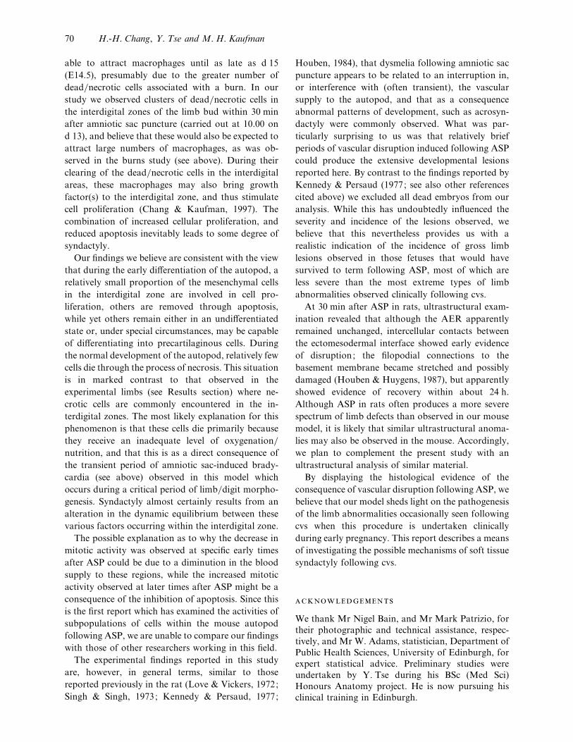
(a)
(b)
(c)
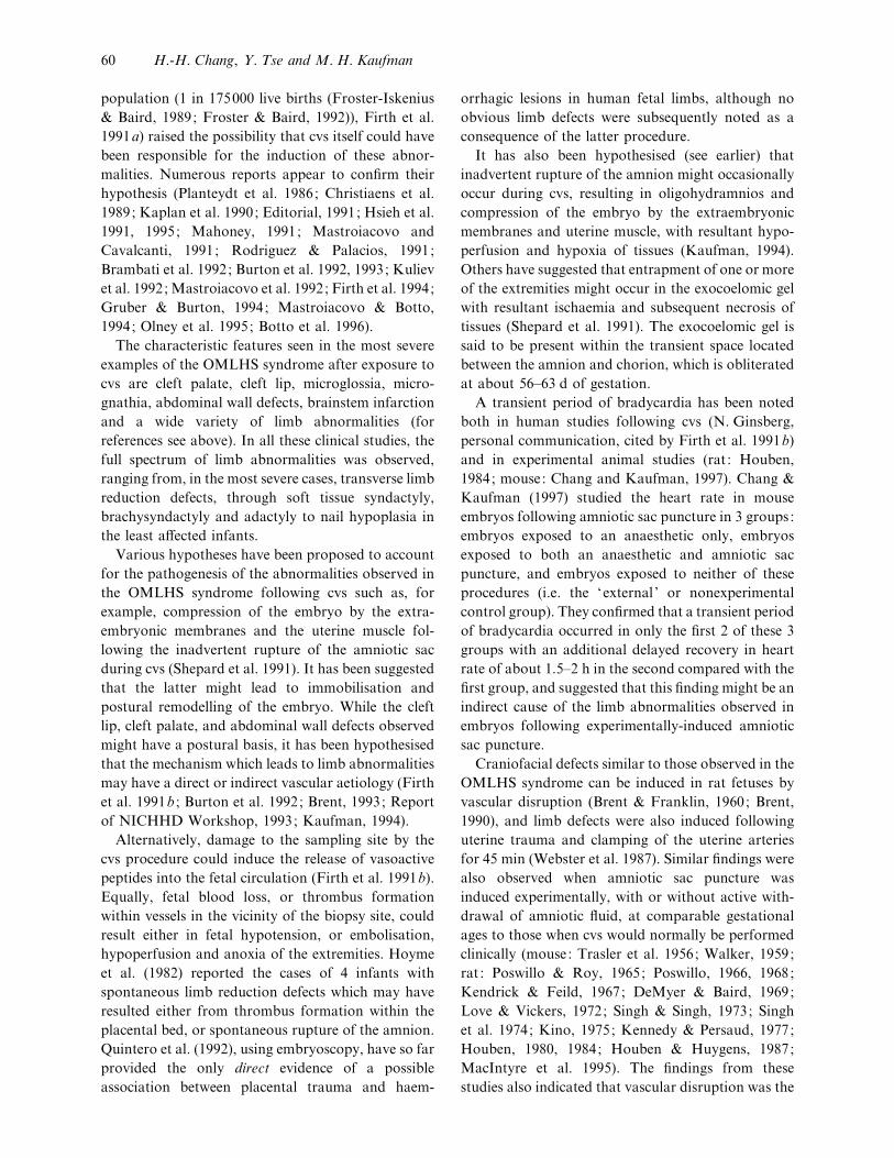
Fig. 1. Representative photomicrographs of interdigital spaces
displaying cells in mitosis (a, arrows), a number of pyknotic cells (b,
arrows) and cells showing the morphological features of apoptosis
(c, arrows). All photographed under ¬100 oil immersion objective
(final magnification ¬1000).
a nominal thickness of 6 µm and stained with Ehrlich’s
haematoxylin and eosin (H&E). The sections were
examined and representative sections photographed
using a Leitz Laborlux K photomicroscope.
While the general incidence of gross limb ab-
normalities reported previously (MacIntyre et al.
1995) was 22% (right forelimb), 20% (left forelimb),
35% (right hindlimb), and 46% (left hindlimb) when
amniotic sac puncture was carried out during the
morning on d 13 p.c., we would expect that the
frequency of abnormalities observed at the cellular
level would be substantially higher. It is for this reason
that we selected the sample sizes for each group
indicated above in the expectation that this would
provide us with one or more samples from each group
which display cellular abnormalities.
The method of analysis of interdigital zones to
determine statistical differences between the various
control and experimental groups was undertaken as
follows. Dividing cells were readily recognised by the
appearance of their mitotic figures (Fig. 1a). A total
of 3 representative histological sections of either
62 H.-H. Chang, Y. Tse and M. H. Kaufman
interdigital spaces II or III were photographed from
each limb. In order to avoid analysing the same cells
on more than one occasion, the 1st, 4th and 7th
sections from a limb that had been serially sectioned
at a nominal thickness of 6 µm were photographed
using the ¬25 objective of a Leitz photomicroscope
and printed to a final magnification of ¬530. Only the
interdigital mesenchyme was analysed; the digital
areas, surface epithelium and the immediately sub-
jacent subepithelial mesenchymal tissues were ex-
cluded from this analysis. The area of interdigital
tissue to be analysed was then measured using a
Kontron image analysis system. Three regions mea-
suring the equivalent of 50 µm¬50 µm, drawn on a
transparent overhead, were randomly placed on the
photomicrographs and the total number of intact
nuclei within each square was counted (see below).
The approach used for the analysis was that formerly
described by Tomasch & Malpass (1958). A knowl-
edge of the number of nuclei scored in the sample
squares and the total areas analysed, provides an
accurate estimate of the total number of nuclei present
in the sample areas analysed. An estimate of the
degree of accuracy of this approach was determined
by manually counting the total number of nuclei in
interdigital spaces from 4 randomly selected limb
samples. In these, the estimated incidence was within
the range 98–106% of the value obtained by manually
counting all of the nuclei present in the total areas
sampled. In addition, a Pearson’s χ# test was then
applied between the observed and the estimated
values. No significant difference was found between
these 2 sets of data, accordingly the estimation method
described above was used. In this study, between
1500–2500 nuclei were scanned for each limb sample
studied. The number of mitotic figures within the total
interdigital area on each of the photomicrographs was
also determined.
To test whether the mitotic indexes in the control
and experimental groups were significantly different,
the log likelihood ratio test and the χ# test (see
Appendix) were employed (W. Adams, personal com-
munication).
Aspects of cellular morphology in the interdigital
zones were examined in this study in order to establish
whether there was (1) evidence of vascular disruption,
and (2) to determine the incidence of cells in mitosis
(i.e. mitotic index), (3) the incidence of apoptotic cells,
and (4) the proportion of dead}necrotic cells. Apop-
totic cells were easily recognised at the light micro-
scopic level based on the descriptions and illustrations
provided in the literature (Saunders et al. 1962;
Glu$ cksmann, 1965; Zakei et al. 1993). To distinguish
between pyknotic cells (Fig. 1b) and apoptotic cells
(Fig. 1c), histological evidence of the features illu-
strated by Hopkinson-Woolley et al. (1994) was
sought. We have also been guided by the account of
necrosis}pyknosis described and illustrated by Burkitt
et al. (1993). As a specific staining technique was not
employed to investigate the incidence of apoptosis,
the occurrence of these cells could not be un-
equivocally determined. However, as such cells were
readily recognised, because of their characteristic
histological morphology, we did not feel able to
undertake a detailed morphometric analysis as under-
taken with regard to the mitotic activity observed in
the digital interspaces (see above), but have instead
provided a descriptive account of their incidence in the
various groups studied. Equally, although the pyk-
notic cells were also readily recognised, because of
their characteristic histological morphology (see Fig-
ure 1b), only a descriptive account is provided of their
incidence in this study.
While appropriate numbers of fore and hindlimbs
from the left side were isolated at each of the time
intervals indicated, technical difficulties were encoun-
tered which precluded the analysis of the 8 h and
24 hASP hindlimb samples. Distortion, principally
in the form of flexion of the distal region of the
footplate in many of the affected limbs meant that it
was often not technically possible to obtain sections of
the required orientation to display the detailed
histological morphology of the digital interspaces.
Because hindlimbs at d 13 of gestation develop about
12 h more slowly than forelimbs (Kaufman, 1992),
forelimbs only will be described unless any substantial
difference between the fore and hindlimbs needs to be
addressed.
Although there is a brief transient episode of
bradycardia observed in the ‘ internal ’ control group
(Chang & Kaufman, 1997), the forelimbs from this
group were found to be histologically indistinguish-
able from those of the ‘external ’ control group.
Accordingly, only the ‘ internal ’ control group is
described below.
Histological observations
Control series. In the 0±5 h control group, the
handplate appears as a pentagon, with precarti-
laginous cells aligned within the digital rays and
undifferentiated mesenchymal cells, associated with
the primitive capillary network, located within the
Interdigital spaces following ASP 63
(a) (b) (c)
(d ) (e) ( f )
(g ) (h) ( i )
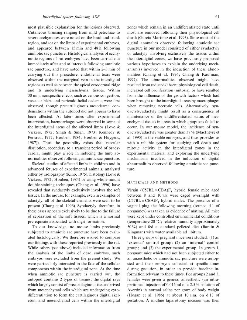
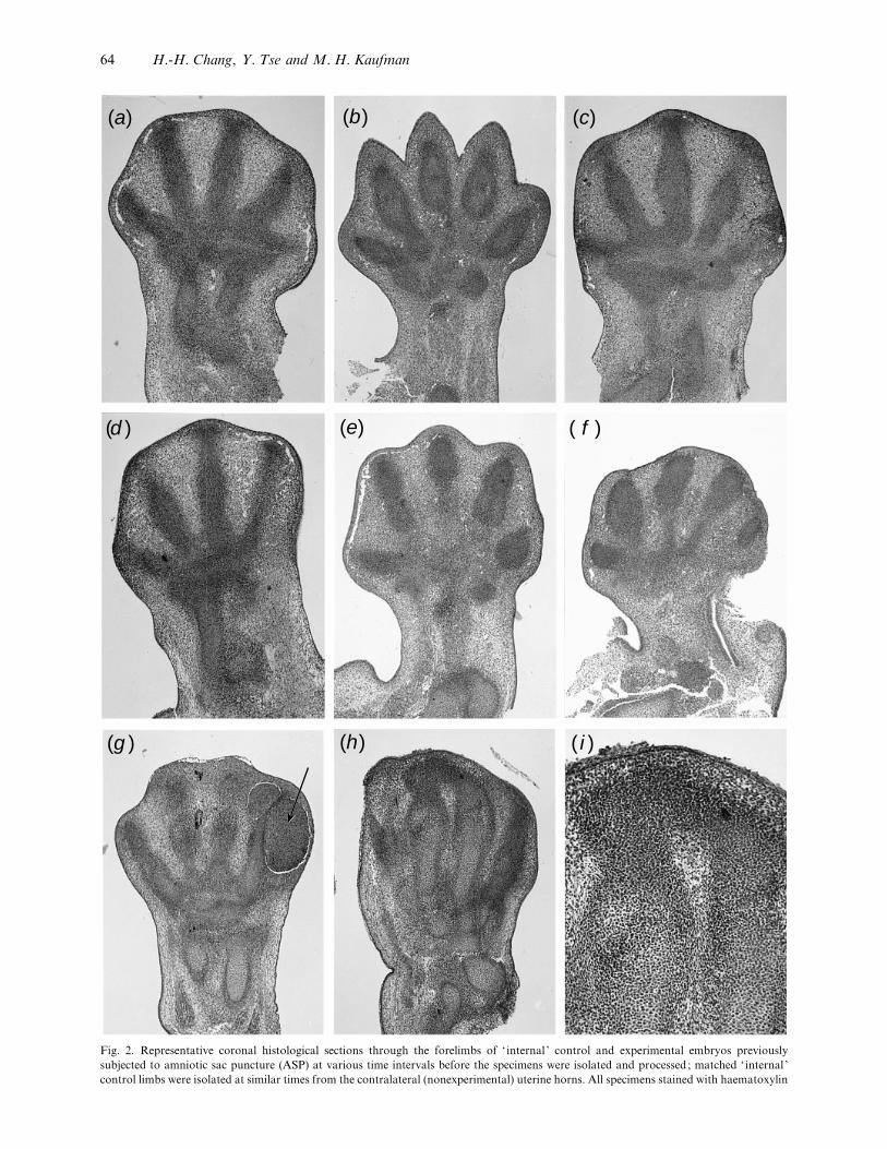
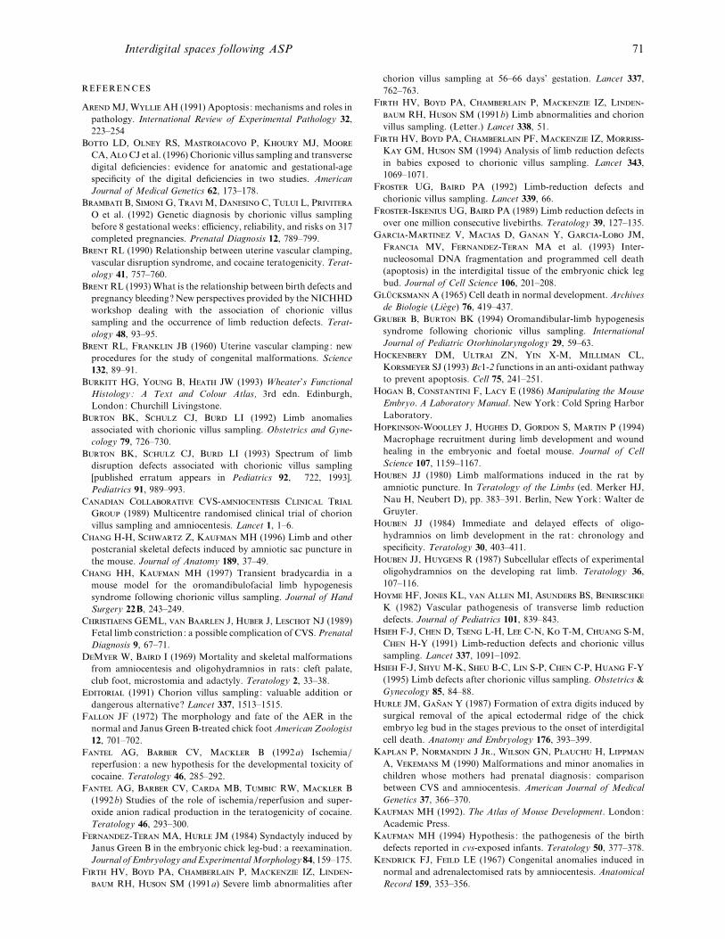
Fig. 2. Representative coronal histological sections through the forelimbs of ‘ internal ’ control and experimental embryos previously
subjected to amniotic sac puncture (ASP) at various time intervals before the specimens were isolated and processed; matched ‘ internal ’
control limbs were isolated at similar times from the contralateral (nonexperimental) uterine horns. All specimens stained with haematoxylin
64 H.-H. Chang, Y. Tse and M. H. Kaufman
interdigital zones. Immediately subjacent to the
surface ectoderm at the distal margin of the handplate,
there are 2–3 layers of compactly arranged mes-
enchymal cells (termed subepithelial mesenchymal
tissue in the following text) beneath which is located
the marginal vein. By contrast, in the footplate, there
are usually 5–6 layers of compactly arranged mes-
enchymal cells in this location. No indication of gross
indentations in the interdigital zones are observed
before about 4 h (Fig. 2a). In the 8–12 h groups, the
precartilaginous cells become increasingly aligned; by
24 h, the interphalangeal joints also show progressive
evidence of differentiation and by 36 h the proximal,
middle, and distal phalanges of digits 2–5, and the
proximal and distal phalanges of digit 1, are recog-
nised. In the interdigital zones, the undifferentiated
mesenchymal cells display no clear evidence of
differentiation until physiological cell death super-
venes. The exact timing of these events varies between
the different interdigital zones, being after about 4 h
within the peripheral area, or after 8–36 h within the
rest of the interdigital spaces. The depth of the
indentation of the interdigital web increases as
progressive cell death occurs (Fig. 2b).
Experimental series. Representative coronal his-
tological sections through the forelimbs of exper-
imental embryos previously subjected to ASP at
30 min (Fig. 2c), 4 h (Fig. 2d ) and 24 h (Fig. 2e, f )
clearly demonstrate that a progressive reduction in the
degree of indentation normally seen in the interdigital
zones of matched control limbs occurs. Equally, by
24 h after ASP, the marginal vein is (abnormally) still
present in the experimental limbs. At this time it is
also possible to recognise gross abnormalities in the
shape of the limb (Fig. 2 f ) and the presence of
extensive areas of haemorrhagic disruption (Fig. 2g).
In the most severely affected of the experimental
limbs an extensive necrotic core often associated with
haemorrhage and gross tissue disruption was charac-
teristically seen which often obliterated the fine
structure of the digital rays and interdigital zones
and eosin. Magnifications: a–h¬40, i¬100. (a) Control limb isolated at 4 h after ASP carried out to embryos in contralateral uterine horn.
Overview of intact limb. (b) Overview of intact control limb isolated at 24 h after ASP carried out to embryos in contralateral uterine horn.
Note the increasing depth of the interdigital zones compared with the situation illustrated previously. The marginal vein is no longer evident
in control limbs at this time. (c) Experimental limb isolated 30 min after ASP. Overview of intact limb. (d ) Experimental limb isolated 4 h
after ASP. Overview of intact limb. (e) Overview of intact experimental limb isolated at 24 h after ASP. Note that the marginal vein is still
present, and that the degree of indentation seen in the interdigital spaces is substantially less than that seen in matched control limbs (see
b). ( f ) Overview of another intact experimental limb isolated at 24 h after ASP. In this example, the gross morphology of the limb bud is
abnormal, and there is little evidence of indentations at the peripheral margins of the interdigital spaces. (g) Overview of intact experimental
limb isolated at 36 h after ASP. Note extensive areas of haemorrhage (arrow) in the lateral part of this limb, and minimal evidence of
indentations in the peripheral parts of the interdigital spaces. (h) Overview, and higher magnification (i) views of hindlimb isolated at 36 h
after ASP, showing an example of a precartilaginous ‘bridge’ between 2 distal phalanges. The latter links the distal parts of 2 of the central
digits and spans the intervening interdigital zone. This would have given rise to a form of acrosyndactyly in which the 2 distal phalanges
would have been joined by a skeletal ‘bridge’.
(Fig. 2g) ; in some instances, minimal evidence of
interdigital indentations are observed. In the majority
of the less affected limbs, the whole autopod appeared
to become compressed, and this usually resulted in the
presence of distorted cartilaginous and, later, skeletal
elements, though the gross structure of the digital rays
was similar to that seen in the matched control limbs.
The proximal-distal dimensions of the autopod of
the experimental limbs were invariably observed to be
smaller than those of developmentally matched
controls ; though this information was not quantified
in the present study, it is planned that this will form
the basis of a subsequent report.
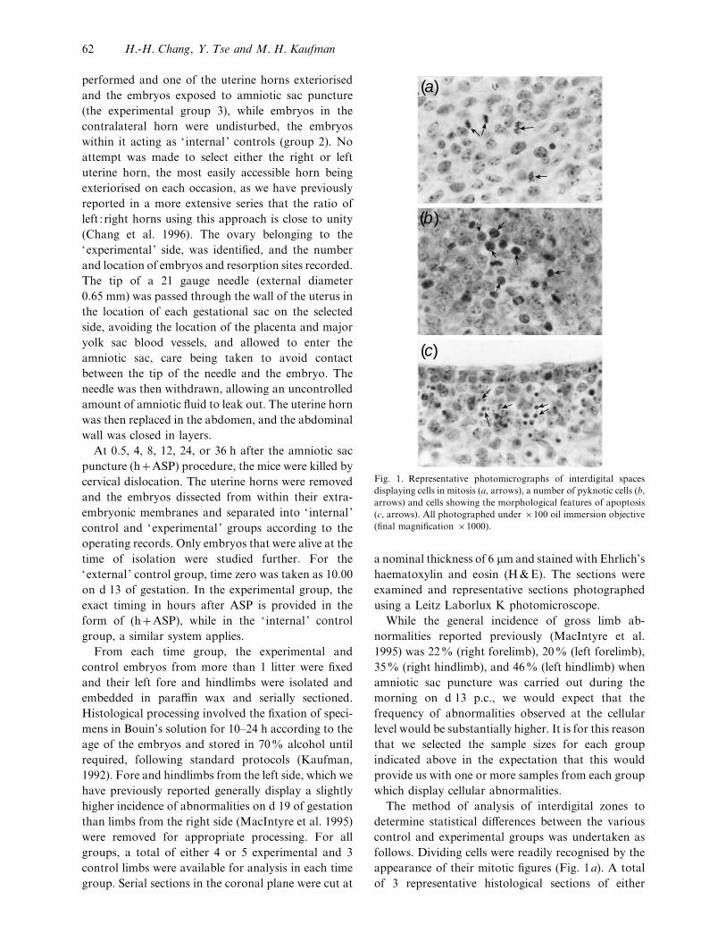
The vascular events
Control series. Within the control limbs, the mar-
ginal vein initially runs along the peripheral margin of
the handplate, being located between the subepithelial
mesenchyme and the rest of the mesenchymal tissue of
the autopod (Fig. 3a, b). As the digits differentiate,
the continuity of the marginal vein gradually becomes
disrupted at the margins of the interdigital zones,
being replaced by about 12 h by a diffuse capillary
network which spreads out within the interdigital
zones. Although the diameter of the marginal vein
remains fairly constant, indirect evidence of an
increased blood supply was noted; the number of
blood cells in the marginal vein in the forelimb in the
4–8 h groups increased by a factor of 2–3 as observed
in the 12 h group. Nucleated primitive red blood cells
are observed within the capillaries, there being no
evidence of vascular congestion in either the marginal
vein or in the capillary network during this period.
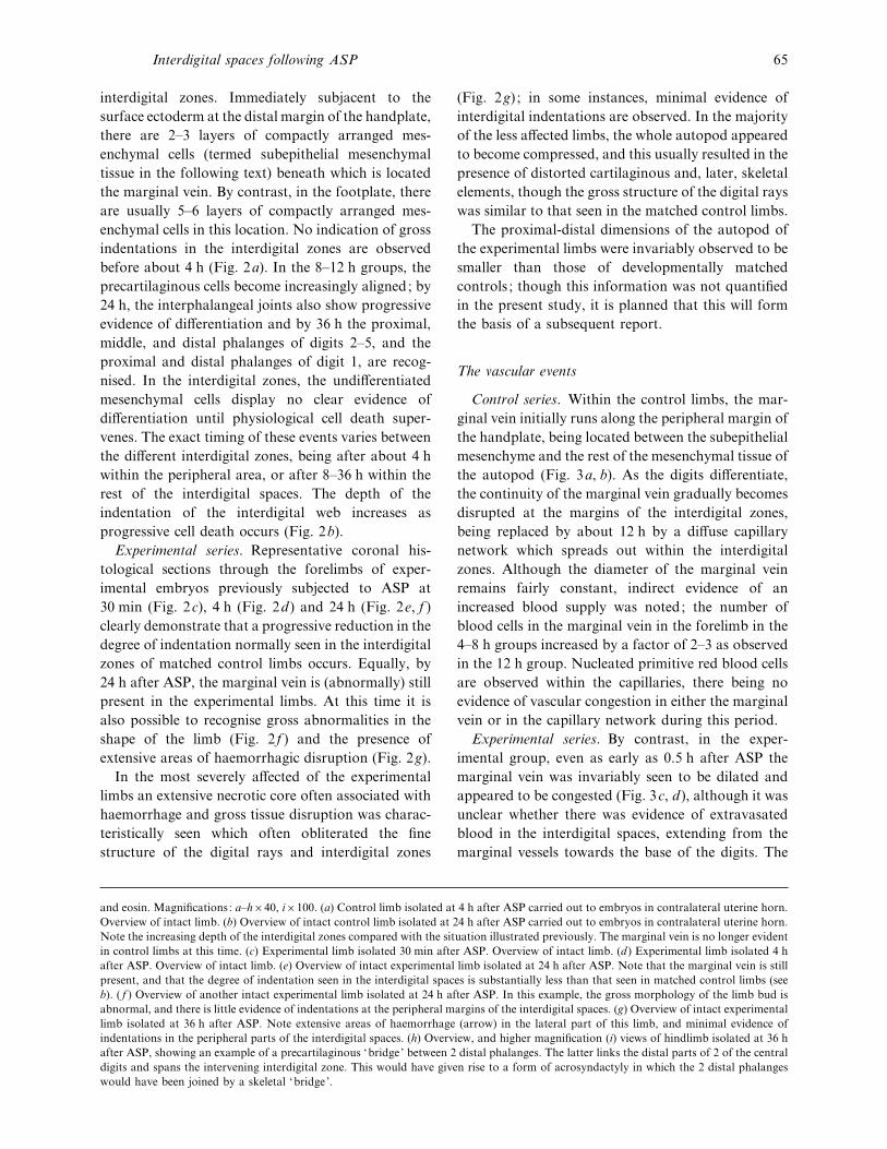
Experimental series. By contrast, in the exper-
imental group, even as early as 0±5 h after ASP the
marginal vein was invariably seen to be dilated and
appeared to be congested (Fig. 3c, d ), although it was
unclear whether there was evidence of extravasated
blood in the interdigital spaces, extending from the
marginal vessels towards the base of the digits. The
Interdigital spaces following ASP 65
(a) (c)
(b) (d)
Fig. 3. Low (a) and higher magnification (b) views of the marginal vein at the peripheral margin of a control and experimental forelimb (c
and d, respectively) following ASP carried out 8 h previously. Note the considerable degree of vascular congestion seen in the marginal vein
in the experimental but not in the control limb. a, c, ¬400; b, d, ¬1000.
fact that it was often difficult to see the endothelial
lining of the primitive capillaries precluded a definitive
judgement on this point in most instances. The
formation of enormous haematomas in the interdigital
spaces appeared to be a late event, as none was found
before ASP36 h, though extensive fluid filled spaces
possibly containing serum were sometimes seen in
these areas between 4–12 hASP.
Physiological cell death (apoptosis)
Apoptotic bodies are characterised by their dense,
pyknotic nuclei (Arend & Wyllie, 1991; also see
figures in the Materials and Methods section). These
are eventually engulfed by monocyte-derived macro-
phages (Hopkinson-Woolley et al. 1994), in the
majority of which apoptotic debris is characteristically
found. While we fully appreciate that specific stains
are now available to demonstrate apoptotic cells
unequivocally, it is our view that such cells are readily
recognised in standard histological sections, because
of their characteristic morphological features. Because
of doubts as to the exact incidence of apoptotic cells
in the interdigital spaces in our conventionally stained
material, this component of our study could not be
quantified, and is therefore based on the qualitative
assessment of our findings.
Control series. During d 13 p.c. in the 4 h, 8 h and
12 h control series when the marginal vein is still
intact, clusters of apoptotic bodies were found distal
to, and closely associated with the vein. During d 14
p.c., in the 24 h and 36 h groups, as massive cell death
occurs in the interdigital space, large numbers of
apoptotic bodies are observed in the interdigital
zones, initially being seen in the distal part of spaces
I and IV, then in spaces II and III. Subsequently, with
the progressive loss of interdigital tissue, these bodies,
while still found close to the interdigital margin,
become more extensively distributed throughout the
basal region of the space.
Experimental series. In the experimental autopods
apoptotic cells may either be completely absent or
dramatically reduced in number compared to those
observed in the corresponding areas of control limbs.
This reduction in the extent of apoptosis seen is first
evident at 4 h after ASP and becomes increasingly
obvious over the next 24–36 h. At 24 h after ASP,
while the control limb possesses extensive evidence of
66 H.-H. Chang, Y. Tse and M. H. Kaufman
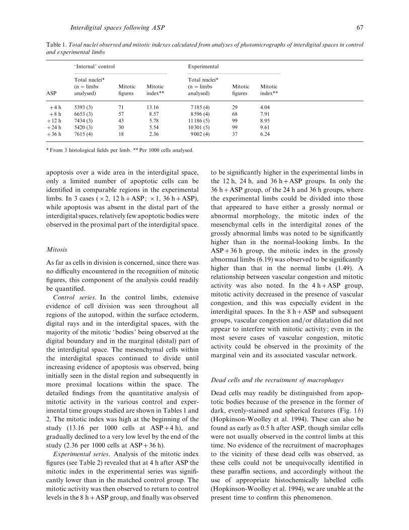
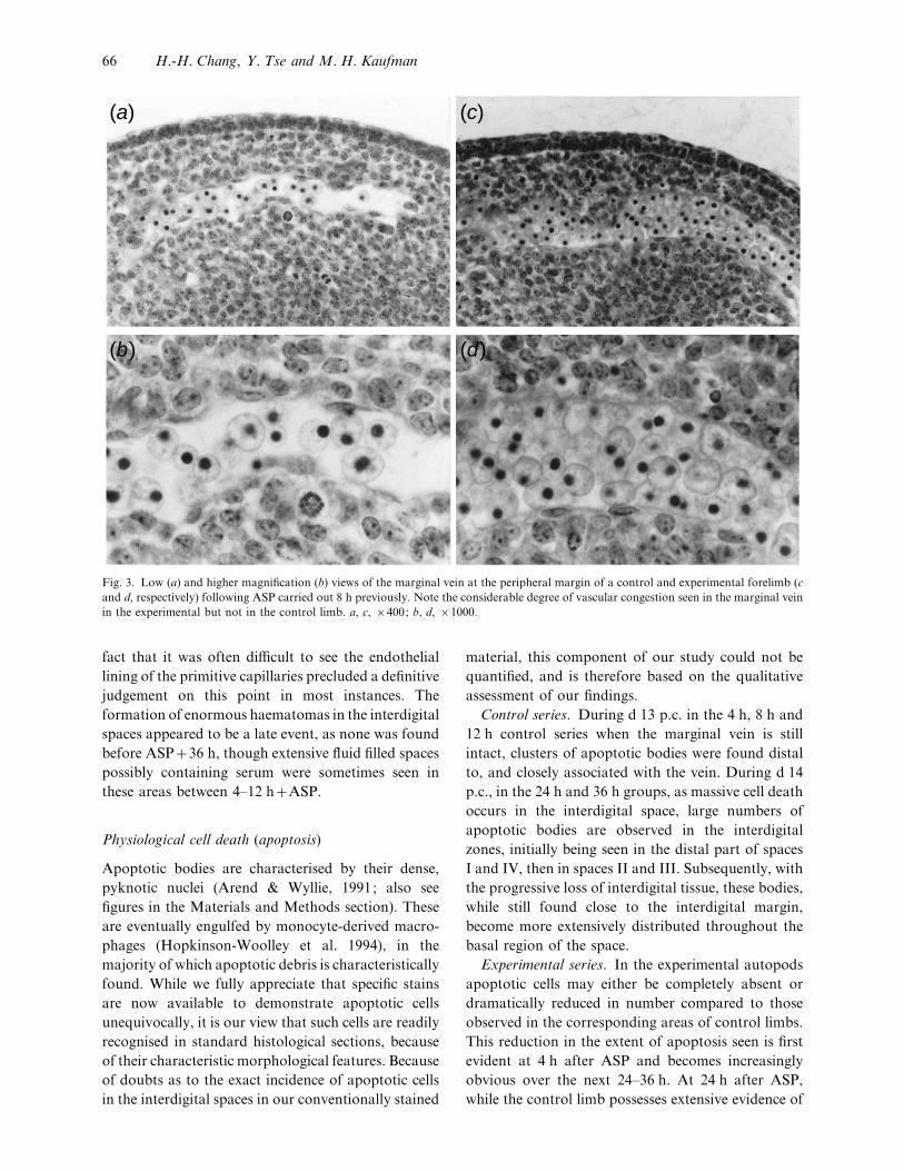
Table 1. Total nuclei observed and mitotic indexes calculated from analyses of photomicrographs of interdigital spaces in control
and experimental limbs
‘ Internal ’ control Experimental
ASP
Total nuclei*
(n¯ limbs
analysed)
Mitotic
figures
Mitotic
index**
Total nuclei*
(n¯ limbs
analysed)
Mitotic
figures
Mitotic
index**
4 h 5393 (3) 71 13±16 7185 (4) 29 4±04
8 h 6653 (3) 57 8±57 8596 (4) 68 7±91
12 h 7434 (3) 43 5±78 11186 (5) 99 8±95
24 h 5420 (3) 30 5±54 10301 (5) 99 9±61
36 h 7615 (4) 18 2±36 9002 (4) 37 6±24
*From 3 histological fields per limb. **Per 1000 cells analysed.
apoptosis over a wide area in the interdigital space,
only a limited number of apoptotic cells can be
identified in comparable regions in the experimental
limbs. In 3 cases (¬2, 12 hASP; ¬1, 36 hASP),
while apoptosis was absent in the distal part of the
interdigital spaces, relatively few apoptotic bodieswere
observed in the proximal part of the interdigital space.
Mitosis
As far as cells in division is concerned, since there was
no difficulty encountered in the recognition of mitotic
figures, this component of the analysis could readily
be quantified.
Control series. In the control limbs, extensive
evidence of cell division was seen throughout all
regions of the autopod, within the surface ectoderm,
digital rays and in the interdigital spaces, with the
majority of the mitotic ‘bodies ’ being observed at the
digital boundary and in the marginal (distal) part of
the interdigital space. The mesenchymal cells within
the interdigital spaces continued to divide until
increasing evidence of apoptosis was observed, being
initially seen in the distal region and subsequently in
more proximal locations within the space. The
detailed findings from the quantitative analysis of
mitotic activity in the various control and exper-
imental time groups studied are shown in Tables 1 and
2. The mitotic index was high at the beginning of the
study (13±16 per 1000 cells at ASP4 h), and
gradually declined to a very low level by the end of the
study (2±36 per 1000 cells at ASP36 h).
Experimental series. Analysis of the mitotic index
figures (see Table 2) revealed that at 4 h after ASP the
mitotic index in the experimental series was signifi-
cantly lower than in the matched control group. The
mitotic activity was then observed to return to control
levels in the 8 hASP group, and finally was observed
to be significantly higher in the experimental limbs in
the 12 h, 24 h, and 36 hASP groups. In only the
36 hASP group, of the 24 h and 36 h groups, where
the experimental limbs could be divided into those
that appeared to have either a grossly normal or
abnormal morphology, the mitotic index of the
mesenchymal cells in the interdigital zones of the
grossly abnormal limbs was noted to be significantly
higher than in the normal-looking limbs. In the
ASP36 h group, the mitotic index in the grossly
abnormal limbs (6±19) was observed to be significantly
higher than that in the normal limbs (1±49). A
relationship between vascular congestion and mitotic
activity was also noted. In the 4 hASP group,
mitotic activity decreased in the presence of vascular
congestion, and this was especially evident in the
interdigital spaces. In the 8 hASP and subsequent
groups, vascular congestion and}or dilatation did not
appear to interfere with mitotic activity ; even in the
most severe cases of vascular congestion, mitotic
activity could be observed in the proximity of the
marginal vein and its associated vascular network.
Dead cells and the recruitment of macrophages
Dead cells may readily be distinguished from apop-
totic bodies because of the presence in the former of
dark, evenly-stained and spherical features (Fig. 1b)
(Hopkinson-Woolley et al. 1994). These can also be
found as early as 0±5 h after ASP, though similar cells
were not usually observed in the control limbs at this
time. No evidence of the recruitment of macrophages
to the vicinity of these dead cells was observed, as
these cells could not be unequivocally identified in
these paraffin sections, and accordingly without the
use of appropriate histochemically labelled cells
(Hopkinson-Woolley et al. 1994), we are unable at the
present time to confirm this phenomenon.
Interdigital spaces following ASP 67
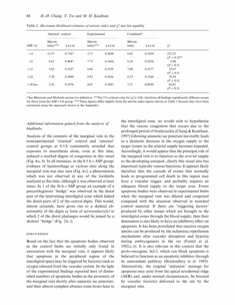
Table 2. Maximum likelihood estimates of mitotic index and χ# test for equality
‘Internal ’ control Experimental Combined*
ASPh
Mitotic
index*** ³...
Mitotic
index*** ³...
Mitotic
index ³... χ#
4 13±27 0±7367 3±71 0±2030 8±42 0±3284 252±32
(P! 0±5)**
8 8±61 0±4847 7±75 0±3364 8±10 0±2934 2±04
(P" 0±5)
12 5±85 0±3187 8±86 0±3559 7±80 0±3177 35±87
(P! 0±5)
24 5±39 0±2909 9±85 0±3826 8±33 0±3360 76±34
(P! 0±5)
36 hrs 2±43 0±1076 4±01 0±1803 3±21 0±0930 62±85
(P! 0±5)
* See Materials and Methods section for definition. **The 5% critical value for χ#
"is 3±86; therefore all findings significantly different except
for those from the ASP8 h group. ***These figures differ slightly from the mitotic index figures shown in Table 1 because they have been
calculated using the approach shown in the Appendix.
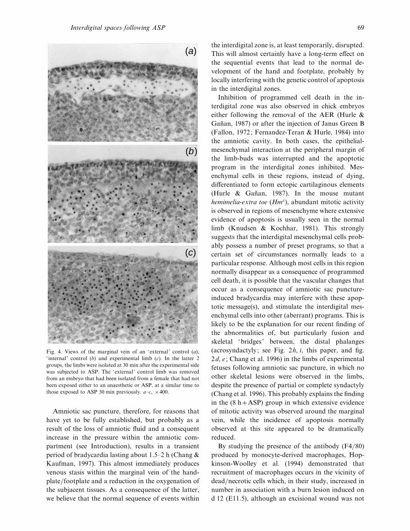
Additional information gained from the analysis of
hindlimbs
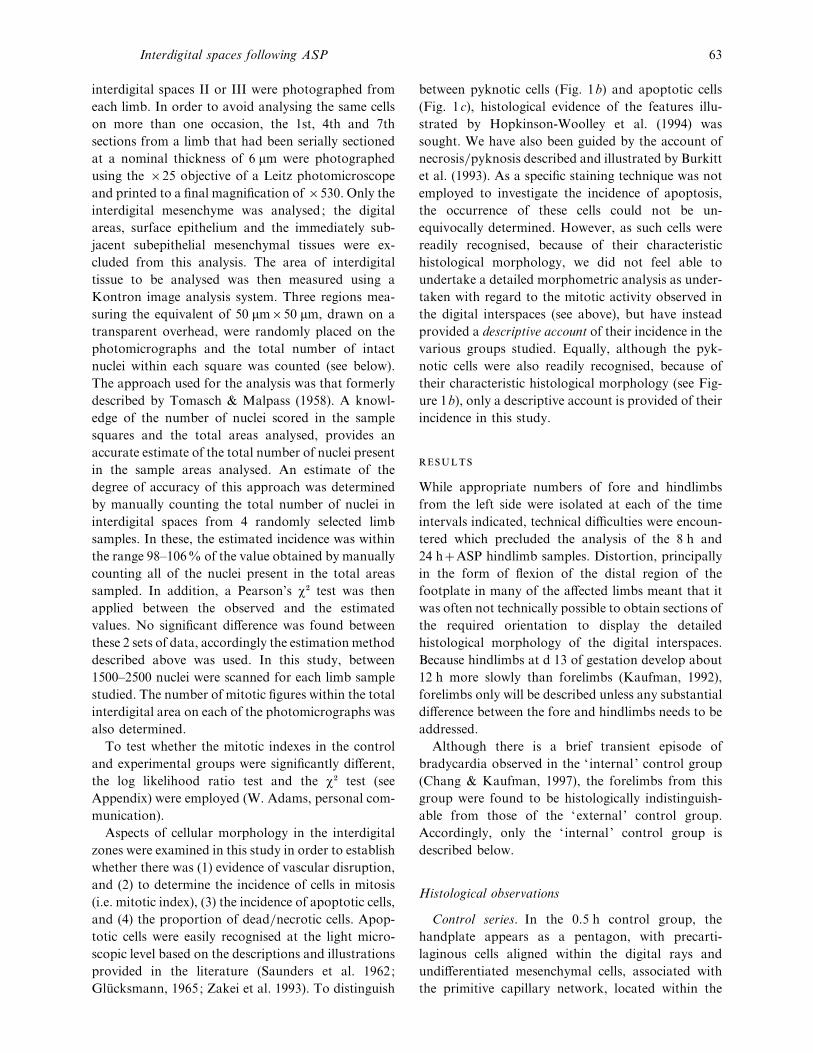
Analysis of the contents of the marginal vein in the
nonexperimental ‘external ’ control and ‘ internal ’
control groups at 0±5 h consistently revealed that
exposure to anaesthesia alone, even at this time,
induced a marked degree of congestion in this vessel
(Fig. 4a, b). In all instances, in the 0±5 hASP group,
evidence of haemorrhage at various sites along the
marginal vein was also seen (Fig. 4c), a phenomenon
which was not observed in any of the forelimbs
analysed at this time, although it was observed at later
times. In 1 of the 36 hASP group an example of a
precartilaginous ‘bridge’ was observed in the distal
part of the intervening interdigital zone which linked
the distal parts of 2 of the central digits. This would,
almost certainly, have given rise to a skeletal ab-
normality of the digits (a form of acrosyndactyly) in
which 2 of the distal phalanges would be joined by a
skeletal ‘bridge’ (Fig. 2h, i).
Based on the fact that the apoptotic bodies observed
in the control limbs are initially only found in
association with the marginal vein, it appears likely
that apoptosis in the peripheral region of the
interdigital space may be triggered by factor(s) such as
oxygen released from the vascular system. In the light
of the experimental findings reported here of dimin-
ished numbers of apoptotic bodies in the proximity of
the marginal vein shortly after amniotic sac puncture,
and their almost complete absence some hours later in
the interdigital zone, we would wish to hypothesise
that the venous congestion that occurs due to the
prolonged period of bradycardia (Chang & Kaufman,
1997) following amniotic sac puncture inevitably leads
to a dramatic decrease in the oxygen supply to the
target tissues as the arterial supply becomes impeded.
Accordingly, it would appear that the principal role of
the marginal vein is to function as the arterial supply
to the developing autopod; clearly this vessel also has
important typically venous functions. It appears likely
therefore that the cascade of events that normally
leads to programmed cell death in this region may
have a vascular trigger, and probably requires an
adequate blood supply to the target area. Fewer
apoptotic bodies were observed in experimental limbs
when the marginal vein was dilated and congested
compared with the situation observed in matched
control material. If there are ‘ triggering factors ’
produced by other tissues which are brought to the
interdigital zones through the blood supply, then their
diminution is also likely to have an inhibitory effect on
apoptosis. It has been postulated that reactive oxygen
species can be produced by the ischaemia}reperfusion
mechanisms after vascular disruption and hypoxia
during embryogenesis in the rat (Fantel et al.
1992a, b). It is also relevant in this context that the
proto-oncogene, bcl-2, which can block apoptosis is
believed to function as an apoptotic inhibitor through
its antioxidant pathway (Hockenbery et al. 1993).
Alternatively, the original ‘ initiation’ message for
apoptosis may arise from the apical ectodermal ridge
(AER) and, under normal circumstances, be boosted
by vascular factor(s) delivered to the site by the
marginal vein.
68 H.-H. Chang, Y. Tse and M. H. Kaufman
(a)
(b)
(c)
Fig. 4. Views of the marginal vein of an ‘external ’ control (a),
‘ internal ’ control (b) and experimental limb (c). In the latter 2
groups, the limbs were isolated at 30 min after the experimental side
was subjected to ASP. The ‘external ’ control limb was removed
from an embryo that had been isolated from a female that had not
been exposed either to an anaesthetic or ASP, at a similar time to
those exposed to ASP 30 min previously. a–c, ¬400.
Amniotic sac puncture, therefore, for reasons that
have yet to be fully established, but probably as a
result of the loss of amniotic fluid and a consequent
increase in the pressure within the amniotic com-
partment (see Introduction), results in a transient
period of bradycardia lasting about 1±5–2 h (Chang &
Kaufman, 1997). This almost immediately produces
venous stasis within the marginal vein of the hand-
plate}footplate and a reduction in the oxygenation of
the subjacent tissues. As a consequence of the latter,
we believe that the normal sequence of events within
the interdigital zone is, at least temporarily, disrupted.
This will almost certainly have a long-term effect on
the sequential events that lead to the normal de-
velopment of the hand and footplate, probably by
locally interfering with the genetic control of apoptosis
in the interdigital zones.
Inhibition of programmed cell death in the in-
terdigital zone was also observed in chick embryos
either following the removal of the AER (Hurle &
Gan4 an, 1987) or after the injection of Janus Green B
(Fallon, 1972; Fernandez-Teran & Hurle, 1984) into
the amniotic cavity. In both cases, the epithelial-
mesenchymal interaction at the peripheral margin of
the limb-buds was interrupted and the apoptotic
program in the interdigital zones inhibited. Mes-
enchymal cells in these regions, instead of dying,
differentiated to form ectopic cartilaginous elements
(Hurle & Gan4 an, 1987). In the mouse mutant
hemimelia-extra toe (Hmx), abundant mitotic activity
is observed in regions of mesenchyme where extensive
evidence of apoptosis is usually seen in the normal
limb (Knudsen & Kochhar, 1981). This strongly
suggests that the interdigital mesenchymal cells prob-
ably possess a number of preset programs, so that a
certain set of circumstances normally leads to a
particular response. Although most cells in this region
normally disappear as a consequence of programmed
cell death, it is possible that the vascular changes that
occur as a consequence of amniotic sac puncture-
induced bradycardia may interfere with these apop-
totic message(s), and stimulate the interdigital mes-
enchymal cells into other (aberrant) programs. This is
likely to be the explanation for our recent finding of
the abnormalities of, but particularly fusion and
skeletal ‘bridges ’ between, the distal phalanges
(acrosyndactyly ; see Fig. 2h, i, this paper, and fig.
2d, e ; Chang et al. 1996) in the limbs of experimental
fetuses following amniotic sac puncture, in which no
other skeletal lesions were observed in the limbs,
despite the presence of partial or complete syndactyly
(Chang et al. 1996). This probably explains the finding
in the (8 hASP) group in which extensive evidence
of mitotic activity was observed around the marginal
vein, while the incidence of apoptosis normally
observed at this site appeared to be dramatically
reduced.
By studying the presence of the antibody (F4}80)
produced by monocyte-derived macrophages, Hop-
kinson-Woolley et al. (1994) demonstrated that
recruitment of macrophages occurs in the vicinity of
dead}necrotic cells which, in their study, increased in
number in association with a burn lesion induced on
d 12 (E11±5), although an excisional wound was not
Interdigital spaces following ASP 69
able to attract macrophages until as late as d 15
(E14±5), presumably due to the greater number of
dead}necrotic cells associated with a burn. In our
study we observed clusters of dead}necrotic cells in
the interdigital zones of the limb bud within 30 min
after amniotic sac puncture (carried out at 10±00 on
d 13), and believe that these would also be expected to
attract large numbers of macrophages, as was ob-
served in the burns study (see above). During their
clearing of the dead}necrotic cells in the interdigital
areas, these macrophages may also bring growth
factor(s) to the interdigital zone, and thus stimulate
cell proliferation (Chang & Kaufman, 1997). The
combination of increased cellular proliferation, and
reduced apoptosis inevitably leads to some degree of
syndactyly.
Our findings we believe are consistent with the view
that during the early differentiation of the autopod, a
relatively small proportion of the mesenchymal cells
in the interdigital zone are involved in cell pro-
liferation, others are removed through apoptosis,
while yet others remain either in an undifferentiated
state or, under special circumstances, may be capable
of differentiating into precartilaginous cells. During
the normal development of the autopod, relatively few
cells die through the process of necrosis. This situation
is in marked contrast to that observed in the
experimental limbs (see Results section) where ne-
crotic cells are commonly encountered in the in-
terdigital zones. The most likely explanation for this
phenomenon is that these cells die primarily because
they receive an inadequate level of oxygenation}nutrition, and that this is as a direct consequence of
the transient period of amniotic sac-induced brady-
cardia (see above) observed in this model which
occurs during a critical period of limb}digit morpho-
genesis. Syndactyly almost certainly results from an
alteration in the dynamic equilibrium between these
various factors occurring within the interdigital zone.
The possible explanation as to why the decrease in
mitotic activity was observed at specific early times
after ASP could be due to a diminution in the blood
supply to these regions, while the increased mitotic
activity observed at later times after ASP might be a
consequence of the inhibition of apoptosis. Since this
is the first report which has examined the activities of
subpopulations of cells within the mouse autopod
following ASP, we are unable to compare our findings
with those of other researchers working in this field.
The experimental findings reported in this study
are, however, in general terms, similar to those
reported previously in the rat (Love & Vickers, 1972;
Singh & Singh, 1973; Kennedy & Persaud, 1977;
Houben, 1984), that dysmelia following amniotic sac
puncture appears to be related to an interruption in,
or interference with (often transient), the vascular
supply to the autopod, and that as a consequence
abnormal patterns of development, such as acrosyn-
dactyly were commonly observed. What was par-
ticularly surprising to us was that relatively brief
periods of vascular disruption induced following ASP
could produce the extensive developmental lesions
reported here. By contrast to the findings reported by
Kennedy & Persaud (1977; see also other references
cited above) we excluded all dead embryos from our
analysis. While this has undoubtedly influenced the
severity and incidence of the lesions observed, we
believe that this nevertheless provides us with a
realistic indication of the incidence of gross limb
lesions observed in those fetuses that would have
survived to term following ASP, most of which are
less severe than the most extreme types of limb
abnormalities observed clinically following cvs.
At 30 min after ASP in rats, ultrastructural exam-
ination revealed that although the AER apparently
remained unchanged, intercellular contacts between
the ectomesodermal interface showed early evidence
of disruption; the filopodial connections to the
basement membrane became stretched and possibly
damaged (Houben & Huygens, 1987), but apparently
showed evidence of recovery within about 24 h.
Although ASP in rats often produces a more severe
spectrum of limb defects than observed in our mouse
model, it is likely that similar ultrastructural anoma-
lies may also be observed in the mouse. Accordingly,
we plan to complement the present study with an
ultrastructural analysis of similar material.
By displaying the histological evidence of the
consequence of vascular disruption following ASP, we
believe that our model sheds light on the pathogenesis
of the limb abnormalities occasionally seen following
cvs when this procedure is undertaken clinically
during early pregnancy. This report describes a means
of investigating the possible mechanisms of soft tissue
syndactyly following cvs.
We thank Mr Nigel Bain, and Mr Mark Patrizio, for
their photographic and technical assistance, respec-
tively, and Mr W. Adams, statistician, Department of
Public Health Sciences, University of Edinburgh, for
expert statistical advice. Preliminary studies were
undertaken by Y. Tse during his BSc (Med Sci)
Honours Anatomy project. He is now pursuing his
clinical training in Edinburgh.
70 H.-H. Chang, Y. Tse and M. H. Kaufman
A MJ, W AH (1991) Apoptosis : mechanisms and roles in
pathology. International Review of Experimental Pathology 32,
223–254
B LD, O RS, M P, K MJ, M
CA, A CJ et al. (1996) Chorionic villus sampling and transverse
digital deficiencies : evidence for anatomic and gestational-age
specificity of the digital deficiencies in two studies. American
Journal of Medical Genetics 62, 173–178.
B B, S G, T M, D C, T L, P
O et al. (1992) Genetic diagnosis by chorionic villus sampling
before 8 gestational weeks: efficiency, reliability, and risks on 317
completed pregnancies. Prenatal Diagnosis 12, 789–799.
B RL (1990) Relationship between uterine vascular clamping,
vascular disruption syndrome, and cocaine teratogenicity. Terat-
ology 41, 757–760.
B RL (1993) What is the relationship between birth defects and
pregnancy bleeding? New perspectives provided by the NICHHD
workshop dealing with the association of chorionic villus
sampling and the occurrence of limb reduction defects. Terat-
ology 48, 93–95.
B RL, F JB (1960) Uterine vascular clamping: new
procedures for the study of congenital malformations. Science
132, 89–91.
B HG, Y B, H JW (1993) Wheater’s Functional
Histology: A Text and Colour Atlas, 3rd edn. Edinburgh,
London: Churchill Livingstone.
B BK, S CJ, B LI (1992) Limb anomalies
associated with chorionic villus sampling. Obstetrics and Gyne-
cology 79, 726–730.
B BK, S CJ, B LI (1993) Spectrum of limb
disruption defects associated with chorionic villus sampling
[published erratum appears in Pediatrics 92, 722, 1993].
Pediatrics 91, 989–993.
C C CVS- C T
G (1989) Multicentre randomised clinical trial of chorion
villus sampling and amniocentesis. Lancet 1, 1–6.
C H-H, S Z, K MH (1996) Limb and other
postcranial skeletal defects induced by amniotic sac puncture in
the mouse. Journal of Anatomy 189, 37–49.
C HH, K MH (1997) Transient bradycardia in a
mouse model for the oromandibulofacial limb hypogenesis
syndrome following chorionic villus sampling. Journal of Hand
Surgery 22B, 243–249.
C GEML, B J, H J, L NJ (1989)
Fetal limb constriction: a possible complication of CVS. Prenatal
Diagnosis 9, 67–71.
DM W, B I (1969) Mortality and skeletal malformations
from amniocentesis and oligohydramnios in rats : cleft palate,
club foot, microstomia and adactyly. Teratology 2, 33–38.
E (1991) Chorion villus sampling: valuable addition or
dangerous alternative? Lancet 337, 1513–1515.
F JF (1972) The morphology and fate of the AER in the
normal and Janus Green B-treated chick foot American Zoologist
12, 701–702.
F AG, B CV, M B (1992a) Ischemia}reperfusion: a new hypothesis for the developmental toxicity of
cocaine. Teratology 46, 285–292.
F AG, B CV, C MB, T RW, M B
(1992b) Studies of the role of ischemia}reperfusion and super-
oxide anion radical production in the teratogenicity of cocaine.
Teratology 46, 293–300.
F-T MA, H JM (1984) Syndactyly induced by
Janus Green B in the embryonic chick leg-bud: a reexamination.
Journal ofEmbryology andExperimentalMorphology 84, 159–175.
F HV, B PA, C P, M IZ, L-
RH, H SM (1991a) Severe limb abnormalities after
chorion villus sampling at 56–66 days’ gestation. Lancet 337,
762–763.
F HV, B PA, C P, M IZ, L-
RH, H SM (1991b) Limb abnormalities and chorion
villus sampling. (Letter.) Lancet 338, 51.
F HV, B PA, C PF, M IZ, M-
K GM, H SM (1994) Analysis of limb reduction defects
in babies exposed to chorionic villus sampling. Lancet 343,
1069–1071.
F UG, B PA (1992) Limb-reduction defects and
chorionic villus sampling. Lancet 339, 66.
F-I UG, B PA (1989) Limb reduction defects in
over one million consecutive livebirths. Teratology 39, 127–135.
G-M V, M D, G Y, G-L JM,
F MV, F-T MA et al. (1993) Inter-
nucleosomal DNA fragmentation and programmed cell death
(apoptosis) in the interdigital tissue of the embryonic chick leg
bud. Journal of Cell Science 106, 201–208.
G$ A (1965) Cell death in normal development. Archives
de Biologie (Lie[ ge) 76, 419–437.
G B, B BK (1994) Oromandibular-limb hypogenesis
syndrome following chorionic villus sampling. International
Journal of Pediatric Otorhinolaryngology 29, 59–63.
H DM, U ZN, Y X-M, M CL,
K SJ (1993) Bc1-2 functions in an anti-oxidant pathway
to prevent apoptosis. Cell 75, 241–251.
H B, C F, L E (1986) Manipulating the Mouse
Embryo. A Laboratory Manual. New York: Cold Spring Harbor
Laboratory.
H-W J, H D, G S, M P (1994)
Macrophage recruitment during limb development and wound
healing in the embryonic and foetal mouse. Journal of Cell
Science 107, 1159–1167.
H JJ (1980) Limb malformations induced in the rat by
amniotic puncture. In Teratology of the Limbs (ed. Merker HJ,
Nau H, Neubert D), pp. 383–391. Berlin, New York: Walter de
Gruyter.
H JJ (1984) Immediate and delayed effects of oligo-
hydramnios on limb development in the rat : chronology and
specificity. Teratology 30, 403–411.
H JJ, H R (1987) Subcellular effects of experimental
oligohydramnios on the developing rat limb. Teratology 36,
107–116.
H HF, J KL, A MI, A BS, B
K (1982) Vascular pathogenesis of transverse limb reduction
defects. Journal of Pediatrics 101, 839–843.
H F-J, C D, T L-H, L C-N, K T-M, C S-M,
C H-Y (1991) Limb-reduction defects and chorionic villus
sampling. Lancet 337, 1091–1092.
H F-J, S M-K, S B-C, L S-P, C C-P, H F-Y
(1995) Limb defects after chorionic villus sampling. Obstetrics &
Gynecology 85, 84–88.
H JM, G4 Y (1987) Formation of extra digits induced by
surgical removal of the apical ectodermal ridge of the chick
embryo leg bud in the stages previous to the onset of interdigital
cell death. Anatomy and Embryology 176, 393–399.
K P, N J J., W GN, P H, L
A, V M (1990) Malformations and minor anomalies in
children whose mothers had prenatal diagnosis : comparison
between CVS and amniocentesis. American Journal of Medical
Genetics 37, 366–370.
K MH (1992). The Atlas of Mouse Development. London:
Academic Press.
K MH (1994) Hypothesis : the pathogenesis of the birth
defects reported in cvs-exposed infants. Teratology 50, 377–378.
K FJ, F LE (1967) Congenital anomalies induced in
normal and adrenalectomised rats by amniocentesis. Anatomical
Record 159, 353–356.
Interdigital spaces following ASP 71

K LA, P TVN (1977) Pathogenesis of developmental
defects induced in the rat by amniotic sac puncture. Acta
Anatomica 97, 23–35.
K Y (1975) Clinical and experimental studies of the congenital
constriction band syndrome, with an emphasis on its etiology.
Journal of Bone and Joint Surgery 57A, 636–643.
K TB, K DM (1981) The role of morphogenetic cell
death during abnormal limb-bud outgrowth in mice heterozygous
for the dominant mutation Hemimelia-extra toe (Hmx). Journal
of Embryology and Experimental Morphology 65 (Suppl.),
289–307.
K AM, M B, J L (1992) Limb abnormalities and
chorionic villus sampling. Lancet 340, 668.
L AM, V TH (1972) Amniocentesis dysmelia in rats.
British Journal of Experimental Pathology 53, 435–444.
MI DJ, C HH, K MH (1995) Teratogenic
effects of amniotic sac puncture: a mouse model. Journal of
Anatomy 186, 527–539.
M MJ (1991) Limb abnormalities and chorionic villus
sampling. Lancet 337, 1422–1423.
M P, C DP (1991) Limb-reduction defects
and chorion villus sampling. Lancet 337, 1091.
M P, B LK, C DP, L F,
S A, T AE et al. (1992) Limb anomalies following
chorionic villus sampling: a registry based case-control study.
American Journal of Medical Genetics 44, 856–864.
M P, B LD (1994) Chorionic villus sampling and
transverse limb deficiencies : maternal age is not a confounder.
American Journal of Medical Genetics 53, 1–8.
MRC W P E C V
S (1991) Medical Research Council European Trial of
Chorion Villus Sampling. Lancet 337, 1491–1499.
O RS, K MJ, A CJ, C P, E LD, F
TJ et al. (1995) Increased risk for transverse digital deficiency
after chorionic villus sampling: results of the United States
multistate case-control study, 1988–1992. Teratology 51, 20–29.
P HT, V MJ, V H (1986) Amniotic
bands and malformation in child born after pregnancy screened
by chorionic villus biopsy. Lancet ii, 756–757.
P D (1966) Observations of fetal posture and caudal
mechanisms of congenital deformity of palate, mandible, and
limbs. Journal of Dental Research 45, 584–596.
P D (1968) The aetiology and surgery of cleft palate with
micrognathia. Annals of the Royal College of Surgeons of England
43, 61–88.
P D, R LJ (1965) The pathogenesis of cleft palate. British
Journal of Surgery 52, 902–913.
Q RA, R R, M MJ, V M, H J,
H JC (1992) Fetal haemorrhagic lesions after chorionic
villus sampling [letter]. Lancet i, 193.
R NICHHD W (1993) Report of National
Institute of Child Health and Human Development workshop on
chorionic villus sampling and limb and other defects, October 20,
1992. Teratology 48, 7–13.
R GG, J LG, S SE, C FF,
D RJ, G MS et al. (1989) The safety and efficacy of
chorionic villus sampling for early prenatal diagnosis of cyto-
genetic abnormalities. New England Journal of Medicine 320,
609–617.
R JI, P J (1991) Pathogenetic mechanisms of fetal
akinesia deformation sequence and oligohydramnios sequence.
American Journal of Medical Genetics 40, 284–289.
S JW J., G MT, S LC (1962) Cell death
in morphogenesis of the avian wing. Developmental Biology 5,
147–178.
S TH, K RP, F AG (1991) Limb-reduction defects
and chorion villus sampling [letter]. Lancet 337, 1092.
S S, S G (1973) Haemorrhages in the limbs of fetal rats
after amniocentesis and their role in limb malformations.
Teratology 8, 11–18.
S S, M MM, S G (1974) Congenital anomalies in
rat foetuses induced by amniocentesis. Indian Journal of Medical
Research 62, 394–401.
T J, M AJ (1958) The human motor trigeminal
nucleus. Anatomical Record 130, 91–102.
T DG, W BE, F FC (1956) Congenital mal-
formations produced by amniotic-sac puncture. Science 124, 439.
W BE (1959) Effects on palate development of mechanical
interference with the fetal environment. Science 130, 981.
W WS, L AH, B-W PDC (1987) Uterine
trauma and limb defects. Teratology 35, 253–260.
Z ZF, Q D, L T, L RA (1993) Delayed
internucleosomal DNA fragmentation in programmed cell death.
FASEB Journal 7, 470–478.
Statistical methods for evaluation of mitotic indexes
The mitotic index (I ) is defined as follows:
I¯m¬1000
s¬r(1)
where m is mitosis and s a multiplication factor; r is
the mean cell count.
This equation is rewritten as follows:
ri¯
ki
Iwhere k
i¯
mi¬1000
si
. (2)
Thus the index is a function of the mean cell counts.
Cell counts are distributed as a Poisson distribution
given by
Pr[Ri¯ r
i]¯
µriie−µ
i
ri!
where for this case µi¯
ki
I.
The best estimate of I is the maximum likelihood
estimator Iq which is given by
Iq ¯Σk
i
Σ ri
. (3)
The standard error (s.e.) of Iq is
s.e. (Iq )¯I $
#
o(Σki). (4)
To test whether 2 indexes are equal we use the log
likelihood ratio test :
log lik ratio¯ (Σ r",i
Σ r#,i
) log Iq$®(Σ r
",i) log Iq
"
®(Σ r#,i
) log Iq#
(5)
where Iq$¯
Σk",i
Σk#,i
Σ r",i
Σ r#,i
the combined group index derived from (3).
It is known that 2¬ the log likelihood ratio is
distributed as χ# with for this case, 1 degree of
freedom.
72 H.-H. Chang, Y. Tse and M. H. Kaufman