Embed Size (px)
Citation preview

ANALYSIS OF HEPATITIS DELTA VIRUS RNA STRUCTURE: EFFECTS ON RNA-PROTEIN INTERACTIONS
AND VIRAL REPLICATION
A Dissertation submitted to the Faculty of the
Graduate School of Arts and Sciences of Georgetown University
in partial fulfillment of the requirements for the degree of
Doctor of Philosophy in Microbiology and Immunology
By
Dawn Angela Defenbaugh, B.S.
Washington, DC December 5, 2008

ii
ANALYSIS OF HEPATITIS DELTA VIRUS RNA STRUCTURE AND THE EFFECT ON RNA-PROTEIN INTERACTION AND VIRAL REPLICATION
Dawn Angela Defenbaugh, B.S.
Thesis Advisor: John L. Casey, Ph.D.
ABSTRACT
Hepatitis delta virus (HDV) is a unique human pathogen whose RNA structures
are critical to viral function. The genome is a circular single-stranded RNA with high
levels of intramolecular complementarity (~ 74%) leading to formation of the unbranched
rod structure characteristic of HDV RNA. The complementary antigenome, which has
similar structural features, encodes the open reading frame from which mRNAs of the
two forms of HDV protein are transcribed: the short form of the delta antigen (HDAg-S)
is absolutely required for initiation and maintenance of viral replication, and the long
form (HDAg-L) is necessary and sufficient for virus particle production in cells
expressing hepatitis B surface antigens (HBsAg). HDV is considered a subviral satellite
of hepatitis B virus (HBV) due to its dependence on HBsAg. In fact, HDV also relies on
host functions to complete its replication cycle. Exploitation of cellular functions may be
directly related to the structure of the ribonucleoprotein complex (RNP) formed through
interaction of HDV RNA and viral proteins. This dissertation analyzes the effects of
HDV RNA structure on RNA-protein interactions and viral replication. The first half
examines the direct biochemical interaction between native bacterially expressed HDAg
and in vitro transcribed HDV RNA. These data identify features of the RNA structure

iii
that are critical to binding by HDAg. Through electrophoretic mobility shift assay, it was
discovered that HDV RNAs containing at least 311 total nucleotides (nt) were stably
bound while those with fewer than 298 nt were not bound. This feature was shown to be
biologically relevant in transfected cells. Furthermore, micrococcal nuclease digestion
indicates the size of the RNP protected is the same as that formed by the smallest RNA
bound by HDAg. The second half of this dissertation focuses on effects of RNA structure
on viral function specifically, replication. Mutational analysis, specifically designed to
preserve the predicted RNA structure of the interior of the unbranched rod structure,
indicates sensitivity to nucleotide deletion. Deletions of various sizes (totaling 6 – 53 nt)
in 6 different unrelated segments showed reduced viral replication. These results, and
others, suggest most of the HDV RNA structure is pivotal to RNP formation and viral
function.

iv
ACKNOWLEDGEMENTS
Like many of us, I owe most of what I am to my parents. Only the good parts –
the bad, is my own undoing. The youngest daughter of Bobbie and Jim Defenbaugh, who
liked school the least, couldn’t figure out how to get out – until now. By extension, much
thanks goes to my sisters – always there with a good meal and a good story. I have plenty
of friends that have supported this effort. A huge thanks to Stella North and Kimberly
Batty, without whom, I may have never started the program and an even bigger thanks to
Clare Thibodeaux without whom, I may have never finished. I have to thank all the
people that have come through the Casey lab over the years but none was a better
colleague than Sarah Linnstaedt. She was a great labmate, stylist, paparazzo, biscotti-
maker, and DJ – and she will always be a great friend.
I’ve always said, “I’d rather be lucky than smart.” Hands down, the luckiest thing
that happened during my time here at Georgetown was stumbling upon Dr Casey. Having
completed three lab rotations and not falling in love with any of them, I met with Dr
Casey. I immediately liked him. He agreed to let me do a fourth rotation and my
comprehensive exam with him and I the rest is history. Never once did I want to be
somewhere else, and never once did I wish he was something different than he was. He
leads by example, encouraging you to be a better scientist. Not pushy, not judgmental,
never disappointed (at least, not that he let on). You could present him with the most
pitiful data and he’d find the good in it. Science in the Casey lab is rarely hypothesis-
driven, it’s wager-driven – you take a position on the outcome of an experiment, he

v
commonly takes the other side (just to make things interesting), and the loser pays in
cookies. It’s a wildly successful technique. He has fostered my healthy scientific
skepticism even if he’s mostly a Pollyanna. He taught me to be critical of data – your
data, my data, even published data. He taught me how to think.
No list of thanks is complete without including my husband, Eddie Czarnetzky. It
is no exaggeration to say that this would not have been possible without him. ‘Thank
you’ doesn’t begin to say how grateful I am for your love and support – usually, I don’t
deserve it.

vi
TABLE OF CONTENTS
Introduction …………………………………………………………………… 1
The HDV Genome …………………………………………………… 3
The Delta Antigens ……………………………………………………. 7
HDV Replication Cycle ……………………………………………….. 12
Experimental Goals …………………………………………………… 19
Hepatitis Delta Antigen Binds the HDV Unbranched Rod Structure with
High Affinity and Requires a Minimum Length RNA for Stable Binding …… 23
Abstract ……………………………………………………………….. 24
Introduction …………………………………………………………… 25
Methods ……………………………………………………………… 27
Results ………………………………………………………………. 34
Discussion …………………………………………………………….. 56
Acknowledgements …………………………………………………… 61
Addendum …………………………………………………………….. 61
HDV RNA Replication is Sensitive to Small Deletions at
Numerous Locations Along the Unbranched Rod Structure ………………… 65
Abstract ……………………………………………………………….. 66
Introduction …………………………………………………………… 67
Methods ……………………………………………………………… 70
Results ………………………………………………………………. 72
Discussion …………………………………………………………….. 91
Acknowledgements …………………………………………………… 95
Addendum …………………………………………………………….. 95
Perspectives …………………………………………………………………….. 103
References Cited ……………………………………………………………… 113

1
INTRODUCTION
Hepatitis delta virus (HDV) was discovered in 1977. At the time, HDV infections
were thought to be a severe form of liver disease caused by a variant of hepatitis B virus
(HBV) that expressed the ‘delta antigen’. In 1980, the delta antigen was identified as a
component of a separate pathogen. HDV particles isolated from infected patients were
capable of eliciting infection in chimpanzees chronically infected with HBV while
chimpanzees without HBV were unable to establish HDV infection. In 1983, hepatitis
delta virus was established as the causative agent of this severe liver disease and in 1986,
the HDV genome was sequenced. HDV is characterized as a subviral human pathogen
that increases the severity of liver disease in patients coinfected with HBV.
HDV is considered a ‘satellite’ of HBV. Without HBV infection, HDV is
incapable of producing infectious virus particles. For this reason, it is said to be a
‘defective’ virus or even a ‘subviral pathogen’ suggesting that it isn’t a virus at all. In this
relationship, HBV is termed the ‘helper’ virus. In cells coinfected with HBV, HDV
ribonucleoprotein complexes (RNPs) bud from cellular membranes enveloped by
hepatitis B surface antigen (HBsAg), which provides both viruses means to target and
invade cells. Only one other satellite-helper virus relationship is known in humans:
adeno-associated virus (AAV), a member of the Parvoviridae family, is only replicated in
cells coinfected with adenovirus. AAV is non-pathogenic in humans, initiates a low
immune response, and the single-stranded DNA genome can incorporate into host

2
chromosomes. For these reasons, a substantial body of research has been aimed at
developing AAV as a vector for gene therapy. AAV shares little/no similarity to HDV.
HDV is a blood-borne pathogen. HDV infection requires that HBV is already
present (superinfection) in cells of HBV carriers, or that HBV is delivered simultaneously
(coinfection) to naïve cells. Coinfection is usually self-limiting. The severe liver disease
that occurs due to HDV infection is commonly due to superinfection. According to the
Centers for Disease Control and Prevention, 350 million people are infected with HBV
worldwide and as many as 10 – 20% are thought to also be infected with HDV. More
than 70% will develop chronic type D hepatitis and as many as 20% of cases are fatal.
Widespread use of an effective HBV vaccine has reduced HBV infection from as high as
20% in some endemic areas to < 2%. By extension, this vaccine has also led to a
welcome reduction in HDV infection.
Liver disease caused by HDV is not the only reason for studying this virus. As a
result of the HBV vaccine, reasons for continued analysis of HDV are directed at the
contribution to basic science and may be broadly applicable. For example, HDV has the
smallest genome known to be pathogenic in man. Within this small genome, a single
open reading frame encodes the two forms of viral protein required for critical viral
function in the cell. Interestingly, neither of these proteins is an RNA-dependent RNA
Polymerase (RdRP). In fact, HDV is the only known RNA virus infecting animals that
does not encode an RdRP. It has been shown that the HDV RNA genome is replicated by
host cell RNA polymerase II (RNA pol II), a DNA-dependent RNA Polymerase. Lastly,

3
structure analysis has identified post-transcriptional processing, such as ribozyme and
editing activity, that is directly related to RNA structure. These unique features
distinguish HDV as an interesting RNA model and represent the inspiration of continued
HDV research.
The HDV Genome
At 1679 nucleotides (nt), HDV has the smallest genome of any known animal
virus. The circular HDV RNA is technically single-stranded however, high levels of
intramolecular complementarity (~ 74%) collapse the unit-length transcripts into the
unbranched rod structure characteristic of HDV RNA (56, 113) (Fig 1.1). Infectious
virions deliver genomic RNA complexed with viral protein to susceptible cells. Viral
replication of the genome yields a complementary antigenome upon which the open
reading frame for the delta protein is encoded; thus, HDV is a negative-strand RNA virus.
Both the genome and the antigenome are self-cleaved by internal ribozymes.

4
Figure 1.1. Diagram of HDV unbranched rod structure. The HDAg coding sequence is
indicated by the open rectangle; the asterisk denotes the location of the amber/W RNA
editing site at nt 1012; the location of the antigenomic ribozyme site is indicated by a
triangle. Vertical lines represent partial base pairing between the two sides of the
unbranched rod. The predicted secondary structure (mfold) of the antigenomic RNA is
shown below the schematic (shaded rectangle), with sequence numbering for the
corresponding positions in the genome RNA shown. Watson-Crick base pairs are
indicated by vertical lines between the nucleotides; unconventional base pairs are
indicated by black dots between the nucleotides; unpaired nucleotides are shown above
and below paired bases; the five-membered loop on the left is shown unpaired
(5'–ACAGA–3'). The location of the AUG for the methionine start codon of the mRNA is
indicated.

5

6
At least eight HDV genotypes have been identified. The vast majority of HDV
research focuses on two, genotypes I and III. Genotype I is the most common among
infected patients in the United States and Europe (9) while genotype III is associated with
the most severe form of disease (12, 15). Genotype I RNA is known to form only one
main structure, the unbranched rod, while genotype III may form many complex
branched structures (11, 69). Genotype I is ideally suited to the study of very small
changes in RNA sequence and structure. Accordingly, all of the work in this dissertation
investigates HDV genotype I.
Post-transcriptional processing
The structure of HDV RNA is directly related to many functions of the virus life
cycle. Genomic and antigenomic ribozymes, for example, fold into very defined
structures (91, 117) during RNA replication that allows for cis-cleavage at precise
nucleotides (103). Discovery of the crystal structure has launched extensive research and
manipulation of the pseudoknot-like RNA (38). The self-cleavage and subsequent
ligation of HDV RNAs are critical to HDV function. Single point mutations at the
cleavage site result in complete elimination of viral replication (74).
A second post-transcriptional event allows the single open reading frame to
encode for the two forms of HDV protein. During replication, a population of the full-
length complementary antigenome undergoes adenosine deamination by host ADAR1
within the amber stop codon of the open reading frame. The result is conversion of the

7
stop codon to a codon for tryptophan and, ultimately, translation of a second, longer
protein. The efficiency with which ADAR1 recognizes the RNA and catalyzes
deamination is directly related to the structure in and around the editing site.
Delta Antigens
From the early days of its discovery, the protein encoded by HDV is still referred
to as the delta antigen (HDAg). As mentioned, RNA editing allows the single open
reading frame to encode for two forms of HDAg. The 24 kDa short form of the delta
antigen (HDAg-S) is 195 amino acids (aa) and the 27 kDa long form (HDAg-L) is 214 aa
(Fig. 1.2). The first 195 aa are common to both HDAg-S and HDAg-L and the proteins
share common functional domains. For example, near the amino-terminus, an extended
coiled coil domain shared by both proteins allows for dimerization and multimerization
of HDAg monomers. The high resolution crystal structure of this region (aa 12 – 60) has
been solved and predicts anti-parallel dimerization as well as the possible formation of an
octamer in which the dimers interact near the ends to form a hole in the center (122). This
interaction is required for viral function (31). For instance, oligomerization of HDAg-S is
required for activation of HDV replication (119) and HDAg-L dimerization mutants do
not efficiently package HDV particles (89). Furthermore, the two proteins contain a
bipartite nuclear localization signal (aa 68 – 88) to allow for nuclear import of HDAg
(18) as well as HDV RNPs (30). Both proteins have two arginine-rich motifs (ARMs) (aa
97 – 107 and aa 136 – 146) separated by a helix-loop-helix motif that participate in RNA

8
binding and are required for replication initiation by HDAg-S (64). The 50 aa C-terminal
to the ARMs (aa 145 – 195) are known to be highly proline/glycine-rich but no specific
function has yet been identified. This region has recently been shown to interact with
RNA pol II however, the nature of the residues may contribute to non-specific protein
interactions (120). Lastly, RNA editing extends the 3` end of the mRNA transcript to
encode for 19 additional amino acids at the C-terminus of HDAg-L. This region is known
to contain a prenylation site.
The two forms of HDAg have been discovered to have distinctly different roles in
the HDV life cycle. Briefly, HDAg-S is absolutely required for initiation and
maintenance of viral replication (21). HDV RNA defective for synthesis of HDAg is
replicated in cells expressing HDAg-S indicating that HDAg can act in trans (55).
HDAg-S alone is sufficient for replication while expression of HDAg-L alone yields no
HDV RNA replication (21). In fact, expression of both proteins indicates an ability of
HDAg-L to inhibit replication under certain experimental conditions (21, 72, 101). More
importantly, HDAg-L is known to be necessary and sufficient for viral assembly in the
presence of HBsAg while HDAg-S plays a minimal role in packaging (58).

9
Figure 1.2. Diagrams of HDAg-S and HDAg-L. Dimerization and oligomerization
domains included within aa 12-60 are shaded in light gray and indicate the residues of the
solved high-resolution crystal structure. The bi-partite nuclear localization signal (NLS)
is indicated in medium gray within aa 68-88. Two arginine-rich motifs (ARM) (in dark
gray) are spaced by a helix-loop-helix motif (horizontal line). The amber editing site at nt
1012 is indicated by an asterisk and the C-terminal 19 aa of HDAg-L are indicated in
black. Sites of post-translational modifications are identified. P, phosphorylation; M,
methylation; A, acetylation; F, farnesylation.

10

11
Post-translational modification
Both forms of HDAg have been shown to be post-translationally modified. A few
of these modifications play a direct role in their function. For example, without post-
translational modification, HDAg-L does not facilitate packaging or inhibit replication.
Farnesylation of HDAg-L has been shown to occur at Cysteine-211 within the
isoprenylation motif CaXX (C, cysteine; a, aliphatic residue; X, any amino acid) (41, 49).
This modification is required for direct interaction between HDAg-L and HBsAg (48)
and disruption of this interaction results in elimination of packaged virions (41).
However, isoprenylation alone is not sufficient for virus assembly. Lee et al. established
that almost all of the C-terminal 19 aa of HDAg-L are required for packaging (62).
Additionally, loss of prenylation has been correlated with a reduction in the inhibition by
HDAg-L on viral replication (47).
Both forms of HDAg have been shown to be phosphorylated (17). HDAg-S is
phosphorylated on both serine and threonine residues while HDAg-L appears to only be
phosphorylated on serines (86). Furthermore, phosphorylation of HDAg-S on Serine-177
is required for efficient viral replication (26, 109). Mutational analysis changing the
serine to alanine (S177A) indicates no genomic RNA synthesis from the antigenomic
template by Northern blot analysis (84). Interestingly, phosphorylation of HDAg-L
appears to be affected by disruption of the prenylation motif suggesting a requirement
that farnesylation occurs prior to phosphorylation (4).

12
Both forms of HDAg have been shown to be acetylated in vivo. Acetylation of
Lysine-72 has been shown to affect nuclear transport and possibly, viral replication (85,
109). Point mutation to arginine (K72A) resulted in inefficient nuclear transport of both
HDAgs contributing to dramatic reduction in viral replication.
HDAg-S is methylated at Arginine-13 and this modification is important for viral
replication (109). Expression of an mRNA containing a mutation that translates an
alanine rather than an arginine at aa 13 results in a HDAg-S unable to establish
replication of genomic RNA from a wild type antigenomic template. The same effect on
replication occurs when the mutation is expressed by the replicating template and the
mRNA is wild type. Interestingly, effects of this mutation are only seen when replicating
genomic RNA from an antigenomic template; synthesis of antigenomic RNA from a
genomic template appears relatively unaffected by unmethylated HDAg-S (67).
HDV Replication Cycle
Natural infection of HDV likely begins through unknown receptor-ligand
interactions of HBsAg and human or non-human primate hepatocytes. Unlike HBV,
replicating HDV RNA is only found in liver tissues (88) whereas, HBV has been found to
replicate in many cell types (76). The HDV RNP is delivered to the nucleus where viral
replication of HDV RNA proceeds via a double rolling circle mechanism (7, 57, 73, 108).
The schematic in Figure 1.3 shows the HDV RNA replication cycle. HDAg-S facilitates
replication of the infecting genomic RNA (indicated by a 1) by host RNA pol II (a DNA-

13
dependent RNA polymerase) (16, 39, 65, 80, 83). Replication synthesizes a
complementary linear RNA that is self-cleaved by the HDV ribozyme and ligated to form
the circular antigenomic RNA (50, 51, 59, 66, 74, 96). From this antigenome, many
copies of genomic HDV RNA are transcribed. During viral replication, the ratio of
antigenomic to genomic RNA is approximately 1:10 (27). Early in the cycle, HDAg-S
mRNA is transcribed from the genomic RNA and exported to the cytoplasm. This mRNA
is capped and polyadenylated like host mRNAs and is translated by host ribosomes.
Later, editing of a fraction of the antigenomic RNA results in transcription of an edited
genome which replicates to an edited antigenome. Again, the edited genomic RNA serves
as the template for mRNA transcription leading to HDAg-L synthesis.

14
Figure 1.3. Schematic of HDV RNA replication. Early in the cycle, infectious genomic
RNA (indicated by a 1) is replicated (filled arrows) to a complementary antigenomic
copy and transcribed to the mRNA for translation of HDAg-S. Late in the cycle, some of
the antigenome will be edited (open arrow) from adenosine to inosine at nt 1012.
Replication leads to a second genomic RNA which is replicated to an antigenomic copy
and transcribed to the mRNA for HDAg-L.

15

16
No practical cell culture system is available for infection by HDV. Only primary
hepatocytes of chimpanzees and woodchucks have been demonstrated to be infected by
HDV in cell culture (29, 105). However, HDV replication is supported by a number of
cell types and a few techniques have been demonstrated to establish replication in cell
culture. The method that most closely mimics the natural route of infection is by
transfection of HDV virions or the RNPs of virions (5). A more common method
involves transfection of in vitro transcribed RNAs. In this case, HDAg-S must be
supplied independently; the transfected cells can be transiently or stably expressing
HDAg-S (40), purified or partially purified recombinant protein can be pre-mixed with
the RNA creating an RNP prior to transfection (35), or an in vitro transcribed, capped and
polyadenylated HDAg-S mRNA can be cotransfected (79). RNA transfections are
appropriate for studying HDV cycle control. For example, HDAg-L has been shown to
inhibit replication only when the protein is expressed abnormally early in the replication
cycle (72). In a natural infection, HDAg-L may have little regulatory control of
replication. A third transfection method involves transfection of two HDV cDNA
expression plasmids, one expressing an HDV RNA defective for HDAg synthesis and
one expressing the HDAg mRNA. Expression of HDAg in trans allows for significant
manipulation of the HDV RNA transcript without affecting the open reading frame for
translation of wild type protein. This project is focused on the early stages of the HDV
replication cycle when HDV RNA and HDAg-S are thought to be simultaneously

17
synthesized. In this case, DNA transfections represent an appropriate and effective
method of HDV expression in cell culture.
Comparison of HDV to Closely Related Viruses
HDV shares little in common with most animal viruses however, the HDV
genome shares many characteristics with the genomes of viroids, infectious RNAs of
plants, and viroid-like satellite RNAs. Like HDV, viroids and satellite RNAs contain
single-stranded circular RNA genomes that collapse into rod-like structures (71). For
each, replication by a host DNA-dependent RNA polymerase occurs by a rolling circle
mechanism in the nuclei of infected cells (7, 33, 73, 106). Additionally, a few viroids use
ribozymes that catalyze cis-cleavage of their RNA (33, 45, 46, 70, 87). However, HDV is
the only human pathogen known to require a catalytic ribozyme for viability (38) and
ribozyme domains of viroids form remarkably different structures than those of the HDV
ribozymes. Viroids are replicated autonomously (34) however, replication of viroid-like
satellite RNAs requires coinfection by specific helper viruses. HDV relies on its helper,
HBV, for encapsidation, not viral replication. In fact, encapsidation is a common
characteristic of viroid-like satellite RNAs but viroids are not encapsidated. HDV and
satellite RNAs share little or no sequence homology with their specific helper viruses
(98). In contrast to HDV, viroids and viroid-like satellite RNAs do not encode protein
which may explain why the HDV genome is 3 – 4 times bigger than viroid genomes (<
400 nt); HDV may have a larger genome in order to accommodate the HDAg open

18
reading frame. Although no sequence homology has been elucidated between HDV and
plant viroids or viroid-like satellite RNAs, the similarities are striking.
The most closely related, although not genetically, negative-strand RNA virus that
infects mammals might be the influenza viruses, members of the Orthomyxoviridae
family. The linear single-stranded RNA of influenza viruses is at least 7 times bigger than
that of HDV and shares no known sequence homology to HDV. However, the genome is
divided into eight segments, at least four of which are comparable in size (or smaller) to
HDV RNA. While these RNA segments are linear, short sequences (<15 nt) common to
both the 5' and 3' ends are partially complementary allowing for hairpin formation;
similarities can be drawn if HDV RNA is considered to be a long hairpin ligated to form
a closed circle. Within the 12 – 15,000 nt of Influenza A, for example, eight genes encode
for at least 11 proteins; at least four are structural proteins, three make up the RNA
polymerase, and one is the nucleoprotein that binds single-stranded RNA. HDV relies on
a helper virus protein (HBsAg) for targeting of susceptible cells and host polymerase (at
least RNA pol II) functions for transcribing viral mRNA and infectious genomes. The
nucleoprotein (NP) of influenza viruses exhibits characteristics somewhat similar to
HDAg. NP is known to bind cooperatively to the phosphate backbone of single-stranded
RNA in a non-sequence specific manner with high affinity (Kd = ~ 20 nM) (1, 121). NP
also has protein binding properties. It is known to form NP–NP oligomers as well as bind
both viral polymerases and one of the viral matrix proteins. Binding of NP to influenza
RNA during transcription is required for high processivity of the polymerase but NP is

19
dispensable for initiation of transcription. Lastly, NP is responsible for targeting
influenza RNPs to the nucleus. These are all similar contributions as those made by
HDAg in the HDV life cycle.
Experimental Goals
It has become increasingly apparent that the structure of the HDV RNA is critical
to viral function. Not surprisingly, HDV ribozymes are quite intolerant of small base
changes. Research shows that any mutation at C75 (nt 760) of the genomic ribozyme
eliminates enzymatic activity (54, 92, 107). Interestingly, enzymatic activity is
completely unaffected by sequences upstream of the break site (between nt 685 and nt
686) making the HDV ribozyme a useful tool for precise cleavage of non-HDV RNAs (3,
52, 104, 121). RNA structure also plays a critical role in HDV RNA editing. Subtle single
base changes around the editing site can dramatically impact the efficiency with which
ADAR1 recognizes and deaminates the specific adenosine at nt 1012 (69). In fact, HDV
may have evolved imperfect editing substrates as a means of replication control; editing
sites in which the target adenosine is too easily identified, bound and deaminated by
ADAR1, might progress the virus replication cycle into the packaging phase long before
transcription of new RNAs was complete. In contrast to stringent RNA sequence and
structure requirements for enzymatic activity, packaging of virus particles by HDAg-L
appears to be fairly flexible. Significant disruption of the unbranched rod structure seems
to have little effect on virion production in the presence of HBsAg. In fact, a linear HDV

20
hairpin of the ribozyme end containing only 311 nt of the unbranched rod was efficiently
packaged and secreted from cells expressing HDAg-L and HBsAg (19).
Binding of HDAg to these HDV RNA structures has been shown to influence all
of these processes (28, 50, 51, 110). However, analysis of the direct relationship between
HDV RNA and HDAg has been limited. Interpretation of early in vitro analysis was
complicated by the use of large fusion partners to facilitate protein purification; fusion
proteins were abundant in the insoluble fraction requiring denaturation and renaturation.
More recently, Dingle et al. showed that replication is initiated in cells transfected with
full-length in vitro transcribed HDV RNA complexed with purified bacterially-expressed
amino-terminally His-tagged HDAg (35) (Dingle 1998). The nature of these RNPs was
not fully explored. This dissertation begins with in vitro analysis of the direct interaction
between HDV RNA and HDV protein. We hypothesized that HDAg is specific for the
unbranched rod structure of HDV RNA and critical binding features can be characterized.
Development of an electrophoretic mobility shift assay facilitated quantitative analysis of
the relationship between HDV RNA and HDAg. Moreover, investigation aimed at
addressing this hypothesis led to some predictions of the size and nature of the protein
complex bound.
The replication of HDV RNA is a complicated process and a great deal of effort
has been focused on understanding the specifics. Data have been presented regarding
sequence and structure requirements of the HDV unbranched rod. Some mutational
analysis has been shown either without specific intention to maintain the rod structure or

21
with specific intention to disrupt the rod. For example, beyond simply forming an
unbranched rod RNA, replication has been shown to be sensitive to insertion of
nucleotides on one side of the rod structure. Insertion of 8 to 12 nt in many locations to
one side of the rod eliminated viral replication in transfected cells (111). Using an RNA
folding algorithm (mfold), these insertions were predicted to add local stem-loop
structures to the mostly undisturbed unbranched rod. Recently, similar effects on
replication have been shown in mutant RNAs with large one-sided insertions (< 1,000
nt). Interestingly, serial RNA transfection resulted in excision of the non-HDV sequences
and replication could be detected due to recovery of the rod structure, in some cases
reverting to wild type HDV sequence (44).
RNA mutations in which the unbranched rod structure has been carefully
preserved have also been analyzed. RNA truncations from the left end of the rod that
maintain ribozyme and ligation activity to form circularized unbranched rod structures
are not replicated in transfected cells (58). Analysis of the stem loops of either end of the
rod indicated discordant sensitivity to insertion or deletion. The right end of the rod
appears fairly tolerant – a 16 nt deletion indicated replication levels ~ 40% of wild type.
Alternatively, the left end is acutely sensitive – a 1 bp deletion virtually eliminated
replication (118). The sensitivity at the left end of the rod may be a result of mRNA
transcription initiation in this region resulting in stringent structural requirements (43,
79). RNA sequence or structure requirements for the interior of the rod have not been
examined using methods in which the unbranched rod structure has been maintained. The

22
second aim of this dissertation examines the HDV RNA structure as it relates to viral
replication. A cellular replication assay allowed for characterization of specific RNA
sequences and structures within the interior of the HDV unbranched rod where the
overall rod structure is preserved. Furthermore, examination of these RNAs using the in
vitro electrophoretic mobility shift assay can provide some insight into the role of HDAg-
S in the replication cycle.

23
HEPATITIS DELTA ANTIGEN BINDS THE
HDV UNBRANCHED ROD STRUCTURE WITH HIGH AFFINITY AND
REQUIRES A MINIMUM LENGTH RNA FOR STABLE BINDING
Dawn A. Defenbaugh, Matthew Johnson, Renxiang Chen, Ying Yi Zheng and John L.
Casey*
Department of Microbiology and Immunology, Georgetown University Medical Center,
Washington, DC 20007
*Corresponding author
Department of Microbiology and Immunology
Georgetown University Medical Center
3900 Reservoir Road, NW
Washington, DC 20007
Phone: (202) 687-1052
Fax: (202) 687-1800
email: [email protected]

24
ABSTRACT
Hepatitis delta virus (HDV) is a subviral pathogen that increases the severity of
liver disease caused by hepatitis B virus. In both the small circular RNA genome and its
complement, the antigenome, about 70% of the nucleotides base-pair to form a
characteristic unbranched rod structure. These RNAs are associated with the sole virally
encoded protein, hepatitis delta antigen (HDAg), in infected cells and, for the genome, in
virus particles. RNA-protein interactions are critical to viral function; however, the nature
of the ribonucleoprotein complexes (RNPs) is not well understood. Previous in vitro
analyses have indicated that HDAg specifically binds the unbranched rod structure;
however these studies were limited by the use of large fusion partners and methods
requiring denaturation of the purified protein (23, 68). Analyses of binding using native,
bacterially expressed HDAg have been complicated by a lack of specificity for HDV
RNA(35). Here, we show that native, bacterially expressed HDAg-160, from which the
C-terminal 35 amino acids of HDAg have been removed, binds a segment of the HDV
unbranched rod RNA with high affinity (apparent Kd = 2.5 nM). In an electrophoretic
mobility shift assay, this protein produced a discrete, micrococcal nuclease-resistant
complex with a ~ 400 nt segment of HDV unbranched rod RNA. Binding was specific for
the unbranched rod structure and occurred with several segments of HDV RNA, although
with varying affinities and efficiencies. Analysis of the effects of deleting segments of the
unbranched rod indicated that binding did not require one or two specific binding sites
within the RNA. Rather, a minimum length HDV RNA unbranched rod, approximately

25
311 nt, was essential for complex formation. The results are consistent with a model in
which HDAg binds HDV unbranched rod RNA as multimers of fixed size rather than as
individual subunits.
INTRODUCTION
Hepatitis delta virus (HDV) is a unique human pathogen that increases the
severity of liver disease in those infected with its helper virus, hepatitis B virus. HDV
RNA replication is unique among known animal viruses in that it uses the DNA-
dependent RNA polymerase activity of the host to replicate its genome without a DNA
intermediate. The negative-stranded HDV genome is a single-stranded circular RNA that
collapses to form a characteristic unbranched rod structure due to ~ 70% Watson-Crick
base pairing. Genome replication involves a double rolling circle mechanism in which the
antigenome, with a similar rod structure, serves as a replication intermediate (7, 57, 73,
108). Arranged along one side of the unbranched rod structure is the single HDV open
reading frame, which encodes the hepatitis delta antigen, HDAg.
HDAg plays several critical functions in the HDV replication cycle: it has pivotal
roles in both RNA replication and packaging (21, 58). Central to these functions are the
ability of HDAg to form multimeric complexes and to bind HDV RNA (28, 31, 51, 89,
110, 119). In infected cells and in HDV particles, both the genome and antigenome are
associated with HDAg. However, the nature of the ribonucleoprotein (RNP) complex
formed is not fully understood. For example, attempts to determine the relative amounts

26
of HDAg and HDV RNA complexed in RNPs in viral particles and in cells have
produced ratios ranging from 22 to 200 (42, 99).
Previous in vitro investigations of the interaction between HDAg and HDV RNA
indicated that HDAg could specifically bind several segments of the HDV RNA
unbranched rod (23, 68). However, these studies used HDAg fusion proteins that were
denatured prior to analysis of RNA binding; thus, these analyses could not provide
quantitative binding data. Moreover, the particular binding determinants in the RNA have
not yet been identified. More recent studies have demonstrated that native bacterially
expressed HDAg can form complexes with HDV RNA in vitro and that these complexes
can initiate HDV replication when transfected into cultured cells (35). However, the
structure of the complexes formed has not been fully addressed and it is not clear whether
HDAg expressed in E. coli binds HDV RNA specifically.
Here, we show that bacterially expressed, native HDAg forms heterogeneous
complexes, possibly aggregates, with HDV RNAs as well as non-HDV RNAs. However,
these obstacles to analysis of binding were overcome by using a truncated version of
HDAg from which the C-terminal 35 amino acids of HDAg had been removed. In an
electrophoretic mobility shift assay, this protein produced a discrete, micrococcal
nuclease-resistant complex with a ~ 400 nt segment of HDV unbranched rod RNA.
Binding was observed with several different segments of the unbranched rod with
variable affinities, but no binding was observed with non-HDV RNAs. Deletional
mapping indicated that binding did not require a particular site or a limited set of sites

27
within the RNA; rather, a minimum length HDV RNA unbranched rod, approximately
311 nt, was essential for complex formation. Our results are consistent with a model in
which HDAg binds HDV RNA as multimer of fixed size rather than as individual
subunits.
MATERIALS AND METHODS
Plasmids. In vitro transcription reactions used PCR products amplified from one
of three plasmids as transcription templates. pCMV-DC1×1.2-AgS(–) is an antigen-
defective expression plasmid in which the HDAg reading frame is disrupted by a single
base insertion at codon 7. pCMV-DC1-NRΔApaI+LeftLoop is a non-replicating
expression plasmid in which the ApaI fragment has been deleted and remaining bases of
the ApaI site are replaced with the sequence of the HDV loop at the left end (opposite the
ribozyme). The gel purified and digested PCR product was ligated into the NheI/PstI site
of the M34 DC1ΔApaI Xba M30 vector.
Cloning of 395L and 311L expression plasmids began by annealing two
oligonucleotides containing the antigenomic HDV ribozyme (equal amounts in 10 mM
Tris, pH 7.5, 50 mM NaCl, 1 mM EDTA). This annealed insert was ligated into the
SacI/BamHI site of the digested pCMV-DC1×1.2-AgS(–) vector resulting in the empty
vector control, pCMV-Ribo. Insert fragments for 395L and 311L were PCR amplified,
digested and ligated into the SacI/NotI-digested pCMV-Ribo vector to make pCMV-
Ribo-395L and pCMV-Ribo-311L.

28
In vitro transcription templates and T7 transcription. 395L, 207L, 311L, and
253L were transcribed from templates amplified from pCMV-DC1×1.2-AgS(–). 395L
includes the left 395 nt from nt 153 (5') to 1441 (3'). 207L includes nt 59 (5') to 1536 (3').
311L includes nt 111 (5') to 1484 (3'). And 253L includes nt 81 (5') to 1511 (3').
The template used to make 395R, 316R and 298R requires that the left loop of the
HDV unbranched rod be moved to the opposite end of the transcript. The 395R template
was amplified using two-piece PCR. The first round generated two PCR products from
pCMV-DC1×1.2-AgS(–) plasmid. The second round of PCR used equal amounts of these
two gel purified PCR products as templates with the outermost primers and then gel
purified again. This second round product was used as the transcription template for
395R which includes nt 1643 (5') to 153, ACAGA loop, nt 1441 to 1637 (3'). A third
round of PCR amplification resulted in the templates for 316R and 298R. 316R includes
nt 3 (5') to 153, ACAGA loop, nt 1441 to 1597 (3') and 298R includes nt 13 (5') to 153,
ACAGA loop, nt 1441 to 1589 (3').
390NL and 229NL were each made by annealing two transcripts. Templates were
amplified from pCMV-DC1×1.2-AgS(–). The two products for 390NL were amplified to
transcribe nt 153 to 1643 and nt 1637 to 1441, and the products for 229NL were
amplified to transcribe nt 111 to 3 and nt 1597 to 1484. In vitro RNA transcripts were
purified by native polyacrylamide gel electrophoresis. Equimolar amounts were annealed
by overnight incubation at 45°C in 40 mM PIPES (pH 6.7,) 400 mM NaCl, 1 mM DTT
after denaturation at 75°C for 5 minutes. Annealed RNAs were gel purified again.

29
The template for 207L+207M transcription was generated using three-piece PCR.
The first round amplified three products from pCMV-DC1×1.2-AgS(–). After gel
purification, these three products acted as template for the second round using the
outermost primers. This second round product was used for T7 transcription.
207L+207M includes nt 314 (5') to 213, nt 59 to 1536, and nt 1379 to 1283 (3').
The transcription templates for 384M, g384M and 207M were amplified from the
pCMV-DC1-NRΔApaI+LeftLoop plasmid. 384M and g384M include nt 401 (5') to 213,
ACAGA loop, nt 1379 to 1193 (3') while 207M includes nt 314 (5') to 213, ACAGA
loop, nt 1379 to 1283 (3'). 405E includes nt 680 (5') to 482 (at the SmaI site), nt 1109 to
907 (3').
Transcription reactions included RNA Polymerase Buffer (New England
Biolabs), 10 mM DTT, 1 U/µl RNase Inhibitor (Applied Biosystems), 500 µM ATP, 500
µM GTP, 500 µM UTP, 32 µM CTP (Invitrogen), DNA template, 0.8 µCi/µl α32P-CTP
(Perkin Elmer), 2.5 U/µl T7 RNA Polymerase (New England Biolabs). Reactions were
incubated at 37°C no less than 1 h then chilled on ice no less than 1 h. Native transcripts
were resolved on 6% native polyacrylamide gel (BioRad) before Amicon Separations
column purification with Ultrafree-MC PVDF 0.22 µm and Ultracel YM-30 30,000
MWCO.

30
Table 2.1. Amplification primers for RNA transcription.
395L RNA – PCR Template: pCMVDC1-1.2×Ag(-)
MiniD1F – TAATACGACTCACTATAGGGTTCTCCGGCGTTGTGGGGAT MiniD1R – TTCCCCAGCCAGGGATTTTC
311L RNA – PCR Template: pCMVDC1-1.2×Ag(-)
MiniD2F – TAATACGACTCACTATAGGGTCTTTGCTTCTTGGGAGTAG
MiniD2R – TCTTTGTCTTCCGGAGGTCT
253L RNA – PCR Template: pCMVDC1-1.2×Ag(-)
MiniD2bF – TAATACGACTCACTATAGGGGTTCCAATGCTCTTTACCGTGACAT
MiniD2bR – GTTCCTCTAACTTCTTTCTTCCGGCCA
207L RNA – PCR Template: pCMVDC1-1.2×Ag(-)
MiniD3F – TAATACGACTCACTATAGGGCATCCCCTCTCGGGAGCTGA
MiniD3R – CACCCACTGCTCGAGGATCT
384M RNA – PCR Template: pCMV-DC1-NRΔApaI+LeftLoop
D1-like Forward – TAATACGACTCACTATAGGGCTTCGGTCTCCTCCTACTCC
D1-like Reverse – CCTCGGTCAACCTCCTGAGT
g384M RNA – PCR Template: pCMV-DC1-NRΔApaI+LeftLoop
genomicD1-like F – TCGGTCTCCTCCTACTCCTA
genomicD1-like R – TAATACGACTCACTATAGGGTCGGTCAACCTCCTGAGTTC
207M RNA – PCR Template: pCMV-DC1-NRΔApaI+LeftLoop
D3-like Forward – TAATACGACTCACTATAGGGTCCTCTGGAGGTGATTTCTC
D3-like Reverse – TCCTTCGTCGGTGATCCTGC
395R – 1st Round PCR Product A – PCR Template: pCMVDC1-1.2×Ag(-)
MiniD1-IOF – GGCTGGGGAAACAGATTCTCCGGCGTTGTGGGGAT
D4-SL-Long-R – AAAGAGGAGACTGCTGGACTCGCCG
395R – 1st Round PCR Product B – PCR Template: pCMVDC1-1.2×Ag(-)
D4-SL-Long-F – TAATACGACTCACTATAGGGAAAGAGTAAGAGTACTGAGGACTGCCGCCT
MiniD1-IOR – CGCCGGAGAATCTGTTTCCCCAGCCAGGGATTTTCGTCCT
395R RNA – PCR Template: equimolar 395R 1st Round Products A and B
D4-SL-Long-F – TAATACGACTCACTATAGGGAAAGAGTAAGAGTACTGAGGACTGCCGCCT
D4-SL-Long-R – AAAGAGGAGACTGCTGGACTCGCCG

31
316R RNA – PCR Template: equimolar 395R 1st Round Products A and B
MiniD10aF – TAATACGACTCACTATAGGGTGAGCCGGTCCGAGTCGAGG MiniD10aR – TGAGCCAAGTTCCGAGCGAG
298R RNA – PCR Template: equimolar 395R 1st Round Products A and B
MiniD10F – TAATACGACTCACTATAGGGTCCGAGTCGAGGAAGAACCGCGGAG
MiniD10R – TCCGAGCGAGGAGACGCGGG
390NL A RNA – PCR Template: pCMVDC1-1.2×Ag(-)
MiniD1F – TAATACGACTCACTATAGGGTTCTCCGGCGTTGTGGGGAT
D4-SL-Long-R – AAAGAGGAGACTGCTGGACTCGCCG
390NL B RNA – PCR Template: pCMVDC1-1.2×Ag(-)
D4-SL-Long-F – TAATACGACTCACTATAGGGAAAGAGTAAGAGTACTGAGGACTGCCGCCT
MiniD1R – TTCCCCAGCCAGGGATTTTC
229NL A RNA – PCR Template: pCMVDC1-1.2×Ag(-)
MiniD2F – TAATACGACTCACTATAGGGTCTTTGCTTCTTGGGAGTAG
MiniD10aR – TGAGCCAAGTTCCGAGCGAG
229NL B RNA – PCR Template: pCMVDC1-1.2×Ag(-)
MiniD10aF – TAATACGACTCACTATAGGGTGAGCCGGTCCGAGTCGAGG
MiniD2R – TCTTTGTCTTCCGGAGGTCT
395LΔ53 – 1st Round PCR Product A – PCR Template: pCMVDC1-1.2×Ag(-)
D3-like Forward – TAATACGACTCACTATAGGGTCCTCTGGAGGTGATTTCTC
D3/3 B Reverse – TCAGCTCCCGAGAGGGGATGGCCCAACCTCCAGATCTGGA
395LΔ53 – 1st Round PCR Product B – PCR Template: pCMVDC1-1.2×Ag(-)
D3/3 B Forward – TCCAGATCTGGAGGTTGGGCCATCCCCTCTCGGGAGCTGA
D3/3 A Reverse – TCCATCTGGTCCGTTCGGGCCACCCACTGCTCGAGGATCT
395LΔ53 – 1st Round PCR Product C – PCR Template: pCMVDC1-1.2×Ag(-)
D3/3 A Forward – AGATCCTCGAGCAGTGGGTGGCCCGAACGGACCAGATGGA
D3-like Reverse – TCCTTCGTCGGTGATCCTGC
395LΔ53 RNA – PCR Template: 1:2:1 395LΔ53 1st Round Products A, B and C
D3-like Forward – TAATACGACTCACTATAGGGTCCTCTGGAGGTGATTTCTC
D3-like Reverse – TCCTTCGTCGGTGATCCTGC

32
Protein expression and purification. Open reading frames of HDAg-195,
HDAg-160 and HDAg-145 were cloned into the pET30 expression plasmid with N-
terminal 6xHis-tag. Proteins were expressed in E.coli Rosetta(DE3)pLysS competent
cells (Novagen). His-tagged proteins were purified on Ni-NTA (ProBond) column
according to manufacturer’s recommendations. Purified proteins were dialyzed against
phosphate buffer, pH 7.2 – 7.5. Protein purity and concentrations were assessed using
SDS-PAGE and SYPRO (Invitrogen) staining. Proteins were determined to be greater
than 95% pure.
Electrophoretic mobility shift assays. Electrophoretic mobility shift assays were
performed as follows. Binding reactions were done in 25 µl including 10 mM Tris-HCl
pH 7.0, 25 mM KCl, 10 mM NaCl, 0.1 µg/µl bovine serum albumin (New England
Biolabs), 5% glycerol, 0.5 mM DTT, 0.2 U/µl RNase Inhibitor (Applied Biosystems),
and 1 mM Phenylmethanesulfonyl fluoride solution (Fluka BioChemika). Radiolabeled
RNA and protein concentrations were as indicated in figure legends. Reactions were
assembled on ice, incubated at 37°C for 1 h and electrophoresed on 6% native
polyacrylamide at 240 V for 2.5 h unless otherwise noted. Levels of free and bound RNA
were determined by radioanalytic scanning with a Molecular Dynamics Storm 475
phosphorimager. Binding was calculated as the intensity of unbound RNA relative to the
intensity of the entire lane less background.
Cell transfection. HEK293T cells were maintained in Dulbecco’s modified
Eagle’s medium supplemented with 10% fetal bovine serum and 1 mM glutamine. 3.8

33
x105 cells were transfected in suspension with a total of 0.5 µg plasmid DNA and
LipofectAMINE PLUS (Invitrogen) according to the manufacturer’s recommendations.
Expression of HDAg-S was supplied in trans by including 0.02 µg of the HDAg-S-
expression plasmid pCMV-AgS (94). All transfections were performed in duplicate, and
most were repeated multiple times. Transfection efficiencies varied by less than 50%.
Northern blot analysis. Total cellular RNA was harvested 2 and 3 days post
transfection using QIAshredder and RNeasy Mini Kit (Qiagen) following manufacturer’s
protocols. RNA (0.5 µg) was electrophoresed through 1.5% agarose gels containing 1.25
M glyoxal, transferred to positively charged nylon membranes, and hybridized with an
antigenomic-sense 32P-labeled probe. To reduce interference by HDAg-S mRNA, the
probe was designed to hybribize the non-coding side of the HDV RNA. The
hybridization temperature was 65°C and the post-hybridization wash temperature was
70°C. Relative levels of HDV RNA were determined by radioanalytic scanning of blots
with a Molecular Dynamics Storm 475 phosphorimager. The integrity of the RNA
samples and equivalency of loading were assessed by visualization of ethidium bromide-
stained ribosomal RNA bands and by the non-specific hybridization of the probe to 28S
rRNA.
Micrococcal nuclease assay. Binding reactions were assembled as above using
5.2 pM radiolabeled RNA bound by 3.26 nM or 32.6 nM HDAg-160. Binding was
achieved by incubation at 37°C for 30 m. 0.2 – 200 U micrococcal nuclease (New
England Biolabs) prepared in nuclease buffer were added to each reaction and incubation

34
continued for 15 m. Digestion was stopped by immediate electrophoresis on 6%
polyacrylamide at 240 V for 2.5 h.
RESULTS
C-terminally truncated HDAg binds a segment of the HDV unbranched rod
RNA with high affinity and specificity in vitro. We characterized the interaction
between HDV RNA and HDAg using bacterially expressed N-terminally His-tagged
HDAg (Fig. 2.1A; HDAg-195) in an electrophoretic mobility shift assay. The N-terminal
tag does not interfere with HDAg function; Dingle et al. showed that bacterially
expressed N-terminally His-tagged HDAg forms complexes with HDV RNA in vitro that
can initiate HDV replication following transfection into cultured cells (35). To simplify
the analysis of complexes formed, we used a 395 nt RNA, referred to here as 395L,
derived from the left end of the antigenomic unbranched rod, rather than the full-length
1679 nt RNA (Fig. 2.1A; L395). Analysis of this RNA sequence with the RNA
secondary structure folding algorithm mfold (78, 123) predicts that it forms an
unbranched rod structure; after gel purification, greater than 95% migrates as a single
band on a non-denaturing polyacrylamide gel. Natively purified HDAg-195 was
incubated with 53 pM 32P-labeled 395L RNA for 1 h at 37ºC, prior to electrophoresis for
2 h at room temperature in a 6% polyacrylamide gel. We observed that increasing
concentrations of HDAg-195 led to the disappearance of unbound 395L RNA and the
appearance of more slowly migrating RNA-protein complexes (Fig. 2.1B, left panel). At

35
Figure 2.1. Mobility shift assays of full-length and truncated HDAg binding HDV and
non-HDV RNAs. (A) Diagrams of wt HDAg-S and native bacterially expressed His-
tagged HDAg-195 and HDAg-160. Dimerization and oligomerization domains included
within aa 12-60 are shaded in gray and indicate the residues of the solved high-resolution
crystal structure. The arginine-rich motifs are indicated in black (ARM1 and ARM2). The
HDV unbranched rod antigenomic RNA is illustrated as an oval. Partial base pairing is
indicated by light vertical lines. The HDAg open reading frame reads left to right on the
top side of the rod with the editing site (asterisk) and ribozyme (diamond) to the right.
The 395 nt unbranched rod segment (395L) is indicated below the full-length HDV RNA.
(B) Mobility shift assays of HDAg-195 binding HDV 395L RNA (left) and non-specific
K12 RNA (right). RNA concentrations are 20 pM each. HDAg-195 concentrations
increase 4-fold left to right (filled triangle); specifically, 0, 0.25, 1, 4, 16, 64 nM. 6% non-
denaturing polyacrylamide gels were run at 250 V for 2 h (395L) or 3.5 h (K12) and are
aligned from the top by the location of the well (open arrowhead). Free RNAs (open
circles) and bound RNAs (closed circles) are designated. (C) Mobility shift assays of
HDAg-160 bound to HDV 395L and K12 RNAs. As in Fig. 2.1B except HDAg-160
concentrations increase 7.33-fold left to right (filled triangle); specifically, 0, 0.039,
0.282, 2.07, 15.18, 111.2 nM.

36

37
a concentration of 1 nM HDAg-195, we observed the appearance of a complex with
about one-third the mobility of free 395L RNA. With higher HDAg-195 concentrations
(> 16 nM), a high molecular weight complex formed that barely entered the gel. That the
intensities of the shifted complexes did not equal those seen with the unbound 395L,
could indicate that the majority of the RNA is not bound in discrete complexes by
HDAg-195. The apparent dissociation constant (Kd), determined by non-linear regression
(GraphPad Prism) analysis of the disappearance of unbound 395L, was found to be 1 – 2
nM.
To examine the specificity of HDAg-195 for HDV RNA, we analyzed binding to
an RNA derived from the K12 gene of human herpesvirus 8. This RNA is similar in size
(393 nt) to 395L, but is not known or predicted to form a rod-like structure. Consistent
with the different structure, the migration of this RNA in a native polyacrylamide gel in
the absence of protein is significantly slower than that of 395L. We observed that HDAg-
195 bound this non-HDV RNA (Fig. 2.1B, right panel) with affinity indistinguishable
from that observed for 395L. Moreover, similar to the behavior of 395L RNA, K12 RNA
formed high molecular weight complexes with limited or no mobility at HDAg-195
concentrations above 4 nM. We have observed comparable results with other non-HDV
RNAs. Chao et al. observed a similar lack of specificity of HDAg for HDV RNA in a
mobility shift assay using a bacterially expressed HDAg-fusion protein (23). Overall,
these results indicate that HDAg-195 binds HDV RNA; however there is little or no

38
specificity to the binding, and much of the bound RNA is present in large heterogeneous
complexes.
The failure of complexes formed at high HDAg-195 concentrations to migrate
into the gel suggests the formation of aggregates, which could also affect the specificity
of binding and complex heterogeneity. We therefore examined the effects of adding
various detergents to the binding assay. We found that low concentrations of sodium
dodecyl sulfate (SDS) (e.g. 0.02%) eliminated the formation of complexes that did not
enter the gel but did not abolish complex formation. Furthermore, this concentration of
SDS resulted in increased specificity of HDAg-195 for HDV RNA (data not shown).
These results could indicate that the formation of aggregates promotes non-specific
binding of HDAg-195 to RNA. We considered that the proline/glycine-rich C-terminal 35
amino acids of HDAg, which are not known to have any RNA-binding activity, could be
contributing to aggregate formation and removed these residues from the coding
sequence to express HDAg-160 (Fig. 2.1A).
We observed that this C-terminal truncation of HDAg substantially improved the
binding characteristics of the protein in the electrophoretic mobility shift assay (Fig.
2.1C). Unlike HDAg-195, HDAg-160 bound 395L RNA as a single discrete complex,
which migrated with about half the mobility of free 395L RNA, and slightly faster than
the complex formed by HDAg-195 and 395L RNA (Fig. 2.1C). Greater than 95% of
395L RNA was bound by HDAg-160 in this complex at the highest protein concentration
tested. No high molecular weight complexes with limited or no mobility were observed,

39
even at HDAg-160 concentrations as high as 1.1 µM (data not shown). Binding was
highly specific for the unbranched rod structure of HDV RNA; in contrast to full-length
HDAg-195, HDAg-160 showed no evidence of binding K12 RNA. Furthermore, we also
observed that fully double-stranded RNA formed by two annealed complementary
segments of HDV RNA did not bind, nor did segments of HDV RNA that could not form
the unbranched rod structure (data not shown). The apparent Kd for HDAg-160 binding to
395L RNA was determined to be 2.46 nM +/– 0.31 nM, similar to that of HDAg-195.
Thus, the C-terminal truncation substantially improved the quality of the binding
observed without reducing the binding activity of the protein for HDV RNA.
HDAg-160 binds several sub-genomic segments of the HDV unbranched rod.
Previous analyses have indicated that HDAg binds to different segments of the HDV
genome and antigenome that have the ability to form the unbranched rod structure (23,
68). However, these studies used bacterially expressed fusion proteins that had been
denatured prior to binding analysis. Thus, it was not possible to quantitatively compare
binding of different RNAs or to characterize the nature of any complexes formed. We
therefore analyzed the binding of native HDAg-160 to two additional segments of the
antigenomic RNA unbranched rod, 384M and 405E, that are similar in size to 395L (Fig.
2.2). We also analyzed binding to the genomic RNA segment complimentary to 384M,
named g384M.
We observed that HDAg-160 bound each of these unbranched rod RNAs,
consistent with the results of Chao et al. and Lin et al. (Fig. 2.2) (23, 68). Moreover, all

40
Figure 2.2. HDAg-160 binding to several segments of HDV RNA. Diagram of the
unbranched rod antigenomic RNA, as in Fig. 2.1A. Locations of 384M and 405E are
indicated. Mobility shift assays of HDAg-160 bound to 5.2 pM 384M, 405E and genomic
384M (g384M) RNA. As in Fig. 2.1C except HDAg-160 concentrations for 384M are 0,
0.18, 0.46, 2.85, 17.8, 111.2 nM. Free RNAs (open circles) and bound RNAs (closed
circles) are indicated.

41

42
three RNAs formed discrete complexes with HDAg-160 that migrated with mobilities
similar to that of the complex formed by 395L and HDAg-160. Nevertheless, in terms of
both the affinity and the amount of RNA bound at the highest HDAg-160 concentration,
these RNAs exhibited distinct binding behaviors. L395 and g384M (apparent Kd, 2.46
nM and 1.53 nM, respectively) bound about 2 – 4-fold more tightly than 384M and 405E.
Also, while most of the 395L and 384M RNAs were bound by HDAg-160 at the highest
concentrations tested, less than half of the 405E and g384M RNAs formed complexes,
even at protein concentrations considerably higher than the apparent Kd. Overall, 395L,
derived from the left end of the antigenome, was the best binder.
The length of the HDV unbranched rod RNA plays a critical role in HDAg-
160-binding. To characterize the minimal determinants within 395L for binding, we
created truncated RNAs by shortening the transcription templates from both the 5' and 3'
ends, such that the predicted unbranched rod structure of the RNA transcripts was
preserved. We observed that 207L RNA, 207 nt in length, was not bound by HDAg-160,
even at the highest protein concentration tested, 1.1 µM, which bound > 95% of 395L
RNA (Fig. 2.3). We therefore examined binding of HDAg-160 to a panel of RNAs
progressively shortened from the right or the left to identify the structures required for
binding. RNAs were named according to nucleotide length, and the location of the loop
sequence (L for left, R for right, NL for no loop). Analysis of RNAs truncated on the
right indicated that HDAg-160 efficiently bound 311L RNA, but not 253L (Fig. 2.3A). In
order to examine truncations from the left end of the 395L unbranched rod structure, we

43
Figure 2.3. Panel of unbranched rod mutations and truncations examined by mobility
shift assay. (A) Diagram of the HDV unbranched rod as in Fig. 2.1A. 395L and mutant
RNAs are enlarged for clarity. Mutations that maintain the wt HDV left loop and are
truncated from the right are named 311L, 253L and 207L. Mutations and truncated RNAs
in which the wt HDV left loop is moved to the right end of the RNA segment are named
395R, 316R and 298R. 395LΔ53 contains an internal deletion from nt 110 to 82 and nt
1512 to 1484. Two RNAs have no loop: 390NL and 229NL. RNAs bound by HDAg-160
are indicated by a + and RNAs indicating no binding by HDAg-160 are indicated by a – .
(B) Mobility shift assays of HDAg-160 binding to 5.2 pM 395L and 207L in vitro.
HDAg-160 concentrations as in Fig. 2.1C. Free RNAs (open circles) and bound RNAs
(closed circles) are indicated.

44

45
first created RNA 395R, in which the HDV loop sequence was moved from the left side
to the right. Binding of HDAg-160 to RNAs 395L and 395R was indistinguishable,
indicating that the context of the loop does not affect protein binding. Mobility shift
assays performed on RNAs truncated on the left indicated that HDAg-160 forms a
complex with 316R but not with 298R.
The binding results obtained for 311L and 253L indicate that a critical binding
feature within 311L has been deleted in 253L. Similarly, binding results for RNAs
truncated on the right show that 298R has lost a critical binding feature that is present in
316R. However, the sequences deleted from 253L are present in 298R and, conversely,
the sequences deleted from 298R are present in 253L; yet, neither 253L nor 298R are
bound by HDAg-160. Thus, there is no single unique feature that serves as a binding site
for HDAg-160 within this HDV segment. Perhaps two binding features are present within
395L RNA – one that includes sequences between the ends of 311L and 253L, and
another that includes sequences between the ends of 316R and 298R – and neither one
alone is sufficient for binding. Contradicting this explanation, an RNA (395LΔ53) with
the same 5' and 3' ends as 395L, but lacking the sequences deleted from 311L to create
253L, exhibited high affinity binding similar to 395L RNA. The determinants of binding
were further examined by creating an RNA substrate (229NL) that includes just the 229
nt region common to the two shortest truncations that bind, 311L and 316R. The 229NL
RNA was made by annealing two ~ 115 nt RNA strands and contains no loop. As a
control, we analyzed binding of a 390 nt RNA, which also contains two annealed RNA

46
Figure 2.4. Length of the HDV RNA and its role in HDAg-160-binding. Diagram of the
HDV unbranched rod and 395L, 207L and 384M RNAs as in previous figures. Regions
of 207M and 207L+207M are indicated. Mobility shift assays of 5.2 pM 207L, 207M and
207L+207M RNAs. HDAg-160 concentrations in lanes 2 and 4 are 1.1 µM. HDAg-160
concentrations increase 7-fold left to right (filled triangle); specifically, 0, 0.46, 3.25,
22.8, 159.3 nM. Free RNAs (open circles) and bound RNAs (closed circles) are
indicated.

47

48
strands, but no loop. While 390NL was bound by HDAg-160 with the same affinity as
395L and 395R, 229NL exhibited no binding by HDAg-160. These data indicate that
structural features critical for binding of 311L and 316R by HDAg-160 are, alone, not
sufficient for binding.
What structural features, then, determine the high affinity specific binding of
HDAg-160 to HDV RNA? Inspection of the HDV unbranched rod RNAs analyzed in
Figure 2.3 reveals that all of the RNAs bound by HDAg-160 are 311 nt or longer, and
that all RNAs not bound are shorter than 298 nt. We tested the possibility that binding
requires HDV RNA substrates with a minimum length of unbranched rod structure by
analyzing the binding of an RNA formed by joining two short, non-contiguous segments
of the unbranched rod that are not bound by HDAg-160 (Fig. 2.4). The short segments
used were 207L, which is derived from 395L, and 207M, which is derived from 384M;
neither of these 207 nt RNAs formed complexes with HDAg-160 (Figs. 2.3, 2.4). The
lack of binding by 207M indicates that the dependence of binding on the size of the RNA
is not restricted to the left end of the unbranched rod structure. We made a 405 nt hybrid
RNA, 207L+207M, by joining the sequences of 207L and 207M. We observed that this
RNA forms a complex with HDAg-160 in the mobility shift assay (Fig. 2.4). While this
binding was not as complete as that of 395L, it was comparable to that shown by other
similarly sized unbranched rod RNAs analyzed (Fig. 2.2). The observed binding of
207L+207M supports the conclusion that HDAg-160 requires a minimum size

49
unbranched rod structure for binding. Based on the RNA truncations analyzed in Figure
2.3, this minimum size is between 298nt and 311nt, or about 300nt.
Binding of HDAg-160 to HDV RNA in vitro correlates with RNA
accumulation in cells expressing HDAg-S. To examine whether the size-dependent
interaction between HDV RNA and HDAg-160 observed in vitro also occurs in cells, we
relied on the observation that non-replicating segments of HDV RNA accumulate to
higher levels in cells expressing HDAg in trans (58). This accumulation is most likely
due to protection of the RNA from cellular nucleases by HDAg binding. Whereas
Lazinski et al. studied the effects of HDAg expression on the accumulation of circular
RNAs (58), we chose to study whether HDAg could affect the accumulation of linear
RNAs, which are less stable in the cell than circular RNAs (95). HEK293T cells were
transfected with expression constructs for either 395L or 207L RNA, with or without an
expression construct for full-length HDAg-S. Cellular RNA levels were assessed by
Northern blot analysis on Days 2 and 3 post transfection (Fig. 2.5). We observed that
395L RNA accumulated to higher levels in cells cotransfected with HDAg (Fig. 2.5). In
contrast, no increase in the amount of 207L RNA was observed in the presence of HDAg.
The accumulation of 395L in the presence of HDAg is consistent with the interpretation
that this RNA is bound by HDAg in cells and is thereby protected against cellular RNase
activity. The lack of any observed accumulation of 207L in the presence of HDAg could
indicate that this RNA is rapidly degradation in the cell because it is not bound by HDAg.
This interpretation is consistent with the observation that HDAg-160 did not bind this

50
Figure 2.5. Correlation of in vitro RNA-binding by HDAg-160 with RNA accumulation
in cells expressing HDAg-S. (A and B) 395L RNA accumulates in cells cotransfected
with HDAg-S but 207L does not. Northern blot analysis of total cellular RNA harvested
2 and 3 d post transfection. HEK293T cells were transfected with pCMV-Ribo (empty
vector), pCMV-Ribo-395L (395L) or pCMV-Ribo-207L (207L) with or without
cotransfection of the HDAg-S expression plasmid. Representative blot in Fig. 2.5A
shows RNA harvested 3 d pt. In vitro transcribed 395L and 207L (lanes 7 and 8) are
shown for comparison.

51

52
RNA in vitro (Fig. 2.3). Thus, binding of HDAg-160 to these RNAs in vitro was
correlated with their accumulation in the presence of HDAg in cells. This correlation
underscores the biological significance of the RNA binding activity of native bacterially
expressed HDAg-160.
The complex formed by HDAg-160 and HDV RNA is resistant to micrococcal
nuclease digestion. As noted above, the effect of HDAg on the accumulation of 395L
RNA in cells could indicate that the complex formed by HDAg-S and 395L RNA
protects the RNA from nuclease activity (58). To examine this possibility in vitro, we
examined the micrococcal nuclease sensitivity of the complex formed by HDAg-160 and
HDV RNA. HDAg-160 and 395L RNA were allowed to bind, then incubated with
increasing amounts of micrococcal nuclease prior to electrophoresis on a native
polyacrylamide gel. We observed that the RNA-protein complex was highly resistant to
micrococcal nuclease. Whereas 2 units of nuclease extensively degraded 395L RNA in
the absence of HDAg-160, there was no effect on the complex formed by HDAg-160 and
395L (Fig. 2.6, lanes 3 and 8). A considerable amount of complexed RNA was still
observed in the presence of 20, and even 200, units of the nuclease, whereas unbound
RNA was completely degraded. The nuclease-resistance of the complex formed by
HDAg-160 and 395L is consistent with the conclusion that the accumulation of 395L
RNA in cells in the presence of HDAg-S is due to the protection of bound RNA against
cellular nuclease activities.

53
All of the HDV RNAs analyzed that bound HDAg-160 (Figs. 2.2 – 2.4) produced
just one band of reduced mobility in the shift assay at any HDAg-160 protein
concentration, indicating that a single discrete complex was formed. One interpretation of
this finding is that the amount of HDAg-160 bound is the same for RNAs between 311
and 395nt in length. Another possibility is that the amount of protein bound increases for
longer RNAs, but this binding is highly cooperative for the accumulation of additional
protein units, such that no partially bound intermediates are observed. The former model
is supported by inspection of the effects of the highest concentration of micrococcal
nuclease used in Figure 2.6A. The increased mobility of the complex following treatment
with 200 units of enzyme is consistent with digestion of portions of the RNA that are not
in direct contact with HDAg-160.
To more directly examine how HDAg is assembled on the RNA, we compared the
mobilities of complexes formed by HDAg-160 and either 395L or 311L, both before and
after treatment with increasing concentrations of nuclease. In the absence of micrococcal
nuclease, the complex formed by HDAg-160 and 311L migrated just ahead of that
formed by 395L (Fig. 2.6B; lanes 3 and 4). Similar to the results shown in Figure 2.6A,
395L and 311L RNAs that remained unbound were degraded by micrococcal nuclease,
but HDAg-160 complexes formed by both RNAs were protected against digestion (Fig.
2.6B; lanes 5 and 6). With 200 units of nuclease, the intensities of the bands due to both
395L and 311L complexes decreased, possibly due to some degradation of the RNA in
these complexes, or to the formation of larger complexes with the nuclease (115), as

54
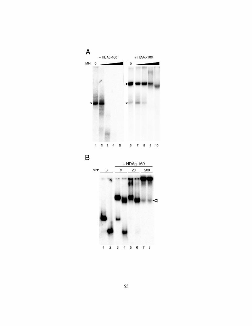
Figure 2.6. (A) HDAg-160 protection of 395L RNA from micrococcal nuclease
digestion. 395L RNA in the absence (left panel) or presence (right panel) of HDAg-160
is treated with increasing amounts of micrococcal nuclease (filled triangle); specifically,
0, 0.2, 2, 20, 200 units. Free RNAs (open circles) and bound RNAs (closed circles) are
indicated. (B) Micrococcal nuclease digestion of the exposed RNA complexed with
HDAg-160. Mobility shift assay on 395L (odd lanes) and 311L (even lanes) in the
absence (lanes 1 and 2) or presence (lanes 3 – 8) of HDAg-160. Amounts of micrococcal
nuclease increase; specifically, 0, 20, 200 units. Change in migration of 395L complexed
with HDAg-160 in the presence of 200 units of micrococcal nuclease (lane 7) is indicated
by the open arrowhead.
55

56
suggested by the increased intensity in the well. Most notable, however, was that the
mobility of the complex formed by 395L in the presence of 200 units of micrococcal
nuclease increased such that it was identical to that of 311L (Fig. 2.6B; lanes 7 and 8).
The mobility of the 311L–HDAg-160 complex was unaffected by the nuclease (Fig.
2.6B; lanes 4, 6, 8). Thus, for both RNAs, the size of the complex protected from
nuclease digestion is the same as that formed by the smallest RNA bound by HDAg-160.
Furthermore, these results suggest that the amount of HDAg-160 bound is the same for
RNAs between 311 and 395nt in length.
DISCUSSION
We have found that a C-terminally truncated form of HDAg, HDAg-160,
expressed in E. coli and purified under native conditions, exhibits specific binding to
unbranched rod segments of HDV RNA, as indicated by the formation of a discrete
complex in an electrophoretic mobility shift assay. The use of HDAg-160 in binding
assays is a significant improvement over the full-length protein, which produced
heterogeneously migrating complexes, bound to non-HDV RNAs, and formed
aggregates. Secondary structure prediction analyses performed on the segment of the
protein removed to create HDAg-160 indicate no defined structures. Possibly, this
proline/glycine-rich 35 aa region contributes to structural variability and/or aggregate
formation in the full-length protein. Removal of this region is unlikely to directly affect
RNA binding: it is not among those regions of the protein implicated in RNA binding by

57
other studies that have used either site-directed mutagenesis or deletional analysis (60,
93, 118). In fact, an HDAg protein created by a 50 aa C-terminal truncation yielded
binding results similar to those of HDAg-160, except that the discrete complex formed
migrated slightly faster (not shown).
Binding of HDAg-160 was specific for the HDV RNA unbranched rod structure;
fully double-stranded RNAs and single-stranded RNAs incapable of forming this
structure, even if derived from HDV, were not bound (data not shown). The specific
requirements for HDAg-160 binding within the unbranched rod are not yet clear.
Deletion analysis indicated that binding is not determined by just one or two unique
features; rather, complex formation in vitro required that the unbranched rod be greater
than 298 nt (Fig. 2.3). We also observed a size-dependent relationship in cells via
analysis of RNA stabilization by HDAg (Fig. 2.5). Relevant to these findings, Chang et
al. observed that segments of HDV RNA 311 nt or longer were efficiently packaged into
virus-like particles by the long form of the delta antigen, while a 258 nt segment was not
(19). The nature of the difference in packaging was not further explored at the time, but
could be explained by our in vitro binding results: the failure of the shorter 258 nt RNA
to be efficiently packaged could be due to its inability to be bound by HDAg-L. One
significant consequence of the length requirement for the virus is that it may provide a
means by which HDAg discriminates between binding to its cognate RNA rather than
shorter similarly structured RNAs (i.e. extended hairpins) in infected cells.

58
The results presented in Figure 2.2 indicate that HDAg-160 bound several
different unbranched RNA segments derived from the HDV antigenome and genome.
This result is similar to that reported by Chao et al. and Lin et al (23, 68). However, these
previous binding studies, which used denatured bacterially expressed HDAg, were
qualitative, and could not distinguish binding characteristics of different RNAs. With
natively expressed HDAg-160, we found that binding to different unbranched RNAs was
not identical, in terms of either affinity or the maximum levels of binding (Fig. 2.2).
Variations in affinity could be due to subtle structural differences in the RNAs that
modulate binding activity. It is not clear why some RNAs failed to be completely bound
during the assay. Perhaps, structural heterogeneity in the RNA that affects binding,
without affecting gel mobility, is responsible. Another possibility is that binding requires
conformational changes – in the RNA, the protein or both – and the time required for
these changes varies among the different RNA-protein interactions. Consistent with these
models, we have observed that binding is strongly dependent on both the temperature and
time of incubation (DAD, JLC, unpublished data). Regardless of the mechanistic
explanation, the variable binding to different segments of the RNA suggests that binding
in the context of the full-length unbranched rod RNA might occur preferentially at certain
sites. Such preferential binding could lead to an ordered assembly of HDAg on the RNA
in cells.
HDAg is known to form dimers, and several reports have indicated that the
protein assembles into higher order structures, possibly octamers (31, 112, 122). The

59
results of our electrophoretic mobility shift assays are most consistent with the formation
of an HDV RNA-HDAg complex containing a large HDAg multimer, rather than an RNP
consisting of either an HDAg monomer or dimer. The mobility of the complex formed by
HDAg-160 and 395L, a 395 nt RNA derived from the left end of the antigenomic
unbranched rod, was approximately half that of unbound 395L RNA (Fig. 2.1). The
molecular mass of an HDAg-160 monomer is about 19 kDa; complexes formed with
395L and either a single monomer or dimer of HDAg-160 would have molecular masses
15% (149 kDa) to 30% (168 kDa) greater than the RNA alone, respectively. On the other
hand, a complex involving 395L RNA and an HDAg-160 octamer (152 kDa), for
example, would produce an RNA-protein complex with a mass more than twice the size
of the RNA alone (280 kDa vs. 130 kDa), consistent with the large shift in mobility
observed. Of course, in addition to effects of the mass of protein on RNP mobility,
conformational changes in the RNA that might occur upon protein binding could also
alter the mobility in a native gel. However, two additional findings support the
conclusion that a larger mass of protein is bound. Comparison of the mobility of RNA-
protein complexes formed by HDAg-195 and HDAg-160 (Fig. 2.1) and HDAg-145 (not
shown), demonstrate readily detectable changes in mobility, consistent with a large
contribution of the protein mass to the mobility of the complex. Furthermore, the
resistance of RNA-protein complexes to nuclease digestion (Fig. 2.6) is more consistent
with binding of a large amount of protein to the RNA. Because no RNPs of intermediate
mobility were observed as the level of HDAg-160 increased (Fig. 2.1), the protein either

60
assembles on the RNA with a high degree of cooperativity, or binds as a pre-assembled
protein complex. While others have suggested that HDAg exists as an octameric structure
in cells and in vitro, even in the absence of HDV RNA (89, 122), our results presented
here cannot distinguish between these two possibilities, nor can we determine the number
of HDAg subunits involved.
The complex formed between HDAg-160 and 395L RNA was resistant to
digestion with micrococcal nuclease (Fig. 2.6). Interestingly, high concentrations of
nuclease reduced the size of the complex formed by 395L RNA, which is greater than the
minimum length RNA required for binding, to the same as that formed by 311L RNA,
which is the smallest RNA bound by HDAg-160 (Fig. 2.6). These results indicate that: 1)
most of the bound RNA is tightly associated with HDAg-160; and, 2) the 395 nt RNA
does not bind more protein than the 311 nt RNA. This result, together with the minimum
length requirement for binding, and our conclusion that HDAg binds as a large multimer,
suggests that binding of the multimeric unit occurs in discrete amounts. Thus, given that
the minumim length for binding of HDAg-160 to HDV unbranched rod RNA is about
300 nt, we expect 5 HDAg-160 multimers are able to bind per full-length HDV RNA. If
the multimeric unit is an octamer (as suggested by Zuccola et al.) then we would expect
relative amounts of protein to RNA of 40. This number is near the low end of the range
of RNA-protein ratios determined by previous analyses of HDV RNA-protein complexes
in cells and in virions. In future studies, determination of the exact size of the HDAg
multimer involved in binding, as well as the number of such complexes bound to

61
progressively larger RNAs, will permit a more precise evaluation of the composition of
the HDV RNA-protein complex.
ACKNOWLEDGEMENTS
This work was supported in part by grant R01-AI42324 from the National
Institutes of Health. We extend our thanks to Alex Israel for protein preparation and
Dunkin’ Donuts.
ADDENDUM
RNA length has been established as a structural feature critical to the interaction
with HDAg-160. In vitro binding assays show fragments of the left quarter of the HDV
unbranched rod RNA greater than 311 nt are bound by HDAg-160 while those less than
298 nt remain unbound. Furthermore, RNPs formed in which the RNA is greater than 311
nt indicate degradation of exposed RNA by micrococcal nuclease suggesting that HDAg-
160 bind to HDV RNA in discrete complexes – longer RNAs do not appear to be bound
by more HDAg-160 than shorter RNAs. That said, if the RNP is comprised of one
HDAg-160 complex per 311 nt HDV RNA, an RNA substrate at least twice as long
should accommodate binding of two protein complexes. To test the hypothesis, two
RNAs were examined for in vitro binding by HDAg-160. PCR products amplified from
the pCMV-DC1×1.2-AgS(–) plasmid using the same primers as those used for 207M and

62
384M were transcribed by T7 RNA polymerase. The 717nt (717L) RNA and the 891 nt
(891L) RNA were analyzed for binding by HDAg-160 as in previous experiments. Both
long RNAs were bound by HDAg-160 with similar affinities as seen with previous HDV
RNAs (Fig. 2.7). 717L RNA indicates binding by a single complex of HDAg-160 as has
been seen on all shorter HDV RNAs containing 311 nt or greater. Binding of 891L
however, resulted in a primary shift similar to previously analyzed RNAs, as well as the
appearance of a secondary shift with slower migration than the first. The secondary
complex appeared at HDAg-160 concentrations at which the majority of the 891L RNA
had been bound to form the primary complex.

63
Figure 2.7. Mobility shift assays of HDAg-160 binding long HDV RNAs. Diagram of
the HDV unbranched rod as in previous figures. 717L and 891L are indicated. Mobility
shift assays of 5.3 pM 717L and 891L HDV RNAs. HDAg-160 concentrations increase
3.67-fold left to right (filled triangle); specifically, 0, 0.046, 0.168, 0.617, 2.26, 8.28,
30.4, 111.2 nM. Free RNAs (open circles), primary RNPs (single closed circles), and
secondary RNPs (double closed circles) are indicated.

64

65
HDV RNA REPLICATION IS SENSITIVE TO SMALL DELETIONS AT NUMEROUS
LOCATIONS ALONG THE UNBRANCHED ROD STRUCTURE
Dawn A. Defenbaugh1, Robert L. Judson1, Marcy E. Beildeck and John L. Casey*
Department of Microbiology and Immunology, Georgetown University Medical Center,
Washington, DC 20007
1D.A.D. and R.L.J. contributed equally to this work.
*Corresponding author
Department of Microbiology and Immunology
Georgetown University Medical Center
3900 Reservoir Road, NW
Washington, DC 20007
Phone: (202) 687-1052
Fax: (202) 687-1800
email: [email protected]

66
ABSTRACT
Hepatitis delta virus (HDV) is a defective human pathogen that requires
concurrent infection with hepatitis B virus. The genome structure and replication strategy
of HDV are unique among animal viruses. HDV is a negative strand RNA virus and the
viral RNA is associated with a virally encoded protein, hepatitis delta antigen. However,
the structure of the genome and its use of host DNA-dependent RNA polymerase in a
rolling circle replication mechanism are more similar to viroids, infectious RNAs of
plants that do not encode proteins. As a result of extensive Watson-Crick base-pairing,
the circular HDV RNA genome (and antigenome) forms a characteristic unbranched rod
structure that is required for viral RNA replication. Previous analyses of the role of this
structure in HDV RNA replication have shown that HDV RNA replication is diminished
by disruptive one-sided insertions at many locations. Furthermore, structure-preserving
base pair deletions and insertions substantially reduce replication when placed at the non-
ribozyme end of the unbranched rod, but have little effect at the other end. Here, we
describe the creation of mutations that delete base-paired segments of the interior of the
unbranched rod such that the predicted secondary structure is not disrupted. In transfected
human hepatoma cells with the viral protein provided exogenously, these deletions
substantially reduced viral RNA replication, even though the predicted base-paired
structure is preserved. This result was observed for deletions of various sizes (total of 6 –
53 nt), and appeared to be a feature common to the interior of the unbranched rod –
similar small deletions of between 6 and 13 nt from 6 different unrelated segments of the

67
unbranched rod had the same effect. Our results suggest that, unlike the genomes of other
mammalian viruses, most of the HDV RNA plays a critical, structural role in replication
of the viral genome that is distinct from its function in viral protein production.
INTRODUCTION
Hepatitis delta virus (HDV) is a subviral human pathogen that increases the
severity of liver disease in those infected with its helper, hepatitis B virus (HBV) (97).
The virus is classified as the sole member of the floating genus Deltavirus (37). Several
features of the 1.7kb HDV genome and its replication are similar to viroids, infectious
RNAs of plants: the circular HDV RNA collapses into an unbranched rod structure and it
is replicated by a host RNA polymerase in the cell nucleus via a rolling circle
mechanism. Unlike viroids, HDV produces a protein, the hepatitis delta antigen (HDAg),
which is encoded on the antigenome; HDV is thus a negative strand RNA virus. HDAg is
required for replication of the viral RNA (55). The protein forms dimers and higher order
multimers (31, 82, 112, 119, 122), and binds the unbranched rod structure of the viral
RNA (22, 36, 63) to form a ribonucleoprotein complex (6, 99, 112). Both its ability to
form multimers and bind RNA are essential for HDV RNA replication (60, 63, 82, 119).
The characteristic unbranched rod structure that is assumed by both genomic and
antigenomic HDV RNAs is formed by Watson-Crick base-pairing between HDAg-
coding sequences along one side of the rod and non-coding sequences on the other (56,
114). Approximately 70% of the bases in the circular RNA form base pairs in this

68
structure. The pairing is distributed along the structure, such that small 1 – 4 nt bulges
and 2 – 8 nt internal loops separate short regions of 2 to 11 base pairs.
Several reports have shown that HDV RNA replication is blocked when the
unbranched rod structure is disrupted by mutations that create unpaired bulges or short
stem-loops on one side of the unbranched rod. For example, Wang et al. found that RNA
replication was eliminated by one-sided sequence insertions of 8 to12 nt at numerous
sites along the RNA (111). In a more recent study, Gudima et al. also reported that one-
sided insertion of extraneous sequences in the unbranched rod strongly reduced
replication; high levels of replication were restored when the extraneous sequences were
spontaneously lost, regenerating the unbranched rod structure (44). Finally, two studies
that were primarily devoted to analysis of RNA editing reported that replication was
strongly reduced by site-directed mutations that disrupted several base pairs, thereby
creating ca. 12 – 20 nt internal bulges in the unbranched rod (10, 101). Importantly, in
both cases, replication was fully restored by additional mutations that re-established base
pairing, even though the primary sequence was changed.
Although the studies cited above indicate that disruption of the unbranched rod
structure by the introduction of one-sided bulges interferes with HDV RNA replication,
there is little information about whether specific regions of the intact structure are
required for RNA replication. One approach to address this question is to delete segments
of the unbranched rod in such a way that the base-paired structure of the remainder is
maintained. Lazinski et al. (58) reported that removal of a large segment from the non-

69
ribozyme end of the unbranched rod structure (referred to as the left end in this
manuscript) eliminated RNA replication, even though the overall unbranched rod
structure was maintained. This result suggests that some structure in the deleted segment
of the RNA is required for replication. However, this large deletion removed nearly one-
third of the genome, including the entire left end of the unbranched rod. In fact, Wu et al.
found that replication is sensitive to very minor changes at the left end of the unbranched
rod: removal of even 1 or 2 bp from the base-paired stem at the end reduced replication
100-fold or more (118). Beard et al. (2) also showed that some site-directed mutations
near the left end of the rod eliminated RNA replication; however, these mutations altered
the predicted secondary structure.
In contrast to results obtained for the left end of the unbranched rod, replication
appears to be more tolerant of deletions and insertions at the right end. Wu et al. reported
that modest site-directed deletions (ca. 8 bp) from the right end of the unbranched rod had
little or no effect on replication, and that insertions of at least 6 bp could also be tolerated
(118). Overall, these data have contributed to the idea that the left end of the unbranched
rod structure is particularly important for replication. Because viral mRNA transcription
initiates in this region (43, 79), mutations could interfere with replication by either
disrupting mRNA production or genome (or antigenome) synthesis.
While the studies cited above analyzed the effects of deletions (and insertions)
positioned at the left and right ends of the unbranched rod, the role of interior regions of
the structure in HDV RNA replication has not been addressed in experiments in which

70
the unbranched rod structure was maintained. Here, we investigate the role in RNA
replication of multiple segments of the interior of the unbranched rod structure by
creating matched deletion mutations on both sides of the rod that were designed to
maintain the pattern of paired and unpaired bases that make up the structure. We find that
deletions within the interior of the rod significantly reduce viral RNA replication, even
though the predicted base-paired structure is preserved. This result was observed for
deletions of various sizes (total of 6 – 53 nt), and appeared to be a feature common to the
interior of the unbranched rod – similar small deletions of between 6 and 13 nt from 6
different unrelated segments of the unbranched rod had the same effect. Our results
indicate that, unlike most viral genomes, much of the HDV RNA genome plays a critical
role in HDV RNA replication.
MATERIALS AND METHODS
Plasmids. All mutations were introduced into the plasmid pCMV3-DC1×1.2-
AgS(–) (13). On transfection, this plasmid produces a 1.2mer antigenomic RNA, in
which the HDAg reading frame is disrupted by a single base insertion at codon 7.
Replication of this RNA can be supported by co-transfection of an expression construct
for HDAgS (13). Deletions ∆1 through ∆6, ∆3u and the flip mutation f3 were made using
PCR primers containing the Apa I sites at positions 215 and 1380 (numbering refers to
genome coordinates; ref. 114); mutated products were cloned into the Apa I sites of
pCMV3-DC1×1.2-AgS(–). Other mutations were made using overlap PCR. For deletion

71
mutations ∆7 through ∆10 (as well as the related mutations ∆7u, f7, ∆7+i7a through
∆7+i7d, ∆9u, f9, ∆9+i9a, and ∆9+i9b) products were subsequently cloned between the
Stu I and Bgl II sites of pCMV3-DC1×1.2-AgS(–). For ∆11 and f11, amplified fragments
were cloned between the Sal I site at position 963 and an additional Sal I site introduced
at position 628 by site-directed mutagenesis. All cloned fragments were verified by
sequencing.
Transfection. Huh7 (human hepatoma) cells were maintained in Dulbecco’s
modified Eagle’s medium supplemented with 10% fetal bovine serum and 1 mM
glutamine. One day prior to transfection, cells were plated at a density of 4 x 10⁵ per well
in 12-well plates. Transfections were carried out with a total of 0.5 µg plasmid DNA and
LipofectAMINE PLUS (Invitrogen), according to the manufacturer’s recommendations.
For replication assays, expression of HDAgS was supplied in trans by including 0.02 µg
of the HDAgS-expression plasmid pCMV-AgS (94). All transfections were performed in
duplicate, and most were repeated multiple times. In order to normalize for transfection
efficiency, some transfections included a secreted alkaline phosphatase (SEAP)
expression construct (32); transfection efficiencies varied by less than 50%.
Northern blot analysis. Total cellular RNA was harvested using QIAshredder
and RNeasy Mini Kit (Qiagen) following manufacturer’s protocols. RNA (1 µg) was
electrophoresed through 1.5% agarose gels containing 2.2 M formaldehyde, transferred to
positively charged nylon membranes, and hybridized with an antigenomic-sense 32P-
labeled probe, as described previously (14). The hybridization temperature was 65oC and

72
the post-hybridization wash temperature was 70oC. Relative levels of HDV RNA were
determined by radioanalytic scanning of blots with a Molecular Dynamics Storm 475
phosphorimager. The integrity of the RNA samples and equivalency of loading were
assessed by visualization of ethidium bromide-stained ribosomal RNA bands and by the
non-specific hybridization of the probe to 28S rRNA.
Analysis of HDV isolates. The sequences of 7 HDV genotype I isolates (12, 24,
25, 61, 75, 100, 114) were used to compare sequence and secondary structure
conservation. The accession numbers of the isolates are: X04451, M28267, L22066,
M92448, D01075, M84917 and M58629. The predicted lowest energy secondary
structure of each isolate was determined using mfold (77, 124) and these structures were
aligned manually for comparison. The percent sequence conservation in a segment was
defined as the number of identities in all isolates divided by the total number of sequence
positions. To compare conservation of secondary structure, each nucleotide position was
considered as base-paired or unpaired. The percent structure conservation was defined as
the number of identical configurations for a position divided by the total number of
positions. In some cases, structure conservation was lower than sequence conservation
because of structural contributions from varying neighboring sequences.
RESULTS
Small deletions from the interior of the unbranched rod structure impair
HDV RNA replication. To investigate the requirements of the interior of the unbranched

73
rod for RNA replication, we created deletions within a region near convenient Apa I sites
in the HDV RNA expression construct pCMV3-DC1×1.2-AgS(–) (Fig. 3.1). This
construct is designed to generate antigenomic RNA containing two copies of the
ribozyme domain that will be processed to form circular RNAs that are functional for
replication in transfected cells. Because this construct contains a site-directed mutation
that prevents HDAg synthesis, HDAg must be supplied in trans. Co-transfection of the
HDAg expression construct pCMV-AgS leads to replication of this RNA, as indicated by
the accumulation of genomic RNA that can be detected by blot hybridization (13). As
HDAg-S expression is provided in trans by pCMV-AgS, potential effects of site-directed
mutations on HDAg expression and function are avoided.
The sequences that make up the Apa I sites in the pCMV3-DC1×1.2-AgS(–)
cDNA clone are derived from coding and non-coding sequences that are predicted to be
base-paired in the unbranched rod structure; they are located approximately one-third of
the way from the non-ribozyme end of the unbranched rod (the left end, as depicted in
Fig. 3.1). Deletions were designed to maintain the unbranched rod structure in that the
total number of contiguous paired or unpaired bases was not changed (Fig. 3.1). For
example, in the deletion mutant ∆1, the left side of the segment deleted is base-paired and
on the right it ends with an unpaired adenosine; the predicted structure of the deletion
mutant contains two unpaired uridines flanked by base pairs, as in the wild type
sequence.

74
Figure 3.1. HDV RNA replication is sensitive to small deletions from the interior of the
HDV RNA unbranched rod. A. Schematic of HDV RNA. The HDAg coding sequence is
indicated by the open rectangle; the asterisk denotes the location of the amber/W RNA
editing site; the location of the antigenomic ribozyme site is indicated by a triangle.
Vertical lines represent base pairing between the two sides of the unbranched rod. The
region in which sequences were deleted is indicated by a shaded rectangle; the predicted
secondary structure of the antigenomic RNA for this region is shown below the
schematic, with sequence numbering for the corresponding positions in the genome RNA
shown. Sequences in boldface correspond to the Apa I sites in the cDNA. Deletions ∆1 –
∆6 are indicated diagrammatically, with deleted segments represented by a horizontal
dashed line. B. Northern blot analysis of HDV genomic RNA in cells transfected with
wild-type and mutant antigenomic RNA expression constructs. Huh7 cells were
cotransfected with the antigenomic RNA expression plasmid pCMV3-DC1×1.2-AgS(–)
containing either wild type or indicated mutations in the unbranched rod along with the
HDAg expression plasmid pCMV-AgS, as indicated; as a negative control, one
transfection included no pCMV-AgS. RNAs were harvested 6 days post transfection and
analyzed for HDV genomic RNA by electrophoresis followed by blot hybridization with
a radioactively labeled antigenomic RNA. Open and filled arrowheads indicate positions
of 28S ribosomal RNA and HDV RNA, respectively.

75

76
Huh7 cells were cotransfected with wild type pCMV3-DC1×1.2-AgS(–) or
plasmids containing deletion mutants, along with the HDAgS-expression plasmid pCMV-
AgS. Total cellular RNA was harvested 6 days post transfection and analyzed via blot
hybridization for the accumulation of genomic HDV RNA as an indication of replication.
The results shown in Figure 3.1 are representative of multiple transfections that gave
identical results; transfection efficiencies varied by less than 2-fold, as determined by
SEAP expression from a co-transfected construct. The initial deletion mutant, ∆1, which
removed 53 nt (22bp), prevented accumulation of genomic RNA, as detected by Northern
blot hybridization (Fig. 3.1). To determine whether a particular region within ∆1 was
responsible for this decreased accumulation of genomic RNA, we created a series of
nested deletions, ∆2 – ∆4 (Fig. 3.1). The accumulation of genomic RNA was strongly
reduced by all of these deletions, including ∆4, in which just 2 base pairs and 2 unpaired
bases (6 nt in all) were removed. To determine whether other regions of the ∆1 deletion
could also be contributing to the inability to accumulate genomic RNA, we created the
non-overlapping deletion mutants ∆5 and ∆6, which, along with ∆3, covered the entire
region removed in ∆1. Similarly, both of these mutations also eliminated accumulation of
genomic RNA (Fig. 3.1).
The reduced accumulation of genomic RNA (Fig. 3.1) suggests that mutations ∆1
through ∆6 have dramatically reduced the ability of the RNA to replicate. However, it is
important to consider whether the mutations affected RNA processing or stability, and
that these effects could have resulted in the decreased accumulation of genomic RNA.

77
Others have shown that much larger deletions from the left end of the unbranched rod
and insertions at other locations do not interfere with processing of HDV transcripts into
circular RNAs (44, 58); thus, it is unlikely that the modest mutations studied here affect
processing of the RNA. To confirm that the failure to detect genomic RNA in cells
transfected with the deletion mutants was not due to the inherent instability of the
mutated antigenomic RNA transcripts synthesized from the CMV promoter, we
cotransfected cells with expression constructs for both mutated antigenomic RNAs and
the long form of HDAg, which binds and stabilizes HDV RNA (58), but does not support
RNA replication (21). We observed that levels of antigenomic RNA were identical for
wild type and deletion mutant constructs (not shown), indicating that the initial
transcripts were stable. Thus, the decreased accumulation of genomic RNA for the
mutations analyzed is not due to transcript instability. From these results, together with
the results presented in Figure 3.1, we conclude that deletions as small as 6 nt within this
region of the unbranched rod prevent HDV RNA replication, even though the structure of
the unbranched rod is maintained.
Deletion of many different small segments of the unbranched rod reduces
replication. The sequence and predicted secondary structure of the nucleotides deleted in
the ∆1 mutant are highly conserved among HDV genotype I isolates; this conservation is
highest for the sequences and structures that were removed in deletions ∆3 and ∆5 (Table
3.1). Thus, the sensitivity of replication to these deletions could indicate that the deleted
sequences provide particular critical functions for replication, for example, by forming

78
RNA secondary structures that serve as specific binding sites for cellular or viral factors
necessary for replication.
Table 3.1. Sequence and structure conservation of deleted segments Deletion Mutant
Sequence
Conservation Structure
Conservation Location
Δ3 100% 97% 207-210, 1382-1387
Δ5 99% 99% 183-199, 1393-1406
Δ6 93% 83% 200-206, 1388-1392
Δ7 100% 93% 48-51, 1545-1549
Δ8 90% 98% 63-66, 1528-1533
Δ9 99% 100% 99-105, 1491-1495
Δ10 70% 63% 172-178, 1411-1416
Δ11 48% 69% 603-607, 987-990
In order to determine whether replication was equally sensitive to similar small
deletions in other regions of the HDV RNA unbranched rod and to determine whether
such sensitivity was correlated with sequence and structure conservation, we selected 5
additional segments of 9 to 13 nt (3 – 5 bp) for deletion (Fig. 3.2). These segments were
chosen based on location along the unbranched rod and on comparison of the sequences
and predicted secondary structures of 7 HDV genotype I isolates. They exhibit varying
levels of sequence and predicted secondary structure conservation, but no clear sequence
or structural similarities to each other (Table 3.1, Fig. 3.2). Four of the segments are to
the left of the ∆1 region, and one is toward the right end of the unbranched rod (Fig. 3.2).

79
The least conserved sequences and structures were those deleted in the ∆10 and ∆11
mutations (Table 3.1), for which structure conservation was 63% and 69%, respectively.
Conservation was ≥ 90% for segments removed in the ∆3, ∆5, ∆7, ∆8 and ∆9 deletions.
The replicative ability of HDV RNA containing deletion mutations ∆7 – ∆11 was
assessed by detection of genomic RNA 6 days following cotransfection of Huh7 cells, as
in Figure 3.1. We observed that, for all 5 deletion mutations, viral replication was
dramatically reduced (Fig. 3.2). Thus, HDV RNA replication is sensitive to deletion of
many different small segments from the interior of the unbranched rod, regardless of the
extent of sequence and structural conservation of the deleted segment or the location of
the segment on the unbranched rod. It is notable that there are no apparent similarities
among the deleted segments (Table 3.1), and that deletion of weakly conserved segments
inhibited replication to an extent similar to deletion of highly conserved segments.

80
Figure 3.2. HDV RNA replication is sensitive to small deletions from many different
regions of the unbranched rod structure. A. Schematic of the predicted unbranched rod
structure of HDV RNA, with the locations of deletions indicated by numbered rectangles.
For reference, the sequences deleted in ∆1 are indicated by a shaded box. The predicted
secondary structure of the antigenome is indicated for regions surrounding deletions, with
the deleted sequences contained in boxes; numbers indicate numbering of corresponding
positions in the genome. B. Replication of HDV RNA after cotransfection of Huh7 cells
with either wild type or mutant HDV antigenomic RNA expression plasmids along with
the HDAg expression plasmid pCMV-AgS. RNAs were harvested 6 days post
transfection and analyzed for genomic RNA by Northern blot hybridization. Levels of
HDV genomic RNA shown in the bar graph were determined relative to wild-type.
Individual mutations were analyzed in at least 4 independent experiments; error bars
indicate standard deviation.

81

82
Flipping base-paired nucleotides or deleting unpaired nucleotides did not
affect viral RNA replication. The results shown in Figures 3.1 and 3.2 indicate that
replication is sensitive to many different small deletions that preserve the unbranched rod
structure. Although two of the deletions studied removed segments with weakly
conserved sequences and structures, most segments analyzed are highly conserved among
different genotype I isolates. The high conservation of these segments could arise from
their contributions to RNA replication by acting as specific binding sites for proteins, or
by participating in multiple alternative RNA secondary structures, such as proposed for
certain segments of potato spindle tuber viroid RNA (71, 102). We therefore created
additional mutations to determine to what extent the contribution of the deleted segments
to replication relies on the sequence of the deleted bases. We first removed just the
unpaired bases from the conserved segments deleted in ∆3, ∆7 and ∆9 to create the
mutations ∆3u, ∆7u and ∆9u, respectively, and analyzed the effects of these deletions on
replication (Fig. 3.3). We found that the ∆7u and ∆9u deletions had no affect on RNA
replication (Fig. 3.3B). The ∆3u mutation, which removed 2 unpaired adenosines,
reduced replication to about 60% of wild-type; this level was far higher than that
observed for the ∆3 deletion, for which replication levels were 7% of wild-type (Fig. 3.1).
These results show that the loss of activity in the ∆3, ∆7 and ∆9 deletion mutants was not
due solely to the removal of the unpaired bases. Because sequence-specific contacts
between RNA and protein typically involve unpaired bases, this result suggests that the
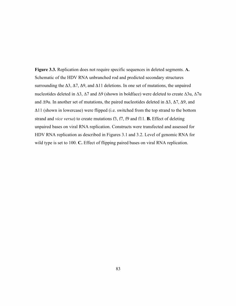
83
Figure 3.3. Replication does not require specific sequences in deleted segments. A.
Schematic of the HDV RNA unbranched rod and predicted secondary structures
surrounding the Δ3, Δ7, Δ9, and Δ11 deletions. In one set of mutations, the unpaired
nucleotides deleted in ∆3, ∆7 and ∆9 (shown in boldface) were deleted to create ∆3u, ∆7u
and ∆9u. In another set of mutations, the paired nucleotides deleted in Δ3, Δ7, Δ9, and
Δ11 (shown in lowercase) were flipped (i.e. switched from the top strand to the bottom
strand and vice versa) to create mutations f3, f7, f9 and f11. B. Effect of deleting
unpaired bases on viral RNA replication. Constructs were transfected and assessed for
HDV RNA replication as described in Figures 3.1 and 3.2. Level of genomic RNA for
wild type is set to 100. C. Effect of flipping paired bases on viral RNA replication.

84

85
segments deleted in ∆3, ∆7 and ∆9 do not participate in sequence-specific protein binding
required for RNA replication.
In order to examine possible sequence-specific contributions of paired bases in
deleted segments, we created the “flip” mutants f3, f7, f9 and f11, in which the paired
nucleotides from both sides of the unbranched rod in the sequences deleted in ∆3, ∆7, ∆9
and ∆11 were replaced by their respective base pair counterparts (Fig. 3.3A). The
sequences changed in f3, f7 and f9 are nearly identical among different HDV genotype I
isolates. Nevertheless, none of these mutations interfered with HDV RNA replication
(Fig. 3.3C). In all of these mutations the predicted base-paired structure of the
unbranched rod was preserved. There is no clear evidence that the segments examined
would form alternative branched RNA secondary structures that deviate from the
unbranched rod in the wild type, but, if such structures were to form, we would expect
them to be destabilized by the flip mutations. Thus, overall, we interpret the results in
Figure 3.3 to indicate that the primary contributions to replication of the bases removed
in the deletion constructs examined in Figures 3.1 and 3.2 involve the unbranched rod
structure, rather than sequence-specific interactions with cellular or viral proteins, or
potential alternative RNA structures.
Gudima et al. (44) reported that HDV genomes containing extraneous sequences
could change during the course of replication, resulting in the formation of modified
genomes that had replicative abilities similar to the wild type. We therefore considered
that the levels of replication shown in Figure 3.3 could be due to revertants or other

86
sequence changes that occurred during replication, rather than to the mutated sequences
that were transfected. However, sequencing of RT-PCR products that included the
mutated positions showed that, in all cases, the mutated positions did not change during
replication.
Re-inserting deleted base-paired segments near the deletion site could restore
RNA replication of deletion mutants. Some negative strand RNA viruses exhibit length
restrictions such that replication is optimal when the length of the genome is a multiple of
the number of bases bound by the nucleocapsid protein (53, 90). The nature of the
complex formed by HDV RNA and HDAg is not fully understood, but we considered
that a similar spacing requirement might also apply for HDV. In order to determine
whether the loss of replication caused by some of our deletions could be explained by
spacing requirements, we re-inserted the base pairs deleted in the ∆7 and ∆9 mutations
back into the unbranched rod structure. Re-insertions of the base pairs deleted in ∆7 were
positioned 5 nt, 14 nt, or 25 nt to the left or 6 nt to the right of the ∆7 deletion site (Fig.
3.4A); the base pairs removed in ∆9 were re-inserted 9 nt to the right or 6 nt to the left of
the ∆7 deletion site (Fig. 3.4B).
Remarkably, we found that several of the re-insertions rescued replication of the
deletion constructs (Fig. 3.4); for example, the ∆7+i7a and ∆9+i9b re-insertion mutations
replicated at approximately 70% of wild-type levels. As a control for the ∆7+i7a re-
insertion, we also inserted the 4 bp deleted in ∆7 at the i7a position in the wild type
construct pCMV3-DC1×1.2-AgS(–); this mutation reduced replication as strongly as the

87
Figure 3.4. Re-insertion of deleted bases can restore replication of deletion mutants. A.
Effects of deletion ∆7, insertion i7a and combined insertion/deletion mutations ∆7+i7a
through ∆7+i7d on RNA replication. B. Effects of deletion ∆9, and the combined
insertion/deletion mutations ∆9+i9a and ∆9+i9b on RNA replication. Top panels.
Predicted secondary structure of antigenomic RNA surrounding deletion and insertion
sites. A horizontal dashed line represents deleted sequences (boxed); inserted base pairs
are indicated above the position in the structure at which they were inserted. For ∆7+i7a
through ∆7+i7d and ∆9+i9b, only the insertion site is indicated. Bottom panels. Levels of
genomic HDV RNA in Huh7 cells cotransfected with pCMV-AgS and wild type or
mutant pCMV3-DC1×1.2-AgS(–) expression plasmids, determined by Northern blot
hybridization. Values are normalized to wild-type replication, which is set as 100.

88

89

90
∆7 deletion mutant (Fig. 3.4A). Thus, the increased replication observed for the ∆7+i7a
re-insertion was not due solely to an enhancing effect of the inserted base pairs.
Moreover, the reduced replication of the i7a insertion mutant demonstrates that the
interior of the unbranched rod is sensitive to base-pair insertions as well as deletions and
that the loss of activity of the i7a insertion mutant was also rescued by combination with
the ∆7 deletion.
The results obtained for the ∆7+i7a and ∆9+i9b mutations are consistent with the
idea that the sequences removed in the ∆7 and ∆9 deletions are required for appropriate
spacing along the unbranched rod structure in order for replication to occur, and that the
re-insertions restore proper spacing. However, not all re-insertion mutations restored
replication (Fig. 3.4). For the ∆7 deletion, re-insertions became less effective as the re-
insertion site was moved farther from the deletion site. Moreover, for both ∆7 and ∆9, the
location of re-insertions on either side of the deletion site had different effects. The
insertion i7a, located 5nt from the ∆7 deletion site, successfully rescued the ∆7 mutation;
however, the insertion i7d, which was located 6 nt away on the right side, did not rescue
replication at all. For ∆9, the insertion i9b, located on the left, was more effective than
i9a, located on the right. These results indicate that, while spacing along the unbranched
rod can be important, particularly over a short span, additional structural features of the
unbranched rod also contribute to replication.
91
DISCUSSION
We have found that small (6 – 13 nt) deletions and insertions at 6 different sites in
the interior of the HDV unbranched rod structure strongly reduced the accumulation of
HDV genomic RNA from antigenomic RNA in transfected cells. This decreased
accumulation is most likely due to a defect in RNA replication, rather than reduced
efficiency of processing linear transcripts into circles via ribozyme cleavage and
subsequent ligation, or reduced stability of the primary antigenomic transcript. Others
have shown that deletions and insertions much larger and more disruptive of the
unbranched rod than those studied here do not interfere with the processing of primary
transcripts into circular RNAs (44, 58); thus, there is no reason to expect that the
mutations analyzed in this study interfere with the ribozyme cleavage and ligation
processing steps. We analyzed several deletions and observed that the production and
stability of primary antigenomic transcripts was not affected (data not shown). Thus, we
conclude that the decreased accumulation of genomic RNA is due to decreased RNA
replication. Although we have analyzed production of genomic RNA from antigenomic
RNA, it is possible that the observed decreased accumulation of genomic RNA could be
due to defects in RNA-templated synthesis of either genomic or antigenomic RNA (or
both).
Previous studies of the role of the unbranched rod structure in HDV RNA
replication have either analyzed the effects of disrupting the structure (2, 44, 111) or of
inserting/deleting base pairs in a manner that preserves the overall structure (118). Our

92
study is most similar to that of Wu et al. (118) in that we have attempted to maintain not
only base-pairing within the unbranched rod, but also the pattern of paired and unpaired
bases (Figs. 3.1 and 3.2). The most dramatic effects observed by Wu et al. were for
mutated constructs that produced both replicating RNA and the mRNA for HDAg; for
such constructs the reduced accumulation of full-length HDV RNA could have been due
to decreased mRNA production, limited full-length RNA synthesis, or both. Our study
differs from that of Wu et al. in that we used an RNA expression construct defective for
production of HDAg, which was supplied in trans. Thus, our observed effects cannot be
attributed to a loss of mRNA synthesis and are therefore due to decreased ability of the
RNA to replicate. Wu et al. focused on the effects of insertions and deletions at the two
ends of the unbranched rod structure and found that HDV RNA replication was much less
tolerant of insertions and deletions at the left end than at the right end (top and bottom,
respectively, in their nomenclature). Here, we show that replication is also intolerant of
small (6 – 13 nt) deletions and insertions in the interior of the unbranched rod, even when
the predicted structure is maintained. There are no obvious similarities among the
segments deleted in this study, and we expect that most, if not all, similar deletions from
the interior of the unbranched rod will interfere with RNA replication.
The sensitivity of HDV RNA replication to such small deletions at multiple sites
in the RNA is remarkable, and places the HDV genome in sharp contrast to most other
viral genomes, which can be extensively manipulated. Even viroid RNAs, which are less
than one-fourth the size of HDV RNA, have been shown to tolerate an 18 nt deletion that

93
preserves the unbranched rod structure (116). Why is HDV RNA replication so sensitive
to these deletions? It seems unlikely that the observed sensitivity is due to requirements
that the RNA must adopt alternative secondary structures, in addition to the unbranched
rod, in order for RNA replication to occur. When we analyzed predicted secondary
structures in the deletion regions using mfold (77, 124), no such stable alternative
conformations were seen that involved the deleted segments. Moreover, we would expect
flip mutations such as those analyzed in Figure 3.3 to affect the formation of most
alternative RNA structures (the exception being alternative structures in which mutated
sequences from both sides of the unbranched rod are unpaired in the alternative
structure). That all four flip mutations replicated well (Fig. 3.3) suggests that the ∆3, ∆7,
∆9, and ∆11 mutations do not prevent replication by interfering with the ability of the
RNA to form alternative secondary structures that are required for replication.
While it is conceivable that some of our deletions could have disrupted specific
binding sites for HDAg or for cellular factors required for RNA replication, it seems
highly unlikely that all of them had this effect. First, we would expect highly specific
binding sites to be highly conserved among different isolates; while some deleted
segments were highly conserved, others, particularly ∆10 and ∆11, were not (Table 3.1).
Second, RNA-protein interactions are typically mediated by interactions with bases that
are unpaired; we found that removal of just the unpaired positions in three deletion
segments had little or no effect on RNA replication (Fig. 3.3B). Furthermore, flipping the
paired bases in four deletion segments did not interfere with replication (Fig. 3.3C),

94
indicating that even if sequence-specific contacts occurred between proteins and base-
paired positions in the RNA, these hypothetical interactions are not important for
replication. Finally, the lack of clear similarities among the deleted structures suggests
that if each of these sites did bind specifically to viral or cellular proteins, most sites
would bind factors unique to that site. It seems unlikely that the entire RNA would be
involved in a series of interactions with multiple unique host factors that are required for
RNA replication.
Perhaps small deletions from the interior of the unbranched rod interfere with
replication because they affect the shape of the structure. The simplest type of shape
effect would be spacing along the unbranched rod. In support of this explanation, we
observed that replication was restored for two deletion mutations, ∆7 and ∆9, by re-
insertion of the deleted base pairs 5 – 9 nt away from the deletion site (Fig. 3.4).
However, not all re-insertion mutations restored replication (Fig. 3.4) and the ability of
insertion mutations to restore the activity of deletion mutants decreased as the insertion
and deletion sites were moved further apart. Thus, although HDV is a negative strand
RNA virus, spacing affected by our mutations does not appear to be related to the entire
length of the genome, as it is for some negative strand RNA viruses (8, 53, 90). Because
of the partial double-stranded structure of the HDV RNA, insertions and deletions could
affect not only the length of the RNA, but also bends and twists introduced by the
numerous bulges and internal loops. Thus, for the HDV RNA, it might be necessary to
consider the effects of deletions and insertions on the 3-dimensional structure of the

95
RNA; restoration of activity lost by certain deletions could require not only restoration of
the length of the region removed, but also the 3-dimensional shape, including bends and
twists of the structure. Certainly, further analysis of the effects of deletions and insertions
on the interaction of HDV RNA with HDAg and on the 3-dimensional shape of the RNA
could help to better understand why these mutations in the interior of the HDV RNA
unbranched rod restrict RNA replication.
ACKNOWLEDGEMENTS
This work was supported by grant R01-AI42324 from the National Institutes of
Health. We wish to thank Dr. Renxiang Chen and Sarah Linnstaedt for helpful comments
on the work and manuscript.
ADDENDUM
During sequence identification of HDV RNA from virus particles isolated from
patient sera, a naturally occurring deletion mutant was discovered. Isolate R, as it was
labeled, exhibited 22 nt deleted from the coding (13 nt) and non-coding (9 nt) sides of the
unbranched rod. Since this mutation introduced a significant disruption of the HDAg
coding sequence, this patient must also have been infected with wild type HDV RNA.
This virus not only relied on HBV to provide HBsAg but also on wild type HDV to
provide HDAg. In fact, this virus was a satellite of a satellite – a super-defective virus.
That said, it is highly likely that this mutation occurred as a result of replication and was

96
allowed to persist in an environment in which HBsAg and HDAg were both provided in
trans. Existence of this HDV RNA supports the hypothesis that deletion of defined
segments with specific structural characteristics can be removed from the unbranched rod
without completely eliminating viral replication.
As expected, folding predictions of this mutated RNA suggested no significant
disruption of the remaining rod structure as shown in Figure 3.5. In fact, the mutated
RNA appeared similar to the experimental mutants previously analyzed in the replication
assay. To test this natural mutation, the deleted nucleotides were cloned into our
expression plasmid and analyzed for replication in cell culture. Not surprisingly, this
deletion mutant replicated to levels comparable to wild type. This was the only RNA
mutant containing a deletion larger than 4 nt (2 bp) we found that exhibited viral
replication in cell culture. Taking the lead from Isolate R, additional deletions were
analyzed to determine what segments or features of this region were critical. Design of
these RNAs was similar to previous deletions; the unbranched rod structure was
maintained and the pattern of paired and unpaired nucleotides was preserved. Two
mutants, R4 and R5, contained the same number of bases deleted in the original isolate
although aligned like our previous mutants. R4 has 12 nt deleted from the coding side and
10 nt from the non-coding side while R5 has 11 nt deleted from each side. The mutant
with the smallest deletion, R2, deleted only the bases common to both R4 and R5, and all
of the deleted bases common to both sides of the unbranched rod in the original isolate.
Surprisingly, none of our deletion mutants exhibited viral replication; similar to previous

97
mutants, replication levels were reduced to < 20% those of wild type. Altogether, these
data further demonstrate the sensitivity of the replication process to the structure of the
HDV unbranched rod RNA.

98
Figure 3.5. Natural deletions can support HDV RNA replication when HDAg is supplied
in trans. A. Schematic of the predicted unbranched rod structure of HDV RNA, with the
location of the Isolate R deletion indicated by the shaded rectangle labeled R. The
predicted secondary structure of the antigenomic RNA for this region is shown below the
schematic, with the deleted sequences contained in boxes; numbers indicate numbering of
corresponding positions in the genome. Isolate R is depicted twice. The first indicates the
deleted sequences (boxed). The second is the predicted secondary structure of this region
after removal of the bases; unaffected nucleotides are aligned as in the other sequences;
bases whose paired/unpaired nature is changed relative to wt are in bold. B. Effects of nt
deletion on RNA replication. Constructs were transfected as described in Figure 3.1.
Open and filled arrowheads indicate positions of 28S ribosomal RNA and HDV RNA,
respectively.

99

100
HDV replication has been shown to require RNA-binding by HDAg-S (20, 64,
68). Briefly, point mutation and deletional analysis within the RNA binding domains of
HDAg-S resulted in elimination of in vitro binding and viral replication, as measured by
blot analysis. The loss of replication observed for mutant RNAs may have been due to an
inability of these RNAs to be bound by HDAg-S in transfected cells. This idea was tested
through use of the in vitro electrophoretic mobility shift assay. PCR amplification of T7
transcription templates was carried out as previously described using expression plasmids
containing deletions ∆7, ∆8, ∆9 and Isolate R, and the same primer pairs as those used for
395L. Migration of these mutant RNAs in the absence of protein on non-denaturing
polyacrylamide gel was indistinguishable from wild type 395L RNA; gross structural
differences are not likely affecting the ability of these RNAs to be replicated in cells.
Similarly, these RNAs (395L-∆7, 395L-∆8, 395L-∆9, and 395L-R) were equivalently
bound by HDAg-160 as measured by mobility shift assay (Fig. 3.6). Furthermore, shift
assays and affinities of HDAg-160 for these mutant RNAs were comparable to those seen
with wild type 395L RNA.

101
Figure 3.6. Binding of HDAg-160 to deletion mutants of 395L RNA. Diagram of the
antigenomic HDV unbranched rod RNA as in Fig. 2.1A. Locations of deleted nts within
Regions 7, 8, 9, and Isolate R are indicated. RNA concentrations are 5.2 pM each.
HDAg-160 concentrations increase 6.3-fold left to right (filled triangle); specifically, 0,
0.445, 2.80, 17.7, 111.2 nM. Free RNAs (open circles) and bound RNAs (closed circles)
are designated.

102

103
PERSPECTIVES
Data presented in this dissertation has significantly contributed to the body of
HDV research regarding our understanding of RNA sequence and structure requirements.
The impacts of HDV RNA structure on the direct interaction between HDAg and RNA
are much better elucidated by this work. Specific methods, developed in the Casey lab,
have been outlined to provide quantitative analysis of HDAg-binding in vitro. Careful
interpretation of previous in vitro studies was pivotal to the development of my work. As
a result, the quality of in vitro data has been greatly improved and high specificity of
HDAg for HDV RNA allows for quantitative measurements of binding affinities.
Moreover, sensitivity of the assay facilitates comparison of various HDV RNA fragments
and, possibly, mutated RNAs.
Development of the electrophoretic mobility shift assay for analysis of the
interaction between HDAg and HDV RNA proved more challenging than anticipated. In
the beginning, nuclear proteins extracted from human hepatoma cells (Huh7) transiently
transfected with an HDAg-S expression plasmid were examined for HDV RNA binding.
Generally, these assays showed a gradual shift from free RNA to a complex unable to
migrate into the gel. As the titration of extract increased, a smear of radioactivity was
visible; however, no clear band of any significance appeared to indicate a specific RNA-
protein interaction. Furthermore, little if any, difference was seen with mobility shift
assays in which the protein was extracted from untransfected Huh7 cells. It was clear that
the protein source needed to be improved.

104
The Casey lab was fortunate to employ Alex Israel that summer who successfully
expressed and purified full-length amino-terminally His-tagged HDAg-195 from E. coli.
Through many attempts to optimize polyacrylamide gel conditions, binding assays with
this protein gradually improved. 395L RNA complexed with this same HDAg-195
preparation was presented in Figure 2.1. As shown, binding was not specific for HDV
RNA. Fortuitously, it was discovered in the lab that residual SDS from gel purification of
radiolabeled HDV RNAs affected the nature of the RNA-protein complex. Upon closer
examination, I found that a low concentration of SDS (0.02%) appeared to improve not
only the specificity of HDAg-195 for HDV RNA but eliminated formation of the
complex that failed to migrate into the gel. As it turned out, addition of SDS to binding
reactions proved to be a valuable tool. For example, the discovery that a minimum length
RNA was required for stable binding was originally learned through analysis of HDV
RNAs bound by HDAg-195 in the presence of SDS. Furthermore, interpretation of data
gathered with this method bolstered the idea that the large difference in gel migration
between free and bound RNA was due to binding of a large multimeric unit of HDAg-
195. Results of binding reactions containing SDS commonly indicated many bands
representing RNA-protein complexes migrating faster than the bands seen in the absence
of SDS. These faster migrating bands were interpreted to represent smaller RNA-protein
complexes formed with smaller units of HDAg-195 such as monomers or, more
consistent with results, dimers.

105
The most valuable information gained through exploration of mobility shift assays
in the presence of SDS may have been the elimination of the complex unable to enter the
gel. Addition of 0.02% SDS almost completely eliminated the appearance of these
assumedly high molecular weight complexes, even at high protein concentrations. High
SDS concentrations (1 – 2%) are understood to denature proteins and provide a uniformly
negative charge for separation on denaturing polyacrylamide gels based on protein size.
However, the interaction of SDS and protein, at low SDS concentrations, is not well
understood. If the complex trapped in the well was due to non-specific protein-protein
interactions, low concentrations of SDS may have disrupted protein aggregation that
interfered with specific RNA-binding by HDAg-195. This interpretation led to the idea
that removal of the proline/glycine-rich region, C-terminal to the arginine-rich motifs
required for RNA-binding, may reduce HDAg-195 aggregation. The truncated protein,
HDAg-160, was pivotal to the in vitro binding data presented in this dissertation.
The mobility shift assay revealed some characteristics of the HDAg complex
bound to HDV RNA. HDAg has been predicted to form higher order multimers,
specifically octamers (122). While my in vitro binding results cannot confirm the number
of monomers in the protein complex, they are in agreement with multimer formation. For
example, the RNP formed in the presence of HDAg-160 migrated significantly slower
than free RNA. This characteristic was consistent on every RNA that was bound by
HDAg-160. Through micrococcal nuclease digestion, I discovered that a fixed unit of
HDAg-160 bound HDV RNAs of different sizes (311 nt – 395 nt). Available space on

106
longer RNAs was not occupied by more protein than bound to shorter RNAs.
Furthermore, data support the prediction that this protein complex either assembles on the
RNA in a highly cooperative manner, or protein complexes are formed in solution and
bind RNA as a complete unit. To distinguish between these possibilities, two different
truncated HDAg proteins were evaluated. In addition to HDAg-160, a second protein was
constructed in which the C-terminal 50 amino acids were removed (HDAg-145). These
two proteins bound a given HDV RNA similarly, although RNPs formed by HDAg-145
migrated slightly faster than those formed by HDAg-160. This difference allowed for
evaluation of protein assembly. Experiments in which equimolar amounts of these two
proteins were mixed prior to binding HDV RNA indicated RNPs formed with only
HDAg-160 or with HDAg-145 (data not shown). No RNPs were formed that migrated
with intermediate mobility indicating that the complex was made up of both protein
species. This result suggested that protein complexes are pre-formed in solution and bind
HDV RNA as discrete multimeric units. Added to the aforementioned SDS results, these
data suggest HDAg-160 binds the HDV unbranched rod RNA as a discrete multimeric
unit.
Mobility shift assays evaluating the relationship of HDAg-160 to various HDV
RNAs and micrococcal nuclease digestion of RNA-protein complexes identified some
critical features of the RNA structure. For example, the shift assay illustrated that stable
RNA-protein interactions were established only with HDV RNAs greater than 298 nt.
And nuclease digestion indicated that the size of the RNA fragment protected by HDAg-

107
binding was the same as the smallest RNA bound by HDAg-160. Considering that 311L
was the smallest RNA bound by HDAg-160, I predicted that an RNA at least twice as
long would be bound by more than one multimer of protein. To examine these
possibilities, a 717 nt RNA (717L) and an 891 nt RNA (891L) were analyzed for in vitro
binding by HDAg-160. Based on previous data, I anticipated that both of these HDV
RNAs would be bound by more than one multimer of protein however, the shorter of the
two, 717L, appeared to be bound by only a single unit of HDAg-160. This result may
provide information regarding the nature of the RNA-protein interaction. It is possible
that a high affinity site or structure is located near the center of this 717 nt RNA. When
HDAg-160 binds at this site, there is not enough RNA to the left or right available to be
stably bound by a second multimer. Alternatively, while ~ 300 nts are bound by HDAg-
160 and protected from nuclease digestion, the shape of the protein may be such that the
distance between two units, relative to the RNA, is actually greater than 300 nts. In
theory, since 717L cannot be bound by two multimers, the linking RNA must be greater
than 95 nts (717 less 311 times 2) and, if 891L was the shortest RNA on which two
multimers could bind, the linking RNA would not be greater than 269 nts (891 less 311
times 2). Both theories can be tested. For example, if a high affinity site exists near the
center of 717L, truncation of ~ 200 nts from the left end of 891L (i.e. 691R) may result in
a ~ 690 nt RNA on which two HDAg-160 units are bound. The second theory could be
examined by binding HDAg-160 to full-length HDV RNA. If the ratio of one HDAg-160
unit per 311 nt HDV RNA holds, the 1679 nt RNA would accommodate 5 multimers.

108
However, if linker RNA is required and ~ 100 nts bridge two multimers, only 4 HDAg-
160 units would be able to bind. And lastly, if the required linker RNA is ~ 250 nts, 3
multimers of protein would be accommodated on full-length HDV RNA.
As recently as six months ago, I envisioned the HDV RNA as a straight rod (like
those in the figures) enveloped in octamers of HDAg. However, the idea that HDAg
requires a certain length of RNA conflicts with this model – if the RNA structure is
straight, a protein complex bound at a given position would have no contact with
complexes 150 nts to the left or right. I began to consider that flexibility of the RNA may
allow it to wrap around the protein. It is possible that a loop of RNA wraps around a
multimer of HDAg, and only a loop long enough to completely circle the protein allows
for stabilization of the RNA-protein complex. Perhaps a better understanding of HDV
RNA-binding by individual HDAg-160 monomers or dimers would address this issue.
Currently in the Casey lab, native bacterially expressed HDAg-160 mutants predicted to
disrupt dimer and mulitmer formation are being analyzed for binding. The tryptophan
residue at aa 50 has been predicted to contribute to dimerization and oligomerization of
HDAg-S and HDAg-L (122). Mutation at aa 50 to alanine significantly impacts protein
functions in transfected cells (81). Preliminary data suggest in vitro binding by an HDAg-
160-W50A mutant is substantially altered. Rather than one shifted band, mobility shift
assays show the W50A mutant protein bound to 395L RNA as many discrete shifted
bands migrating faster than that seen with wild type HDAg-160 protein. In fact, at least 8
bands are clearly visible. At least two possibilities explain this result. One, W50A

109
monomers are binding to at least 8 different sites within 395L RNA. Or two, the mutation
at W50 alone is not able to completely overcome multimerization of the protein and the
assay is indicating stepwise formation of a multimer. These ideas can be explored using
RNAs that I have shown to be too short to be stably bound by HDAg-160 complexes. If
W50A-binding is indicative of monomer–RNA interactions, 207L RNA, for example,
should be bound by as many W50A monomers as will fit on the RNA. If however, the
data is indicative of weakened multimer formation, 207L would not be expected to be
stably bound by W50A protein. This is just one of many RNA-protein interactions that
could be evaluated using the mobility shift assay.
As previously discussed, one challenge encountered during development of the
mobility shift assay was that of specificity of native bacterially expressed HDAg-195 for
HDV RNA. The non-specific nature of this protein was thought to be exacerbated by
aggregate formation of the proline/glycine-rich region at the C-terminus. HDAg is not
expected to bind non-specifically in cells so the problem of specificity may be due to
experimental design. As discussed, HDAg is post-translationally modified during natural
HDV infection. While at least one phosphorylation site is found within the
proline/glycine-rich region, any of the post-translational modifications could affect
protein–protein and protein–RNA interactions. Therefore, purification of a native, post-
translationally modified HDAg expressed in mammalian cells that shows specificity for
HDV RNA in the mobility shift assay would further enhance our understanding of
HDAg-binding.

110
Subtle mutations in the RNA sequence and structure were found to have dramatic
effect on the ability of viral RNA to accumulate via replication in transfected cells.
Previously published data evaluating the interior of the unbranched rod indicated the high
level of sensitivity of replication for the RNA structure. My data confirmed this
sensitivity by investigating mutations specifically designed to preserve the RNA
structure. Unlike previous methods, I was able to provide information regarding the cause
of this sensitivity. Deletion of unpaired nucleotides and flipping of base-paired
nucleotides had no significant effect on viral replication. These data, combined with the
observation that functional sensitivity to RNA structure does not appear to be related to
levels of sequence conservation, suggest that nucleotide sequence is not critical to viral
replication. In fact, mutants designed to compensate for the deleted nucleotides by
inserting base pairs nearby recover reasonable levels of viral replication by apparently re-
establishing local structures.
Having established that specific HDV RNA structures are critical to efficient viral
replication, no data has been presented that suggests a mechanism behind this
observation. Here, through use of the in vitro electrophoretic mobility shift assay, we
were able to begin to address how these mutations are affecting the ability of the RNA to
undergo replication in the cell. While replication appears to be highly sensitive to RNA
structure, the mobility shift assay suggests HDAg-binding may be fairly tolerant to
variations in RNA structure. Mobility shift assays indicate no significant deficiencies in
the ability of mutant RNA to be bound by HDAg-160. Similar flexibility would be
111
expected regarding HDV RNA bound by HDAg-S in transfected cells. However, subtle
differences in in vitro binding have been detected on some HDV RNAs indicating
improvements on the mobility shift assay may help emphasize these differences. In the
future, it may become clear that these small deletions impact the nature of the
relationship between HDAg and HDV RNA.
By my calculations, viral replication of some deletion mutants was not completely
eliminated; for some mutants, levels consistently measured as high as 20%, relative to
wild type RNA. Previous studies have shown through serial RNA transfection, that HDV
RNA mutations can be altered in the cell leading to increased levels of RNA (44). Some
of these altered RNAs reverted to wild type sequence while many became replication-
competent mutant RNAs. This same approach could be applied to my deletion mutants.
Through serial RNA transfection, recovery of replication may occur allowing for
sequence and structure analysis of the altered RNA. This technique may enhance our
understanding of RNA structure requirements.
Furthermore, these RNAs harvested from cells migrated similarly to wild type on
denaturing formaldehyde gels suggesting that the RNA accumulating is nearly full-
length. These data imply that replication occurs with these RNAs but at significantly low
levels suggesting significant impact on initiation by RNA polymerase, or on the ability of
the polymerase to traverse the mutation site. According to several studies, transcription
initiation seems likely to occur at the end(s) of the unbranched rod structure (65). It
seems unlikely that all of the deletion mutants examined for viral replication affected

112
RNA polymerase initiation. A more reasonable interpretation is that the polymerase
slows down or dissociates from the RNA leading to two RNA products as it approaches
mutated sequences: a small amount of a long RNA product and a larger amount of a
small RNA product. A nuclear run-on assay could be used to distinguish levels of these
two HDV RNA species.
Altogether, data presented in this dissertation contribute greater understanding of
the effects of HDV RNA sequence and structure on RNA-protein interactions and viral
replication. A great deal of information is yet to be learned regarding the functions of
HDV and techniques outlined here can have a significant impact on the future of HDV
research.
113
REFERENCES CITED
1. Baudin, F., C. Bach, S. Cusack, and R. W. Ruigrok. 1994. Structure of
influenza virus RNP. I. Influenza virus nucleoprotein melts secondary structure in
panhandle RNA and exposes the bases to the solvent. Embo J 13:3158-65.
2. Beard, M. R., T. B. MacNaughton, and E. J. Gowans. 1996. Identification and
characterization of a hepatitis delta virus RNA transcriptional promoter. J Virol
70:4986-95.
3. Ben Abdeljelil, N., B. Delmas, and H. Mardassi. 2008. Replication and
packaging of an infectious bursal disease virus segment A-derived minigenome.
Virus Res 136:146-51.
4. Bichko, V., S. Barik, and J. Taylor. 1997. Phosphorylation of the hepatitis delta
virus antigens. J Virol 71:512-8.
5. Bichko, V., H. J. Netter, and J. Taylor. 1994. Introduction of hepatitis delta
virus into animal cell lines via cationic liposomes. J Virol 68:5247-52.
6. Bichko, V. V., S. M. Lemon, J. G. Wang, S. Hwang, M. M. Lai, and J. M.
Taylor. 1996. Epitopes exposed on hepatitis delta virus ribonucleoproteins. J
Virol 70:5807-11.
7. Branch, A. D., and H. D. Robertson. 1984. A replication cycle for viroids and
other small infectious RNA's. Science 223:450-5.
8. Calain, P., and L. Roux. 1993. The rule of six, a basic feature for efficient
replication of Sendai virus defective interfering RNA. J Virol 67:4822-30.
9. Casey, J. L. 1998. Hepatitis delta virus: molecular biology, pathogenesis and
immunology. Antivir Ther 3:37-42.
10. Casey, J. L. 2002. RNA Editing in Hepatitis Delta Virus Genotype III Requires a
Branched Double-Hairpin RNA Structure. J Virol 76:7385-97.
11. Casey, J. L. 2002. RNA editing in hepatitis delta virus genotype III requires a
branched double-hairpin RNA structure. J Virol 76:7385-97.

114
12. Casey, J. L., T. L. Brown, E. J. Colan, F. S. Wignall, and J. L. Gerin. 1993. A
genotype of hepatitis D virus that occurs in northern South America. Proc Natl
Acad Sci U S A 90:9016-20.
13. Casey, J. L., and J. L. Gerin. 1998. Genotype-specific complementation of
hepatitis delta virus RNA replication by hepatitis delta antigen. J Virol 72:2806-
14.
14. Casey, J. L., and J. L. Gerin. 1995. Hepatitis D virus RNA editing: specific
modification of adenosine in the antigenomic RNA. J Virol 69:7593-600.
15. Casey, J. L., G. A. Niro, R. E. Engle, A. Vega, H. Gomez, M. McCarthy, D.
M. Watts, K. C. Hyams, and J. L. Gerin. 1996. Hepatitis B virus
(HBV)/hepatitis D virus (HDV) coinfection in outbreaks of acute hepatitis in the
Peruvian Amazon basin: the roles of HDV genotype III and HBV genotype F. J
Infect Dis 174:920-6.
16. Chang, J., X. Nie, H. E. Chang, Z. Han, and J. Taylor. 2008. Transcription of
hepatitis delta virus RNA by RNA polymerase II. J Virol 82:1118-27.
17. Chang, M. F., S. C. Baker, L. H. Soe, T. Kamahora, J. G. Keck, S. Makino, S.
Govindarajan, and M. M. Lai. 1988. Human hepatitis delta antigen is a nuclear
phosphoprotein with RNA-binding activity. J Virol 62:2403-10.
18. Chang, M. F., S. C. Chang, C. I. Chang, K. Wu, and H. Y. Kang. 1992.
Nuclear localization signals, but not putative leucine zipper motifs, are essential
for nuclear transport of hepatitis delta antigen. J Virol 66:6019-27.
19. Chang, M. F., C. H. Chen, S. L. Lin, C. J. Chen, and S. C. Chang. 1995.
Functional domains of delta antigens and viral RNA required for RNA packaging
of hepatitis delta virus. J Virol 69:2508-14.
20. Chang, M. F., C. J. Chen, and S. C. Chang. 1994. Mutational analysis of delta
antigen: effect on assembly and replication of hepatitis delta virus. J Virol
68:646-53.

115
21. Chao, M., S. Y. Hsieh, and J. Taylor. 1990. Role of two forms of hepatitis delta
virus antigen: evidence for a mechanism of self-limiting genome replication. J
Virol 64:5066-9.
22. Chao, M., S. Y. Hsieh, and J. Taylor. 1991. The antigen of hepatitis delta virus:
examination of in vitro RNA- binding specificity. J Virol 65:4057-62.
23. Chao, M., S. Y. Hsieh, and J. Taylor. 1991. The antigen of hepatitis delta virus:
examination of in vitro RNA-binding specificity. J Virol 65:4057-62.
24. Chao, Y., M. Chang, I. Gust, and M. Lai. 1990. Sequence conservation and
divergence of hepatitis delta virus RNA. Virology 178:384-92.
25. Chao, Y., C. Lee, H. Tang, S. Govindarajan, and M. Lai. 1991. Molecular
cloning and characterizaton of an isolate of hepatitis delta virus from Taiwan.
Hepatology 12:345-52.
26. Chen, C. W., Y. G. Tsay, H. L. Wu, C. H. Lee, D. S. Chen, and P. J. Chen.
2002. The double-stranded RNA-activated kinase, PKR, can phosphorylate
hepatitis D virus small delta antigen at functional serine and threonine residues. J
Biol Chem 277:33058-67.
27. Chen, P. J., G. Kalpana, J. Goldberg, W. Mason, B. Werner, J. Gerin, and J.
Taylor. 1986. Structure and replication of the genome of the hepatitis delta virus.
Proc Natl Acad Sci U S A 83:8774-8.
28. Cheng, Q., G. C. Jayan, and J. L. Casey. 2003. Differential inhibition of RNA
editing in hepatitis delta virus genotype III by the short and long forms of
hepatitis delta antigen. J Virol 77:7786-95.
29. Choi, S. S., R. Rasshofer, and M. Roggendorf. 1988. Propagation of
woodchuck hepatitis delta virus in primary woodchuck hepatocytes. Virology
167:451-7.
30. Chou, H. C., T. Y. Hsieh, G. T. Sheu, and M. M. Lai. 1998. Hepatitis delta
antigen mediates the nuclear import of hepatitis delta virus RNA. J Virol
72:3684-90.

116
31. Cornillez-Ty, C. T., and D. W. Lazinski. 2003. Determination of the
multimerization state of the hepatitis delta virus antigens in vivo. J Virol
77:10314-26.
32. Cullen, B. R., and M. H. Malim. 1992. Secreted placental alkaline phosphatase
as a eukaryotic reporter gene. Methods in Enzymology 216:362-368.
33. Daros, J. A., J. F. Marcos, C. Hernandez, and R. Flores. 1994. Replication of
avocado sunblotch viroid: evidence for a symmetric pathway with two rolling
circles and hammerhead ribozyme processing. Proc Natl Acad Sci U S A
91:12813-7.
34. Diener, T. O. 1991. Subviral pathogens of plants: viroids and viroidlike satellite
RNAs. Faseb J 5:2808-13.
35. Dingle, K., V. Bichko, H. Zuccola, J. Hogle, and J. Taylor. 1998. Initiation of
hepatitis delta virus genome replication. J Virol 72:4783-8.
36. Dingle, K., G. Moraleda, V. Bichko, and J. Taylor. 1998. Electrophoretic
analysis of the ribonucleoproteins of hepatitis delta virus. J Virol Methods
75:199-204.
37. Fauquet, C. M., M. A. Mayo, J. Maniloff, U. Desselberger, and L. A. Ball
(ed.). 2005. Virus Taxonomy: VIIIth Report of the International Committee on
Taxonomy of Viruses Elsevier Academic Press, Amsterdam.
38. Ferre-D'Amare, A. R., K. Zhou, and J. A. Doudna. 1998. Crystal structure of a
hepatitis delta virus ribozyme. Nature 395:567-74.
39. Filipovska, J., and M. M. Konarska. 2000. Specific HDV RNA-templated
transcription by pol II in vitro. Rna 6:41-54.
40. Glenn, J. S., J. M. Taylor, and J. M. White. 1990. In vitro-synthesized hepatitis
delta virus RNA initiates genome replication in cultured cells. J Virol 64:3104-7.
41. Glenn, J. S., J. A. Watson, C. M. Havel, and J. M. White. 1992. Identification
of a prenylation site in delta virus large antigen. Science 256:1331-3.

117
42. Gudima, S., J. Chang, G. Moraleda, A. Azvolinsky, and J. Taylor. 2002.
Parameters of human hepatitis delta virus genome replication: the quantity,
quality, and intracellular distribution of viral proteins and RNA. J Virol 76:3709-
19.
43. Gudima, S., S. Y. Wu, C. M. Chiang, G. Moraleda, and J. Taylor. 2000.
Origin of hepatitis delta virus mRNA. J Virol 74:7204-10.
44. Gudima, S. O., J. Chang, and J. M. Taylor. 2006. Restoration in vivo of
defective hepatitis delta virus RNA genomes. RNA 12:1061-73.
45. Hernandez, C., and R. Flores. 1992. Plus and minus RNAs of peach latent
mosaic viroid self-cleave in vitro via hammerhead structures. Proc Natl Acad Sci
U S A 89:3711-5.
46. Hutchins, C. J., P. D. Rathjen, A. C. Forster, and R. H. Symons. 1986. Self-
cleavage of plus and minus RNA transcripts of avocado sunblotch viroid. Nucleic
Acids Res 14:3627-40.
47. Hwang, S. B., and M. M. Lai. 1994. Isoprenylation masks a conformational
epitope and enhances trans-dominant inhibitory function of the large hepatitis
delta antigen. J Virol 68:2958-64.
48. Hwang, S. B., and M. M. Lai. 1993. Isoprenylation mediates direct protein-
protein interactions between hepatitis large delta antigen and hepatitis B virus
surface antigen. J Virol 67:7659-62.
49. Hwang, S. B., C. Z. Lee, and M. M. Lai. 1992. Hepatitis delta antigen expressed
by recombinant baculoviruses: comparison of biochemical properties and post-
translational modifications between the large and small forms. Virology 190:413-
22.
50. Jeng, K. S., A. Daniel, and M. M. Lai. 1996. A pseudoknot ribozyme structure
is active in vivo and required for hepatitis delta virus RNA replication. J Virol
70:2403-10.

118
51. Jeng, K. S., P. Y. Su, and M. M. Lai. 1996. Hepatitis delta antigens enhance the
ribozyme activities of hepatitis delta virus RNA in vivo. J Virol 70:4205-9.
52. Kato, Y., T. Kuwabara, H. Toda, M. Warashina, and K. Taira. 2000.
Suppression of BCR-ABL mRNA by various ribozymes in HeLa cells. Nucleic
Acids Symp Ser:283-4.
53. Kolakofsky, D., T. Pelet, D. Garcin, S. Hausmann, J. Curran, and L. Roux.
1998. Paramyxovirus RNA synthesis and the requirement for hexamer genome
length: the rule of six revisited. J Virol 72:891-9.
54. Kumar, P. K., Y. A. Suh, H. Miyashiro, F. Nishikawa, J. Kawakami, K.
Taira, and S. Nishikawa. 1992. Random mutations to evaluate the role of bases
at two important single-stranded regions of genomic HDV ribozyme. Nucleic
Acids Res 20:3919-24.
55. Kuo, M. Y., M. Chao, and J. Taylor. 1989. Initiation of replication of the
human hepatitis delta virus genome from cloned DNA: role of delta antigen. J
Virol 63:1945-50.
56. Kuo, M. Y., J. Goldberg, L. Coates, W. Mason, J. Gerin, and J. Taylor. 1988.
Molecular cloning of hepatitis delta virus RNA from an infected woodchuck liver:
sequence, structure, and applications. J Virol 62:1855-61.
57. Lai, M. M. 1995. The molecular biology of hepatitis delta virus. Annu Rev
Biochem 64:259-86.
58. Lazinski, D. W., and J. M. Taylor. 1994. Expression of hepatitis delta virus
RNA deletions: cis and trans requirements for self-cleavage, ligation, and RNA
packaging. J Virol 68:2879-88.
59. Lazinski, D. W., and J. M. Taylor. 1995. Intracellular cleavage and ligation of
hepatitis delta virus genomic RNA: regulation of ribozyme activity by cis-acting
sequences and host factors. J Virol 69:1190-200.
60. Lazinski, D. W., and J. M. Taylor. 1993. Relating structure to function in the
hepatitis delta virus antigen. J Virol 67:2672-80.

119
61. Lee, C., F. Bih, Y. Chao, S. Govindarajan, and M. Lai. 1992. Evolution of
hepatitis delta virus RNA during chronic infection. Virology 188:265-73.
62. Lee, C. Z., P. J. Chen, M. M. Lai, and D. S. Chen. 1994. Isoprenylation of large
hepatitis delta antigen is necessary but not sufficient for hepatitis delta virus
assembly. Virology 199:169-75.
63. Lee, C. Z., J. H. Lin, M. Chao, K. McKnight, and M. M. Lai. 1993. RNA-
binding activity of hepatitis delta antigen involves two arginine- rich motifs and is
required for hepatitis delta virus RNA replication. J Virol 67:2221-7.
64. Lee, C. Z., J. H. Lin, M. Chao, K. McKnight, and M. M. Lai. 1993. RNA-
binding activity of hepatitis delta antigen involves two arginine-rich motifs and is
required for hepatitis delta virus RNA replication. J Virol 67:2221-7.
65. Lehmann, E., F. Brueckner, and P. Cramer. 2007. Molecular basis of RNA-
dependent RNA polymerase II activity. Nature 450:445-9.
66. Li, Y. J., T. Macnaughton, L. Gao, and M. M. Lai. 2006. RNA-templated
replication of hepatitis delta virus: genomic and antigenomic RNAs associate with
different nuclear bodies. J Virol 80:6478-86.
67. Li, Y. J., M. R. Stallcup, and M. M. Lai. 2004. Hepatitis delta virus antigen is
methylated at arginine residues, and methylation regulates subcellular localization
and RNA replication. J Virol 78:13325-34.
68. Lin, J. H., M. F. Chang, S. C. Baker, S. Govindarajan, and M. M. Lai. 1990.
Characterization of hepatitis delta antigen: specific binding to hepatitis delta virus
RNA. J Virol 64:4051-8.
69. Linnstaedt, S. D., W. K. Kasprzak, B. A. Shapiro, and J. L. Casey. 2006. The
role of a metastable RNA secondary structure in hepatitis delta virus genotype III
RNA editing. Rna 12:1521-33.
70. Liu, Y. H., and R. H. Symons. 1998. Specific RNA self-cleavage in coconut
cadang cadang viroid: potential for a role in rolling circle replication. Rna 4:418-
29.

120
71. Loss, P., M. Schmitz, G. Steger, and D. Riesner. 1991. Formation of a
thermodynamically metastable structure containing hairpin II is critical for
infectivity of potato spindle tuber viroid RNA. Embo J 10:719-27.
72. Macnaughton, T. B., and M. M. Lai. 2002. Large hepatitis delta antigen is not a
suppressor of hepatitis delta virus RNA synthesis once RNA replication is
established. J Virol 76:9910-9.
73. Macnaughton, T. B., S. T. Shi, L. E. Modahl, and M. M. Lai. 2002. Rolling
circle replication of hepatitis delta virus RNA is carried out by two different
cellular RNA polymerases. J Virol 76:3920-7.
74. Macnaughton, T. B., Y. J. Wang, and M. M. Lai. 1993. Replication of hepatitis
delta virus RNA: effect of mutations of the autocatalytic cleavage sites. J Virol
67:2228-34.
75. Makino, S., M. Chang, C. Shieh, T. Kamahara, D. Vannier, S. Govindarajan,
and M. Lai. 1987. Molecular Cloning and Sequencing of a human hepatitis delta
virus RNA. Nature 329:343-6.
76. Mason, A., M. Wick, H. White, and R. Perrillo. 1993. Hepatitis B virus
replication in diverse cell types during chronic hepatitis B virus infection.
Hepatology 18:781-9.
77. Mathews, D., J. Sabina, M. Zuker, and D. Turner. 1999. Expanded sequence
dependence of thermodynamic parameters improves prediction of RNA secondary
structure. J. Mol. Biol. 288:911-940.
78. Mathews, D. H., J. Sabina, M. Zuker, and D. H. Turner. 1999. Expanded
sequence dependence of thermodynamic parameters improves prediction of RNA
secondary structure. J Mol Biol 288:911-40.
79. Modahl, L. E., and M. M. Lai. 1998. Transcription of hepatitis delta antigen
mRNA continues throughout hepatitis delta virus (HDV) replication: a new model
of HDV RNA transcription and replication. J Virol 72:5449-56.

121
80. Modahl, L. E., T. B. Macnaughton, N. Zhu, D. L. Johnson, and M. M. Lai.
2000. RNA-Dependent replication and transcription of hepatitis delta virus RNA
involve distinct cellular RNA polymerases. Mol Cell Biol 20:6030-9.
81. Moraleda, G., K. Dingle, P. Biswas, J. Chang, H. Zuccola, J. Hogle, and J.
Taylor. 2000. Interactions between hepatitis delta virus proteins. J Virol 74:5509-
15.
82. Moraleda, G., K. Dingle, P. Biswas, J. Chang, H. Zuccola, J. Hogle, and J.
Taylor. 2000. Interactions between hepatitis delta virus proteins. J Virol 74:5509-
15.
83. Moraleda, G., and J. Taylor. 2001. Host RNA polymerase requirements for
transcription of the human hepatitis delta virus genome. J Virol 75:10161-9.
84. Mu, J. J., D. S. Chen, and P. J. Chen. 2001. The conserved serine 177 in the
delta antigen of hepatitis delta virus is one putative phosphorylation site and is
required for efficient viral RNA replication. J Virol 75:9087-95.
85. Mu, J. J., Y. G. Tsay, L. J. Juan, T. F. Fu, W. H. Huang, D. S. Chen, and P. J.
Chen. 2004. The small delta antigen of hepatitis delta virus is an acetylated
protein and acetylation of lysine 72 may influence its cellular localization and
viral RNA synthesis. Virology 319:60-70.
86. Mu, J. J., H. L. Wu, B. L. Chiang, R. P. Chang, D. S. Chen, and P. J. Chen.
1999. Characterization of the phosphorylated forms and the phosphorylated
residues of hepatitis delta virus delta antigens. J Virol 73:10540-5.
87. Navarro, B., and R. Flores. 1997. Chrysanthemum chlorotic mottle viroid:
unusual structural properties of a subgroup of self-cleaving viroids with
hammerhead ribozymes. Proc Natl Acad Sci U S A 94:11262-7.
88. Negro, F., B. E. Korba, B. Forzani, B. M. Baroudy, T. L. Brown, J. L. Gerin,
and A. Ponzetto. 1989. Hepatitis delta virus (HDV) and woodchuck hepatitis
virus (WHV) nucleic acids in tissues of HDV-infected chronic WHV carrier
woodchucks. J Virol 63:1612-8.

122
89. O'Malley, B., and D. W. Lazinski. 2005. Roles of carboxyl-terminal and
farnesylated residues in the functions of the large hepatitis delta antigen. J Virol
79:1142-53.
90. Ortega, J., J. Martin-Benito, T. Zurcher, J. M. Valpuesta, J. L. Carrascosa,
and J. Ortin. 2000. Ultrastructural and functional analyses of recombinant
influenza virus ribonucleoproteins suggest dimerization of nucleoprotein during
virus amplification. J Virol 74:156-63.
91. Perrotta, A. T., and M. D. Been. 1991. A pseudoknot-like structure required for
efficient self-cleavage of hepatitis delta virus RNA. Nature 350:434-6.
92. Perrotta, A. T., and M. D. Been. 1996. Core sequences and a cleavage site
wobble pair required for HDV antigenomic ribozyme self-cleavage. Nucleic
Acids Res 24:1314-21.
93. Poisson, F., P. Roingeard, and A. Goudeau. 1995. Direct investigation of
protein RNA-binding domains using digoxigenin-labelled RNAs and synthetic
peptides: application to the hepatitis delta antigen. J Virol Methods 55:381-9.
94. Polson, A. G., H. L. Ley, 3rd, B. L. Bass, and J. L. Casey. 1998. Hepatitis delta
virus RNA editing is highly specific for the amber/W site and is suppressed by
hepatitis delta antigen. Mol Cell Biol 18:1919-26.
95. Puttaraju, M., A. T. Perrotta, and M. D. Been. 1993. A circular trans-acting
hepatitis delta virus ribozyme. Nucleic Acids Res 21:4253-8.
96. Reid, C. E., and D. W. Lazinski. 2000. A host-specific function is required for
ligation of a wide variety of ribozyme-processed RNAs. Proc Natl Acad Sci U S
A 97:424-9.
97. Rizzetto, M. 1983. The delta agent. Hepatology 3:729-37.
98. Roossinck, M. J., D. Sleat, and P. Palukaitis. 1992. Satellite RNAs of plant
viruses: structures and biological effects. Microbiol Rev 56:265-79.
99. Ryu, W. S., H. J. Netter, M. Bayer, and J. Taylor. 1993. Ribonucleoprotein
complexes of hepatitis delta virus. J Virol 67:3281-7.

123
100. Saldanha, J. A., H. C. Thomas, and J. P. Monjardino. 1990. Cloning and
sequencing of RNA of hepatitis delta virus isolated from human serum. J Gen
Virol 71:1603-6.
101. Sato, S., C. Cornillez-Ty, and D. W. Lazinski. 2004. By inhibiting replication,
the large hepatitis delta antigen can indirectly regulate amber/W editing and its
own expression. J Virol 78:8120-34.
102. Schroder, A. R., and D. Riesner. 2002. Detection and analysis of hairpin II, an
essential metastable structural element in viroid replication intermediates. Nucleic
Acids Res 30:3349-59.
103. Sharmeen, L., M. Y. Kuo, G. Dinter-Gottlieb, and J. Taylor. 1988.
Antigenomic RNA of human hepatitis delta virus can undergo self-cleavage. J
Virol 62:2674-9.
104. Sheng, J., F. Al-Anouti, and S. Ananvoranich. 2004. Engineered delta
ribozymes can simultaneously knock down the expression of the genes encoding
uracil phosphoribosyltransferase and hypoxanthine-xanthine-guanine
phosphoribosyltransferase in Toxoplasma gondii. Int J Parasitol 34:253-63.
105. Sureau, C., J. R. Jacob, J. W. Eichberg, and R. E. Lanford. 1991. Tissue
culture system for infection with human hepatitis delta virus. J Virol 65:3443-50.
106. Symons, R. H. 1997. Plant pathogenic RNAs and RNA catalysis. Nucleic Acids
Res 25:2683-9.
107. Tanner, N. K., S. Schaff, G. Thill, E. Petit-Koskas, A. M. Crain-Denoyelle,
and E. Westhof. 1994. A three-dimensional model of hepatitis delta virus
ribozyme based on biochemical and mutational analyses. Curr Biol 4:488-98.
108. Taylor, J. M. 2006. Structure and replication of hepatitis delta virus RNA. Curr
Top Microbiol Immunol 307:1-23.
109. Tseng, C. H., K. S. Jeng, and M. M. Lai. 2008. Transcription of subgenomic
mRNA of hepatitis delta virus requires a modified hepatitis delta antigen that is
distinct from antigenomic RNA synthesis. J Virol 82:9409-16.

124
110. Wang, H. W., P. J. Chen, C. Z. Lee, H. L. Wu, and D. S. Chen. 1994.
Packaging of hepatitis delta virus RNA via the RNA-binding domain of hepatitis
delta antigens: different roles for the small and large delta antigens. J Virol
68:6363-71.
111. Wang, H. W., H. L. Wu, D. S. Chen, and P. J. Chen. 1997. Identification of the
functional regions required for hepatitis D virus replication and transcription by
linker-scanning mutagenesis of viral genome. Virology 239:119-31.
112. Wang, J. G., and S. M. Lemon. 1993. Hepatitis delta virus antigen forms dimers
and multimeric complexes in vivo. J Virol 67:446-54.
113. Wang, K. S., Q. L. Choo, A. J. Weiner, J. H. Ou, R. C. Najarian, R. M.
Thayer, G. T. Mullenbach, K. J. Denniston, J. L. Gerin, and M. Houghton.
1986. Structure, sequence and expression of the hepatitis delta (delta) viral
genome. Nature 323:508-14.
114. Wang, K. S., Q. L. Choo, A. J. Weiner, J. H. Ou, R. C. Najarian, R. M.
Thayer, G. T. Mullenbach, K. J. Denniston, J. L. Gerin, and M. Houghton.
1986. Structure, sequence and expression of the hepatitis delta viral genome.
Nature 323:508-14.
115. Wang, M. J., and P. Gegenheimer. 1990. Substrate masking: binding of RNA
by EGTA-inactivated micrococcal nuclease results in artifactual inhibition of
RNA processing reactions. Nucleic Acids Res 18:6625-31.
116. Wassenegger, M., S. Heimes, and H. L. Sanger. 1994. An infectious viroid
RNA replicon evolved from an in vitro-generated non-infectious viroid deletion
mutant via a complementary deletion in vivo. EMBO J 13:6172-7.
117. Wu, H. N., Y. J. Wang, C. F. Hung, H. J. Lee, and M. M. Lai. 1992. Sequence
and structure of the catalytic RNA of hepatitis delta virus genomic RNA. J Mol
Biol 223:233-45.

125
118. Wu, T. T., H. J. Netter, D. W. Lazinski, and J. M. Taylor. 1997. Effects of
nucleotide changes on the ability of hepatitis delta virus to transcribe, process, and
accumulate unit-length, circular RNA. J Virol 71:5408-14.
119. Xia, Y. P., and M. M. Lai. 1992. Oligomerization of hepatitis delta antigen is
required for both the trans-activating and trans-dominant inhibitory activities of
the delta antigen. J Virol 66:6641-8.
120. Yamaguchi, Y., J. Filipovska, K. Yano, A. Furuya, N. Inukai, T. Narita, T.
Wada, S. Sugimoto, M. M. Konarska, and H. Handa. 2001. Stimulation of
RNA polymerase II elongation by hepatitis delta antigen. Science 293:124-7.
121. Yamanaka, R., and K. G. Xanthopoulos. 2004. Development of improved
Sindbis virus-based DNA expression vector. DNA Cell Biol 23:75-80.
122. Zuccola, H. J., J. E. Rozzelle, S. M. Lemon, B. W. Erickson, and J. M. Hogle.
1998. Structural basis of the oligomerization of hepatitis delta antigen. Structure
6:821-30.
123. Zuker, M., and A. B. Jacobson. 1995. "Well-determined" regions in RNA
secondary structure prediction: analysis of small subunit ribosomal RNA. Nucleic
Acids Res 23:2791-8.
124. Zuker, M., D. Mathews, and D. Turner. 1999. Algorithms and thermodynamics
for RNA secondary structure prediction: a practical guide. In J. Barciszewski and
B. Clark (ed.), RNA biochemistry and biotechnology. Kluwer Academic
Publishers, Amsterdam.


![Scalzini VIRUS EMERGENTI [modalità compatibilità] · - Hepatitis G virus - TTV. West Nile Virus: The Agent • Flaviviridae – Flavivirus • Single stranded RNA ... hepatitis](https://img.pdfslide.us/doc/110x75/5c68c8ef09d3f29b758c1ec0/scalzini-virus-emergenti-modalita-compatibilita-hepatitis-g-virus-ttv.jpg)